Báo cáo y học: " Activation of the SPHK/S1P signalling pathway is coupled to muscarinic receptor-dependent regulation of peripheral airways" pot
Bạn đang xem bản rút gọn của tài liệu. Xem và tải ngay bản đầy đủ của tài liệu tại đây (1.15 MB, 14 trang )
BioMed Central
Page 1 of 14
(page number not for citation purposes)
Respiratory Research
Open Access
Research
Activation of the SPHK/S1P signalling pathway is coupled to
muscarinic receptor-dependent regulation of peripheral airways
Melanie Pfaff
1
, Norbert Powaga
1
, Sibel Akinci
1
, Werner Schütz
1
,
Yoshiko Banno
2
, Silke Wiegand
1
, Wolfgang Kummer
1
, Jürgen Wess
3
and
Rainer Viktor Haberberger*
1
Address:
1
Institute for Anatomy and Cell Biology Justus-Liebig-University Giessen, Germany,
2
Department of Cell Signaling, Graduate School of
Medicine, Gifu University, Gifu, Japan and
3
Laboratory of Bioorganic Chemistry, National Institute of Diabetes and Digestive Kidney Diseases,
Bethesda, Maryland 20892, USA
Email: Melanie Pfaff - ; Norbert Powaga - ; Sibel Akinci - ;
Werner Schütz - ; Yoshiko Banno - ; Silke Wiegand -
giessen.de; Wolfgang Kummer - ; Jürgen Wess - ;
Rainer Viktor Haberberger* -
* Corresponding author
Abstract
Background: In peripheral airways, acetylcholine induces contraction via activation of muscarinic
M2-and M3-receptor subtypes (M
2
R and M
3
R). Cholinergic hypersensitivity is associated with
chronic obstructive pulmonary disease and asthma, and therefore the identification of muscarinic
signaling pathways are of great therapeutic interest. A pathway that has been shown to be activated
via MR and to increase [Ca
2+
]
i
includes the activation of sphingosine kinases (SPHK) and the
generation of the bioactive sphingolipid sphingosine 1-phosphate (S1P). Whether the SPHK/S1P
signaling pathway is integrated in the muscarinic control of peripheral airways is not known.
Methods: To address this issue, we studied precision cut lung slices derived from FVB and M
2
R-
KO and M
3
R-KO mice.
Results: In peripheral airways of FVB, wild-type, and MR-deficient mice, SPHK1 was mainly
localized to smooth muscle. Muscarine induced a constriction in all investigated mouse strains
which was reduced by inhibition of SPHK using D, L-threo-dihydrosphingosine (DHS) and N, N-
dimethyl-sphingosine (DMS) but not by N-acetylsphingosine (N-AcS), a structurally related agent
that does not affect SPHK function. The initial phase of constriction was nearly absent in peripheral
airways of M
3
R-KO mice when SPHK was inhibited by DHS and DMS but was unaffected in M
2
R-
KO mice. Quantitative RT-PCR revealed that the disruption of the M
2
R and M
3
R genes had no
significant effect on the expression levels of the SPHK1-isoform in peripheral airways.
Conclusion: These results demonstrate that the SPHK/S1P signaling pathway contributes to
cholinergic constriction of murine peripheral airways. In addition, our data strongly suggest that
SPHK is activated via the M
2
R. Given the important role of muscarinic mechanisms in pulmonary
disease, these findings should be of considerable therapeutic relevance.
Published: 31 May 2005
Respiratory Research 2005, 6:48 doi:10.1186/1465-9921-6-48
Received: 12 November 2004
Accepted: 31 May 2005
This article is available from: />© 2005 Pfaff et al; licensee BioMed Central Ltd.
This is an Open Access article distributed under the terms of the Creative Commons Attribution License ( />),
which permits unrestricted use, distribution, and reproduction in any medium, provided the original work is properly cited.
Respiratory Research 2005, 6:48 />Page 2 of 14
(page number not for citation purposes)
Background
Acetylcholine (ACh), released from parasympathetic
nerve fibres, leads to bronchoconstriction via stimulation
of muscarinic acetylcholine receptors (MRs) and subse-
quent increase in intracellular calcium levels [Ca
2+
]
i
. The
MR-family consists of five molecularly distinct subtypes
(M
1–5
R) that are coupled to heterotrimeric G-proteins
[1,2]. Activation of the M
2
R and M
4
R subtypes generally
decreases intracellular cAMP levels, whereas stimulation
of the M
1
R, M
3
R and M
5
R subtypes leads to the activation
of phospholipase Cβ (PLCβ) with subsequent generation
of the second messenger inositol 1, 4, 5-trisphosphate
(IP
3
) and an increase in [Ca
2+
]
I
[1,2]. Asthmatic patients
show hypersensitivity to MR agonists, and consequently
antimuscarinic agents are commonly used in treatment of
upper and lower airway diseases [3].
Several studies suggest that the PLC-IP
3
-signalling path-
way is not solely responsible for changes in [Ca
2+
]
I
. For
example, activation of the cyclic ADP-ribose pathway
abolishes ACh-induced Ca
2+
oscillations in smooth mus-
cle of porcine airways (White et al. 2003). Another alter-
native pathway that has been shown to increase [Ca
2+
]
i
involves the activation of sphingosine kinases (SPHK)
and the subsequent generation of the bioactive sphingol-
ipid sphingosine 1-phosphate (S1P) [4-6]. While S1P is
well known as an important extracellular mediator of
many biological pathways, including cell survival, angio-
genesis and cell migration, recent studies indicate that S1P
is also an intracellular second messenger which is coupled
to changes in intracellular Ca
2+
levels [4-6]. In the airways,
S1P has been shown to stimulate airway smooth muscle
proliferation and cytokine release [7]. When applied to
cultured human tracheal myocytes, S1P also increases
[Ca
2+
]
i
and evokes contractile responses [7,8]. Moreover,
muscarinic activation of M
2
R-and M
3
R-transfected
HEK293 cells stimulates S1P synthesis [4,5]. In the
present study we therefore tested the hypothesis that acti-
vation of the SPHK/S1P signalling pathway may contrib-
ute to MR-dependent regulation of peripheral airway
diameter. For all studies we used airways from wild-type
(wt) and M
2
R and M
3
R mutant mice that were ~200 µm
in diameter. It is well known that smaller airways are
mainly responsible for airway resistance [9,10]. Moreover
previous studies clearly demonstrated differences between
larger and smaller airways concerning functional
responses, receptor expression or ion conductance [11].
In murine tissues, S1P is synthesized after activation of
two SPHK isoforms, SPHK1 and SPHK2. SPHK1 is highly
expressed in adult lung [12-14], whereas the SPHK2 iso-
form shows much lower expression in lung tissue [15].
SPHK2 contains a nuclear localization sequence and has
recently been identified as a nuclear protein capable of
inhibiting DNA synthesis, whereas SPHK1 is mainly local-
ized in the cytosol [14,16]. In the present study we there-
fore focused exclusively on the expression and potential
functional role of the SPHK1 isoform in MR-mediated air-
way constriction.
The expression of the SPHK1 isoform was investigated by
means of quantitative RT-PCR and immunohistochemis-
try using precision cut lung slices (PCLS) of murine lungs
[17]. Using murine PCLS we also measured the constric-
tion of small intraparenchymal airways in response to MR
activation. Coupling of MRs to SPHK-activation was
investigated by blocking SPHK with D, L-threo-
dihydrosphingosine (DHS) or N, N-dimethyl-sphingo-
sine (DMS, [18]). Moreover, the role of MR-dependent
intracellular Ca
2+
release on airway diameter was studied
by inhibiting Ca
2+
-influx by La
3+
and SKF 96365. Recent
evidence indicates that MR agonists induce bronchocon-
striction by activating a mixture of M
2
R and M
3
R subtypes,
present on smooth muscle cells of extra-and intraparen-
chymal airways [19,20]. To study the potential roles of the
M
2
R and M
3
R subtypes in SPHK1-dependent peripheral
airway responses, we therefore also carried out functional
studies with PCLS prepared from M
2
R-and M
3
R-deficient
mice (M
2
R-KO, M
3
R-KO) and their corresponding wild
type controls (M
2
R-wt, M
3
R-wt) [21,22].
Methods
Animals
Lungs were taken from 8-12 wk old FVB mice (Harlan-
Winkelmann, Borchen, Germany), mice deficient in M
2
R
or M
3
R (M
2
R-KO, M
3
R-KO) and their corresponding wild-
type strains (M
2
R-wt, M
3
R-wt). The generation of M
2
R-
KO-and M
3
R-KO mice has been described previously
[21,22]. The M
2
R-KO mice and the M
2
R-wt mice had the
following genetic background: 129J1 (50%) × CF1 (50%).
The M
3
R-KO mice and the corresponding wild-type mice
had the following genetic background: 129SvEv (50%) ×
CF1 (50%). The animals were killed by cervical disloca-
tion. The mice were kept under specific pathogen free con-
ditions until the experiments.
Quantitative RT-PCR
Real-time quantitative PCR (iCycler, Bio-Rad, München,
Germany) was used to quantify levels of SPHK1 mRNA in
PCLS of FVB, M
2
R-KO, M
3
R-KO, M
2
R-wt, and M
3
R-wt
mice. Lung slices were transferred into lysis buffer (Qia-
gen, Heiden, Germany) and homogenized using a mixer
mill with a frequency of 300 Hz (Qiagen). Total RNA was
isolated according to the protocol recommended by the
manufacturer (Rneasy kit, Qiagen). Contaminating DNA
was removed using DNase (1 U/µg total RNA, Gibco-BRL,
Life Technologies, Karlsruhe, Germany) in the presence of
20 mM Tris-HCl (pH 8.4), 2 mM MgCl
2
, 50 mM KCl for
15 min at 25°C. Equal amounts of RNA were reverse tran-
scribed in the presence of 3 mM MgCl
2
, 75 mM KCl, 50
Respiratory Research 2005, 6:48 />Page 3 of 14
(page number not for citation purposes)
mM Tris-HCl (pH 8.3), 10 mM dithiothreitol, 0.5 mM
dNTPs (Gibco-BRL) and 25 µg oligo (dT) (MWG Biotech,
Ebersberg, Germany), with 200 U of Superscript RNase H
-
Reverse transcriptase (Gibco-BRL) for 50 min at 42°C.
Gene specific PCR primers for mouse SPHK1 and β
2
-
microglobulin (SPHK1, gi:22094104, fw TCCAGAAAC-
CCCTGTGTAGC, rev GCTCCCTAGGGCCAGTAAAC
product size 188 bp, β2-microglobulin gi:12861272 fw
ATGGGAAGCCGAACATACTG, rev CAGTCTCAGT-
GGGGGTGAAT, product size 176 bp) were designed
using Primer Express™ software (Applied Biosystems, Fos-
ter City, USA). All PCR-reactions were prepared in tripli-
cate from four to eight animals using a ready-to-use kit
according to the manufacturers protocol (QuantiTect™
SYBR Green PCR Kit, Qiagen). Primers specific for β-
microglobulin were used for standardisation. The data
were normalised by subtracting the threshold cycle (CT)
levels between SPHK1 and β
2
-microglobulin. In each
independent experiment qRT-PCR reactions were per-
formed in triplicate.
Double-labelling immunofluorescence
Slices (220 µm thick) prepared for videomorphometry
were fixed for 20 min in ice-cold acetone and washed
repeatedly in 0.1 M phosphate buffer. Sections were cov-
ered for 1 h with blocking medium (50 % normal porcine
serum in PBS) followed by overnight incubation with an
antiserum directed against the SPHK1 isoform (1:400,
[23]) in combination with a monoclonal FITC-labelled
anti-α-smooth muscle actin antibody (1:500, clone 1A4,
Sigma, Deisenhofen, Germany). The sections were then
washed in PBS and covered for 1 h with Cy3-conjugated
donkey anti-rabbit Ig antiserum (1:3000, Dianova, Ham-
burg, Germany). After incubation with the secondary anti-
body, the slides were washed in PBS and coverslipped in
carbonate-buffered glycerol at pH 8.6. Omission of pri-
mary antisera or preabsorption of the SPHK1a antiserum
with the corresponding synthetic peptide (20–100 µg
antigen/ml diluted antiserum) abolished immunolabel-
ling. The slides were evaluated by sequential confocal
laser scanning microscopy (TCPSP, Leica, Bensheim, Ger-
many) using the appropriate laser for Cy3 (excitation 543
nm) and FITC (excitation 488 nm).
Videomorphometry
PCLS were prepared using a slightly modified version of
the protocol described by Martin et al. [24]. Briefly, the
mice were killed by cervical dislocation and the lungs were
perfused via the right ventricle with 37°C Krebs-Ringer
buffer containing heparin (1000 I.U.), penicillin/strepto-
mycin (1 %) and sodium nitroprusside (0.075 µM). The
airways were filled via the cannulated trachea with agarose
(low melting point agarose, 1.6 % in Krebs-Ringer buffer,
Sigma, Deisenhofen, Germany). Subsequently, the lungs
and heart were removed en bloc, placed in ice-cold HEPES-
Ringer buffer and cut in 200–250 µm thick slices using a
vibratome (VT1000S, Leica). Subsequently, the precision
cut lung slices (PCLS) were incubated in minimal essen-
tial medium (MEM) at 37°C for 4–7 h. Experiments were
performed in a lung slice superfusion chamber (Hugo
Sachs Elektronik, March, Germany) mounted on an
inverted microscope (Leica). Images were recorded using
a CCD-camera (Stemmer Imaging, Puchheim, Germany)
and analyzed using the Optimas 6.5 image analysis soft-
ware (Stemmer). The slices were fixed in the chamber with
nylon strings that were connected to a platinum ring. Via-
ble airways of about 200 µm in diameter were examined
and incubated in the slide chamber for 5 min in HEPES-
Ringer buffer until the first image was acquired. The area
of the airway lumen at the beginning of the experiment
was defined as 100 % and bronchoconstriction or dilata-
tion were expressed as relative decrease or increase of this
area. Data from FVB mice and wt-strains were used only
from those experiments where the reduction of luminal
area in response to 10
-6
M muscarine reached at least 25
%. Muscarine, [propoxy]-ethyl-1H-imidazole]
(SKF96365), lanthanum chloride and N-acetylspingosine
(N-AcS) were purchased from Sigma. D, L-threo-
dihydrosphingosine (DHS) and N,N-dimethyl-sphingo-
sine (DMS) were purchased from Biomol (Hamburg,
Germany).
Statistical analysis
Data are presented as means ± standard error of the mean
(SEM) of 5–10 slices obtained from five to nine animals.
Matched pairs were evaluated by Wilcoxon's rank sum
test. In the case of more than 2 non-matched groups,
Mann-Whitney U-test for comparison between two
groups was conducted only when statistically significant
differences were reached by the global Kruskal-Wallis test
that was performed first. Differences were considered as
statistically significant when p < 0.05.
Results
The goal of the present study was to determine the poten-
tial involvement of SPHK1 activation in the MR-mediated
constriction of small peripheral airways. To be able to
measure the diameter of small murine airways, we used
videomicroscopy of viable precision-cut lung slices
(PCLS; [24,20]). Specifically, we analyzed the effects of
SPHK1 blockade in FVB and M
2
R and M
3
R single-knock-
out mice (M
2
R-KO and M
3
R-KO mice, respectively) as
well as the corresponding wild-type control animals
(M
2
R-wt and M
3
R-wt mice, respectively).
qRT-PCR
We used qRT-PCR analysis to quantitate and compare
SPHK1-mRNA levels between FVB mice, M
2
R-and M
3
R-
deficient mice (M
2
R-KO, M
3
R-KO), and the correspond-
ing wt control mice (M
2
R-wt, M
3
R-wt). Expression of β
2
-
Respiratory Research 2005, 6:48 />Page 4 of 14
(page number not for citation purposes)
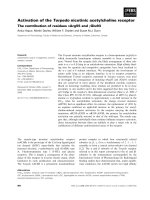
A) Quantitative RT-PCR analysis of SPHK1 expression in PCLS from different mouse strainsFigure 1
A) Quantitative RT-PCR analysis of SPHK1 expression in PCLS from different mouse strains. The relative expression of
SPHK1-mRNA in relation to the house-keeping gene β
2
microglobulin is shown. Data are given as means ± S.E.M. of four inde-
pendent experiments each carried out in triplicate. B) Indirect immunofluorescence studies examining SPHK1 expression in
PCLS of FVB-mice. Immunoreactivity for SPHK1 was present in the wall of larger (a) and smaller (b) airways and in the media
of pulmonary vessels (arrows in a, b). Consecutive sections (c, d) showing that preabsorption of the SPHK1 antiserum abol-
ished immunolabelling (Pre, d). Bars = 50 µm.
b
SPHK1a
a
d
c
SPHK1a Pre
B
0,0
2,0
4,0
6,0
8,0
10,0
FVB M4-WT M3k.o. M2-WT M2k.o
FVB M
3
R-KO M
3
R-wt M
2
R-KO M
2
R-wt
Relative expression
A
*
Respiratory Research 2005, 6:48 />Page 5 of 14
(page number not for citation purposes)
microglobulin served as an internal control. We found
that inactivation of the M
2
R and M
3
R genes had no signif-
icant effect on the relative expression levels of SPHK1
compared to their corresponding wt controls (Mann-
Whitney U-test, Fig. 1A). This analysis also showed that
SPHK1 expression was significantly lower in M
2
R-wt and
M
2
R-KO mice, as compared to FVB, M
3
R-wt, and M
3
R-KO-
mice (p < 0.05, Mann-Whitney U-test, Fig. 1A), perhaps
due to the fact that the M
2
R-KO/wt mice have a genetic
background that is somewhat different from that of the
other mice used (for details see "Materials and Methods").
Immunohistochemistry
We used single-and double-labeling immunohistochem-
istry to examine the distribution of SPHK1 in murine
peripheral lung. Strong SPHK1-immunoreactivity was
detected in the smooth muscle cells of intraparenchymal
bronchi (Figs. 1B, 2) and pulmonary arteries (Fig. 1B) and
veins as confirmed by double-staining of a marker of
smooth muscle cells, α-smooth muscle actin. Preabsorp-
tion of the SPHK1 antiserum abolished immunolabelling
(Fig. 1B). Immunoreactivity for SPHK1 was absent in
bronchial epithelium (Figs. 1B, 2). SPHK1 showed a gran-
ular cytoplasmatic localisation but was also found in
smooth muscle membranes (Fig. 2). The pattern of
SPHK1 immunoreactivity was very similar in PCLS of FVB,
M
2
R-KO/wt, and M
3
R-KO/wt mice
Videomorphometry
FVB mice In PCLS preparations from FVB mice, the lumi-
nal area of peripheral bronchi rapidly decreased within
the first 2 minutes after application of 10
-6
M muscarine,
followed by a sustained decrease in presence of the ago-
nist (Tab. 1, Fig. 3). Application of the SPHK inhibitors
DMS and DHS alone for ten minutes prior to coadminis-
tration of 10
-6
M muscarine had no significant effect on
bronchial luminal diameter (Figs. 3A, B, 5A–D). Coad-
ministration of 10
-6
M muscarine with DHS (10
-4
M-10
-10
M) significantly reduced both the initial and the sustained
phase of the muscarine induced bronchoconstriction
(Tab. 1, Fig. 3B). Similarly, coadministration of 10
-6
M
muscarine with 10
-6
-10
-10
M DMS reduced both phases of
the muscarine response. However 10
-4
M DHS had no
effect (Tab. 1). Coadministration of 10
-6
M muscarine
with the structurally related sphingolipid N-acetylsphin-
gosine (10
-6
-10
-10
M) which has no effect on SPHK1 func-
tion [5] did not alter the muscarine induced constriction
(Tab. 1).
Inhibition of Ca
2+
entry by La
3+
(10
-6
M) considerably
reduced the initial and strongly inhibited the sustained
constriction of peripheral airways following the adminis-
tration of 10
-6
M muscarine (Tab. 2, Fig. 4A, E, G). Com-
bination of 10
-8
M DHS or 10
-8
M DMS with La
3+
(10
-6
M)
almost completely abolished the initial and the sustained
phase of the muscarine-dependent bronchoconstriction
(Tab. 2, Fig. 4C, E, G). The combination of DHS or DMS
with La
3+
further significantly reduced both phases com-
pared to La
3+
alone (Wilcoxon's rank sum test p < 0.05)
Blockade of Ca
2+
-entry by SKF96365 (10
-5
M), which
inhibits G-protein activated and voltage gated Ca
2+
-chan-
nels, greatly reduced both the initial and the sustained
phase of muscarine-induced bronchoconstriction (Tab. 3,
Fig. 4B, F, H). Combination of SKF96365 (10
-5
M) with
10
-8
M DMS or 10
-8
M DHS exerted no further reduction
of luminal airway area (Wilcoxon's rank sum test, Tab. 2,
Fig. 4F, H).
M
2
R-KO/wt mice Muscarine (10
-6
M) stimulation of PCLS
preparations from M
2
R-KO mice resulted in an initial con-
striction response that was followed by a slight relaxation
(4–8 % relaxation of luminal airway area within 3–15
min, 28 slices/27 lungs, Tab. 3, Fig. 5A, B). In contrast, in
PCLS preparations from the corresponding wt mice (M
2
R-
wt), the initial bronchoconstriction was followed by a sus-
tained constriction response. In PCLS of M
2
R-KO and
M
2
R-wt mice, DHS and DMS (10
-8
M each) significantly
inhibited the initial and sustained phase of muscarine
induced constriction (Tab. 3, Fig. 5A, B). The initial con-
striction was inhibited by about 54 % (luminal area of
M
2
R-wt: 48 ± 11.5 %; M
2
R-KO: 61.5 ± 9.5 %, Tab. 3, Fig.
5A, B) and the sustained phase by about 50 % (luminal
area of M
2
R-wt: 34 ± 12 %; M
2
R-KO: 67.5 ± 15 %, Tab. 3,
Fig. 5A, B). The degree of inhibition of the initial phase
was comparable between M
2
R-wt and M
2
R-KO mice.
M
3
R-KO/wt mice As in all other experiments, the area of
the airway lumen at the beginning of the experiment was
defined as 100 %. Application of 10
-6
M muscarine con-
stricted peripheral airways in wt-mice by about 66 % (28
slices/25 lungs), whereas in M
3
R-KO mice the constriction
was reduced by about 57 % (29 slices/28 lungs, Tab. 4
Figs. 5C, D, 6). In wt-bronchi, inhibition of SPHK by DHS
or DMS (10
-8
M each) significantly reduced the initial
phase of constriction by about 50 % (58 ± 12 %, Tab. 4,
Figs. 5C, D, 6). Strikingly, treatment of PCLS from M
3
R-
KO mice with DHS (10
-8
M) or DMS (10
-6
and 10
-8
M)
almost completely abolished the initial constriction
response (94 ± 4 %, Tab. 4, Figs. 5, 6). Inhibition of SPHK
by DHS (10
-8
M) or DMS (10
-6
and 10
-8
M) significantly
reduced the sustained phase of constriction to a compara-
ble degree in M
3
R-wt and M
3
R-KO mice (Tab. 4, Figs. 5C,
D, 6).
Discussion
MR signaling pathways play a key role in the regulation of
airway resistance which is determined largely by the diam-
eter of smaller, intrapulmonary airways [25]. A better
understanding of the different pathways underlying MR
Respiratory Research 2005, 6:48 />Page 6 of 14
(page number not for citation purposes)
Expression of SPHK1 in PCLS from different mouse strains studied via confocal laser scanning microscopyFigure 2
Expression of SPHK1 in PCLS from different mouse strains studied via confocal laser scanning microscopy. Representative con-
focal laser scanning micrographs of double labeling immunohistochemistry demonstrates the restriction of SPHK1-immunore-
activity to smooth muscle cells, identified by immunoreactivity for the marker protein α-smooth muscle actin (α-sma).
SPHK1a-immunoreactivity was present in peripheral airway smooth muscle of knock-out animals (M
2
R-KO, a-d) and wild-type
(M
2
R-wt, e-h, FVB, i-l). Granular immunoreactivity could be detected in the cytoplasm and in the membrane of smooth muscle
cells. Bars = 50 µm
SPHK1a
α
αα
α-sma
M
2
R-wt
M
2
R-KO
FVB
ab
cd
ef
gh
ij
kl
Respiratory Research 2005, 6:48 />Page 7 of 14
(page number not for citation purposes)
activation in the intrapulmonary airways is of considera-
ble clinical relevance. In the present study, we examined
the hypothesis that S1P might be involved in the mus-
carinic control of peripheral airways. To address this ques-
tion, we used the PCLS model which has been shown to
maintain the integrity of all components of the peripheral
lung including viable peripheral airways [20].
Using double labelling immunohistochemistry we dem-
onstrated for the first time that the SPHK1 protein is
highly expressed in the cytosol of murine airway smooth
muscle cells with virtually no immunoreactivity in non-
smooth muscle cells. On the other hand, human and
murine lung tissue showed a high expression and activity
of SPHK which was not restricted to smooth muscle alone
[13,14]. The virtual restriction of SPHK1-immununoreac-
tivity to smooth muscle could be due to the presence of
SPHK isoforms other than SPHK1 in murine lung [14]. At
the subcellular level, SPHK1 showed a cytoplasmic locali-
sation but was also found in airway smooth muscle mem-
branes, in agreement with functional and
immunoprecipitation studies of mouse lung membranes
[14].
In the present study, the use of membrane-permeable
SPHK inhibitors, DHS and DMS [18], convincingly dem-
onstrated that MR-mediated constriction of peripheral air-
ways involves the activation of SPHK. In PCLS
preparations from wt-mice, both SPHK inhibitors signifi-
cantly reduced the fast initial constriction response
(induced by 10
-6
M muscarine) which is mainly depend-
ent on Ca
2+
-release from intracellular stores [26]. This
effect was observed with the lowest concentrations of the
inhibitors but was absent when DHS and DMS were given
alone for ten minutes prior to the application of DHS and
DMS in combination with muscarine. This inhibition of
muscarine induced bronchoconstriction was also absent
when we used N-acetylsphingosine (N-AcS), an agent that
is structurally related to DHS and DMS but that does not
affect SPHK function [5]. These data suggest that DHS and
DMS did not exert their inhibitory effects through non-
specific actions. Accordingly, the muscarine-induced ini-
tial bronchoconstriction was partly inhibited but
persisted in presence of the blockers of Ca
2+
-influx, La
3+
or
SKF 96365 [27,28], but was almost abolished when La
3+
was applied in combination with DHS or DMS. The sus-
tained phase of muscarine-induced bronchoconstriction
Table 1: Effects of N-AcS, DHS and DMS on muscarine-induced reductions in luminal airway area in FVB mice. The number of
experiments (lungs/slices), mean airway diameter in µm, and the luminal airway area determined 1 min (1) and 15 min (2) after
stimulation with muscarine (Mus) and 1 min (3) and 15 min (4) after repeated stimulation with muscarine or after repeated
stimulation with muscarine in combination with N-AcS, DHS or DMS are shown. In all experiments, the muscarine concentration was
10
-6
M. Data are given as means ± S.E.M. Matched pairs (1 vs. 3; 2 vs. 4) were evaluated by Wilcoxon's rank sum test. Differences were
considered as statistically significant when p < 0.05 (n.s., not significant).
Lungs /slices Diameter [µm] Area [%] 1 Area [%] 2 Area [%] 3 Area [%] 4 p (1 vs. 3) p (2 vs. 4)
Mus Mus n.s. n.s.
4/4 206 ± 7 31 ± 6 38 ± 4 38 ± 7 28 ± 3
Mus Mus/N-AcS 10
-6
M n.s. n.s.
6/5 182 ± 7 47 ± 8 42 ± 8 50 ± 8 42 ± 9
Mus Mus/N-AcS 10
-8
M n.s. n.s.
5/5 218 ± 5 36 ± 1 28 ± 5 38 ± 6 26 ± 6
Mus Mus/N-AcS 10
-10
M n.s. n.s.
4/4 212 ± 22 21 ± 8 16 ± 5 26 ± 6 30 ± 5
Mus Mus/DHS 10
-4
M n.s. n.s.
4/6 194 ± 9 42 ± 5 37 ± 7 61 ± 8 45 ± 6
Mus Mus/DHS 10
-6
M p < 0.05 p < 0.05
7/7 197 ± 14 45 ± 11 31 ± 7 70 ± 13 50 ± 5
Mus Mus/DHS 10
-8
M p < 0.05 p < 0.05
7/9 181 ± 6 39 ± 7 29 ± 6 50 ± 9 47 ± 7
Mus Mus/DHS 10
-10
M p < 0.05 p < 0.05
4/8 198 ± 6 34 ± 3 24 ± 3 42 ± 4 40 ± 4
Mus Mus/DMS 10
-4
M p < 0.05 p < 0.05
4/7 180 ± 7 30 ± 7 25 ± 6 45 ± 10 41 ± 7
Mus Mus/DMS 10
-6
M p < 0.05 p < 0.05
4/7 201 ± 11 48 ± 7 35 ± 8 70 ± 6 54 ± 9
Mus Mus/DMS 10
-8
M p < 0.05 p < 0.05
4/7 206 ± 11 31 ± 5 25 ± 4 54 ± 8 50 ± 5
Mus Mus/DMS 10
-10
M p < 0.05 p < 0.05
4/6 196 ± 15 43 ± 6 23 ± 4 71 ± 11 52 ± 7
Respiratory Research 2005, 6:48 />Page 8 of 14
(page number not for citation purposes)
Muscarine-induced reductions in luminal area of peripheral bronchi from FVB miceFigure 3
Muscarine-induced reductions in luminal area of peripheral bronchi from FVB mice. (A) Luminal area of peripheral bronchi
after application of 10
-6
M muscarine (Mus) as recorded by videomorphometry. Data (means ± S.E.M) were expressed as %
luminal area. Bronchi from FVB mice (4 slices from 6 lungs) responded to 10
-6
M muscarine until wash out (Wash). Time points
1–4 were chosen as indicators for initial and sustained constriction. 1 = luminal airway area 1 min after muscarine application,
2 = luminal airway area 15 min after muscarine application, 3 = luminal airway area 1 min after repeated muscarine application,
4 = luminal airway area 15 min after repeated muscarine application. Effect of DHS (B) and DMS (C) on muscarine-mediated
reductions in luminal area of peripheral bronchi from FVB mice. The luminal area of peripheral bronchi (expressed in %) was
recorded by videomorphometry after application of 10
-6
M muscarine (Mus) alone and after application of 10
-6
M muscarine
together with (B) 10
-6
M DHS (diamonds, 7 slices from 4 lungs) and 10
-10
M DHS (triangles, 8 slices from 4 lungs) or (C) 10
-6
M
DMS (diamonds, 9 slices from 7 lungs) and 10
-10
M DMS (triangles, 6 slices from 4 lungs). Bronchoconstriction responses were
significantly reduced in the presence of DHS and DMS. The inhibition was more pronounced following coadministration of
DMS. Data are given as means ± S.E.M.
0
20
40
60
80
100
120
121416181
Area [%]
20 40 60 80
Time [min]
Mus
Mus
Wash
Wash
DMS
0
20
40
60
80
100
120
121416181
20 40 60 80
Mus
Mus
Wash
Wash
DHS
Area [%]
A
4
Mus
Mus
Wash
Wash
B
Area [%]
C
0
20
40
60
80
100
120
1 112131415161718191
20 40 60 80
312
Respiratory Research 2005, 6:48 />Page 9 of 14
(page number not for citation purposes)
was also significantly inhibited by DHS and DMS in FVB
mice. Interestingly, the responses that were inhibited by
DHS/DMS were SKF96365-but not La
3+
-sensitive, since
DHS/DMS significantly increased the La
3+
-mediated inhi-
bition of the muscarine-mediated constriction, but had
no effect on the SKF96365-mediated inhibition of the
constriction.
MR-mediated constriction of peripheral airways has been
shown to be mediated by a mixture of M
2
R and M
3
R sub-
types [20]. It is well known that the M
3
R stimulates IP
3
-
dependent intracellular Ca
2+
-release ([29,2]. However,
like S1P [30], stimulation of M
3
R can also activate RhoA-
dependent signalling pathways leading to increased myo-
filament sensitivity of smooth muscle [31,32]. On the
other hand, activation of M
2
Rs in airway smooth muscle
has been shown to increase the sensitivity of myofila-
ments to Ca
2+
and to inhibit noradrenaline-induced
increases in intracellular cAMP [33,34]. In HEK-293 cells,
DLS and DMS markedly reduced both M2R-and M3R-
mediated increases in [Ca
2+
]
i
[5].
In the present study, we therefore also examined which of
these two receptor subtypes (M
2
R or M
3
R) is involved in
the SPHK1-dependent constriction of peripheral airways.
To address this issue, we carried out a series of functional
experiments using PCLS preparations from M
2
R-and M
3
R-
deficient mice (M
2
R-KO, M
3
R-KO) and their correspond-
ing wild-type controls (M
2
R-wt, M
3
R-wt). To rule out
potential differences in SPHK1 expression between wt and
KO animals we initially performed a set of quantitative
RT-PCR studies. These studies showed that inactivation of
the M
2
R and M
3
R genes had no significant effect on the
relative expression levels of SPHK1 compared to the cor-
responding wt controls. Moreover, confocal laser scan-
ning microscopic studies showed that the localization and
distribution of SPHK1 protein were similar in all mouse
strains. In agreement with the results of a previous study
[20], the peripheral airways of M
3
R-KO mice showed an
about 50 % reduction in the magnitude of the initial mus-
carine induced constriction response, as compared to the
corresponding response obtained with preparations from
M
3
R-wt mice. We previously demonstrated that the
bronchoconstrictor response remaining in the M
3
R-KO
mice is exclusively mediated by M
2
Rs [20].
Changes in [Ca
2+
]
i
are the main trigger in the initiation of
MR-mediated bronchoconstriction. The initial phase of
constriction of intraparenchymal airways is known to be
mediated partly via release of Ca
2+
from intracellular
stores, as shown by the presence of this phase under
blockade of ion influx by La
3+
or SKF96365. The consider-
able reduction of the initial constriction by La
3+
or
SKF96365 further indicates that influx of Ca
2+
is also part
of this phase of muscarine-mediated constriction. We
defined the bronchoconstriction in response to muscarine
as 100 % and analyzed the inhibition under various
experimental conditions. Blockade of SPHK1 by either
DHS or DMS almost completely abolished the initial
phase of constriction in bronchi from M
3
R-KO mice (Fig.
5 Tab. 2). In contrast, DHS or DMS only partially reduced
this early response in bronchi from M
2
R-KO and wt-mice
Table 2: Effects of SKF96365 and La
3+
alone and in combination with DHS or DMS on the muscarine-induced reductions in luminal
airway area in FVB mice Effects of the Ca
2+
-entry inhibitors La
3+
(10
-6
M) and SKF 96365 (10
-5
M) alone and in combination with DHS or
DMS (both 10
-6
M) on the muscarine induced reduction in luminal airway area (expressed in %). The number of experiments (lungs/
slices), mean airway diameter in µm, and the luminal airway area determined 1 min (1) and 15 min (2) after stimulation with
muscarine and 1 min (3) and 15 min (4) and after stimulation with muscarine (Mus) in combination with La
3+
(Mus/La
3+
) or SKF96365
(Mus/SKF), or after stimulation with muscarine and La
3+
or SKF96365 in combination with DHS or DMS are shown. In all experiments,
the muscarine concentration was 10
-6
M. Data are given as means ± S.E.M. Matched pairs (1 vs. 3; 2 vs. 4) were evaluated by Wilcoxon's
rank sum test. Differences were considered as statistically significant when p < 0.05.
Lungs /slices Diameter [µm] Area [%] 1 Area [%] 2 Area [%] 3 Area [%] 4 p (1 vs. 3) p (2 vs. 4)
4/9 212 ± 3 Mus Mus/SKF p < 0.05 p < 0.05
60 ± 7 49 ± 7 76 ± 7 86 ± 3
4/8 193 ± 12 Mus Mus/SKF/DHS p < 0.05 p < 0.05
51 ± 5 43 ± 6 82 ± 6 86 ± 3
5/6 207 ± 11 Mus Mus/SKF/DMS p < 0.05 p < 0.05
65 ± 3 53 ± 6 88 ± 2 85 ± 8
4/7 180 ± 8 Mus Mus/La
3+
p < 0.05 p < 0.05
39 ± 8 49 ± 12 65 ± 7 89 ± 2
4/7 211 ± 12 Mus Mus/La
3+
/DHS p < 0.05 p < 0.05
40 ± 4 33 ± 3 85 ± 2 89 ± 2
4/5 210 ± 15 Mus Mus/La
3+
/DMS p < 0.05 p < 0.05
45 ± 9 38 ± 9 94 ± 2 95 ± 1
Respiratory Research 2005, 6:48 />Page 10 of 14
(page number not for citation purposes)
Effect of various treatments on muscarine-mediated reductions in luminal area of peripheral bronchi from FVB miceFigure 4
Effect of various treatments on muscarine-mediated reductions in luminal area of peripheral bronchi from FVB mice. The lumi-
nal area of peripheral bronchi (expressed in %) was recorded by videomorphometry after application of 10
-6
M muscarine
alone or after application of 10
-6
M muscarine in the presence of (A) 10
-6
M La
3+
(7 slices from 4 lungs), (B) SKF 96365 (SKF 10
-
5
M, 9 slices from 4 lungs), (C) 10
-6
M La
3+
in combination with 10
-6
M DHS (7 slices from 4 lungs), or (D) 10
-5
M SKF 96365 in
combination with 10
-6
M DHS (8 slices from 4 lungs). Data are given as means ± S.E.M.E-F) Summary of effects of various treat-
ments on muscarine-mediated reductions in luminal area of peripheral bronchi from FVB mice. Luminal area (expressed in %)
determined 1 min (initial contraction, E) and 15 min (sustained contraction, G) after application of 10
-6
M muscarine alone or of
muscarine in combination with either La
3+
(both 10
-6
M) or La
3+
in combination with DHS or DMS (both 10
-8
M). Luminal area
(expressed in %) determined 1 min (initial contraction, F) and 15 min (sustained contraction, H) after application of muscarine
alone or of muscarine in combination with either SKF 96365 (10
-5
M) or SKF 96365 in combination with 10
-8
M DHS or DMS.
Asterisks indicate p < 0.05 for the comparison with application of muscarine alone (E-F). Daggers indicate p < 0.05 for the
comparison with application of muscarine in combination with La
3+
(E, G). Data are given as means ± S.E.M.
0
20
40
60
80
100
120
020406080100
0
20
40
60
80
100
120
0 20406080100
0
20
40
60
80
100
120
020406080100
Mus
Mus
Wash Wash
La
3+
Mus
Mus
Wash Wash
DHS/La
3+
Mus
Mus
Wash Wash
DHS/SKF
0
20
40
60
80
100
120
0 20406080100
Mus
Mus
Wash Wash
SKF
A
B
C
D
Time [min] Time [min]
Area [%]Area [%]
0
20
40
60
80
100
120
1234
0
20
40
60
80
100
120
1234
0
20
40
60
80
100
120
1234
0
20
40
60
80
100
120
1234
Mus
La
3+
SKF
DHS
DMS
+
+
+
+
+
+
+
+
+
+
+
+
+
+
+
+
+
+
Area [%]
E
F
G
H
Area [%]
** * ****
***
***
††
††
Respiratory Research 2005, 6:48 />Page 11 of 14
(page number not for citation purposes)
(Fig. 5). This observation strongly suggests that stimula-
tion of M
2
Rs mediates activation of SPHK1, which even-
tually triggers the release of Ca
2+
from intracellular stores
leading to bronchoconstriction. The role of M
2
R in airway
smooth muscle contraction appears multi-functional in
that the receptor can modulate the function of smooth
muscle by activation of multiple signalling pathways
including tyrosine kinase activation and stimulation/inhi-
bition of ion channels [35,32]. Phosphorylation of
myosin light chain and activation of PKC play important
roles in the maintenance of smooth muscle contractions
[36,37]. Since S1P has been shown to stimulate myosin
phosphorylation [8,38], it is likely that this response is
also mediated by the M
2
R subtype.
Our findings strongly suggest that SPHK1 activation is
part of the signalling response to M
2
R stimulation in
peripheral mouse airways. These airways resemble in their
structure and composition of cell types human distal air-
ways [39]. Like human distal airways, murine peripheral
airways about 200 µm in diameter that are terminal bron-
chioles lack submucosal glands, cartilage and contain
smooth muscle and Clara cells, in addition to ciliated cells
[39]. In studies using monkey and rat peripheral airways,
the ACh analogue methacholine (MCh) induced similar
responses in large and small mammals [40]. It is therefore
likely that the distal airways of man and mice, both of
which are also MCh-sensitive [17], share similar MR sig-
nalling pathways. In addition, it has been shown that
human airway smooth muscle cells constrict in response
to S1P [7,8], suggesting that the MR-SPHK1-S1P signal-
ling pathway is also present in human peripheral airways.
Conclusion
In conclusion, in this study we demonstrated the existence
of a novel signalling pathway in the regulation of periph-
eral airways. We found that MR-mediated constriction of
murine peripheral airways is mediated, in part, by activa-
tion of SPHK. Our data suggest that muscarinic activation
of SPHK contributes to the initial and sustained phase of
constriction. The SPHK activation in the initial phase is
mainly mediated via the M
2
R subtype. These findings
could be of relevance for the development of novel drugs
useful for the treatment of chronic obstructive pulmonary
disease and asthma. For example, one may speculate that
altered S1P signalling may contribute to the hyperreactiv-
ity of peripheral airways under pathological conditions.
List of abbreviations
ACh acetylcholine
CT threshold cycle
MR muscarinic acetylcholine receptor
wt wild-type control
Table 3: Effects of DHS and DMS on muscarine-induced reductions in luminal airway area in PCLS from M
2
R-KO and
theircorresponding wild-type mice. The number of experiments (lungs/slices), mean airway diameter in µm, and the muscarine-
induced constriction in luminal airway area (expressed in %) determined after 1 min (1) and 15 min (2) after stimulation with
muscarine and 1 min (3) and 15 min (4) after stimulation with muscarine in combination with DHS or DMS (both 10
-6
M or 10
-8
M) are
shown. In all experiments, the muscarine concentration was 10
-6
M. The initial phase (1 vs 3) and the sustained phase (2 vs 4) of
constriction were reduced in the presence of DHS or DMS. Data are given as means ± S.E.M.
M
2
R-wt Lungs /slices Diameter [µm] Area [%] 1 Area [%] 2 Area [%] 3 Area [%] 4 p (1 vs. 3) p (2 vs. 4)
6/7 208 ± 9 Mus Mus/DHS 10
-6
Mp ≤ 0.05 p ≤ 0.05
35 ± 9 32 ± 9 77 ± 11 52 ± 11
7/8 213 ± 8 Mus Mus/DHS 10
-8
M n.s. p = 0.078 p ≤ 0.05
46 ± 6 45 ± 12 61 ± 9 60 ± 9
5/5 202 ± 17 Mus Mus/DMS 10
-6
Mp ≤ 0.05 p ≤ 0.05
40 ± 10 42 ± 9 72 ± 9 66 ± 10
7/7 217 ± 8 Mus Mus/DMS 10
-8
Mp ≤ 0.05 p ≤ 0.05
54 ± 10 41 ± 9 69 ± 10 60 ± 10
M
2
R-KO
6/6 210 ± 8 Mus Mus/DHS 10
-6
Mp ≤ 0.05 p ≤ 0.05
50 ± 11 54 ± 11 79 ± 7 82 ± 7
7/7 194 ± 9 Mus Mus/DHS 10
-8
Mp ≤ 0.05 p ≤ 0.05
44 ± 10 48 ± 8 74 ± 8 74 ± 7
7/8 218 ± 8 Mus Mus/DMS 10
-6
Mp ≤ 0.05 p ≤ 0.05
56 ± 8 62 ± 7 83 ± 6 79 ± 6
7/7 189 ± 9 Mus Mus/DMS 10
-8
Mp ≤ 0.05 p ≤ 0.05
43 ± 13 51 ± 11 71 ± 9 73 ± 9
Respiratory Research 2005, 6:48 />Page 12 of 14
(page number not for citation purposes)
Muscarine-mediated changes in luminal area of peripheral bronchi from wild-type and MR deficient miceFigure 5
Muscarine-mediated changes in luminal area of peripheral bronchi from wild-type and MR deficient mice. Changes in luminal
area of mouse peripheral airways were recorded by videomorphometry after cumulative application of different concentra-
tions of muscarine. (A) Videomorphometric images of precision cut lung slices before (∅), 1 min after stimulation with mus-
carine (10
-6
M), after 15 min wash out (Wash) and 1 min after stimulation with muscarine in presence of DMS (10
-8
M). The
bronchi from wild-type (M
3
R-wt) and M
3
R-KO mice constricted in response to 10
-6
M muscarine. In M
3
R-wt mice, the con-
striction was slightly reduced in presence of DMS but abolished in M
3
R-KO mice. Bar = 200 µm. (B-D) Effect of DHS and DMS
on muscarine-mediated reductions in luminal area of murine peripheral bronchi in the absence of the M
2
R or M
3
R subtypes. (B-
C) Luminal area of peripheral airways from M
2
R-KO (triangles) and M
2
R-wt mice (squares)(expressed in % in response to
application of 10
-6
M muscarine (Mus) alone or after application of muscarine (10
-6
M) in combination with DHS (B) or DMS (C)
(both 10
-8
M). Both DHS and DMS significantly inhibited the muscarine-induced constriction in airways of both M
2
R-KO and
M
2
R-wt mice. (D, E) Luminal area of peripheral airways (expressed in %) from M
3
R-KO (triangles) and M
3
R-wt (squares) mice
in response to application of 10
-6
M muscarine alone or after application of muscarine (10
-6
M) in combination with DHS (D) or
DMS (E) (both 10
-8
M). Both DHS and DMS significantly inhibited the muscarine-induced constriction in airways of both M
3
R-
KO and M
3
R-wt mice. Data are given as means ± S.E.M.
0
20
40
60
80
100
120
1 21416181
0
20
40
60
80
100
120
1 21416181
Mus
Mus
Wash
Wash
DHS
B
Mus
Mus
Wash
Wash
DMS
C
0
20
40
60
80
100
120
121416181
0
20
40
60
80
100
120
1 21416181
Area [%]
Mus
Mus
Wash
Wash
DHS
D
Mus
Mus
Wash
Wash
DMS
E
Time [min] Time [min]
20 40 60 80 20 40 60 80
Mus
Mus/DMSWashØ
M
3
R-wt
M
3
R-KO
A
Area [%]
Respiratory Research 2005, 6:48 />Page 13 of 14
(page number not for citation purposes)
M
2
R-KO muscarinic acetylcholine receptor 2-deficient
mice
M
3
R-KO muscarinic acetylcholine receptor 3-deficient
mice
PLCβ phospholipase C beta
PCLS Precision cut lung slices
SKF96365 1-2-(4-methoxyphenyl)-2-[3-(4-methoxyphe-
nyl) propoxy]-ethyl-1H-imidazole
S1P sphingosine 1-phosphate
SPHK sphingosine kinases
IP
3
inositol 1, 4, 5-trisphosphate
DHS D, L-threo-dihydrosphingosine
DMS N, N-dimethyl-sphingosine
NAcS N-acetylsphingosine
MCh Methacholine
Authors' contributions
PM, NP, SA, WS, SW and RVH carried out the videomor-
phometric experiments and performed qRT-PCR and
immunohistochemistry. JW developed the KO-mice and
participated together with WK and RVH in writing and
preparation of the manuscript and in the statistical analy-
sis. YB was involved in the design of the study and the
immunohistochemical investigations. The data presented
in the manuscript are part of the doctoral thesis of PM,
NP, SA, WS.
Acknowledgements
We thank Ms K. Michael for skilful technical assistance.
References
1. Wess J, Blin N, Mutschler E, Bluml K: Muscarinic acetylcholine
receptors: structural basis of ligand binding and G protein
coupling. Life Sci 1995, 56(11–12):915-922.
2. Caulfield MP, Birdsall NJ: International Union of Pharmacology.
XVII. Classification of muscarinic acetylcholine receptors.
Pharmacol Rev 1998, 50(2):279-290.
3. Jacoby DB, Fryer AD: Anticholinergic therapy for airway
diseases. Life Sci 2001, 68(22–23):2565-2572.
4. Meyer zu Heringdorf D, Lass H, Alemany R, Laser KT, Neumann E,
Zhang C, Schmidt M, Rauen U, Jakobs KH, van Koppen CJ: Sphingo-
sine kinase-mediated Ca2+ signalling by G-protein-coupled
receptors. Embo J 1998, 17(10):2830-2837.
5. van Koppen CJ, Meyer zu Heringdorf D, Alemany R, Jakobs KH:
Sphingosine kinase-mediated calcium signaling by mus-
carinic acetylcholine receptors. Life Sci 2001, 68(22–
23):2535-2540.
6. Olivera A, Spiegel S: Sphingosine kinase: a mediator of vital cel-
lular functions. Prostaglandins Other Lipid Mediat 2001, 64(1–
4):123-134.
Table 4: Effects of DHS and DMS on muscarine-induced reductions in luminal airway area in PCLS from M
3
R-KO and their
corresponding wild-type mice. The number of experiments (lungs/slices), mean airway diameter in µm, and the muscarine-induced
constriction in luminal airway area (expressed in %) determined after 1 min (1) and 15 min (2) after stimulation with muscarine and 1
min (3) and 15 min (4) after stimulation with muscarine in combination with DHS or DMS (both 10
-6
M or 10
-8
M) are shown. In all
experiments, the muscarine concentration was 10
-6
M. The initial phase (1 vs 3) and the sustained phase (2 vs 4) of constriction were
reduced in presence of DHS or DMS. Data are given as means ± S.E.M.
M
3
R-wt Lungs /slices Diameter [µm] Area [%] 1 Area [%] 2 Area [%] 3 Area [%] 4 p (1 vs. 3) p (2 vs. 4)
6/6 221 ± 10 Mus Mus/DHS 10
-6
M n.s. p ≤ 0.05
53 ± 7 47 ± 8 75 ± 7 69 ± 11
8/9 204 ± 10 Mus Mus/DHS 10
-8
Mp ≤ 0.05 p ≤ 0.01
40 ± 6 30 ± 6 64 ± 9 55 ± 6
6/7 203 ± 6 Mus Mus/DMS 10
-6
Mp ≤ 0.05 p ≤ 0.05
41 ± 10 37 ± 8 64 ± 10 56 ± 8
5/6 197 ± 8 Mus Mus/DMS 10
-8
Mp ≤ 0.05 p ≤ 0.05
42 ± 5 41 ± 10 71 ± 7 69 ± 9
M
3
R-KO
4/4 205 ± 18 Mus Mus/DHS 10
-6
M n.s. n.s.
69 ± 2 51 ± 4 77 ± 8 62 ± 12
8/9 188 ± 9 Mus Mus/DHS 10
-8
Mp ≤ 0.05 p ≤ 0.05
75 ± 6 67 ± 6 96 ± 2 88 ± 4
7/7 202 ± 12 Mus Mus/DMS 10
-6
Mp ≤ 0.05 p ≤ 0.05
75 ± 4 67 ± 6 97 ± 1 82 ± 4
9/9 196 ± 7 Mus Mus/DMS 10
-8
Mp ≤ 0.01 p ≤ 0.05
65 ± 5 56 ± 6 93 ± 2 81 ± 3
Publish with BioMed Central and every
scientist can read your work free of charge
"BioMed Central will be the most significant development for
disseminating the results of biomedical researc h in our lifetime."
Sir Paul Nurse, Cancer Research UK
Your research papers will be:
available free of charge to the entire biomedical community
peer reviewed and published immediately upon acceptance
cited in PubMed and archived on PubMed Central
yours — you keep the copyright
Submit your manuscript here:
/>BioMedcentral
Respiratory Research 2005, 6:48 />Page 14 of 14
(page number not for citation purposes)
7. Ammit AJ, Hastie AT, Edsall LC, Hoffman RK, Amrani Y, Krymskaya
VP, Kane SA, Peters SP, Penn RB, Spiegel S, et al.: Sphingosine 1-
phosphate modulates human airway smooth muscle cell
functions that promote inflammation and airway remode-
ling in asthma. Faseb J 2001, 15(7):1212-1214.
8. Rosenfeldt HM, Amrani Y, Watterson KR, Murthy KS, Panettieri RA
Jr, Spiegel S: Sphingosine-1-phosphate stimulates contraction
of human airway smooth muscle cells. Faseb J 2003,
17(13):1789-1799.
9. Lacasse Y, Brosseau L, Milne S, Martin S, Wong E, Guyatt GH, Gold-
stein RS: Pulmonary rehabilitation for chronic obstructive
pulmonary disease. Cochrane Database Syst Rev 2002,
(3):CD003793.
10. Escolar JD, Escolar MA, Guzman J, Roques M: Morphological hys-
teresis of the small airways. Histol Histopathol 2003, 18(1):19-26.
11. Wohlsen A, Uhlig S, Martin C: Immediate allergic response in
small airways. Am J Respir Crit Care Med 2001, 163(6):1462-1469.
12. Kohama T, Olivera A, Edsall L, Nagiec MM, Dickson R, Spiegel S:
Molecular cloning and functional characterization of murine
sphingosine kinase. J Biol Chem 1998, 273(37):23722-23728.
13. Melendez AJ, Carlos-Dias E, Gosink M, Allen JM, Takacs L: Human
sphingosine kinase: molecular cloning, functional character-
ization and tissue distribution. Gene 2000, 251(1):19-26.
14. Fukuda Y, Kihara A, Igarashi Y: Distribution of sphingosine kinase
activity in mouse tissues: contribution of SPHK1. Biochem Bio-
phys Res Commun 2003, 309(1):155-160.
15. Liu H, Sugiura M, Nava VE, Edsall LC, Kono K, Poulton S, Milstien S,
Kohama T, Spiegel S: Molecular cloning and functional charac-
terization of a novel mammalian sphingosine kinase type 2
isoform. J Biol Chem 2000, 275(26):19513-19520.
16. Young KW, Willets JM, Parkinson MJ, Bartlett P, Spiegel S, Nahorski
SR, Challiss RA: Ca2+/calmodulin-dependent translocation of
sphingosine kinase: role in plasma membrane relocation but
not activation. Cell Calcium 2003, 33(2):119-128.
17. Held HD, Martin C, Uhlig S: Characterization of airway and vas-
cular responses in murine lungs. Br J Pharmacol 1999,
126(5):1191-1199.
18. Olivera A, Spiegel S: Sphingosine-1-phosphate as second mes-
senger in cell proliferation induced by PDGF and FCS
mitogens. Nature 1993, 365(6446):557-560.
19. Coulson FR, Fryer AD: Muscarinic acetylcholine receptors and
airway diseases. Pharmacol Ther 2003, 98(1):59-69.
20. Struckmann N, Schwering S, Wiegand S, Gschnell A, Yamada M, Kum-
mer W, Wess J, Haberberger RV: Role of muscarinic receptor
subtypes in the constriction of peripheral airways: studies on
receptor-deficient mice. Mol Pharmacol 2003, 64(6):1444-1451.
21. Gomeza J, Shannon H, Kostenis E, Felder C, Zhang L, Brodkin J, Grin-
berg A, Sheng H, Wess J: Pronounced pharmacologic deficits in
M2 muscarinic acetylcholine receptor knockout mice. Proc
Natl Acad Sci U S A 1999, 96(4):1692-1697.
22. Yamada M, Miyakawa T, Duttaroy A, Yamanaka A, Moriguchi T, Mak-
ita R, Ogawa M, Chou CJ, Xia B, Crawley JN, et al.: Mice lacking the
M3 muscarinic acetylcholine receptor are hypophagic and
lean. Nature 2001, 410(6825):207-212.
23. Murate T, Banno Y, K TK, Watanabe K, Mori N, Wada A, Igarashi Y,
Takagi A, Kojima T, Asano H, et al.: Cell type-specific localization
of sphingosine kinase 1a in human tissues. J Histochem Cytochem
2001, 49(7):845-855.
24. Martin C, Uhlig S, Ullrich V: Videomicroscopy of methacholine-
induced contraction of individual airways in precision-cut
lung slices. Eur Respir J 1996, 9(12):2479-2487.
25. Martin RJ: Therapeutic significance of distal airway inflamma-
tion in asthma. J Allergy Clin Immunol 2002, 109(2 Suppl):S447-460.
26. Hakonarson H, Grunstein MM: Regulation of second messengers
associated with airway smooth muscle contraction and
relaxation. Am J Respir Crit Care Med 1998, 158(5 Pt 3):S115-122.
27. Murray RK, Kotlikoff MI: Receptor-activated calcium influx in
human airway smooth muscle cells. J Physiol 1991, 435:123-144.
28. Oonuma H, Nakajima T, Nagata T, Iwasawa K, Wang Y, Hazama H,
Morita Y, Yamamoto K, Nagai R, Omata M: Endothelin-1 is a
potent activator of nonselective cation currents in human
bronchial smooth muscle cells. Am J Respir Cell Mol Biol 2000,
23(2):213-221.
29. Wess J: Molecular biology of muscarinic acetylcholine
receptors. Crit Rev Neurobiol 1996, 10(1):69-99.
30. Bolz SS, Vogel L, Sollinger D, Derwand R, Boer C, Pitson SM, Spiegel
S, Pohl U: Sphingosine kinase modulates microvascular tone
and myogenic responses through activation of RhoA/Rho
kinase. Circulation 2003, 108(3):342-347.
31. Murthy KS, Zhou H, Grider JR, Brautigan DL, Eto M, Makhlouf GM:
Differential signalling by muscarinic receptors in smooth
muscle: m2-mediated inactivation of myosin light chain
kinase via Gi3, Cdc42/Rac1 and p21-activated kinase 1 path-
way, and m3-mediated MLC20 (20 kDa regulatory light
chain of myosin II) phosphorylation via Rho-associated
kinase/myosin phosphatase targeting subunit 1 and protein
kinase C/CPI-17 pathway. Biochem J 2003, 374(Pt 1):145-155.
32. Ehlert FJ: Contractile role of M2 and M3 muscarinic receptors
in gastrointestinal, airway and urinary bladder smooth
muscle. Life Sci 2003, 74(2-3):355-366.
33. Chiba Y, Sakai H, Misawa M: Characterization of muscarinic
receptors in rat bronchial smooth muscle in vitro. Res Com-
mun Mol Pathol Pharmacol 1998, 102(2):205-208.
34. Janssen LJ, Wattie J, Lu-Chao H, Tazzeo T: Muscarinic excitation-
contraction coupling mechanisms in tracheal and bronchial
smooth muscles. J Appl Physiol 2001, 91(3):1142-1151.
35. Togashi H, Emala CW, Hall IP, Hirshman CA: Carbachol-induced
actin reorganization involves Gi activation of Rho in human
airway smooth muscle cells. Am J Physiol 1998, 274(5 Pt
1):L803-809.
36. Ochsner M: Ca2+ transient, cell volume, and microviscosity of
the plasma membrane in smooth muscle. Biochem Pharmacol
1997, 53(12):1765-1777.
37. Kai T, Yoshimura H, Jones KA, Warner DO: Relationship between
force and regulatory myosin light chain phosphorylation in
airway smooth muscle. Am J Physiol Lung Cell Mol Physiol 2000,
279(1):L52-58.
38. Zhou H, Murthy KS: Distinctive G protein-dependent signaling
in smooth muscle by sphingosine 1-phosphate receptors
S1P1 and S1P2. Am J Physiol Cell Physiol 2004, 286(5):C1130-1138.
39. Parent RA: Comparative biology of the normal lung. Volume 1.
CRC Press; 1991.
40. Kott KS, Pinkerton KE, Bric JM, Plopper CG, Avadhanam KP, Joad JP:
Methacholine responsiveness of proximal and distal airways
of monkeys and rats using videomicrometry. J Appl Physiol
2002, 92(3):989-996.