Báo cáo y học: "Green tea polyphenol extract attenuates lung injury in experimental model of carrageenan-induced pleurisy in mice" ppt
Bạn đang xem bản rút gọn của tài liệu. Xem và tải ngay bản đầy đủ của tài liệu tại đây (897.44 KB, 13 trang )
BioMed Central
Page 1 of 13
(page number not for citation purposes)
Respiratory Research
Open Access
Research
Green tea polyphenol extract attenuates lung injury in
experimental model of carrageenan-induced pleurisy in mice
Rosanna Di Paola
1
, Emanuela Mazzon
1
, Carmelo Muià
1
, Tiziana Genovese
1
,
Marta Menegazzi
2
, Raffaela Zaffini
2
, Hisanory Suzuki
2
and
Salvatore Cuzzocrea*
1
Address:
1
Department of Clinical and Experimental Medicine and Pharmacology, Torre Biologica, Policlinico Universitario, Messina, Italy and
2
Biochemistry Division, Department of Neuroscience and Vision, University of Verona, Verona, Italy
Email: Rosanna Di Paola - ; Emanuela Mazzon - ; Carmelo Muià - ;
Tiziana Genovese - ; Marta Menegazzi - ; Raffaela Zaffini - ;
Hisanory Suzuki - ; Salvatore Cuzzocrea* -
* Corresponding author
green tea extractcarrageenan-induced pleurisyneutrophils infiltrationlung injury
Abstract
Here we investigate the effects of the green tea extract in an animal model of acute inflammation,
carrageenan-induced pleurisy. We report here that green tea extract (given at 25 mg/kg i.p. bolus
1 h prior to carrageenan), exerts potent anti-inflammatory effects in an animal model of acute
inflammation in vivo.
Injection of carrageenan (2%) into the pleural cavity of mice elicited an acute inflammatory response
characterized by fluid accumulation in the pleural cavity that contained many neutrophils (PMNs),
an infiltration of PMNs in lung tissues and increased production of nitrite/nitrate, tumour necrosis
factor alpha. All parameters of inflammation were attenuated by green tea extract treatment.
Furthermore, carrageenan induced an up-regulation of the adhesion molecule ICAM-1, as well as
nitrotyrosine and poly (ADP-ribose) synthetase (PARS) formation, as determined by
immunohistochemical analysis of lung tissues. Staining for the ICAM-1, nitrotyrosine, and PARS was
reduced by green tea extract.
Our results clearly demonstrate that treatment with green tea extract exerts a protective effect
and offers a novel therapeutic approach for the management of lung injury.
Introduction
The role of oxyradical formation in various forms of
inflammation is well established [1] Reactive oxygen spe-
cies (ROS) are associated with the inflammatory response
and frequently they contribute to the tissue damaging
effects of inflammatory reactions [2-4]. ROS formation
and degradation are key components of the metabolism
of aerobic organisms. Certain levels of ROS are required
for normal cell functions, but if in surplus, they will cause
oxidative stress [5-7]. ROS like superoxide, hydrogen per-
oxide and lipid hydroperoxides can regulate the activities
Published: 29 June 2005
Respiratory Research 2005, 6:66 doi:10.1186/1465-9921-6-66
Received: 21 April 2005
Accepted: 29 June 2005
This article is available from: />© 2005 Di Paola et al; licensee BioMed Central Ltd.
This is an Open Access article distributed under the terms of the Creative Commons Attribution License ( />),
which permits unrestricted use, distribution, and reproduction in any medium, provided the original work is properly cited.
Respiratory Research 2005, 6:66 />Page 2 of 13
(page number not for citation purposes)
of several kinases, transcription factors, cell death machin-
ery and proteins such as COX-2 and iNOS [8,9].
Recent data demonstrate that the expression of the induc-
ible isoform of nitric oxide (NO) synthase also plays
important pathogenetic roles in various models of inflam-
mation [10-12]. Peroxynitrite, a cytotoxic oxidant species
formed from the reaction of NO and superoxide [13], may
mediate part of the oxidative injury associated with simul-
taneous production of NO and oxyradicals. Peroxynitrite
formation has been demonstrated in various inflamma-
tory disorders [14,15] and in circulatory shock [16]. Per-
oxynitrite is a potent oxidant, and therefore it is
conceivable that endogenous antioxidant mechanisms
counteract its toxicity. In in vitro studies, it has been estab-
lished that antioxidants such as glutathione, ascorbic acid,
and alpha-tocopherol are scavengers of peroxynitrite and
inhibitors of its oxidant capacity [17,18].
Green tea – a minimally processed product of the same
plant that gives us black and oolong teas – is rich in pow-
erful antioxidant compounds called polyphenols. The
polyphenols found in tea are more commonly known as
flavanols or catechins and comprise 30–40 percent of the
extractable solids of dried green tea leaves. The main cate-
chins in green tea are epicatechin, epicatechin-3-gallate,
epigallocatechin, and epigallocatechin-3- gallate (EGCG),
with the latter being the highest in concentration. Green
tea polyphenols have demonstrated significant antioxi-
dant, anticarcinogenic, anti-inflammatory, thermogenic,
probiotic, and antimicrobial properties in numerous
human, animal, and in vitro studies [19,20]. Recently it
has been showed that green tea polyphenols inhibited
tumour necrosis factor-alpha induction in macrophages
by attenuating nuclear factor-kβ NF-Kβ) activation [21].
Similarly [22] showed that EGCG inhibits lipopolysacca-
ride (LPS) – stimulated nitric oxide production and induc-
ibile nitric oxide synthase gene expression in peritoneal
macrophages by decreasing NF-κβ activation. These stud-
ies provide significant evidence that green tea polyphe-
nols have anti-inflammatory effects. Lung inflammation
is characterised by T-cell rich infiltrates and enhanced
expression of pro-inflammatory cytokines. The signalling
pathway of IFN-γ, secreted by type-1 helper lymphocyte
(Th-1), lead to the activation of signal transducer and acti-
vator of transcription-1 (STAT-1) [23]. Moreover, IFN-γ is
involved in the induction of iNOS and ICAM-1 gene
expression by the activation of STAT-1 transcription factor
[24,25]. Thus, upregulation of STAT-1 activity could play
a key role in the pathogenesis of carrageenan-induced
pleurisy. STAT-1 are activated by phosphorylation on con-
served tyrosine and serine residues by the Janus kinases
(JAKs) and MAP kinase families respectively, which allow
the STAT-1 to dimerise and translocate to the nucleus and
there by regulate gene expression [23]. Previously, we
demonstrated, in some epithelial cell cultures, the inhibi-
tory effect of EGCG on iNOS induction by preventing
STAT-1 phosphorylation and activation [26].
In this study we investigated the role of Green tea extract
in rodent model carrageenan-induced pleurisy.
This experimental model has been widely used to investi-
gate the pathophysiology of acute inflammation and also
to evaluate the efficacy of drugs in inflammation. Injec-
tion of carrageenan into the pleural space leads to pleu-
risy, infiltration by polymorphonuclear leukocytes
(PMN), and lung injury. In this study, we have investi-
gated the effect of the green tea on: PMN infiltration [mye-
loperoxidase (MPO) activity]; STAT-1 activity (by EMSA),
up-regulation of ICAM-1 (by immunohistochemistry);
the nitration of tyrosine residues (an indicator of the for-
mation of peroxynitrite) (by immunohistochemistry) and
lung damage (histology).
Materials and methods
Animals
Mice (4–5 weeks old, 20–22 g) were purchased from Jack-
son Laboratories (Harlan Nossan, Italy). The animals
were housed in a controlled environment and provided
with standard rodent chow and water. Animal care was in
compliance with Italian regulations on protection of ani-
mals used for experimental and other scientific purposes
(D.M. 116192) as well as with the EEC regulations (O.J.
of E.C. L 358/1 12/18/1986).
Green tea extract
Green tea extract (GTE) was a kind gift of Indena (Milano,
Italy), and it was defined by the producer as having a
polyphenolic content of 75 ± 5% with the major constit-
uent being epigallocatechin-3-gallate at 62% and the
minor ones being epicatechin-3-gallate, epigallocatechin
and epicathechin.
Carrageenan-induced pleurisy
Mice were anaesthetised with isoflurane and submitted to
a skin incision at the level of the left sixth intercostals
space. The underlying muscle was dissected and saline
(0.1 ml) or saline containing 2%λ-carrageenan (0.1 ml)
was injected into the pleural cavity. The skin incision was
closed with a suture and the animals were allowed to
recover. At 4 h after the injection of carrageenan, the ani-
mals were killed by inhalation of CO
2
. The chest was care-
fully opened and the pleural cavity rinsed with 1 ml of
saline solution containing heparin (5 U/ml) and
indomethacin (10 µg/ml). The exudate and washing solu-
tion were removed by aspiration and the total volume
measured. Any exudate, which was contaminated with
blood, was discarded. The amount of exudate was calcu-
lated by subtracting the volume injected (1 ml) from the
Respiratory Research 2005, 6:66 />Page 3 of 13
(page number not for citation purposes)
total volume recovered. The leukocytes in the exudate
were suspended in phosphate-buffer saline (PBS) and
counted with an optical microscope in a Burker's chamber
after Blue Toluidine staining.
Experimental groups
Mice were randomly allocated into the following groups:
(i) CAR + saline group. Mice were subjected to carrageenan-
induced pleurisy (N = 10), (ii) Green Tea group Same as the
CAR + saline group but Green Tea (25 mg/kg i.p) were
administered 1 h prior to carrageenan (N = 10), (iii)
Sham+saline group. Sham-operated group in which identi-
cal surgical procedures to the CAR group was performed,
except that the saline was administered instead of carra-
geenan (N = 10), (iv) Sham + Green Tea group. Same as the
Sham+saline group but Green Tea (25 mg/kg i.p) were
administered 1 h prior to carrageenan (N = 10). The doses
of Green Tea used here to reduce acute lung injury have
been reported by us to reduce the tissue injury caused by
ischemia-reperfusion in the gut (dose-response curve
study) (Muià et al submitted 2005).
Determination of myeloperoxidase activity
Myeloperoxidase (MPO) activity, an indicator of poly-
morphonuclear leukocyte (PMN) accumulation, was
determined as previously described [27]. At 4 h after intra-
pleural injection of carrageenan lung tissues, were
obtained and weighed. Each piece of tissue was homoge-
nised in a solution containing 0.5% hexa-decyl-trimethyl-
ammonium bromide dissolved in 10 mM potassium
phosphate buffer (pH 7) and centrifuged for 30 min at
20,000 × g at 4°C. An aliquot of the supernatant was then
allowed to react with a solution of tetra-methyl-benzidine
(1.6 mM) and 0.1 mM H
2
O
2
. The rate of change in
absorbance was measured spectrophotometrically at 650
nm. MPO activity was defined as the quantity of enzyme
degrading 1 µmol of peroxide min at 37°C and was
expressed in mill units per gram weight of wet tissue.
Measurement of TNF-
α
levels
TNF-α levels were evaluated in the exudates at 4 h after the
induction of pleurisy by carrageenan injection. The assay
was carried out by using a colorimetric, commercial ELISA
kit (Calbiochem-Novabiochem Corporation, USA).
Measurement of nitrite/nitrate
Nitrite/nitrate (NOx) production, an indicator of NO syn-
thesis, was measured in pleural exudate. At the first nitrate
in the supernatant was incubated with nitrate reductase
(0.1 U/ml) and NADPH (1 mM) and FAD (50 µM) at
37°C for 15 min. Then followed another incubation with
LDH (100 U/ml) and sodium pyruvate (10 mM) for 5
min. The nitrite concentration in the samples was meas-
ured by the Griess reaction, by adding 100 µl of Griess rea-
gent (0.1% naphthylethylenediamide dihydrochloride in
H
2
O and 1% sulphanilamide in 5% concentrated H
2
PO
4
;
vol. 1: 1) to 100 µl samples. The optical density at 550 nm
(OD
550
) was measured using ELISA microplate reader
(SLT- Lab instruments Salzburg, Austria). Nitrate concen-
trations were calculated by comparison with OD
550
of
standard solutions of sodium nitrate prepared in saline
solution.
Immunohistochemical localisation of ICAM-1, PAR and
Nitrotyrosine
At 4 h after carrageenan administration, the lungs were
fixed in 10% buffered formaldehyde and 8 µm sections
were prepared from paraffin embedded tissues. After
deparaffinization, endogenous peroxidase was quenched
with 0.3% H
2
O
2
in 60% methanol for 30 min. The sec-
tions were permeabilized with 0.1% Triton X-100 in PBS
for 20 min. Non-specific adsorption was minimised by
incubating the section in 2% normal goat serum in phos-
phate buffered saline for 20 min. Endogenous biotin or
avidin binding sites were blocked by sequential incuba-
tion for 15 min with avidin and biotin. The sections were
then incubated overnight with primary anti-ICAM-1 anti-
body (1:500), with 1:1000 dilution of primary antinitro-
tyrosine antibody (DBA), and anti-PAR antibody (1:500)
or with control solutions. Controls included buffer alone
or non-specific purified rabbit IgG.
To confirm that the immunoreaction for the nitrotyrosine
was specific, some sections were also incubated with the
primary antibody (anti-nitrotyrosine) in the presence of
excess nitrotyrosine (10 mM) to verify the binding specif-
icity. To verify the binding specificity for PARS, sections
were also incubated with only the primary antibody (no
secondary) or with only the secondary antibody (no pri-
mary). In these situations, no positive staining was found
in the sections, indicating that the immunoreaction was
positive in all the experiments carried out.
Immunocytochemistry photographs (n = 5) were assessed
by densitometry. The assay was carried out by using Opti-
lab Graftek software on a Macintosh personal computer
(CPU G3-266).
Histological examination
Lung biopsies were taken at 4 h after injection of carra-
geenan. The biopsies were fixed for 1 wk in buffered for-
maldehyde solution (10% in PBS) at room temperature,
dehydrated by graded ethanol and embedded in Paraplast
(Sherwood Medical, Mahwah, N.J.). Tissue sections
(thickness 7 µm) were deparaffinized with xylene, stained
with trichromic Van Gieson, and studied using light
microscopy (Dialux 22 Leitz). Blood was passed on the
slide, fixed at 37°C, stained with May Grunward-Giensa,
and studied using light microscopy.
Respiratory Research 2005, 6:66 />Page 4 of 13
(page number not for citation purposes)
Electrophoretic mobility shift assay
The lung samples have been collected in liquid nitrogen
and stored at -80°C until use. Nuclear extracts have been
prepared according to [28] in the presence of 10 µg/ml
leupeptin, 5 µg/ml antipain and pepstain, and 1 mM
PMSF (Sigma-Aldrich Company, Milan, Italy). Protein
concentration in the nuclear extracts was determined
using the method of [29]. Ten µg of nuclear extract have
been incubated at room temperature for 20 min with (2–
5 × 10
4
cpm) of
32
P-labeled double stranded oligonucle-
otide, containing the STAT-1 binding site (sis-inducible
factor-binding recognition element, SIE/M67) from the c-
Fos promoter (5'-GTCGACATTTCCCGTAAATCG-3'), the
PARP-1 binding site (5'-TTCCTTGCCCCTCCCATTTTTC-
3') from the Reg promoter [30] or the SP-1 consensus
sequence (5'GGGGCGGGGC-3', Santa Cruz Biotechnol-
ogy, CA) in a 15 µl of binding reaction buffer (20 mM
HEPES, pH 7.9, 50 mM KCl, 10% glycerol, 0.5 mM DTT,
0.1 mM EDTA, 2 µg poly(dI-dC), 1 µg salmon sperm
DNA). Products have been fractioned on a non denatur-
ing 5% polyacrilamide gel in TBE (Tris-Borate-EDTA
buffer, 0.5X). The intensity of the retarded bands has been
measured with a Phosphorimager (Molecular Dynamic,
Milan, Italy).
Materials
Unless otherwise stated, all compounds were obtained
from Sigma-Aldrich Company (Milan, Italy). Primary
monoclonal ICAM-1 (CD54) for immunoistochemistry
was purchases by Pharmingen. Reagents and secondary
and nonspecific IgG antibody for immunohistochemical
analysis were from Vector Laboratories InC. Primary mon-
oclonal anti-poly (ADP-ribose) antibody was purchased
by Alexis. All other chemicals were of the highest commer-
cial grade available. All stock solutions were prepared in
non pyrogenic saline (0.9% NaCl; Baxter Healthcare Ltd.,
Thetford, Norfolk, U.K.).
Data analysis
All values in the figures and text are expressed as mean ±
standard error (s.e.m.) of the mean of n observations. For
the in vivo studies n represents the number of animals
studied. In the experiments involving histology or immu-
nohistochemistry, the figures shown are representative of
at least three experiments performed on different experi-
mental days. The results were analysed by one-way
ANOVA followed by a Bonferroni post-hoc test for multiple
comparisons. A p-value less than 0.05 was considered
significant.
Results
Effects of green tea extract in carrageenan-induced
pleurisy
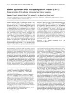
Histological examination of lung sections revealed signif-
icant tissue damage (Fig. 1B). Thus, when compared with
lung sections taken from saline-treated animals (Fig. 1A),
histological examination of lung sections of mice treated
with carrageenan showed oedema, tissue injury (Fig 1B),
and infiltration of the tissue with neutrophils (PMNs)
(Fig. 1B1). GTE significantly reduced the degree of injury
as well as the infiltration of PMNs (Fig. 1C). Furthermore,
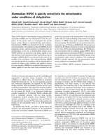
injection of carrageenan into the pleural cavity of mice
elicited an acute inflammatory response characterized by
the accumulation of fluid (oedema) that contained large
amounts of PMNs (Fig. 2A,B). Oedema and PMNs infil-
tration in pleural cavity were attenuated by the i.p. injec-
tion of GTE (Figs. 2A,B).
Effect of green tea extract on TNF-
α
levels
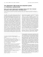
The levels of TNF-α were significantly elevated in the exu-
dates from vehicle-treated mice at 4 h after carrageenan
administration (Fig. 3). In contrast, the levels of this pro-
inflammatory cytokine was significantly lower in carra-
geenan-treated mice treated with GTE (Fig. 3). No signifi-
cant increased of TNF-α levels was observed in the
exudates of sham-operated mice.
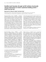
Effects of green tea extract on MPO activity
The above histological pattern of lung injury appeared to
be correlated with the influx of leukocytes into the lung
tissue. Therefore, we investigate the role of GTE on the
neutrophils infiltration by measurement of the activity of
myeloperoxidase. Myeloperoxidase activity was signifi-
cantly elevated (p < 0.001) at 4 h after carrageenan admin-
istration in vehicle-treated mice (Fig. 4). In Mice treated
with green tea extract lung myeloperoxidase activity was
significantly reduced (p < 0.01) in comparison to those of
vehicle-treated mice (Fig. 4).
Effects of green tea extract on the expression of adhesion
molecule (ICAM-1)
Staining of lung tissue sections obtained from saline-
treated mice with anti-ICAM-1 antibody showed specific
staining along bronchial epithelium, demonstrating that
ICAM-1 is constitutively expressed (data not shown). At 4
h after carrageenan injection, the staining intensity sub-
stantially increased along the bronchial epithelium (see
arrows, Fig. 5A, 6). Sections from GTE-treated mice did
not reveal any up-regulation of constitutively expressed
ICAM-1, which was normally expressed in the epithelium
(see arrows, Fig. 5B, 6). To verify the binding specificity
for ICAM-1, some sections were also incubated with only
the primary antibody (no secondary). In these situations,
no positive staining was found in the sections, indicating
that the immunoreaction was positive in all the experi-
ments carried out.
Effects of green tea extract on nitric oxide production
The levels of NO
x
were significantly (P < 0.01) increased
in the exudate from carrageenan-treated mice (Fig. 7). In
Respiratory Research 2005, 6:66 />Page 5 of 13
(page number not for citation purposes)
contrast, levels of NO
x
were significantly lower in the exu-
date of mice treated with GTE (Fig. 7).
Effects of green tea extract on nitrotyrosine and PARS
At 4 h after carrageenan injection, lung sections were
taken in order to determine the immunohistological
staining for nitrotyrosine or PARS. Sections of lung from
saline-treated mice did not reveal any immunoreactivity
for nitrotyrosine or PARS within the normal architecture
(data not shown). A positive staining for nitrotyrosine
(Fig. 6, 8A) and PARS (Fig. 6, 8C) was localized primarily
in the vessels and in the bronchial epithelium. GTE
reduced the staining for both nitrotyrosine (Fig. 6, 8B)
and PARS (Fig. 6, 8D). Therefore, no differences between
groups were shown for SP-1 DNA binding activity (data
not shown). It was also shown the DNA binding capacity
of PARP-1 to the promoter sequence of the Reg gene [30].
The retarded bands of the carraggeenan-treated mice were
reduced in comparison to those of vehicle-treated or GTE
pre-treated mice (Fig. 9A, B)
Effects of green tea extract on the STAT-1 activation
To examine the molecular mechanisms responsible for
mediating the anti-inflammatory effects of GTE we meas-
ured, by EMSA, the changes in activation of the
transcription factors STAT-1 and SP-1. DNA-binding activ-
ity of STAT-1 was significantly elevated at 4 h after carra-
geenan administration in vehicle-treated mice (Fig. 10A,
Effect of GTE on lung injuryFigure 1
Effect of GTE on lung injury. When compared with lung sections taken from control animals (A), lung sections from carra-
geenan-treated mice (B) demonstrate interstitial haemorrhage and polymorphonuclear leukocyte accumulation (B1). Lung sec-
tions from a carrageenan-treated mice that received GTE (C) exhibit reduced interstitial haemorrhage and a lesser cellular
infiltration. Figure is representative of all the animals in each group.
Respiratory Research 2005, 6:66 />Page 6 of 13
(page number not for citation purposes)
B). In Mice treated with green tea extract lung STAT-1
activity was similar to those of sham-operated group and
significantly reduced in comparison to those of vehicle-
treated mice (Fig. 10A, B).
Discussion
Polyphenols are the most significant group of tea compo-
nents, especially the catechin group of the flavonols. The
major tea catechins are EGCG, EGC, ECG, EC, (+)-gallo-
catechin, and (+)-catechin.
Many biological functions of tea polyphenols have been
studied [31], including anti-inflammatory, antioxidative
[32-34], antimutagenic [35], and anticarcinogenic [36]
effects.
This study provides the evidence that pretreatment of
mice with green tea extract attenuates 1) the development
of carrageenan-induced pleurisy, 2) the infiltration of the
lung with PMNs (histology and MPO activity), 3) the
degree of lung injury (histology) caused by injection of
carrageenan. All of these findings support the view that
green tea extract attenuates the degree of acute inflamma-
tion in mice. What, then, is the mechanism by which
green tea extract reduces acute inflammation?
The generation of oxidative and nitrosative species, which
exert their effects both directly and indirectly, is a impor-
tant contributor to inflammatory injury. The terms oxida-
tive and nitrosative refer to the formation of reactive
oxygen species (ROS), such as superoxide (O
2
),
Effect of GTE on carrageenan-induced inflammationFigure 2
Effect of GTE on carrageenan-induced inflammation. The increase in volume exudate (A) and accumulation of poly-
morphonuclear cells (PMNs, B) in pleural cavity 4 h after carrageenan injection was inhibited by GTE. Data are means ± SEM of
10 mice for each group. *P < 0.01 vs. sham. °P < 0.01 vs. carrageenan.
Respiratory Research 2005, 6:66 />Page 7 of 13
(page number not for citation purposes)
hydrogen peroxide (H
2
O
2
), and hydroxyl radicals, and
reactive nitrogen species (RNS), such as nitric oxide (NO),
peroxynitrite (ONOO
-
), and nitrogen dioxide.
Oxidants are generated as a result of the inflammatory
response by phagocytic cells, such as mononuclear cells.
Oxidants that are generated in excess of antioxidant
defences or that are lacking in antioxidant defences can
result in severe pulmonary inflammation leading to acute
lung injury Additionally, NO plays a multifaceted role in
mediating inflammatory processes [37]. Potential sources
of NO in the lungs include expression of iNOS, activated
neutrophils, [38] alveolar type-IIcells [39] endothelial
cells [40] and airway cells [41]. It has been demonstrated
that levels of NO
2
-
, increase markedly during acute and
chronic inflammation [42]. Recent study had demon-
strated that green tea polyphenols inhibit NO production
in peritoneal exudate (macrophage) cells [43] and EGCG
inhibits lipopolysaccharide (LPS)-induced NO produc-
tion and iNOS gene expression in isolated peritoneal
macrophages by decreasing NF-KB activation [22]. In
agreement with these observations in this study we shown
that the treatment with green tea extract in vivo reduce NO
formation.
Simultaneous generation of NO
.
and O
2
favours the pro-
duction of a toxic reaction product, peroxynitrite anion
(ONOO
-
)[13] and this product may account for some of
the deleterious effects associated with NO
.
production.
The pro-inflammatory and cytotoxic effects of ONOO- are
numerous [44]. Peroxynitrite nitrosates tyrosine residues
in proteins and nitrotyrosine formation has been used as
a marker for the detection of the endogenous formation of
peroxynitrite [45]. Using nitrotyrosine as a marker for the
presence of ONOO- has been challenged by the demon-
stration that other reactions can also induce tyrosine nitra-
tion; e.g., the reaction of nitrite with hypochlorous acid
and the reaction of myeloperoxidase with hydrogen per-
oxide can lead to the formation of nitrotyrosine [46].
Thus, increased nitrotyrosine staining is considered, as an
indicator of "increased nitrosative stress" rather than a
specific marker of the generation of peroxynitrite [47]. We
have found that nitrotyrosine is indeed present in lung
sections taken after carrageenan injection and that green
tea extract reduced the staining in these tissues.
ROS and peroxynitrite produce cellular injury and necro-
sis via several mechanisms including protein denatura-
tion, and DNA damage ROS produce strand breaks in
DNA that trigger energy-consuming DNA repair
mechanisms and activate the nuclear enzyme PARS,
resulting in the depletion of its substrate NAD in vitro and
a reduction in the rate of glycolysis. As NAD functions as
a cofactor in glycolysis and the tricarboxylic acid cycle,
Effect of GTE on TNF-α, levelFigure 3
Effect of GTE on TNF-α, level. Pleural injection of carra-
geenan caused by 4 h an increase in the release of the pro-
inflammatory cytokines, tumour necrosis factor alpha (TNF-
α). GTE significantly inhibited TNF-α. Data are means ± SEM
of 10 mice for each group. *P < 0.01 vs. sham. °P < 0.01 vs.
carrageenan.
Effect of GTE on myeloperoxidase (MPO) activity in the lungFigure 4
Effect of GTE on myeloperoxidase (MPO) activity in
the lung. Within 4 h, pleural injection of carrageenan led to
an increase in neutrophil accumulation in the lung (as meas-
ured by MPO activity) GTE treatment significantly inhibited
neutrophil infiltration. Data are means ± SEM of 10 mice for
each group. *P < 0.01 vs. sham. °P < 0.01 vs. carrageenan.
Respiratory Research 2005, 6:66 />Page 8 of 13
(page number not for citation purposes)
Immunohistochemical localization of ICAM-1 in the lungFigure 5
Immunohistochemical localization of ICAM-1 in the lung. Section obtained from carrageenan-treated mice showed
intense positive staining for ICAM-1 (A, see arrows). The degree of bronchial epithelium (see arrows) staining for ICAM-1 (B)
was markedly reduced in tissue section obtained from GTE-treated mice. Figure is representative of all the animals in each
group.
Typical Densitometry evaluationFigure 6
Typical Densitometry evaluation. Densitometry analy-
sis of immunocytochemistry photographs (n = 5) for ICAM-1,
Nitrotyrosine and PAR from lung was assessed. The assay
was carried out by using Optilab Graftek software on a Mac-
intosh personal computer (CPU G3-266). Data are
expressed as % of total tissue area. ND: not detectable. *P <
0.01 vs. sham. °P < 0.01 vs. carrageenan.
Effect of GTE on NO productionFigure 7
Effect of GTE on NO production. Nitrite and nitrate
concentrations in pleural exudate at 4 h after carrageenan
administration. Nitrite and nitrate levels in carrageenan-
treated mice was significantly increased vs. sham group. GTE
treatment significantly reduced the carrageenan-induced ele-
vation of nitrite and nitrate levels. Data are means ± SEM of
10 mice for each group. *P < 0.01 vs. sham. °P < 0.01 vs.
carrageenan.
Respiratory Research 2005, 6:66 />Page 9 of 13
(page number not for citation purposes)
NAD depletion leads to a rapid fall in intracellular ATP.
This process has been termed the 'PARS suicide hypothe-
sis'. There is recent evidence that the activation of PARS
may also play an important role in inflammation [48,49].
We demonstrate here that green tea extract treatment
reduced the activation of PARS during carrageenan-
induced pleurisy in the lung. In light of the role of PARS
in inflammation, it is possible that PARS inhibition by
green tea extract accounts for the anti-inflammatory
response.
Besides attenuating ONOO- production and PARS activa-
tion, green tea extract also reduced the development of
oedema, neutrophil accumulation and had an overall pro-
tective effect on the degree of lung injury as assessed by
histological examination.
A possible mechanism by which green tea extract attenu-
ates PMNs infiltration is by down-regulating adhesion
molecules ICAM-1
The activation and expression of adhesion molecules
allows for the adhesion, conformational change, and
extravasation (emigration) of the neutrophil that may
Immunohistochemical localization for nitrotyrosine and PARS in the lungFigure 8
Immunohistochemical localization for nitrotyrosine and PARS in the lung. Immunohistochemistry for nitrotyrosine
(A) and PARS (C) show positive staining along the vessels and in the bronchial epithelium from a carrageenan-treated mice.
The intensity of the positive staining for nitrotyrosine (B) and PARS (C) was significantly reduced in the lung from GTE-treated
mice. Figure is representative of all the animals in each group.
Respiratory Research 2005, 6:66 />Page 10 of 13
(page number not for citation purposes)
Effect of GTE on PARP-1 activationFigure 9
Effect of GTE on PARP-1 activation. (A) DNA binding activity of PARP-1 in sham operated, carrageenan-treated (CAR)
and GTE pre-treated mice (CAR + GTE). Nuclear extracts (10 µg) from lung sample were incubated with a
32
P-labeled double-
stranded oligonucleotide containing binding sequence for PARP-1 and separated by nondenaturing PAGE. The specificity of the
retarded bands was demonstrated by competition with 100-fold excess of specific unlabeled oligonucleotide (not shown). (B)
The intensity of retarded bands (measured by phosphoimager) in carrageenan-treated mice was significantly increased vs. sham
group. GTE treatment significantly reduced the carrageenan-induced elevation of PARP-1 activity. Data are means ± SEM of 5
mice for each group. *P < 0.01 vs. sham. °P < 0.001 vs. carrageenan.
Respiratory Research 2005, 6:66 />Page 11 of 13
(page number not for citation purposes)
Effect of GTE on STAT-1 activationFigure 10
Effect of GTE on STAT-1 activation. (A) DNA binding activity of STAT-1 in sham operated, carrageenan-treated (CAR)
and GTE pre-treated mice (CAR + GTE). Nuclear extracts (10 µg) from lung sample were incubated with a
32
P-labeled double-
stranded oligonucleotide containing binding sequence for STAT-1 and separated by nondenaturing PAGE. The specificity of the
retarded bands was demonstrated by competition with 100-fold excess of specific unlabeled oligonucleotide (not shown). (B)
The intensity of retarded bands (measured by phosphoimager) in carrageenan-treated mice was significantly increased vs. sham
group. GTE treatment significantly reduced the carrageenan-induced elevation of STAT-1 activity. *P < 0.01 vs. sham. °P <
0.001 vs. carrageenan.
Respiratory Research 2005, 6:66 />Page 12 of 13
(page number not for citation purposes)
induce local injury and participate in the orchestration of
systemic inflammation and all of its consequences. ICAM-
1 in particular plays a role in inflammatory processes and
in the T cell-mediated host defense system. Within the
endothelium, ICAM-1 has an important role in migration
of leukocytes to sites of inflammation, enabling the firm
adhesion and diapedesis of leukocytes. In accordance
with these findings, we observed that green tea extract
reduce the upregulation the surface expression of ICAM-1
on endothelial cells and prevents the infiltration of neu-
trophils at inflamed sites.
Lung inflammation is associated with enhanced expres-
sion of proinflammatory cytokines which serve as inter-
cellular signals that recruit cells and modulate cell
function. Cytokines produced predominantly by activated
macrophages and lymphocytes mediate many inflamma-
tory processes [49]. These proinflammatory cytokines
include interferon-γ (IFN-γ), interleukin-1 (IL-1), tumour
necrosis factor-α (TNFα) and chemokines (e.g. inter-
leukin-8 [IL-8], macrophage chemotactic and activating
factor [MCAF]). Though all of these cytokines play impor-
tant roles in the evolving inflammatory response, TNFα
appears to be a critical mediator of the inflammatory cas-
cade. Numerous studies show that TNFα rises rapidly fol-
lowing acute trauma/inflammation/infection and that
blocking TNFα activity reduces injury [50-52]. Recently it
has been shown that, EGCG inhibits okadaic acid-
induced TNFα production and gene expression in BALB/
3T3 cells [53]. In agreement with these observations we
have found in this study that green tea pre-treatment
reduce exudates level of TNF-alpha. Moreover cytokines
activate, several signaling pathways, leading to the down-
stream activation of NF-κB transcription factors. In this
study we have placed our attention on STAT-1. Signal
transducers and activators of transcription (STAT) factors
are a family of cytoplasmic transcription factors that
mediate intracellular signaling initiated at cytokine cell
surface receptors and transmitted to the nucleus. Recently,
it has been reported that the polyphenolic agent epigallo-
catechin-3-gallate (EGCG), a major constituent of green
tea, is a potent inhibitor of STAT-1 phosphorylation and
activation [26]. The JAK/STAT pathway has been shown to
be essential for human and murine iNOS expression [24]
and in ICAM-1 induction [25]. We demonstrate here that
green tea extract pretreatment reduced STAT-1 activation
in carrageenan-treated mice. The fall in STAT-1 activity
may further account for the repression of ICAM-1 and
iNOS lung expression with decrease in nitrite/nitrate con-
centration in the pleural exudate. Thus, the down-regula-
tion of STAT-1 action provides a molecular basis for the
anti-inflammatory effect of GTE.
Data generated from the present study indicate that green
tea extract cause a substantial reduction of carraggenan
induced-pleurisy in the mice suggesting toxicity from oxy-
gen metabolites, released by stimulated neutrophils, mac-
rophages, and other cells, as one of the significant
mechanisms of lung injury. These findings support the
potential use of green tea extract as therapeutic agents in
the therapy of conditions associated with acute
inflammation.
Acknowledgements
This study was supported by grant from PRIN 2003. The authors would like
to thank Giovanni Pergolizzi and Carmelo La Spada for their excellent tech-
nical assistance during this study, Mrs Caterina Cutrona and Deborah A.
Schaller for secretarial assistance and Miss Valentina Malvagni for editorial
assistance with the manuscript.
References
1. Cuzzocrea S, Zingarelli B, Gilad E, Hake P, Salzman AL, Szabo C: Pro-
tective effect of melatonin in carrageenan-induced models of
local inflammation: relationship to its inhibitory effect on
nitric oxide production and its peroxynitrite scavenging
activity. J Pineal Res 1997, 23(2):106-16.
2. Pawliczak R: The role of radical oxygen species in airway
inflammation. Pol Merkuriusz Lek 2003, 14:493-6.
3. Cuzzocrea S, McDonald MC, Filipe HM, Costantino G, Mazzon E, San-
tagati S, Caputi AP, Thiemermann C: Effects of tempol, a mem-
brane-permeable radical scavenger, in a rodent model of
carrageenan-induced pleurisy. Eur J Pharmacol 2000, 390:209-22.
4. Leiro J, Alvarez E, Arranz JA, Laguna R, Uriarte E, Orallo F: Effects
of cis resveratrol on inflammatory murine macrophages:
antioxidant activity and down-regulation of inflammatory
genes. J Leukoc Biol 2004, 75:.
5. Los M, Schenk H, Hexel K, Baeuerle PA, Droge W, Schulze-Osthoff
K: IL-2 gene expression and NF-kappa B activation through
CD28 requires reactive oxygen production by 5-lipoxygen-
ase. EMBO J 1995, 14:3731-40.
6. Turpaev KT: Reactive oxygen species and regulation of gene
expression. Biochemistry (Mosc) 2002, 67:281-92.
7. Adcock IM, Brown CR, Kwon O, Barnes PJ: Oxidative stress
induces NF kappa B DNA binding and inducible NOS mRNA
in human epithelial cells. Biochem Biophys Res Commun 1994,
199:1518-24.
8. Schenk H, Klein M, Erdbrugger W, Droge W, Schulze-Osthoff K: Dis-
tinct effects of thioredoxin and antioxidants on the activa-
tion of transcription factors NF-kappa B and AP-1. Proc Natl
Acad Sci USA 1994, 91:1672-6.
9. Mathy-Hartert M, Deby-Dupont GP, Reginster JY, Ayache N, Pujol JP,
Henrotin YE: Regulation by reactive oxygen species of inter-
leukin-1beta, nitric oxide and prostaglandin E(2) production
by human chondrocytes. Osteoarthritis Cartilage 2002, 10:547-55.
10. Moncada S, Palmer RMG, Higgs EA: Nitric oxide: physiology,
pathophysiology and pharmacology. Pharmacol Rev 1991,
43:109-142.
11. Nathan C: Nitric oxide as a secretory product of mammalian
cells. FASEB J 1996, 6:3051-3064.
12. Cuzzocrea S, Zingarelli B, Gilard E, Hake P, Salzman AL, Szabó C:
Anti-inflammatory effects of mercaptoethylguanidine, a
combined inhibitor of nitric oxide synthase and peroxyni-
trite scavenger, in carrageenan-induced models of
inflammation. Free Rad Biol Med 1998, 24:450-459.
13. Beckman JS, Beckman TW, Chen J, Marshall PA, Freeman BA: Appar-
ent hydroxyl radical production by peroxynitrite: implica-
tions for endothelial injury from nitric oxide and superoxide.
Proc Natl Acad Sci U S A 1990, 87(4):1620-4.
14. Halliwell B: Oxygen radicals, nitrite oxide and human inflam-
matory joint disease. Ann Rheum Dis 1995, 54:505-510.
15. Salvemini D, Wang ZQ, Wyatt P, Bourdon DW, Marino MH, Manning
PT, Currie MG: Nitric oxide: a key mediator in the early and
late phase of carrageenan-induced rat paw inflammation. Br
J Pharmacol 1996, 118:829-838.
16. Cuzzocrea S, Zingarelli B, Costantino G, Szabó A, Salzman AL, Caputi
AP, Szabó C: Beneficial effects of 3-aminobenzamide, an inhib-
Publish with BioMed Central and every
scientist can read your work free of charge
"BioMed Central will be the most significant development for
disseminating the results of biomedical research in our lifetime."
Sir Paul Nurse, Cancer Research UK
Your research papers will be:
available free of charge to the entire biomedical community
peer reviewed and published immediately upon acceptance
cited in PubMed and archived on PubMed Central
yours — you keep the copyright
Submit your manuscript here:
/>BioMedcentral
Respiratory Research 2005, 6:66 />Page 13 of 13
(page number not for citation purposes)
itor of poly (ADP-ribose) synthetase in a rat model of
splanchnic artery occlusion and reperfusion. Br J Pharmacol
1997, 121:1065-1074.
17. Radi R, Beckman JS, Bush KM, Freeman BA: Peroxynitrite oxida-
tion of sulfhydryls. The cytotoxic potential of superoxide and
nitric oxide. J Biol Chem 1991, 266:4244-4250.
18. Karoui H, Hogg N, Frejaville C, Tordo P, Kalyanaraman B: Charac-
terization of sulfur-centered radical intermediates formed
during the oxidation of thiols and sulfite by peroxynitrite.
ESR-spin trapping and oxygen uptake studies. J Biol Chem
1996, 271:6000-6009.
19. Alschuler L: Grean Tea: Healing tonic. Am J Natur Med 1998,
5:28-31.
20. Graham HN: Green tea composition, consumption, and
polyphenol chemistry. Prev Med 1992, 21:334-350.
21. Yang F, de Villiers WJ, McClain CJ, Varilek GW: Green tea
polyphenols block endotoxin-induced tumor necrosis factor-
production and lethality in a murine model. J Nutr Dec 1998,
128(12):2334-40.
22. Lin YL, Lin JK: (-)-Epigallocatechin-3-gallate blocks the induc-
tion of nitric oxide synthase by down-regulating lipopolysac-
charide-induced activity of transcription factor nuclear
factor-KB. Mol Pharm 1997, 52:465-472.
23. Shuai K, Schindler C, Prezioso RV, Darnell JE Jr: Activation of tran-
scription by IFN-γ: tyrosine phosphorylation of a 91-kD DNA
binding protein. Science 1992, 258:1808-1812.
24. Gao J, Morrison DC, Parmely TJ, Russel SW, Murphy WJ: An inter-
feron-γ activated site (GAS) is necessary for the full expres-
sion of the mouse iNOS gene in response to interferon-γ and
lipopolysaccharide. J Biol Chem 1997, 272:1226-1230.
25. Roy J, Audette M, Tremblay MJ: Intercellular adhesion molecule-
1(ICAM-1) gene expression in human T cells is regulated by
phosphotyrosyl phosphatase activity. Involvement of NF-kB,
Ets, and palindromic interferon-gamma-responsive ele-
ment-binding site. J Biol Chem 2001, 276:14553-61.
26. Menegazzi M, Tedeschi E, Dussin D, De Prati AC, Cavalieri E, Mari-
otto S, Suzuki H: Anti-interferon gamma action of epigallo-
catechin-3-gallate mediated by specific inhibition of STAT1
activation. FASEB J 2001, 15:1309-1311.
27. Mullane KM, Kraemer R, Smith B: Myeloperoxidase activity as a
quantitative assessment of neutrophil infiltration into
ischemic myocardium. J Pharmacol Meth 1985, 14:157-167.
28. Schreiber E, Matthias P, Muller MM, Schaffner W: Rapid detection
of octamer binding proteins with miniextracts prepared
from a small number of cell. Nucleic Acid Res 1989, 17:6419A.
29. Bradford M: A rapid and sensitive method for the quantitation
of microgram quantities of protein using the principle of pro-
tein-dye binding. Anal Biochem 1976, 72:248-254.
30. Akiyama T, Takasawa S, Nata K, Kobayashi S, Abe M, Shervani NJ,
Ikeda T, Nakagawa K, Unno M, Matsuno S, Okamoto H: Activation
of Reg gene for insulin-producing β-cel regeneration:
poly(ADP-ribose) polymerase binds Reg promoter and reg-
ulates the transcription by autopoly(ADP-ribosyl)ation.
PNAS 2001, 98:48-53.
31. Yang CS, Wang ZY: Tea and cancer. J Natl Cancer Inst 1993,
85:1038-1049.
32. Lin YL, Juan IM, Chen YL, Liang YC, Lin JK: Composition of
polyphenols in fresh tea leaves and associations of their oxy-
gen-radical-absorbing capacity with antiproliferative actions
in fibroblast cells. J Agric Food Chem 1996, 44:1387-1394.
33. Ho CT, Chen Q, Shi H, Zhang KQ, Rosen RT: Antioxidative effect
of polyphenol extract prepared from various Chinese teas.
Prev Med 1992, 21:520-525.
34. Katiyar SK, Agarwal R, Zaim MT, Mukhtar H: Protection against N-
nitrosodiethylamine and benzo(a)pyrene-induced forestom-
ach and lung tumorigenesis in A/J mice by green tea. Carcino-
genesis 1993, 14:849-855.
35. Shiraki M, Hara Y, Osawa T, Kumon H, Nakauama T, Kawakishi S:
Antioxidative and antimutagenic effects of theaflavins from
black tea. Mutat Res 1994, 323:29-34.
36. Huang MT, Ho CT, Wang ZY, Ferraro T, Finnegan-Olive T, Lou YR,
Mitchell JM, Laskin JD, Newmark H, Yang CS, Conney AH: Inhibi-
tory effect of topical application of a green tea polyphenol
fraction on tumor initiation and promotion in mouse skin.
Carcinogenesis 1992, 13:947-954.
37. Haddad IY, Pitt BR, Matalon S: Nitric oxide and lung injury. In Pul-
monary diseases and disorders Edited by: Fishman AP. McGraw-Hill
New York, NY; 1996:337-346.
38. Fierro IM, Nascimento-DaSilva V, Arruda MA: Induction of NOS in
rat blood PMN in vivo and in vitro: modulation by tyrosine
kinase and involvement in bactericidal activity. J Leukoc Biol
1999, 65:508-514.
39. Kooy NW, Royall JA: Agonist-induced peroxynitrite produc-
tion from endothelial cells. Arch Biochem Biophys 1994,
310:352-359.
40. Kobzik L, Bredt DS, Lowenstein CJ: Nitric oxide synthase in
human and rat lung: immunocytochemical and histochemi-
cal localization. Am J Respir Cell Mol Biol 1993, 9:371-377.
41. Hickman-Davis JM, Lindsey JR, Zhu : Surfactant protein A medi-
ates mycoplasmacidal activity of alveolar macrophages. Am
J Physiol 1998, 274:L270-L277.
42. Farrell AJ, Blake DR, Palmer RMJ, Moncada S: Increased concentra-
tions of nitrite in synovial fluid and serum samples suggest
increased nitric oxide synthesis in rheumatic diseases. Ann
Rheum Dis 1992, 51:1219-1222.
43. Chan MMY, Ho CT, Huang HI: Effects of three dietary phyto-
chemicals from tea, rosemary and turmeric on inflamma-
tion-induced nitrite production. Cancer Lett 1995, 96:23-29.
44. Squadrito GL, Pryor WA: The formation of peroxynitrite in vivo
from nitric oxide and superoxide. Chem Biol Interact 1995,
96(2):203-6. Review
45. Beckman JS: Oxidative damage and tyrosine nitration from
peroxynitrite. Chem Res Toxicol 1996, 9(5):836-44. Review
46. Eiserich JP, Hristova M, Cross CE, Jones AD, Freeman BA, Halliwell
B, van der Vliet A: Formation of nitric oxide-derived inflamma-
tory oxidants by myeloperoxidase in neutrophils. Nature
1998, 391:393-7.
47. Cuzzocrea S, Caputi AP, Zingarelli B: Peroxynitrite-mediated
DNA strand breakage activates poly (ADP-ribose) syn-
thetase and causes cellular energy depletion in carrageenan-
induced pleurisy. Immunology 1998, 93:96-101.
48. Cuzzocrea S, Zingarelli B, Gilard E, Hake P, Salzman AL, Szabó C:
Protective effects of 3-aminobenzamide, an inhibitor of poly
(ADP-ribose) synthase in carrageenan-induced models of
local inflammation. Eur J Pharmacol 1998, 342:67-76.
49. Brennan FM, Feldmann M: Cytokines in autoimmunity. Curr Opin
Immunol 1996, 8:872-877.
50. Beutler B, Cerami A: Cachectin and tumour necrosis factor as
two sides of the same biological coin. Nature 1986,
320:584-588.
51. Machleidt T, Kramer B, Adam D, Neumann B, Schutze S, Wiegmann
K, Kronke M: Function of the p55 tumor necrosis factor recep-
tor "death domain" mediated by phophatidylcholine-specific
phospholipase. C J Exp Med 1996, 184:725-733.
52. Pfeffer K, Matsuyama T, Kundig TM, Wakeham A, Kishihara K, Shahi-
nian A, Wiegmann K, Ohashi PS, Kronke M, Mak TW: Mice defi-
cient for the 55 kd tumor necrosis factor receptor are
resistant to endotoxin shock, yet succumb to L. monocy-
togenes infection. Cell 1993, 73:457-467.
53. Suganuma M, Okabe S, Sueoka E, Iida N, Komori A, Kim SJ, Fujiki H:
A new process of cancer prevention mediated through inh-
bition of tumor necrosis factor αexpression. Cancer Res 1996,
56:3711-3715.