Báo cáo y học: " Toll-like receptor 2 expression is decreased on alveolar macrophages in cigarette smokers and COPD patients" docx
Bạn đang xem bản rút gọn của tài liệu. Xem và tải ngay bản đầy đủ của tài liệu tại đây (374.89 KB, 8 trang )
BioMed Central
Page 1 of 8
(page number not for citation purposes)
Respiratory Research
Open Access
Research
Toll-like receptor 2 expression is decreased on alveolar
macrophages in cigarette smokers and COPD patients
Daniel Droemann*
1
, Torsten Goldmann
2
, Thorsten Tiedje
1
, Peter Zabel
1,3
,
Klaus Dalhoff
3
and Bernhard Schaaf
3
Address:
1
Medical Clinic, Research Center Borstel, 23845 Borstel, Germany,
2
Clinical and Experimental Pathology, Research Center Borstel, 23845
Borstel, Germany and
3
Medical Clinic III, University of Lübeck, 23538 Lübeck, Germany
Email: Daniel Droemann* - ; Torsten Goldmann - ; Thorsten Tiedje - ;
Peter Zabel - ; Klaus Dalhoff - ; Bernhard Schaaf -
* Corresponding author
Abstract
Backround: Cigarette smoke exposure including biologically active lipopolysaccharide (LPS) in the
particulate phase of cigarette smoke induces activation of alveolar macrophages (AM) and alveolar
epithelial cells leading to production of inflammatory mediators. This represents a crucial
mechanism in the pathogenesis of chronic obstructive pulmonary disease (COPD). Respiratory
pathogens are a major cause of exacerbations leading to recurrent cycles of injury and repair. The
interaction between pathogen-associated molecular patterns and the host is mediated by pattern
recognition receptors (PRR's). In the present study we characterized the expression of Toll-like
receptor (TLR)- 2, TLR4 and CD14 on human AM compared to autologous monocytes obtained
from patients with COPD, healthy smokers and non-smokers.
Methods: The study population consisted of 14 COPD patients without evidence for acute
exacerbation, 10 healthy smokers and 17 healthy non-smokers stratified according to age. The
expression of TLR2, TLR4 and CD14 surface molecules on human AM compared to autologous
monocytes was assessed ex vivo using FACS analysis. In situ hybridization was performed on
bronchoalveolar lavage (BAL) cells by application of the new developed HOPE-fixative.
Results: The expression of TLR2, TLR4 and CD14 on AM from COPD patients, smokers and non-
smokers was reduced as compared to autologous monocytes. Comparing AM we detected a
reduced expression of TLR2 in COPD patients and smokers. In addition TLR2 mRNA and protein
expression was increased after LPS stimulation on non-smokers AM in contrast to smokers and
COPD patients.
Conclusion: Our data suggest a smoke related change in the phenotype of AM's and the cellular
response to microbial stimulation which may be associated with impairment of host defenses in the
lower respiratory tract.
Backround
COPD patients appear to have underlying pathologic
abnormalities which facilitate bacterial colonisation and
result in an increased rate of respiratory infections.
Published: 08 July 2005
Respiratory Research 2005, 6:68 doi:10.1186/1465-9921-6-68
Received: 18 January 2005
Accepted: 08 July 2005
This article is available from: />© 2005 Droemann et al; licensee BioMed Central Ltd.
This is an Open Access article distributed under the terms of the Creative Commons Attribution License ( />),
which permits unrestricted use, distribution, and reproduction in any medium, provided the original work is properly cited.
Respiratory Research 2005, 6:68 />Page 2 of 8
(page number not for citation purposes)
Bacteria are detected in 40–60 % of exacerbations [1], and
the significance of exacerbations for clinical course and
decline of lung function is increasingly acknowledged [2].
The mechanisms of the increased susceptibility to bacte-
rial infections are poorly understood. In addition to the
impaired mucociliary clearance, deficient functions of the
innate immune system seem to be of importance [3].
Incomplete elimination of bacterial pathogens contrib-
utes to continuing activation of immune effector mecha-
nisms [4], possibly resulting in damage of the mucosa and
parenchyma.
Tobacco smoking is known to induce inflammatory proc-
esses. Recent data demonstrated high concentrations of
lipopolysaccharide (LPS) in cigarette tobacco as well as
biologically active LPS in the particulate phase of cigarette
smoke, suggesting a clinical relevance . AM play an orches-
trating role in the pulmonary immune response. Patho-
gen recognition receptors (PRR) which are expressed on
the macrophage's surface mediate the interaction between
conserved patterns on microorganisms, pathogen associ-
ated molecular patterns (PAMP's), and host cells [6]. TLR-
4 together with CD14 and the MD2 adapter molecule
serves as receptor for components from Gram-negative
bacteria such as LPS. TLR2 predominantly recognizes
components from Gram-positive bacteria such as lipotei-
choic acid (LTA) and peptidoglycan (PGN) [6].
A disturbed regulation of PRR on monocytes and AM may
affect the recognition of bacterial pathogens and the intra-
cellular signaling as well as resulting effector mechanisms.
A change in the PRR expression on AM from COPD
patients may therefore be involved in the process of con-
tinuing inflammation and bacterial colonization. In our
study we asked whether chronic cigarette smoke exposure
alters the expression of PRR's in human monocytes/AM ex
vivo. The surface expression of TLR 2, TLR 4 and CD14 was
phenotypically characterized on circulating monocytes
and AM obtained by BAL in COPD patients, healthy
smokers and non-smokers. In addition, the response to
LPS stimulation was evaluated at mRNA and protein level.
Methods
Study design
The study population consisted of three groups: 14 COPD
patients (11 male, 3 female, FEV1 % predicted: mean 58,
range 35–78), 10 healthy smokers (5 male, 5 female,
FEV1 % predicted: mean 103, range 92–120), 10 young (6
male, 4 female, FEV1 % predicted: mean 108, range 98–
118) and 7 elderly healthy non-smokers (6 male 1 female,
FEV1 % predicted: mean 120, range 113–142). In the
non-smoker group two age groups were recruited to
exclude an age specific effect of PRR expression (young
[A], elderly [B]).
Bronchopulmonary infection was excluded by clinical
examination, systemic inflammatory markers and chest x-
ray. The demographic data of the study population are
summarized in table 1. This study was approved by the
ethical committee of the University of Lübeck.
Bronchoscopy and isolation of BAL cells
After sedation with midazolam (3–10 mg) and local
anesthesia with 2% lidocain bronchoscopically guided
lavage was performed according to standard conditions in
the middle lobe with instillation of 300 ml 0,9% NaCl. 20
ml aliquots were instilled and immediately reaspirated,
recovery was 70–90 % for smokers and non-smokers and
35–68% for COPD patients. The lavage fluid was diluted
to a final volume of 50 ml and filtered through four layers
of gauze to eliminate remaining mucus [7]. Cells were
differentiated counting a minimum of 600 cells on a cyto-
centrifuge smear (Cytospin II, Shandon, Frankfurt)
stained with May-Grünwald/Giemsa solution. Gram-
stains were performed on a cytocentrifuge smear, and cul-
ture for bacteria and yeast was routinely performed which
Table 1: Demographic data of the study population. Data are given as mean ± SD. AM = alveolar macrophages, Ly = lymphocytes,
PMN = polymorphonuclear neutrophils.
COPD (n = 14) Smoker (n = 10) Non-smoker
old (n = 7) Young (n = 10)
Age 64.4 ± 9.2 30 ± 4.5 57.8 ± 6 26.8 ± 2.1
GOLD stage II (n = 9)
III (n = 5)
Pack years 43.6 ± 13.8
#
16.2 ± 5.2 0 0
Cell concentration BAL × 10
6
/100 ml 22.5 ± 10.8* 29.4 ± 19.0 13.0 ± 3.4 10.7 ± 5.1
Diff. Count BAL × 10
6
/100 ml AM 20.1 ± 9.7* 27.7 ± 18.2 11.1 ± 3.3 9.4 ± 4.8
Ly 1.2 ± 0.41 0.92 ± 0.31 1.6 ± 2.1 0.9 ± 0.29
PMN 0.95 ± 0.5
+
0.66 ± 0.2 0.21 ± 0.23 0.21 ± 0.1
Gram stain smear negative negative negative Negative
#
= p < 0.01 vs. smokers, * = p < 0.01 vs. non-smokers,
+
= p < 0.01 vs. smokers and non-smokers.
Respiratory Research 2005, 6:68 />Page 3 of 8
(page number not for citation purposes)
did not show significant growth of pathogenic microor-
ganisms. Viability was determined by trypan blue dye
exclusion and the sample was diluted to a concentration
of 10
6
viable cells/ml.
Studies of AM were always carried out in parallel with
studies of peripheral blood monocytes from the same
subject.
Isolation of peripheral blood mononuclear cells (PBMC)
Peripheral venous blood was drawn 20–40 min. before
bronchoscopy and PBMC were isolated from heparinized
whole blood samples by Percoll density gradient
centrifugation.
Cell culture and in vitro stimulation
BAL cells and PBMCs were cultured in 6-well tissue plates
(Nunc, Wiesbaden, Germany) using endotoxin-free RPMI
1640 medium (Biochrome, Berlin, Germany) supple-
mented with 2 mM L-glutamine (Gibco, Eggenstein, Ger-
many) and 100 mg/ml streptomycin (Gibco, Eggenstein,
Germany) at a density of 1 × 10
6
cells/ ml at 37°C in a 5%
CO
2
humidified atmosphere for a period of 4 h. Nonad-
herent cells were then carefully removed and the pellet
was again cultured in medium which was supplemented
with 1 µg/ml highly purified lipopolysaccharide (LPS, Sal-
monella friedenau, kindly provided by Prof. Brade,
Research Center Borstel) for stimulation experiments. Pre-
liminary experiments testing increasing LPS concentra-
tions demonstrated a dose-dependent effect (TLR
expression on AM in response to 0.1 µg/ml LPS: 12.8 rMFI
vs. 16.1 rMFI after 1 µg LPS/ml, mean of n = 3
experiments).
Flow cytometry
To facilitate flowcytometric analysis of the AM, we used a
previously described quenching technique which reduces
intracellular fluorescence and permits analysis of fluoro-
chrome-labeled antibodies by flow cytometry [8]. After 4
h of in vitro cultivation cells were analyzed for surface anti-
gen expression. The expression of TLR2, TLR4 and CD14
on AM and autologous monocytes was determined using
a fluorescence activated cell sorter (FACS) Calibur (Becton
Dickinson, Heidelberg, Germany). Data acquisition and
analysis were performed with CellQuest software (Becton
Dickinson, Heidelberg, Germany). Each measurement
contained ≥ 20,000 cells in the AM and monocyte popu-
lation determined by characteristic forward/orthogonal
light scattering in a density plot and positive HLA-DR
expression. For compensation of the autofluorescence of
AM, cell preparations were performed using crystal violet
like recommended previously [8]. For permeabilization
Intraprep reagent was used (Beckman Coulter, Krefeld,
Germany). Antibodies against the following epitopes
were used. PE-labeled: TLR2, TLR4, isotype controls (eBi-
oscience, San Diego, USA), CD14, isotype control; PE-CY-
5-labeled: HLA-DR, isotype control (the latter all pur-
chased from BD Pharmingen, Hamburg, Germany). The
expression of surface markers was calculated as relative
mean fluorescence intensity (rMFI = monoclonal anti-
body/ corresponding isotype control) since no bimodal
distribution was found.
In situ hybridization (ISH)
In a subgroup of 13 non-smokers and six COPD patients
BAL cells were attached on SuperFrost Plus microscope
slides (Menzel-Gläser, Braunschweig, Germany) by cen-
trifugation for 5 minutes at 450 rpm at high acceleration
in a Cytospin 2 centrifuge (Shandon, Frankfurt, Germany)
and dried for 10 min at room temperature. After overnight
fixation at 4°C in Hepes-Glutamic acid buffer mediated
Organic solvent Protection Effect (HOPE) solution, cells
were incubated with acetone/glyoxal for 1 hr. at 4°C, 6
times dehydrated with acetone for 30 min. at 4°C, fol-
lowed by two incubations in isopropanol (10 min at
60°C, 2 min. at 60°C) and air dryed. Rehydration was
achieved by incubation in 70% (vol/vol) acetone for 10
min. at 4°C and DEPC treated water for 10 min. at 4°C [9-
11]. Slides were air dried. A TLR2 probe for ISH was pre-
pared as previously described [12], and ISH was carried
out overnight in moist chambers at 46°C. Post hybridiza-
tion washes and the detection of hybrids have been
described previously [9,12]. The generation of signals was
achieved in approximately 10 minutes. Slides were
mounted and digitally photographed.
Statistics
Nonparametric statistics were used throughout the study.
Data are given as mean ± SD, if not otherwise indicated.
Surface antigen expression on AM and monocytes from
COPD patients, smokers and non-smokers were tested by
the analysis of variance followed by Kruskal Wallis test
and the Wilcoxon signed rank test was used for compari-
son of paired samples (pulmonary vs. systemic cells from
the same persons and stimulation experiments). A p value
< 0.05 was considered as significant.
Results
Differential PRR surface pattern of monocytes and AM
The expression of CD14, TLR4 and TLR2 was higher on
monocytes compared to AM in non-smokers (A and B),
smokers and COPD patients (CD14: 42.92 ± 12.15 vs. 5 ±
1.56; 36.1 ± 15.4 vs. 4.7 ± 1.6 [nonsmoker groups], 32.4
± 16.2 vs. 4.09 ± 0.69 and 40.8 ± 10.7 vs. 4.3 ± 1.53 rela-
tive mean fluorescence intensity (rMFI), p < 0.01; TLR4:
10.81 ± 2.88 vs. 5.19 ± 1.58; 8.7 ± 5.2 vs. 5.3 ± 2.1; 12.3 ±
4.6 vs. 4.28 ± 0.7 and 9.2 ± 3.9 vs. 4.62 ± 1.38 rMFI, p <
0.01; TLR2: 29.71 ± 9.01 vs. 13.98 ± 2.54; 24.8 ± 10.6 vs.
12.07 ± 3.5; 32.3 ± 8.4 vs. 6.59 ± 1.42 and 27.3 ± 10.8 vs.
6.08 ± 1.5 rMFI, p < 0.01). There was no significant
Respiratory Research 2005, 6:68 />Page 4 of 8
(page number not for citation purposes)
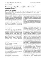
(A) Flow cytometry expression of CD14, TLR4 and TLR2 on monocytes and autologous alveolar macrophages (AM)Figure 1
(A) Flow cytometry expression of CD14, TLR4 and TLR2 on monocytes and autologous alveolar macrophages (AM). rMFI (±
SD) is shown from non-smokers (n = 10). White bars = monocytes, black bars = AM. * = p < 0.01 vs. AM. rMFI = relative
mean fluorescence intensity. (B) Representative histogram from experiments with monocytes.
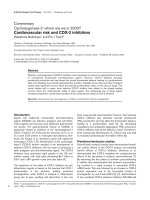
(A) Flow cytometry expression of TLR2 and TLR4 on alveolar macrophages (AM)Figure 2
(A) Flow cytometry expression of TLR2 and TLR4 on alveolar macrophages (AM). rMFI (± SD) is shown from non-smokers
(white bars [young], n = 10, horizontal hatched bars [elderly], n = 7), smokers (diagonal hatched bars, n = 10) and COPD
patients (black bars, n = 14). * = p < 0.01 vs. smokers and COPD patients AM. rMFI = relative mean fluorescence intensity. (B)
Representative histogram from experiments with AM.
Respiratory Research 2005, 6:68 />Page 5 of 8
(page number not for citation purposes)
difference in the PRR expression on monocytes from non-
smokers (A and B), smokers, COPD patients. Figure 1a
shows data from non-smokers (A), a representative histo-
gram demonstrates the ratio of isotype control to surface
molecule fluorescence (figure 1b). In addition there was
also a difference in the percentage of positive cells (mono-
cytes vs. AM, CD14: 96 vs. 9.5; TLR4: 69 vs. 11, TLR2: 87.5
vs. 22.5 % positive).
PRR expression of AM from non-smokers, smokers, COPD
patients
Comparing AM from non-smokers (A and B), smokers
and COPD patients we detected a markedly lower expres-
sion of TLR2 on AM from smokers and COPD patients
(13.98 ± 2.54 and 12.07 ± 3.5 [nonsmoker groups] vs.
6.59 ± 1.42 and 6.08 ± 1.5 rMFI, respectively, p < 0.01)
(figure 2a). There was no difference in the expression of
CD14 and TLR4 between the groups (CD14: 5 ± 1.56 and
4,7 ± 1,6 vs. 4.09 ± 0.69 and 4.3 ± 1.53 rMFI; TLR4: 5.19
± 1.58 and 5,3 ± 2,1 vs. 4.28 ± 0.7 and 4.62 ± 1.38 rMFI,
respectively). Percentage of positive cells showed analo-
gous data (nonsmokers (A and B) vs. smokers, COPD
patients, TLR2: 22.5 and 19.2 vs. 12.9 and 12.2; CD14: 9.5
and 8.5 vs. 8.1 and 8.7; TLR4: 11 and 12.5 vs. 8.9 and 10.3
% positive). A representative histogram demonstrates the
ratio of isotype control to surface molecule fluorescence
(figure 2b).
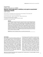
Regulation of PRR expression on AM
To study the regulation of PRR expression after ligand
stimulation, cells were exposed to LPS (1 µg/ml) which
led to an increased expression of TLR2 on AM from non-
smokers (18.35 ± 4.24 vs. 13.98 ± 2.54 rMFI [A] p < 0.04;
21.1 ± 6.23 vs. 12.07 ± 3.5 [B] p < 0.04). In contrast, cells
of smokers and COPD patients did not respond to LPS
stimulation with increased TLR2 expression (7.09 ± 1.42
vs. 6.59 ± 1.42 rMFI [smokers], p = n.s.; 6.63 ± 2.40 vs.
6.24 ± 1.71 rMFI, [COPD patients], p = n.s.) (figure 3a).
Percentage of positive cells showed analogous data (non-
smokers: 28 vs. 22.5 [A], 30.1 vs. 19.2 [B], smokers: 13.8
vs. 12.9, COPD patients: 13.0 vs. 12.2 % positive). There
was no effect of LPS stimulation on the surface expression
of CD14 and TLR4 in all groups. Representative results of
ISH targeting human TLR2-mRNA before and after LPS-
stimulation in non-smokers (A and B) and COPD patients
are photographically displayed in figure 3b–e. HOPE-
fixed specimens showed a good preservation of morphol-
ogy after the ISH procedure. The generation of signals was
achieved in approximately 10 minutes. Strong signals
were found in AM of non-smokers (A and B) after LPS
stimulation. Nonspecific signals were not detected in con-
trol preparations, in which specific DNA probes were sub-
stituted by hybridization buffer alone or an irrelevant
probe.
Discussion
In this study we comparatively evaluated the influence of
chronic smoke exposure on the pattern of TLR2, TLR4 and
CD14 expression in human AM and monocytes in COPD
patients, smokers and non-smokers. We observed a signif-
icantly decreased expression of PRR's on AM compared to
monocytes. The main finding was that AM from COPD
patients and smokers show an equally decreased surface
expression of TLR2 compared to non-smokers of two age
groups. In addition, an upregulation of TLR2 after LPS
stimulation was only observed on non-smokers AM.
An increased TLR2 surface expression on human mono-
cytes in response to LPS-stimulation has been described
previously [13], whereas on the transcriptional level diver-
gent data have been reported showing either upregulation
[14,15], or downregulation [16,17] of TLR2-mRNA
depending on timing and dose of stimulation. In addi-
tion, differential regulation of TLR2 by IL-1, IL-10 and
GM-CSF (upregulation) and IFN-gamma, TNF-α and IL-4
(downregulation) has been observed [13]. Therefore, the
net result of stimulation in vivo will depend on the local
balance of inflammatory mediators. What are the possible
consequences of LPS-stimulated TLR2 expression? Whole
gram-negative bacteria are recognized not only through
TLR4 (by LPS) but also TLR 2 (by bacterial lipopeptides).
In this setting upregulation of TLR2 which occurs mainly
with high LPS doses, may provide an additional mecha-
nism to sensitize cells against large microbial challenges.
Moreover, TLR2 is the main PRR recognizing gram-posi-
tive bacteria, and recent studies have shown that TLR2 -
deficient animals are at high risk for succumbing to inva-
sive pneumococcal infections [18]. Thus, the blunted
TLR2 expression of AM from smokers and COPD patients
after LPS stimulation may impair antimicrobial defenses
in the lower respiratory tract. Recently a reduced TLR
expression in aging mice was demonstrated [19]. However
this factor seems to be without influence on our results
since comparable levels of PRR expression on monocytes
and AM from healthy young and elderly non-smokers
were observed.
The mechanisms of the smoking induced alteration of
pulmonary immune functions are poorly understood. On
the one hand smoking is known to induce inflammatory
processes by activating AM and epithelial cells leading to
production of TNF-α, IL-8 and LTB4 and subsequent neu-
trophil recruitment [20]. Accordingly, in BALF and spu-
tum of patients with COPD neutrophil activation and
elevated levels of proinflammatory cytokines and chem-
okines have been found [21]. On the other hand a
depressed capacity for LPS-induced cytokine release of
TNF-α and IL-6 from AM of smokers was described [22].
Skold and colleagues found a higher expression of
CD11a, CD54 and CD71 in non-smoker's AM compared
Respiratory Research 2005, 6:68 />Page 6 of 8
(page number not for citation purposes)
with smokers [23]. CD11a (LFA-1) and its ligand play an
important role in the interaction between antigen present-
ing cells and T-lymphocytes. In addition the metabolic
response after in vitro stimulation with phorbol myristate
acetate (PMA) was higher in non-smokers than in smok-
ers AM. Dandrea et al. observed a reduced inflammatory
cytokine release in cultured AM from smokers in response
to LPS by simultaneous exposure to NO2 compared to
non-smokers [24]. Cultured human bronchial epithelial
cells from COPD patients release lower levels of inflam-
matory mediators such as TNF-α and IL-8 than similar
preparations from non-smokers or smokers without
COPD, suggesting that downregulation of inflammatory
mediator release may also occur in bronchial epithelial
cells of individuals with COPD [25].
Our data demonstrating an altered AM phenotype with
reduced expression of TLR2 in smokers and COPD
patients suggest that a continuous exposure to microbial
products in this disease provided by bacterial coloniza-
tion and LPS present in tobacco smoke [5] may down-
modulate the pulmonary immune response. Whether this
is due to the selection of a heterogenous macrophage sub-
population in the pulmonary compartment or to a gen-
eral AM phenotype change under the environmental
conditions described cannot be firmly differentiated from
our data. However, with regard to the continous distribu-
tion of TLR expression intensities found by flow cytome-
try the latter possibility seems more likely. In vitro it was
shown that the TLR response is downregulated after repet-
itive stimulation [26]. Interestingly a hyporesponsiveness
of cells to the TLR-4 ligand LPS was shown as well after
preincubation with ligands for TLR2 and vice versa, which
indicates the existence of common signaling pathways of
the TLR system [27]. It is tempting to speculate that this
phenomenon also plays a role under conditions of
chronic stimulation with bacterial components in vivo as
suggested by the missing effect of LPS-stimulation on
TLR2 expression on cells from smokers and COPD
patients. This finding was confirmed using in situ hybrid-
ization targeting TLR2, which was recently demonstrated
by our group on AM and alveolar epithelial cells type II in
the human lung [12]. Although there was only a small
overlap between non-smokers on the one hand and
LPS-stimulation of TLR2 protein and mRNA on alveolar macrophages (AM)Figure 3
LPS-stimulation of TLR2 protein and mRNA on alveolar macrophages (AM). (A) Flow cytometry expression. rMFI (± SD) is
shown from non-smokers (young n = 10, elderly n = 7), smokers (n = 10) and COPD patients (n = 14). rMFI = relative mean
fluorescence intensity. * = p < 0,04. In situ hybridization targeting TLR2 mRNA of HOPE-fixated BAL cells. Cells from non-
smokers before (B) and after (C [young], D [elderly]) LPS-stimulation. Cells from COPD patients (E) after LPS-stimulation
(Anti-DIG-AP-New-fuchsine; 600x).
Respiratory Research 2005, 6:68 />Page 7 of 8
(page number not for citation purposes)
smokers and COPD patients on the other we observed a
large variability in the extent of response to LPS stimula-
tion in the non-smoking group which is a well-known
phenomenon with regard to the release of biologic medi-
ators as proinflammatory cytokines [28]. In contrast, no
difference in TLR4 expression between smokers, non-
smokers, COPD patients was observed in our study which
may be due to the generally weaker expression of this mol-
ecule on monocytes and AM.
Functionally relevant polymorphisms of TLR's have been
found in persons with endotoxin hyporesponsiveness
(TLR-4) and staphylococcal sepsis (TLR-2) [29,30]. Data
regarding their relevance in COPD are not available. The
generally lower expression of PRR's on AM's compared to
monocytes is comparable to data reported for dendritic
cells and may accompany the differentiation process of
monocytes to macrophage populations [15]. Regarding
the effect of chronic smoke exposure on blood cells we did
not find any difference in the PRR expression on mono-
cytes between smokers and non-smokers (data not
shown) suggesting a compartmentalized effect of tobacco
smoking. In contrast Lauener et al. demonstrated a signif-
icantly increased expression of CD14 and TLR2 on blood
cells of farmers' children compared to non-farmers' chil-
dren [31].
In conclusion, the altered phenotype of smokers AM
could play a role in decreased cellular responses to micro-
bial stimulation facilitating persistent infection. Further
studies regarding to the functional relevance of these find-
ings and their contribution to the pathogenesis of COPD
could lead to more effective treatment regimens of this
disease.
Abbreviations
AM = alveolar macrophage; BAL = bronchoalveolar lav-
age; COPD = chronic obstructive pulmonary disease;
FACS = Fluorescence activated cell sorter; GM-CSF = gran-
ulocyte-macrophage colony-stimulating factor; HOPE =
Hepes-Glutamic acid buffer mediated Organic solvent
Protection Effect; IL = interleukin; IFN-γ = interferon-γ;
LTA = lipoteichoic acid; LPS = lipopolysaccharide; PBMC
= peripheral blood mononuclear cell; PE = phycoerythrin;
PGN = peptidoglycan; PMA = phorbol myristate acetate;
PMN = polymorphonuclear neutrophils; PRR = pattern
recognition receptor; rMFI = relative mean fluorescence
intensity; TLR = Toll-like receptor; TNF-α = tumor necrosis
factor-α
Authors' contributions
DD carried out the flow cytometry and was involved in
the design of the study and drafting the manuscript. TG
performed the in situ hybridization and conceived of the
study. TT carried out cell culture experiments and was
involved in drafting the manuscript. PZ, KD and BS con-
ducted the clinical part of the study and were involved in
the design and coordination of the study. All authors read
and approved the final manuscript.
Acknowledgements
The authors thank S. Ross, J. Hofmeister, H. Kühl and W. Martens for
excellent technical assistance.
References
1. Sethi S, Muscarella K, Evans N, Klingman KL, Grant BJ, Murphy TF, et
al.: Airway inflammation and etiology of acute exacerbations
of chronic bronchitis. Chest 2000, 118:1557-1565.
2. Donaldson GC, Seemungal TA, Bhowmik A, Wedzicha JA: Relation-
ship between exacerbation frequency and lung function
decline in chronic obstructive pulmonary disease. Thorax
2002, 57:847-852.
3. Prieto A, Reyes E, Bernstein ED, Martinez B, Monserrat J, Izquierdo
JL, Callol L, de Lucas P, Alvarez-Sala R, Alvarez-Sala JL, Villarrubia VG,
Alvarez-Mon M: Defective natural killer and phagocytic activi-
ties in chronic obstructive pulmonary disease are restored
by glycophosphopeptical (inmunoferon). Am J Respir Crit Care
Med 2001, 163:1578-1583.
4. White AJ, Gompertz S, Bayley DL, Hill SL, O'Brien C, Unsal I, Stockley
RA: Resolution of bronchial inflammation is related to bacte-
rial eradication following treatment of exacerbations of
chronic bronchitis. Thorax 2003, 58:680-685.
5. Hasday JD, Bascom R, Costa JJ, Fitzgerald T, Dubin W: Bacterial
endotoxin is an active component of cigarette smoke. Chest
1999, 115:829-835.
6. Imler JL, Hoffmann JA: Toll receptors in innate immunity. Trends
Cell Biol 2001, 11:304-311.
7. Dalhoff K, Braun J, Wiessmann KJ, Hollandt H, Marre R: Broncho-
scopic diagnosis of pneumonia with quantitative microbial
count determination. Dtsch Med Wochenschr 1990,
115:1459-1465.
8. Hallden G, Skold CM, Eklund A, Forslid J, Hed J: Quenching of
intracellular autofluorescence in alveolar macrophages per-
mits analysis of fluorochrome labelled surface antigens by
flow cytofluorometry. J Immunol Methods 1991, 142:207-214.
9. Goldmann T, Wiedorn KH, Kuhl H, Olert J, Branscheid D, Pechko-
vsky D, Zissel G, Galle J, Muller-Quernheim J, Vollmer E: Assess-
ment of transcriptional gene activity in situ by application of
HOPE-fixed, paraffin-embedded tissues. Pathol Res Pract 2002,
198:91-95.
10. Olert J, Wiedorn KH, Goldmann T, Kuhl H, Mehraein Y, Scherthan
H, Niketeghad F, Vollmer E, Muller AM, Muller-Navia J, HOPE fixa-
tion: a novel fixing method and paraffin-embedding tech-
nique for human soft tissues. Pathol Res Pract 2001, 197:823-826.
11. Umland O, Ulmer AJ, Vollmer E, Goldmann T: HOPE fixation of
cytospin preparations of human cells for in situ hybridization
and immunocytochemistry. J Histochem Cytochem 2003,
51:977-980.
12. Droemann D, Goldmann T, Branscheid D, Clark R, Dalhoff K, Zabel
P, Vollmer E: Toll-like receptor 2 is expressed by alveolar epi-
thelial cells type II and macrophages in the human lung. His-
tochem Cell Biol 2003, 119:103-108.
13. Flo TH, Halaas O, Torp S, Ryan L, Lien E, Dybdahl B, Sundan A, Espe-
vik T: Differential expression of Toll-like receptor 2 in human
cells. J Leukoc Biol 2001, 69:474-481.
14. Liu Y, Wang Y, Yamakuchi M, Isowaki S, Nagata E, Kanmura Y, Kita-
jima I, Maruyama I: Upregulation of toll-like receptor 2 gene
expression in macrophage response to peptidoglycan and
high concentration of lipopolysaccharide is involved in NF-
kappa b activation. Infect Immun 2001, 69:2788-2796.
15. Visintin A, Mazzoni A, Spitzer JH, Wyllie DH, Dower SK, Segal DM:
Regulation of Toll-like receptors in human monocytes and
dendritic cells. J Immunol 2001, 166:249-255.
16. Muzio M, Polentarutti N, Bosisio D, Prahladan MK, Mantovani A:
Toll-like receptors: a growing family of immune receptors
that are differentially expressed and regulated by different
leukocytes. J Leukoc Biol 2000, 67:450-456.
Publish with BioMed Central and every
scientist can read your work free of charge
"BioMed Central will be the most significant development for
disseminating the results of biomedical research in our lifetime."
Sir Paul Nurse, Cancer Research UK
Your research papers will be:
available free of charge to the entire biomedical community
peer reviewed and published immediately upon acceptance
cited in PubMed and archived on PubMed Central
yours — you keep the copyright
Submit your manuscript here:
/>BioMedcentral
Respiratory Research 2005, 6:68 />Page 8 of 8
(page number not for citation purposes)
17. Haehnel V, Schwarzfischer L, Fenton MJ, Rehli M: Transcriptional
regulation of the human toll-like receptor 2 gene in mono-
cytes and macrophages. J Immunol 2002, 168:5629-5637.
18. Echchannaoui H, Frei K, Schnell C, Leib SL, Zimmerli W, Landmann
R: Toll-like receptor 2-deficient mice are highly susceptible
to Streptococcus pneumoniae meningitis because of
reduced bacterial clearing and enhanced inflammation. J
Infect Dis 2002, 186:798-806.
19. Renshaw M, Rockwell J, Engleman C, Gewirtz A, Katz J, Sambhara S:
Cutting edge: impaired Toll-like receptor expression and
function in aging. J Immunol 2002, 169:4697-4701.
20. MacNee W: Oxidants/antioxidants and COPD. Chest 2000,
117:303S-317S.
21. Keatings VM, Collins PD, Scott DM, Barnes PJ: Differences in inter-
leukin-8 and tumor necrosis factor-alpha in induced sputum
from patients with chronic obstructive pulmonary disease or
asthma. Am J Respir Crit Care Med 1996, 153:530-534.
22. McCrea KA, Ensor JE, Nall K, Bleecker ER, Hasday JD: Altered
cytokine regulation in the lungs of cigarette smokers. Am J
Respir Crit Care Med 1994, 150:696-703.
23. Skold CM, Lundahl J, Hallden G, Hallgren M, Eklund A: Chronic
smokeexposure alters the phenotype pattern and the meta-
bolic response in human alveolar macrophages. Clin Exp
Immunol 1996, 106:108-113.
24. Dandrea T, Tu B, Blomberg A, Sandstrom T, Skold M, Eklund A, Cot-
greave I: Differential inhibition of inflammatory cytokine
release from cultured alveolar macrophages from smokers
and non-smokers by NO2. Hum Exp Toxicol 1997, 16:577-588.
25. Mills PR, Davies RJ, Devalia JL: Airway epithelial cells, cytokines,
and pollutants. Am J Respir Crit Care Med 1999, 160:S38-S43.
26. Medvedev AE, Kopydlowski KM, Vogel SN: Inhibition of lipopoly-
saccharide-induced signal transduction in endotoxin-toler-
ized mouse macrophages: dysregulation of cytokine,
chemokine, and toll-like receptor 2 and 4 gene expression. J
Immunol 2000, 164:5564-5574.
27. Sato S, Nomura F, Kawai T, Takeuchi O, Muhlradt PF, Takeda K, Akira
S: Synergy and cross-tolerance between toll-like receptor
(TLR) 2- and TLR4-mediated signaling pathways. J Immunol
2000, 165:7096-7101.
28. Zabel P, Schonharting MM, Schade UF, Schlaak M: Effects of pen-
toxifylline in endotoxinemia in human volunteers. Prog Clin
Biol Res 1991, 367:207-213.
29. Arbour NC, Lorenz E, Schutte BC, Zabner J, Kline JN, Jones M, Frees
K, Watt JL, Schwartz DA: TLR4 mutations are associated with
endotoxin hyporesponsiveness in humans. Nat Genet 2000,
25:187-191.
30. Lorenz E, Mira JP, Cornish KL, Arbour NC, Schwartz DA: A novel
polymorphism in the toll-like receptor 2 gene and its poten-
tial association with staphylococcal infection. Infect Immun
2000, 68:6398-6401.
31. Lauener RP, Birchler T, Adamski J, Braun-Fahrlander C, Bufe A, Herz
U, von Mutius E, Nowak D, Riedler J, Waser M, Sennhauser FH, ALEX
study group: Expression of CD14 and Toll-like receptor 2 in
farmers' and non- farmers' children. Lancet 2002, 360:465-466.