Báo cáo y học: "A role for MCP-1/CCR2 in interstitial lung disease in children" pps
Bạn đang xem bản rút gọn của tài liệu. Xem và tải ngay bản đầy đủ của tài liệu tại đây (443.84 KB, 12 trang )
BioMed Central
Page 1 of 12
(page number not for citation purposes)
Respiratory Research
Open Access
Research
A role for MCP-1/CCR2 in interstitial lung disease in children
Dominik Hartl
1
, Matthias Griese
1
, Thomas Nicolai
1
, Gernot Zissel
2
,
Christine Prell
1
, Dietrich Reinhardt
1
, Dolores J Schendel
3
and
Susanne Krauss-Etschmann*
1
Address:
1
Childrens' Hospital of the Ludwig-Maximilians-University, Munich, Germany,
2
Department of Pneumology, Medical Center, Albert-
Ludwigs-University, Freiburg, Germany and
3
Institute of Molecular Immunology and Immune Monitoring Platform, GSF National Research
Center for Environment and Health, Munich, Germany
Email: Dominik Hartl - ; Matthias Griese - ;
Thomas Nicolai - ; Gernot Zissel - ;
Christine Prell - ; Dietrich Reinhardt - ;
Dolores J Schendel - ; Susanne Krauss-Etschmann* -
* Corresponding author
ChemokinesMCP-1CCR2Bronchoalveolar LavageChildrenInterstitial Lung Diseases
Abstract
Background: Interstitial lung diseases (ILD) are chronic inflammatory disorders leading to
pulmonary fibrosis. Monocyte chemotactic protein 1 (MCP-1) promotes collagen synthesis and
deletion of the MCP-1 receptor CCR2 protects from pulmonary fibrosis in ILD mouse models. We
hypothesized that pulmonary MCP-1 and CCR2
+
T cells accumulate in pediatric ILD and are related
to disease severity.
Methods: Bronchoalveolar lavage fluid was obtained from 25 children with ILD and 10 healthy
children. Levels of pulmonary MCP-1 and Th1/Th2-associated cytokines were quantified at the
protein and the mRNA levels. Pulmonary CCR2
+
, CCR4
+
, CCR3
+
, CCR5
+
and CXCR3
+
T cells
were quantified by flow-cytometry.
Results: CCR2
+
T cells and MCP-1 levels were significantly elevated in children with ILD and
correlated with forced vital capacity, total lung capacity and ILD disease severity scores. Children
with lung fibrosis had significantly higher MCP-1 levels and CCR2
+
T cells in bronchoalveolar lavage
fluid compared to non-fibrotic children.
Conclusion: The results indicate that pulmonary CCR2
+
T cells and MCP-1 contribute to the
pathogenesis of pediatric ILD and might provide a novel target for therapeutic strategies.
Background
Interstitial lung diseases (ILD) are chronic inflammatory
disorders characterized by restrictive lung disease and dif-
fuse pulmonary infiltrates. Although the precise incidence
is not known, ILD are less frequent in children than adults
[1-3]. Lungs of ILD patients show inflammation with alve-
olar wall thickening by leukocytes and pulmonary fibro-
sis. Despite immunosuppressive treatment and
Published: 11 August 2005
Respiratory Research 2005, 6:93 doi:10.1186/1465-9921-6-93
Received: 19 April 2005
Accepted: 11 August 2005
This article is available from: />© 2005 Hartl et al; licensee BioMed Central Ltd.
This is an Open Access article distributed under the terms of the Creative Commons Attribution License ( />),
which permits unrestricted use, distribution, and reproduction in any medium, provided the original work is properly cited.
Respiratory Research 2005, 6:93 />Page 2 of 12
(page number not for citation purposes)
supportive measures, the progressive course leading to
irreversible lung fibrosis sometimes can not be prevented.
Therefore, the development of additional therapeutic
strategies is of high importance.
Monocyte chemotactic protein 1 (MCP-1, CCL2) is pro-
duced in response to inflammatory stimuli by a variety of
cells, including monocytes/macrophages, lymphocytes
and airway epithelial cells [4-6]. MCP-1 stimulates colla-
gen synthesis and production of the pro-fibrotic factor
transforming growth factor β (TGF-β) in fibroblasts, while
MCP-1 antisense oligonucleotides reduce TGF-β produc-
tion[7,8]. Application of MCP-1 into murine lungs
induces an inflammatory cytokine response and pulmo-
nary leukocyte accumulation. In adult patients with ILD,
increased levels of MCP-1 were observed in serum[9,10]
and bronchoalveolar lavage fluid (BALF) [11-14].
Although MCP-1 was originally described for its chemo-
tactic activity on monocytes, in vitro studies revealed an
even higher activity on T cells[15]. This occurs through
MCP-1 binding to its sole receptor CCR2[16]. Deletion of
the CCR2-gene or receptor blockade with anti-CCR2 anti-
bodies leads to a dramatic inhibition of leukocyte accu-
mulation in murine lungs[17]. Furthermore, CCR2-/-
mice are protected from fluorescein (FITC) or bleomycin
induced lung fibrosis[18]. Thus far, CCR2
+
T cells in BALF
of patients with fibrotic lung diseases have not been
determined.
In addition to the MCP-1/CCR2 axis, Th2 cytokines seem
to mediate pulmonary fibrosis [19-22]. IL-4 stimulates
fibroblast proliferation and collagen synthesis[23,24],
while IFN-γ inhibits this process [25-28]. In a Th2 mouse
model fibroblasts expressed more CCR2 protein and
higher levels of MCP-1 and TGF-β as compared to fibrob-
lasts from a Th1-mouse model[8]. Furthermore, increased
levels of IL-4 were observed in animal models of pulmo-
nary fibrosis[29] and lungs of patients with idiopathic
pulmonary fibrosis (IPF)[30] or cryptogenic fibrosing
alveolitis[31].
The contribution of MCP-1 to ILD has been investigated
exclusively in adults. However, the spectrum of ILD differs
considerably between adults and children and some
forms are unique to children while others, such as idio-
pathic pulmonary fibrosis (IPF), are extremely rare in
childhood[32].
Therefore, we asked whether levels of MCP-1 and frequen-
cies of CCR2
+
T cells are increased in BALF of children
with ILD and, if so, how levels of MCP-1 and CCR2
+
T
cells relate to disease severity in pediatric ILD.
To address these questions levels of MCP-1 and frequen-
cies of CCR2
+
T cells in BALF were compared between chil-
dren with ILD and children without lung disease.
To evaluate the contribution of the pulmonary Th1/Th2
micromilieu to the pathogenesis of pediatric ILD, CCR4
+
and CCR3
+
(Th2) and CCR5
+
and CXCR3
+
(Th1) cells
were determined in BALF together with an array of pulmo-
nary Th1- and Th2-associated cytokines.
Our results indicate that pulmonary CCR2
+
T cells and lev-
els of MCP-1 are characteristic components in BALF of
children with ILD. A pathophysiological role in pediatric
ILD seems likely as their levels relate to restrictive lung
function and ILD disease severity.
Methods
Characterization of the patients
Children attending the Department of Pulmonology and
Allergology of the University Children's Hospital of
Munich during 1999–2004 were considered for inclusion
in this study. Children suspective of ILD underwent a
comprehensive clinical evaluation, including patient his-
tory, physical examination, routine laboratory tests, lung
function testing, chest radiography, high resolution com-
puted tomography (HRCT) and bronchoalveolar lavage
(BAL). Children were assigned to the ILD group according
to the criteria of Fan[33]: (i) ≥3 months of respiratory
symptoms characteristic for ILD, i.e. non-productive
cough, dyspnoea, tachypnea, crackles and/or rales, exer-
cise intolerance and/or hypoxemia, (ii) diffuse infiltrates
on chest radiographs and HRCT and (iii) restrictive lung
function (decreased forced vital capacity (FVC) and total
lung capacity (TLC)) according to the ATS criteria[34].
The diagnosis of the specific form of ILD was established
by patient history, physical examination, HRCT, BAL and/
or lung biopsy according to consensus criteria[33,35].
Two thoracic radiologists independently evaluated all
lobes on HRCT for ground glass opacity and pulmonary
fibrosis as described previously[36,37]. A pathologist spe-
cialized on pediatric ILD[38] evaluated the lung sections
systematically[39,40]. Furthermore, the disease severity of
each ILD patient was characterized using the clinical ILD
score of Fan[41]: 1 = asymptomatic, no desaturation; 2 =
symptomatic but normoxic (>90%) under all conditions;
3 = symptomatic with desaturation during sleep or with
exercise; 4 = symptomatic with desaturation at rest. None
of the included children had familial idiopathic pulmo-
nary fibrosis. Patients with congenital heart disease or sus-
pected or proven bacterial pulmonary infection were
excluded from the study.
Twenty-five children with ILD (median age: 7 ± 3.6 years;
male/female = 16/9) were included (Table 1).
Respiratory Research 2005, 6:93 />Page 3 of 12
(page number not for citation purposes)
Ten age-matched children were selected as the control
group (median age: 7.5 ± 2.9 years, m/f: 6/4). These chil-
dren were considered as healthy, i.e. had no systemic dis-
ease, had no suspected or proven pulmonary disease and
were free of respiratory tract infections. These children
underwent elective tonsillectomy under general anaesthe-
sia. BAL was performed prior to the surgical procedure.
Ten age-matched children with chronic severe asthma
(median age: 8.7 ± 1.6 years, m/f: 5/5), from a previous
study[42], who were comparable to the ILD group in
terms of gender and age were included as disease control
group. All parents and/or patients gave their informed
consent prior to bronchoscopy and the institutional
review board approved the study protocol.
Bronchoalveolar lavage
Bronchoscopy with BAL was performed as described pre-
viously[43]. Residual BALF cells were used for flow cytom-
etry. The BALF recovery and the viability of cells did not
differ significantly between the patient groups. Cellular
profiles are shown in Table 2.
Flow cytometry
BALF cells were analyzed by four-colour flow cytometry
(FACSCalibur, Becton-Dickinson, Heidelberg, Germany)
as described previously[42]. The following antibodies
were used: CD4-allophycocyanine (APC) mouse IgG1,
CD8-phycocyanine 5 (PC5) mouse IgG1 (Immunotech,
Marseille, France), CD69-PE mouse IgG1, CCR5-PE
mouse IgG2a, CCR4-PE mouse IgG2a (BD Pharmingen,
Heidelberg, Germany), CCR2-PE mouse IgG2b, CXCR3-
Table 1: Patients' characteristics
No Sex Age
[years]
Interstitial
lung disease
Diagnosis
finding
Radiographic findings Fibrotic
changes
(CT)
ILD
Score*
Dyspnoe Cough Cyanosis Exercise
Intolerance
Failure to
thrive
Medication FVC
[% of pred.]
TLC %
[of pred.]
1 F 7 LIP CT, LB • diffuse interstitial involvement + 4 ++ + + + + CS, AZT 34 56
• reticular-nodular pattern
• follicular bronchiolitis
2 M 14 U-ILD, IPH CT, BAL patchy interstitial involvement - 2 + - - - - CS 77 89
3 M 8 U-ILD CT, LB • ground-glass opacity + 3 ++ - - + - 46 74
4 M 4 IPH CT, BAL, LB interstitial involvement - 3 + - - - - 77 168
5 M 16 U-ILD CT, BAL interstitial involvement + 2 + - - + + 76 95
6 F 7 U-ILD CT, BAL interstitial involvement - 2 + + - - + AZT 50 68
7 M 4 CPI CT, LB • diffuse infiltrates + 3 + - - + + AZT 58 64
• ground-glass opacity
8 F 3 NSIP CT, LB • interstitial involvement + 3 ++ - + + + CS n.d. n.d.
• alveolar infiltrates
9 M 8 Sarcoidosis CT, BAL, LB • interstitial involvement + 2 ++ + - + + CS 56 63
• perivascular nodules
10 F 8 U-ILD CT, BAL, LB ground-glass opacity - 1 - + - + - 76 87
11 F 8 CPI CT, LB • interstitial involvement + 2 + + - + + CS 37 74
• ground-glass opacity
12 M 9 U-ILD CT interstitial involvement - 2 + - - - - 70 98
13 M 5 NSIP CT, LB • interstitial involvement + 3 ++ - - + - CS 61 76
• ground-glass opacity
14 F 6 U-ILD CT reticular-nodular pattern + 3 ++ + - - - AZT 60 68
15 F 4 U-ILD CT interstitial involvement + 2 + + - - + n.d. n.d.
16 M 12 U-ILD CT interstitial involvement - 2 + - - - - 68 75
17 M 3 PAP† CT, BAL, LB • ground glass opacity - 4 +++ + + + + CS n.d. n.d.
18 M 6 NSIP CT, BAL, LB • alveolar infiltrates + 4 +++ + + + + CS, AZT 63 72
PAP • ground glass opacification
19 F 3 PAP† CT, BAL, LB • ground glass opacity + 4 ++ - + + + CS n.d. n.d.
• alveolar infiltrates
20 F 9 NSIP CT, LB • interstitial involvement + 3 ++ + + + + CS, AZT 55 74
• honeycombing
21 M 7 U-ILD CT reticular-interstitial pattern + 3 + + - + + AZT, MT 38 59
22 M 7 Cholesterol CT, BAL, LB • interstitial involvement + 4 +++ + + + + CS 16 24
pneumonitis† • reticular-interstitial pattern
23 M 4 U-ILD CT, LB • interstitial involvement - 2 + - - + + CS 102 99
• honeycombing
24 M 8 U-ILD CT interstitial involvement - 2 + + - + - CS 63 78
25 M 7 NSIP CT, LB • interstitial involvement + 3 + + - + - CS 60 76
ILD-NC: children with interstitial lung disease without systemic corticosteroid treatment; ILD-C: children with interstitial lung disease with systemic
corticosteroid treatment;
U-ILD: undefined/idiopathic interstitial lung disease: no specific diagnosis could be made; PAP: pulmonary alveolar proteinosis; CGD: chronic
granulomatous disease; IPH: idiopathic pulmonary hemosiderosis; LIP: lymphocytic interstitial pneumonia; CPI: Chronic pneumonitis of infancy
CS: corticosteroids, AZT: azathioprine, MT: methotrexat
n.d.: lung function testing not done (children < 5 years); † symbolizes patients who died due to respiratory failure.
CT: Computed tomography; BAL: Bronchoalveolar lavage; LB: Lung biopsy
* ILD score according to Fan[41]
Respiratory Research 2005, 6:93 />Page 4 of 12
(page number not for citation purposes)
fluorescein isothiocyanate (FITC) mouse IgG1 and CCR3-
FITC rat IgG2a (R&D Systems, Wiesbaden, Germany).
Mouse IgG1-FITC, mouse IgG1-PE, mouse IgG2a-PE,
mouse IgG2b-PE (Immunotech, Marseille, France) and rat
IgG2a-FITC (kindly provided by Dr. E. Kremmer, GSF-
Institute of Molecular Immunology, Munich, Germany)
were used as isotype controls.
Detection of MCP-1 and cytokines
Levels of MCP-1 and Th1 (IL-2, IFN-γ), Th2 (IL-4, IL-5, IL-
10) and pro-inflammatory cytokines (TNF-α, IL-6) were
quantified by a multiplex, particle-based assay (Bio-Rad
Laboratories, Minneapolis, USA) as described previ-
ously[42]. The detection limits for all cytokines were 1.5–
2.5 pg/ml (min.) and 1000 pg/ml (max.).
Quantitative RT-PCR
BALF cells were lysed in Trizol LS Reagent (Invitrogen, Life
Technologies, Karlsruhe, Germany) and were stored at -
20°C until mRNA extraction. Total mRNA was isolated
according to the manufacturer's instructions and reverse
transcribed into cDNA. Contamination with genomic
DNA was excluded by mRNA controls without reverse
transcriptase in the cDNA synthesis reaction. The follow-
ing oligonucleotide primers were used: MCP-1 (5-
TGAAGCTCGCACTCTCGCCT-3; 5- GTGGAGTGAGTGT-
TCAAGTC-3); and GAPDH (5-GAGGTGAAGGTCG-
GAGTC-3; 5-AAGATGGTGATGGGATTTC-3). Expression
levels were determined in duplicates by Real time RT-PCR
using SYBR green and the iCycler iQ detection system
(Biorad, Hercules, CA, USA) according to the
manufacturer's instructions. Threshold cycle (CT) values
for genes of interest were normalized to GAPDH and used
to calculate the relative mRNA expression.
Statistical analysis
The non-parametric Mann-Whitney U test was applied.
Correlations were tested with Spearman's rank correlation
test. A probability of p < 0.05 was regarded as signifi-
cant[44] (SPSS statistical program, version 11.5, SPSS Inc.
Chicago, USA).
Results
MCP-1 levels and CCR2
+
T cells in BALF
Levels of MCP-1 were significantly higher in children with
ILD (n = 25) as compared to the control group at protein
and mRNA level (Figure 1A, B). MCP-1 protein and
mRNA expression levels correlated positively with each
other (r = 0.72, p < 0.01). ILD children with pulmonary
fibrosis had significantly higher MCP-1 levels in BALF as
compared to children with non-fibrotic ILD (Figure 1C).
MCP-1 levels related positively to the stage of disease (Fig-
ure 1D). The highest levels of MCP-1 were observed in the
three patients who died after respiratory failure (Table 1;
P17, P19, P22). Furthermore, MCP-1 levels correlated
negatively with restrictive lung function parameters (TLC,
FVC) (Figures 2A, B).
To test whether increased MCP-1 levels are associated with
increased frequencies of CCR2
+
T cells, BALF lymphocytes
were quantified by flow cytometry. CCR2 was expressed
on a higher percentage of CD4
+
than CD8
+
T cells. The
majority of CCR2
+
T cells showed an activated phenotype
(75% CCR2
+
CD69
+
). Children with ILD had significantly
higher percentages of CCR2
+
CD4
+
and CCR2
+
CD8
+
T cells
Table 2: Bronchoalveolar lavage cells
ILD-NC ILD-C Control
Total cells × 10
3
/ml 230 (2.1–1124)** 144 (11–268)* 89 (83–97)
Recovery (%) 55 (25–86) 49 (34–75) 54 (35–70)
Neutrophils (%) 10.5 (1–44)* 8.5 (3–30)* 2 (0–3)
Eosinophils (%) 1 (0–6) 1.5 (0–3) 0 (0–1)
Mast cells (%) 2 (0–43) 2 (1–4) 0 (0-0)
Plasma cells (%) 0 (0–4) 0 (0–4) 0 (0-0)
Macrophages (%) 60 (7–97)* 49 (26–77)* 94 (81–92)
Lymphocytes (%) 24 (2–54)** 22 (5–34)** 4 (2–13)
CD4
+
T cells (%)
†
23 (9–45) 29 (9–82) 23 (15–28)
CD8
+
T cells (%)
†
29 (6–62) 27 (2–83) 25 (15–31)
CD4/8 ratio 0.7 (0.3–6) 1.1 (0.1–55) 0.7 (0.4–0.9)
results are expressed as medians with ranges shown in parenthesis.
ILD-NC: children with interstitial lung disease without systemic corticosteroid treatment;
ILD-C: children with interstitial lung disease with systemic corticosteroid treatment;
*p < 0.05, **p < 0.01 as compared to the control group, Mann-Whitney-U Test.
Total cells and differential cell count were obtained from cytospin slides, CD4
+
, CD8
+
and CD4/CD8 T cells using flow cytometry.
†
CD4
+
T cells and CD8
+
T cells are shown as the percentage of total lymphocytes in BALF, i.e. cells gated in the lymphocyte population.
Neutrophils, eosinophils, mast cells, plasma cells, macrophages and lymphocytes are shown as percentage of total cells in BALF.
Respiratory Research 2005, 6:93 />Page 5 of 12
(page number not for citation purposes)
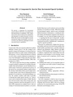
MCP-1 levels in children with ILDFigure 1
MCP-1 levels in children with ILD. MCP-1 levels in bronchoalveolar lavage fluid (BALF) of children with interstitial lung dis-
eases (ILD) and healthy controls are shown at the (A) protein and at the (B) mRNA level. (C) MCP-1 levels in BALF of ILD chil-
dren with and without pulmonary fibrosis. Pulmonary fibrosis was assessed by computed tomography according to [36,37]. (D)
MCP-1 levels in ILD children related to ILD disease severity according to the criteria of Fan [33]. 1 = asymptomatic, no desat-
uration; 2 = symptomatic but normoxic (> 90%) under all conditions; 3 = symptomatic with desaturation during sleep or exer-
cise; 4 = symptomatic with desaturation at rest; MCP-1 protein levels were quantified in BALF by a multiplex, particle-based
assay (Bio-Rad Laboratories, Minneapolis, USA) as described previously [42]. MCP-1 mRNA levels were quantified in BALF
cells by Real time RT-PCR using SYBR green and the iCycler iQ detection system (Biorad, Hercules, CA, USA) and were nor-
malized to GAPDH. Median values are shown by horizontal bars. Differences between the patient groups were tested with the
Mann-Whitney U test; * p < 0.05, *** p < 0.001; Children with systemic corticosteroid therapy are shown as grey circles. P:
Pulmonary alveolar proteinosis; S: Sarcoidosis; † symbolize children who died due to respiratory failure.
Respiratory Research 2005, 6:93 />Page 6 of 12
(page number not for citation purposes)
in BALF as compared to control children (Figure 3A). Sim-
ilar to MCP-1, percentages of CCR2
+
CD4
+
cells were sig-
nificantly higher in ILD children with pulmonary fibrosis
as compared to children with non-fibrotic ILD (Figure
3B). Again, the highest percentages of CCR2
+
CD4
+
T cells
were observed in the three deceased patients and
CCR2
+
CD4
+
cells related positively to the stage of ILD
(Figure 4). Furthermore, percentages of CCR2
+
CD4
+
T
cells correlated negatively with FVC and TLC in ILD
patients (Figures 5A, B). Pulmonary levels of MCP-1 cor-
related positively with CCR2
+
CD4
+
T cells (Figure 5C). No
association between MCP-1/CCR2
+
cells and immuno-
suppressive treatment was found in ILD patients.
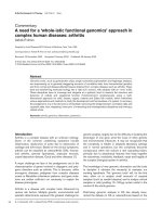
Correlation of MCP-1 levels with lung function parameters in children with ILDFigure 2
Correlation of MCP-1 levels with lung function
parameters in children with ILD. MCP-1 levels in bron-
choalveolar lavage fluid (BALF) correlated with (A) forced
vital capacity (FVC) and (B) total lung capacity (TLC) in chil-
dren with interstitial lung disease (ILD). FVC and TLC are
shown as % of predicted. MCP-1 levels in BALF were quanti-
fied by a multiplex, particle-based assay; P: Pulmonary alveo-
lar proteinosis; S: Sarcoidosis;
CCR2
+
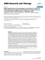
T cells in children with ILDFigure 3
CCR2
+
T cells in children with ILD. (A) Percentages of
CCR2
+
CD4
+
and CCR2
+
CD8
+
T cells in in bronchoalveolar
lavage fluid (BALF) of children with interstitial lung diseases
(ILD) and healthy children. (B) Percentages of CCR2
+
CD4
+
and CCR2
+
CD8
+
T cells in BALF of children with and with-
out pulmonary fibrosis. Percentages of CCR2
+
CD4
+
and
CCR2
+
CD8
+
T cells were analyzed in BALF by flow cytome-
try. Pulmonary fibrosis was assessed by computed tomogra-
phy according to [36,37]. Median values are shown by
horizontal bars. Differences between the patient groups
were tested with the Mann-Whitney U test; * p < 0.05; *** p
< 0.001; Children with systemic corticosteroid therapy are
shown as grey circles. P: Pulmonary alveolar proteinosis; S:
Sarcoidosis; † symbolize the children who died due to respi-
ratory failure.
Respiratory Research 2005, 6:93 />Page 7 of 12
(page number not for citation purposes)
To verify if increased levels of MCP-1 and percentages of
CCR2
+
T cells are characteristic for pediatric ILD, we ana-
lyzed these markers in ten selected age-matched children
with well-characterized allergic asthma who are described
in detail in a previous study[42]. Levels of MCP-1 and
CCR2
+
T cells from asthmatic children were in the range of
the control group and did not correlate with each other
(data not shown).
To assess the value of CCR2
+
CD4
+
T cells and MCP-1 lev-
els in the longitudinal course, three consecutive therapeu-
tical BALs were analyzed in three patients with PAP (P17,
P18, P19) and one patient with cholesterol pneumonitis
(P22). Two PAP patients (P17, P19) and the patient with
cholesterol pneumonitis worsened in the clinical course
continuously (increasing oxygen requirement, increasing
dyspnoe) and died from respiratory failure, while one
PAP patient remained clinically stable (P18). The
deceased PAP patients had continuously rising levels of
MCP-1 and increasing percentages of CCR2
+
CD4
+
T cells
in the three follow-up BALs (Figures 6A, B; black circles)
while the clinically stable patient showed steady levels of
CCR2
+
CD4
+
T cells and ILD disease severityFigure 4
CCR2
+
CD4
+
T cells and ILD disease severity. Percent-
ages of CCR2
+
CD4
+
T cells in bronchoalveolar lavage fluid
(BALF) of children with interstitial lung disease (ILD) related
to ILD disease severity. Percentages of CCR2
+
CD4
+
T cells
were analyzed in BALF by flow cytometry. ILD disease sever-
ity was scored according to the ILD score of Fan(40): 1 =
asymptomatic, no desaturation; 2 = symptomatic but nor-
moxic (> 90%) under all conditions; 3 = symptomatic with
desaturation during sleep or with exercise; 4 = symptomatic
with desaturation at rest; Median values are shown by hori-
zontal bars. Differences between the patient groups were
tested with the Mann-Whitney U test; * p < 0.05, ** p < 0.01;
Children with systemic corticosteroid therapy are shown as
grey circles. P: Pulmonary alveolar proteinosis; S: Sarcoidosis;
† symbolize the children who died due to respiratory failure.
CCR2
+
CD4
+
T cells and lung function parameters in children with ILDFigure 5
CCR2
+
CD4
+
T cells and lung function parameters in
children with ILD. Correlation of CCR2
+
CD4
+
T cells in
bronchoalveolar lavage fluid (BALF) with (A) forced vital
capacity (FVC) and (B) total lung capacity (TLC) in children
with interstitial lung diseases (ILD). Correlation of percent-
ages of CCR2
+
CD4
+
T cells with levels of MCP-1 in BALF of
children with ILD (C). FVC and TLC are shown as % of pre-
dicted. Percentages of CCR2
+
CD4
+
T cells were analyzed in
BALF by flow cytometry. P: Pulmonary alveolar proteinosis;
S: Sarcoidosis
Respiratory Research 2005, 6:93 />Page 8 of 12
(page number not for citation purposes)
MCP-1 and percentages of CCR2
+
CD4
+
T cells (Figures 6A,
B; white circles).
Th1- and Th2-lymphocytes and cytokines in BALF
To test whether increased CCR2
+
T cells and levels of
MCP-1 were paralleled by a pulmonary Th1/Th2-shift,
CCR4
+
and CCR3
+
(Th2) and CCR5
+
an d CXCR3
+
(Th1)
cells were determined in BALF together with an array of
pulmonary Th1/Th2 cytokines.
Children with ILD had significantly higher percentages of
CCR4
+
CD4
+
(Th2) cells as compared to control children
(Figure 7A). CCR4 was predominantly expressed on CD4
+
cells. The majority of CCR4
+
CD4
+
cells had an activated
phenotype (68% CCR4
+
CD69
+
). CCR3
+
(Th2) cells were
not detectable in BALF. Percentages of CCR5
+
and CXCR3
+
T cells (both Th1) were low and did not differ among the
patient groups (Figures 7B, C).
Levels of IFN-γ were increased in ILD patients (p < 0.05),
whereas the remaining cytokines did not differ among the
patient groups (data not shown).
Discussion
The present work demonstrates that BALF levels of MCP-
1 are consistently increased in pediatric ILD. This is
accompanied by increased frequencies of the correspond-
ing CCR2
+
T cells. Levels of MCP-1 and frequencies of
CCR2
+
T cells were higher in fibrotic than in non-fibrotic
forms of ILD and correlated with restrictive lung function
parameters and ILD disease severity, indicating a rele-
vance of the MCP-1/CCR2 axis in the pathogenesis of
pediatric ILD. Infiltrating T cells are a characteristic feature
of pulmonary tissue from ILD patients[45] and T cells in
BALF were found to correlate with T cells in pulmonary
tissue[46]. In line with previous findings[47,48], T cells
were increased in BALF of our children with ILD as com-
pared to control patients, suggesting a contribution of T
cells to the pathogenesis of pediatric ILD. Studies in adult
patients indicated that MCP-1 plays a role in the patho-
genesis of different forms of ILD, including IPF[9,12,13],
PAP[11,14], sarcoidosis[12], scleroderma with lung
involvement[49] and granulomatous lung diseases[50].
Serum levels of MCP-1 were significantly elevated in adult
patients with ILD[9,51] and were closely related to the
clinical course[9]. However, as outlined above, ILD in
children differs noticeably from ILD in adulthood. Pediat-
ric ILD is extremely rare and little data exist with respect to
pathoimmunological mechanisms. Thus, it is very hard to
study a large patient group and to find enough children
for each ILD subtype. We found elevated levels of MCP-1
and CCR2
+
T cells in various etiologies of ILD, which
suggests a common pulmonary T cell response for various
forms of pediatric ILD.
Thus far, frequencies of BALF CCR2
+
T cells in human ILD
have not been determined. The parallel increase of MCP-
1 and CCR2
+
T cells in BALF of ILD children further sub-
stantiates the importance of this chemokine and its recep-
tor in the pathogenesis of ILD, as suggested by mouse
models. In these models, the relevance of the MCP-1/
CCR2 interaction was mainly addressed with respect to
pulmonary fibrosis. Our children with pulmonary fibrosis
had increased levels of MCP-1 and increased percentages
of CCR2
+
cells compared to children with non-fibrotic
Longitudinal analysis of MCP-1 levels and CCR2
+
CD4
+
T cellsFigure 6
Longitudinal analysis of MCP-1 levels and
CCR2
+
CD4
+
T cells. Longitudinal analysis of (A) MCP-1
levels and (B) CCR2
+
CD4
+
T cells in three consecutive bron-
choalveolar lavage fluids (BALF) of four children with intersti-
tial lung diseases, including two children with pulmonary
alveolar proteinosis (P) and one child with cholesterol pneu-
monitis (CP). The child with cholesterol pneumonitis and
one child with pulmonary alveolar proteinosis died by respi-
ratory failure (†), while one child with pulmonary alveolar
proteinosis stayed clinically stable. † symbolize the childen
who died. MCP-1 levels were quantified in BALF by a multi-
plex, particle-based assay. Percentages of CCR2
+
CD4
+
T
cells were analyzed in BALF by flow cytometry.
Respiratory Research 2005, 6:93 />Page 9 of 12
(page number not for citation purposes)
ILD. However, MCP-1 levels and percentages of CCR2
+
T
cells were elevated both in fibrotic and non-fibrotic ILD
children as compared to controls. In addition, MCP-1 and
CCR2
+
T cells were also elevated in pediatric PAP that usu-
ally does not progress to pulmonary fibrosis. In fact, one
of the three patients with the highest levels of MCP-1 and
CCR2
+
T cells had PAP without any indication of fibrosis.
Similar observations were made recently for MCP-1 in
adult PAP patients[11]. A possible biological relevance of
MCP-1 levels and CCR2
+
T cells in pediatric ILD is further
suggested by their correlation with restrictive lung func-
tion parameters and the ILD disease severity score and by
the finding that the deceased children with the most
severe course of disease exhibited the highest BALF levels
of these markers. The possibility that MCP-1 and CCR2
+
T
cells are a general phenomenon of pediatric lung diseases
seems very unlikely, since these markers were present only
at low levels in BALF of children with severe allergic
asthma. This is in line with findings in an Aspergillus-
induced allergic mouse model, where a Th2-mediated
lung pathology occured in the absence of MCP-1 or
CCR2[52].
To assess the value of CCR2
+
T cells and MCP-1 levels in
the longitudinal course of children with ILD, three con-
secutive BALs were performed in four children with ILD
including three ILD patients who died and one patient
who stayed clinically stable. The three deceased children
had high and continuously rising levels of MCP-1 and
CCR2
+
CD4
+
T cells, while the stable patient had low levels
of MCP-1 and percentages of CCR2
+
CD4
+
T cells. Thus,
levels of MCP-1 and percentages of CCR2
+
CD4
+
T cells
might reflect the disease progression in pediatric ILD.
Interestingly, immunosuppressive treatment was not
associated with altered levels of MCP-1 and CCR2
+
T cells
in BALF (data not shown). This is in contrast to a study of
Suga et al.[9] in adult ILD patients where serum levels of
MCP-1 were closely related to the effectiveness of corticos-
teroid therapy. Given the assumption that MCP-1 and
CCR2 are important players in the pathophysiology of
ILD in children, the lack of association with corticosteroid
therapy might explain, at least in part, why corticosteroids
are sometimes unable to control the progression of pedi-
atric ILD.
Several studies indicated that MCP-1 and CCR2 are
involved in Th1[53,54] and Th2 immunity [55-58]. Fur-
thermore, it has been suggested that ILD and pulmonary
fibrosis are associated with a Th2 immune response[20-
22,59-61]. Experiments in mice showed that a lack of
MCP-1[62] leads to decreased Th1 responses while MCP-
1 over-expression[58] results in increased levels of Th2
cytokines. Th1/Th2 cytokine levels in BALF were low or
undetectable in BALF of our children. However,
CCR4
+
CD4
+
T cells were moderately but significantly ele-
vated in ILD patients. On the other hand, CCR4
+
CD4
+
T
cells are clearly less frequent in ILD compared to allergic
Pulmonary CCR4
+
, CCR5
+
, and CXCR3
+
T cellsFigure 7
Pulmonary CCR4
+
, CCR5
+
, and CXCR3
+
T cells. Per-
centages of (A) CCR4
+
CD4
+
, CCR4
+
CD8
+
, (B) CCR5
+
CD4
+
and CCR5
+
CD8
+
and (C) CXCR3
+
CD4
+
and CXCR3
+
CD8
+
T cells in bronchoalveolar lavage fluid (BALF) are shown in
children with interstitial lung diseases (ILD) and healthy con-
trols. Percentages of CCR4
+
CD4
+
, CCR4
+
CD8
+
,
CCR5
+
CD4
+
, CCR5
+
CD8
+
, CXCR3
+
CD4
+
and
CXCR3
+
CD8
+
T cells were analyzed in BALF by flow cytom-
etry. Median values are shown by horizontal bars. Differ-
ences between the patient groups were tested with the
Mann-Whitney U test; * p < 0.05; ** p < 0.01
Respiratory Research 2005, 6:93 />Page 10 of 12
(page number not for citation purposes)
asthma[42]. Thus, a strong Th2 response seems unlikely
in our ILD patients. Beneath T-cells, MCP-1 attracts
CCR2
+
monocytes/macrophages[63]. In mouse models,
MCP-1 was found to attract monocytes to the inflamed
lung, which was accompanied by a concomitant downreg-
ulation of pulmonary MCP-1 levels[64]. We found no
difference in the percentage of CCR2
+
alveolar macro-
phages in BALF between children with ILD and control
children or between fibrotic and non-fibrotic forms of ILD
(data not shown). Instead, we found a strong correlation
between percentages of CCR2
+
T cells and levels of MCP-
1 in BALF of ILD patients. Therefore, we assume that pul-
monary MCP-1 acts on CCR2
+
T cells, which accumulate
in the BALF of children with ILD.
Conclusion
In conclusion, CCR2
+
T cells and levels of MCP-1 are char-
acteristic components in BALF of children with ILD. A
pathophysiological role in pediatric ILD seems likely as
their levels relate to restrictive lung function and ILD
disease severity. Therefore, pharmacological targeting of
the MCP-1/CCR2 axis might represent an additional
option for the treatment of ILD in children.
Abbreviations
BAL(F): Bronchoalveolar lavage (fluid)
CC: CC chemokine receptor
CXC: CXC chemokine receptor
FVC: Forced vital capacity
IFN-γ: Interferon-γ
IL-: Interleukin
IPF: Idiopathic pulmonary fibrosis
IPH: Idiopathic pulmonary hemosiderosis
LIP: Lymphocytic interstitial pneumonia
MCP-1: Monocyte chemotactic protein 1 (CCL2)
PAP: Pulmonary alveolar proteinosis
TGF-β: Transforming growth factor β
Th1/Th2: T helper cell 1/2
TLC: Total lung capacity
TNF-α: Tumor necrosis factor-α
Competing interests
The author(s) declare that they have no competing
interests.
Authors' contributions
DH carried out the experimental analyses and wrote the
manuscript. MG characterized the study population, per-
formed bronchoalveolar lavage and participated in the
study design. TN performed bronchoalveolar lavage and
patient characterization. GZ and CP participated in the
experimental analyses. DR and DJS participated in the
study design and reviewed the manuscript. SKE designed
the study, supervised the experimental analyses and wrote
the manuscript. All authors read and approved the final
manuscript.
Acknowledgements
This work was supported by grants from the Else-Kröner-Fresenius Stif-
tung, the Friedrich-Baur-Stiftung, by a grant of the University and Science
Program of the Ludwig-Maximilians-University (HWP) and by the Clinical
Cooperation Groups "Pediatric Immune Regulation" and "Immune Moni-
toring". We thank Cory M. Hogaboam, Department of Pathology, Univer-
sity of Michigan Medical School, Ann Arbor, for helpful discussions and
critical revision of the mansucript.
References
1. Clement A, Allen J, Corrin B, Dinwiddie R, le Pointe HD, Eber E, et
al.: Task force on chronic interstitial lung disease in immuno-
competent children. European Respiratory Journal 2004,
24:686-697.
2. Bush A: Diagnosis of interstitial lung disease. Pediatric
Pulmonology 1996, 22:81-82.
3. Hartl D, Griese M: Interstitial lung disease in children – genetic
background and associated phenotypes. Respiratory Research
2005, 8:.
4. Yoshimura T, Leonard EJ: Human Monocyte Chemoattractant
Protein-1 (MCP-1) – Secretion by Human-Fibroblasts. Journal
of Leukocyte Biology 1989, 46:331.
5. Yoshimura T, Yuhki N, Moore SK, Appella E, Lerman MI, Leonard EJ:
Human Monocyte Chemoattractant Protein-1 (MCP-1) –
Full-Length Cdna Cloning, Expression in Mitogen-Stimu-
lated Blood Mononuclear Leukocytes, and Sequence Simi-
larity to Mouse Competence Gene Je. FEBS Letters 1989,
244:487-493.
6. Lundien MC, Mohammed KA, Nasreen N, Tepper RS, Hardwick JA,
Sanders KL, et al.: Induction of MCP-1 expression in airway epi-
thelial cells: Role of CCR2 receptor in airway epithelial
injury. Journal of Clinical Immunology 2002, 22:144-152.
7. GharaeeKermani M, Denholm EM, Phan SH: Costimulation of
fibroblast collagen and transforming growth factor beta(1)
gene expression by monocyte chemoattractant protein-1 via
specific receptors. Journal of Biological Chemistry 1996,
271:17779-17784.
8. Hogaboam GM, Bone-Larson CL, Lipinski S, Lukacs NW, Chensue
SW, Strieter RM, et al.: Differential monocyte chemoattractant
protein-1 and chemokine receptor 2 expression by murine
lung fibroblasts derived from Th1-and Th2-type pulmonary
granuloma models. Journal of Immunology 1999, 163:2193-2201.
9. Suga M, Iyonaga K, Ichiyasu H, Saita N, Yamasaki H, Ando M: Clinical
significance of MCP-1 levels in BALF and serum in patients
with interstitial lung diseases. European Respiratory Journal 1999,
14:376-382.
10. Iyonaga K, Suga M, Ichiyasu H, Yamamoto T, Hiraga Y, Ando M:
Measurement of serum monocyte chemoattractant protein-
1 and its clinical application for estimating the activity of
granuloma formation in sarcoidosis. Sarcoidosis Vasculitis and Dif-
fuse Lung Diseases 1998, 15:165-172.
Respiratory Research 2005, 6:93 />Page 11 of 12
(page number not for citation purposes)
11. Bonfield TL, John N, Malur A, Barna BP, Culver DA, Kavuru MS, et al.:
Elevated monocyte chemotactic proteins 1, 2, and 3 in pul-
monary alveolar proteinosis are associated with chemokine
receptor suppression. Clinical Immunology 2005, 114:79-85.
12. Car BD, Meloni F, Luisetti M, Semenzato G, Gialdronigrassi G, Walz
A: Elevated Il-8 and Mcp-1 in the Bronchoalveolar Lavage
Fluid of Patients with Idiopathic Pulmonary Fibrosis and Pul-
monary Sarcoidosis. American Journal of Respiratory and Critical Care
Medicine 1994, 149:655-659.
13. Iyonaga K, Takeya M, Saita N, Sakamoto O, Yoshimura T, Ando M, et
al.: Monocyte Chemoattractant Protein-1 in Idiopathic Pul-
monary Fibrosis and Other Interstitial Lung-Diseases.
Human Pathology 1994, 25:455-463.
14. Iyonaga K, Suga M, Yamamoto T, Ichiyasu H, Miyakawa H, Ando M:
Elevated bronchoalveolar concentrations of MCP-1 in
patients with pulmonary alveolar proteinosis. European Respi-
ratory Journal 1999, 14:383-389.
15. Carr MW, Roth SJ, Luther E, Rose SS, Springer TA: Monocyte Che-
moattractant Protein-1 Acts As A T-Lymphocyte Chemoat-
tractant. Proceedings of the National Academy of Sciences of the United
States of America 1994, 91:3652-3656.
16. Murphy PM, Baggiolini M, Charo IF, Hebert CA, Horuk R, Matsushima
K, et al.: International union of pharmacology. XXII. Nomen-
clature for chemokine receptors. Pharmacological Reviews 2000,
52:145-176.
17. Maus U, von Grote K, Kuziel WA, Mack M, Miller EJ, Cihak J, et al.:
The role of CC chemokine receptor 2 in alveolar monocyte
and neutrophil immigration in intact mice. American Journal of
Respiratory and Critical Care Medicine 2002, 166:268-273.
18. Moore BB, Paine R, Christensen PJ, Moore TA, Sitterding S, Ngan R,
et al.: Protection from pulmonary fibrosis in the absence of
CCR2 signaling. Journal of Immunology 2001, 167:4368-4377.
19. Wynn TA: Fibrotic disease and the Th1/Th2 paradigm. Nature
Reviews Immunology 2004, 4:583-594.
20. Lukacs NW, Hogaboam C, Chensue SW, Blease K, Kunkel SL: Type
1/type 2 cytokine paradigm and the progression of pulmo-
nary fibrosis. Chest 2001, 120:5S-8S.
21. Jakubzick C, Choi ES, Carpenter KJ, Kunkel SL, Evanoff H, Martinez
FJ, et al.: Human pulmonary fibroblasts exhibit altered inter-
leukin-4 and interleukin-13 receptor subunit expression in
idiopathic interstitial pneumonia. American Journal of Pathology
2004, 164:1989-2001.
22. Jakubzick C, Choi ES, Kunkel SL, Evanoff H, Martinez FJ, Puri RK, et
al.: Augmented pulmonary IL-4 and IL-13 receptor subunit
expression in idiopathic interstitial pneumonia. Journal of Clin-
ical Pathology 2004, 57:477-486.
23. Monroe JG, Haldar S, Prystowsky MB, Lammie P: Lymphokine Reg-
ulation of Inflammatory Processes – Interleukin-4 Stimu-
lates Fibroblast Proliferation. Clinical Immunology and
Immunopathology 1988, 49:292-298.
24. Postlethwaite AE, Holness MA, Katai H, Raghow R: Human Fibrob-
lasts Synthesize Elevated Levels of Extracellular-Matrix Pro-
teins in Response to Interleukin-4. Journal of Clinical Investigation
1992, 90:1479-1485.
25. Duncan MR, Berman B: Gamma-Interferon Is the Lymphokine
and Beta-Interferon the Monokine Responsible for Inhibition
of Fibroblast Collagen Production and Late But Not Early
Fibroblast Proliferation. Journal of Experimental Medicine 1985,
162:516-527.
26. Serpier H, Gillery P, SalmonEhr V, Garnotel R, Georges N, Kalis B, et
al.: Antagonistic effects of interferon-gamma and interleukin-
4 on fibroblast cultures. Journal of Investigative Dermatology 1997,
109:158-162.
27. Yuan WH, Yufit T, Li LY, Mori Y, Chen SJ, Varga J: Negative mod-
ulation of (alpha 1(I) procollagen gene expression in human
skin fibroblasts: Transcriptional inhibition by interferon-
gamma. Journal of Cellular Physiology 1999, 179:97-108.
28. Oldroyd SD, Thomas GL, Gabbiani G, El Nahas AM: Interferon-
gamma inhibits experimental renal fibrosis. Kidney International
1999, 56:2116-2127.
29. Buttner C, Skupin A, Reimann T, Rieber EP, Unteregger G, Geyer P,
et al.: Local production of interleukin-4 during radiation-
induced pneumonitis and pulmonary fibrosis in rats: Macro-
phages as a prominent source of interleukin-4. American Jour-
nal of Respiratory Cell and Molecular Biology 1997, 17:315-325.
30. Emura M, Nagai S, Takeuchi M, Kitaichi M, Izumi T: In vitro Produc-
tion of B-Cell Growth-Factor and B-Cell Differentiation Fac-
tor by Peripheral-Blood Mononuclear-Cells and
Bronchoalveolar Lavage Lymphocytes-T from Patients with
Idiopathic Pulmonary Fibrosis. Clinical and Experimental
Immunology 1990, 82:133-139.
31. Wallace WAH, Ramage EA, Lamb D, Howie SEM: A Type-2 (Th2-
Like) Pattern of Immune-Response Predominates in the Pul-
monary Interstitium of Patients with Cryptogenic Fibrosing
Alveolitis (CFA). Clinical and Experimental Immunology 1995,
101:436-441.
32. Fan LL, Langston C: Pediatric interstitial lung disease – Chil-
dren are not small adults. American Journal of Respiratory and Crit-
ical Care Medicine 2002, 165:1466-1467.
33. Fan LL, Kozinetz CA, Deterding RR, Brugman SM: Evaluation of a
diagnostic approach to pediatric interstitial lung disease.
Pediatrics 1998, 101:82-85.
34. [Anon]: Standardization of Spirometry – 1994 Update. Ameri-
can Journal of Respiratory and Critical Care Medicine 1995,
152:1107-1136.
35. Agusti C: American Thoracic Society/European Respiratory
Society International Multidisciplinary Consensus Classifica-
tion of the Idiopathic Interstitial Pneumonias. American Journal
of Respiratory and Critical Care Medicine 2002, 165:.
36. Kazerooni EA, Martinez FJ, Flint A, Jamadar DA, Gross BH, Spizarny
DL, et al.: Thin-section CT obtained at 10-mm increments
versus limited three-level thin-section CT for idiopathic pul-
monary fibrosis: Correlation with pathologic scoring. Ameri-
can Journal of Roentgenology 1997, 169:977-983.
37. Copley SJ, Coren M, Nicholson AG, Rubens MB, Bush A, Hansell DM:
Diagnostic accuracy of thin-section CT and chest radiogra-
phy of pediatric interstitial lung disease. American Journal of
Roentgenology 2000, 174:549-554.
38. Brasch F, Muller KM: Classification of pulmonary alveolar pro-
teinosis in newborns, infants, and children. Pathologe 2004,
25:299-309.
39. [Anon]: Idiopathic pulmonary fibrosis: Diagnosis and treat-
ment – International consensus statement. American Journal of
Respiratory and Critical Care Medicine 2000, 161:646-664.
40. Coren ME, Nicholson AG, Goldstraw P, Bush A: Open lung biopsy
in the investigation of diffuse lung disease in children. Ameri-
can Journal of Respiratory and Critical Care Medicine 1999, 159:A779.
41. Fan LL, Langston C: Chronic Interstitial Lung-Disease in
Children. Pediatric Pulmonology 1993, 16:184-196.
42. Hartl D, Griese M, Nicolai T, Zissel G, Prell C, Konstantopoulos N,
et al.: Pulmonary chemokines and their receptors differenti-
ate children with asthma and chronic cough. Journal of Allergy
and Clinical Immunology 2005, 115:728-736.
43. Griese M, Felber J, Reiter K, Strong P, Reid K, Belohradsky BH, et al.:
Airway inflammation in children with tracheostomy. Pediatric
Pulmonology 2004, 37:356-361.
44. Motulsky H: Intuitive biostatistics New York: Oxford University Press;
1995.
45. Katzenstein ALA, Myers JL: Idiopathic pulmonary fibrosis – Clin-
ical relevance of pathologic classification. American Journal of
Respiratory and Critical Care Medicine 1998, 157:1301-1315.
46. Semenzato G, Bortolin M, Facco M, Tassinari C, Sancetta R, Agostini
C: Lung lymphocytes: Origin, biological functions, and labo-
ratory techniques for their study in immune-mediated pul-
monary disorders. Critical Reviews in Clinical Laboratory Sciences
1996, 33:423-455.
47. Ronchetti R, Midulla F, Sandstrom T, Bjermer L, Zebrak J, Pawlik J, et
al.: Bronchoalveolar lavage in children with chronic diffuse
parenchymal lung disease. Pediatric Pulmonology 1999,
27:395-402.
48. Tessier V, Chadelat K, Baculard A, Housset B, Clement A: BAL in
children – A controlled study of differential cytology and
cytokine expression profiles by alveolar cells in pediatric
sarcoidosis. Chest 1996, 109:1430-1438.
49. Luzina IG, Atamas SP, Wise R, Wigley FM, Xiao HQ, White B: Gene
expression in bronchoalveolar lavage cells from scleroderma
patients. American Journal of Respiratory Cell and Molecular Biology
2002, 26:549-557.
50. Oshima M, Maeda A, Ishioka S, Hiyama K, Yamakido M: Expression
of C-C chemokines in bronchoalveolar lavage cells from
Publish with BioMed Central and every
scientist can read your work free of charge
"BioMed Central will be the most significant development for
disseminating the results of biomedical research in our lifetime."
Sir Paul Nurse, Cancer Research UK
Your research papers will be:
available free of charge to the entire biomedical community
peer reviewed and published immediately upon acceptance
cited in PubMed and archived on PubMed Central
yours — you keep the copyright
Submit your manuscript here:
/>BioMedcentral
Respiratory Research 2005, 6:93 />Page 12 of 12
(page number not for citation purposes)
patients with granulomatous lung diseases. Lung 1999,
177:229-240.
51. Ohnishi H, Yokoyama A, Kondo K, Hamada H, Abe M, Nishimura K,
et al.: Comparative study of KL-6, surfactant protein-A, sur-
factant protein-D, and monocyte chemoattractant protein-1
as serum markers for interstitial lung diseases. American Jour-
nal of Respiratory and Critical Care Medicine 2002, 165:378-381.
52. Koth LL, Rodriguez MW, Bernstein XL, Chan S, Huang XZ, Charo IF,
et al.: Aspergillus antigen induces robust Th2 cytokine pro-
duction, inflammation, airway hyperreactivity and fibrosis in
the absence of MCP-1 or CCR2. Respiratory Research 2004, 5:.
53. Traynor TR, Herring AC, Dorf ME, Kuziel WA, Toews GB, Huffnagle
GB: Differential roles of CC chemokine ligand 2/monocyte
chemotactic protein-1 and CCR2 in the development of T1
immunity. Journal of Immunology 2002, 168:4659-4666.
54. Boring L, Gosling J, Chensue SW, Kunkel SL, Farese RV, Broxmeyer
HE, et al.: Impaired monocyte migration and reduced type 1
(Th1) cytokine responses in C-C chemokine receptor 2
knockout mice. Journal of Clinical Investigation 1997, 100:2552-2561.
55. Chonsue SW, Boring L, Warmington KS, Ruth JR, Charo IF, Kunkel
SL: Effect of C-C chemokine receptor 2 (CCR2) knockout on
Schistosomal egg antigen-elicited granuloma formation and
the regional lymphoid response. Faseb Journal 1998, 12:A646.
56. Gu L, Tseng S, Horner RM, Tam C, Loda M, Rollins BJ: Control of
Th2 polarization by the chemokine monocyte chemoat-
tractant protein-1. Nature 2000, 404:407-411.
57. Kim Y, Sung SSJ, Kuziel WA, Feldman S, Fu SM, Rose CE: Enhanced
airway Th2 response after allergen challenge in mice defi-
cient in CC chemokine receptor-2 (CCR2). Journal of
Immunology 2001, 166:5183-5192.
58. Matsukawa A, Lukacs NW, Standiford TJ, Chensue SW, Kunkel SL:
Adenoviral-mediated overexpression of monocyte chemoat-
tractant protein-1 differentially alters the development of
Th1 and Th2 type responses in vivo. Journal of Immunology 2000,
164:1699-1704.
59. Belperio JA, Dy M, Murray L, Burdick MD, Xue YY, Strieter RM, et al.:
The role of the Th2CC chemokine ligand CCL17 in pulmo-
nary fibrosis. Journal of Immunology 2004, 173:4692-4698.
60. Kunkel SL, Strieter RM: Cytokine Networking in Lung
Inflammation. Hospital Practice 1990, 25:63.
61. Selman M: Plunging into the chaos of the cytokine/chemokine
cocktail in pulmonary fibrosis – How many and how impor-
tant are they? American Journal of Respiratory and Critical Care
Medicine 2003, 168:730-731.
62. Lu B, Rutledge BJ, Gu L, Fiorillo J, Lukacs NW, Kunkel SL, et al.:
Abnormalities in monocyte recruitment and cytokine
expression in monocyte chemoattractant protein 1-deficient
mice. Journal of Experimental Medicine 1998, 187:601-608.
63. Maus U, von Grote K, Kuziel WA, Mack M, Miller EJ, Cihak J, et al.:
The role of CC chemokine receptor 2 in alveolar monocyte
and neutrophil immigration in intact mice. American Journal of
Respiratory and Critical Care Medicine 2002, 166:268-273.
64. Maus UA, Wellmann S, Hampl C, Kuziel WA, Srivastava M, Mack M,
et al.: CCR2-positive monocytes recruited to inflamed lungs
downregulate local CCL2 chemokine levels. American Journal of
Physiology-Lung Cellular and Molecular Physiology 2005, 288:L350-L358.