Báo cáo khoa học: "Transfer of immunoglobulins through the mammary endothelium and epithelium and in the local lymph node of cows during the initial response after intramammary challenge with E. coli endotoxin" ppt
Bạn đang xem bản rút gọn của tài liệu. Xem và tải ngay bản đầy đủ của tài liệu tại đây (310.71 KB, 10 trang )
BioMed Central
Page 1 of 10
(page number not for citation purposes)
Acta Veterinaria Scandinavica
Open Access
Research
Transfer of immunoglobulins through the mammary endothelium
and epithelium and in the local lymph node of cows during the initial
response after intramammary challenge with E. coli endotoxin
Karin Östensson*
1
and Shichun Lun
2
Address:
1
Department of Clinical Sciences, Division of Reproduction, Faculty of Veterinary Medicine and Animal Science, Swedish University of
Agricultural Sciences, Uppsala, Sweden and
2
Department of Medicine, Johns Hopkins University School of Medicine, Baltimore, USA
Email: Karin Östensson* - ; Shichun Lun -
* Corresponding author
Abstract
Background: The first hours after antigen stimulation, interactions occur influencing the outcome of the
immunological reaction. Immunoglobulins originate in blood and/or are locally synthesized. The transfer
of Ig isotypes (Igs) in the udder has been studied previously but without the possibility to distinguish
between the endothelium and the epithelium. The purpose of this study was to map the Ig transfer through
each barrier, separately, and Ig transfer in the local lymph nodes of the bovine udder during the initial
innate immune response.
Methods: The content of IgG1, IgG2, IgM, IgA and albumin (BSA) was examined in peripheral/afferent
mammary lymph and lymph leaving the supramammary lymph nodes, and in blood and milk before (0 h)
and during 4 hours after intramammary challenge with Esherichia coli endotoxin in 5 cows.
Results: Igs increased most rapidly in afferent lymph resulting in higher concentrations than in efferent
lymph at postinfusion hour (PIH) 2, contrary to before challenge. Ig concentrations in milk were lower
than in lymph; except for IgA at 0 h; and they increased more slowly. Afferent lymph:serum and efferent
lymph:serum concentration ratios (CR) of Igs were similar to those of BSA but slightly lower. Milk:afferent
lymph (M:A) CRs of each Ig, except for IgG2, showed strikingly different pattern than those of BSA. The
M:A CR of IgG1, IgM and IgA were higher than that of BSA before challenge and the CR of IgA and IgG1
remained higher also thereafter. At PIH 2 there was a drop in Ig CRs, except for IgG2, in contrast to the
BSA CR which gradually increased. The M:A CR of IgM and Ig A decreased from 0 h to PIH 4, in spite of
increasing permeability.
Conclusion: The transfer of Igs through the endothelium appeared to be merely a result of diffusion
although their large molecular size may hamper the diffusion. The transfer through the epithelium and the
Ig concentrations in milk seemed more influenced by selective mechanisms and local sources, respectively.
Our observations indicate a selective mechanism in the transfer of IgG1 through the epithelium also in
lactating glands, not previously shown; a local synthesis of IgA and possibly of IgM, released primarily into
milk, not into tissue fluid; that IgG2 transfer through both barriers is a result of passive diffusion only and
that the content of efferent lymph is strongly influenced by IgG1, IgM and IgA in the mammary tissue,
brought to the lymph node by afferent lymph.
Published: 2 July 2008
Acta Veterinaria Scandinavica 2008, 50:26 doi:10.1186/1751-0147-50-26
Received: 3 April 2008
Accepted: 2 July 2008
This article is available from: />© 2008 Östensson and Lun; licensee BioMed Central Ltd.
This is an Open Access article distributed under the terms of the Creative Commons Attribution License ( />),
which permits unrestricted use, distribution, and reproduction in any medium, provided the original work is properly cited.
Acta Veterinaria Scandinavica 2008, 50:26 />Page 2 of 10
(page number not for citation purposes)
Background
Bovine mastitis has been extensively studied but mainly as
reflected in milk and circulating blood. Investigations of
the reaction as it appears in the tissue, usually performed
in tissue specimens, have added important information
and improved the understanding of immunological reac-
tions of the mammary gland. For studies of tissue reac-
tions over time when repeated sampling is desirable, it
appears more suitable to examine interstitial fluid that can
be sampled frequently after it has entered the collecting
vessels of the peripheral (afferent) lymphatic system in
the tissue, through application of a semi-permanent cath-
eter. This method was used in the present investigation
parallel to examination of efferent lymph, leaving the
local supramammary lymph nodes and analysis of milk.
It enabled us to follow the inflammatory response along
the entire pathway from the mammary milk compart-
ment, through the interstitial space in the tissue, the affer-
ent lymphatics and the local lymph node; a route where
the immune events are initiated and of significant immu-
nological interest. It further made it possible to separately
study the transfer of various components through, on one
hand the mammary endothelium and on the other hand
the mammary epithelium.
Acute inflammation is the most important innate
immune mechanism, by which antigens can be rapidly
recognized and destroyed. During the first hours after
antigen stimulation important immunological interac-
tions occur with decisive influence on the further develop-
ment and outcome of the immunological reaction. From
the tissue, antigen and locally released immunological
factors like immunoglobulins are rapidly transported
through the afferent lymphatics to the local lymph node
[1,2] which is an important site for antigen-cell and cell-
cell interactions, necessary in the immune defence. Con-
centrations of immunoglobulins in bovine milk and affer-
ent lymph increase shortly after antigen stimulation [3-5]
and injected soluble antigen in tissue has been found to
reach the local draining lymph node already within a few
minutes after injection [6]. The lymph node destroys anti-
gens, but also modulate the leukocyte and immunoglob-
ulin output [7,8].
Soluble antibodies or immunoglobulins play important
roles in the immune defence, through their opsonizing
ability but also by binding and neutralizing antigens and
toxins, and by preventing adherence of microbes to epi-
thelial surfaces. Four Ig isotypes (Igs) are known to influ-
ence mammary gland defence against invading antigens:
IgG1, IgG2, IgM and IgA. Igs in milk and tissue are either
derived from blood through passive diffusion and/or
active transport, synthesized locally, or a combination of
the two. Many studies of the transfer of Igs from blood to
milk under normal and inflammatory conditions have
been performed [3,4,9] with the aim to identify possible
local release and/or selective mechanisms influencing the
transfer. However, these studies have not made it possible
to investigate the transfer through each of the two barriers,
the mammary endothelium and epithelium, separately.
Igs, as well as other factors, arrive to the local lymph node
through two different routes; afferent lymph and blood
[10]. Additionally, some of the incoming substances may
be kept and other may be added by local synthesis in the
node, resulting in a modulation of the output in efferent
lymph. Afferent lymph is, immunologically, significantly
important for the reactions in the lymph nodes through
its content of antigens and immunological factors origi-
nating in the tissue. It could, however, be assumed that
afferent lymph has a minor quantitative impact on effer-
ent lymph, considering the low quantities of various com-
ponents in afferent lymph, compared to blood, and its
low flow rate. Information about characteristics of the
transvascular transfer of different Igs in the lymph nodes
and to what extent afferent lymph and blood derived fac-
tors, respectively, contribute to the content of efferent
lymph is limited. This condition in the local mammary
lymph nodes of cows has, to the best of our knowledge,
never been examined.
The purpose of the present study was to investigate the ini-
tial phase of the innate immune response in the mam-
mary tissue and local lymphatic system of cows, after local
antigen stimulation. To experimentally induce an inflam-
matory reaction endotoxin from Escherichia coli was
infused into the mammary gland. Endotoxin is consid-
ered the key factor in E. coli mastitis and intramammary
infusion of purified endotoxin is known to initiate a pro-
nounced acute inflammatory response in the mammary
gland and local lymphatic system that can be observed
already within a few hours [5,11,12]. In this paper we
describe the simultaneously measured content of Igs in
milk and the local mammary lymphatic compartments,
and the Ig traffic through endothelium and epithelium,
and in the local lymph nodes of the bovine udder. We
believe this has not been described previously. The cell
traffic and content of cytokines in the mammary lymph
compartments and milk during this time period have pre-
viously been reported by our group [12,13].
Methods
Animals
Five primiparous dairy cows of the Swedish Red and
White breed (SRB) were used. All cows were clinically
healthy at the start of the experiment. The cows were in
mid-lactation, producing approximately 20 l milk/day
each. The udders of the cows were pathogen-free prior to
the experiment as determined by bacteriological examina-
tion of quarter milk samples 1 week before the start of
Acta Veterinaria Scandinavica 2008, 50:26 />Page 3 of 10
(page number not for citation purposes)
experiment. At the day before surgery, California Mastitis
Test (CMT) results of left hind quarters were negative, and
those of the other quarters were negative or showed the
lowest degree of a positive CMT reaction ("trace") [14].
The cows had free access to water until they were brought
to the surgery room but did not get any feed in the morn-
ing before the start of the surgery procedure.
Surgical procedure
The surgical and experimental procedure was according to
Lun et al. [12], and was approved by the Ethical Commit-
tee for Experimentation in Animals, Uppsala, Sweden. In
short, the cows were fitted with a semi-permanent catheter
in the jugular vein for blood sample collection and intra-
venous infusions. To enable sampling of lymph, one ves-
sel afferent and one vessel efferent to the left
supramammary lymph node was catheterized. Intubation
was performed, and anaesthesia was maintained with
halothane in oxygen and nitrous oxide. A peripheral
lymph vessel in the udder tissue of the left hind quarter
was catheterized according to a surgical procedure
described by Obel et al. [15]. The efferent vessel was cath-
eterized just before its entry into the inguinal canal,
according to Kottman et al. [16].
A number of vital functions were checked and registered
from the start of the anaesthesia until the end of the exper-
iment to control the general condition and hydration of
the animals: Arterial blood pressure, heart rate, electrocar-
diography, blood gas kinetics and pH in arterial blood,
total and differential leukocyte counts and hematocrit (to
monitor eventual dehydration). Intravenous fluid therapy
(40 ml per kg bodyweight) with a buffered hydration
solution (Ringer-acetat
®
, Pharmacia & Upjohn, Stock-
holm, Sweden) was applied during the anaesthesia. To
ensure adequate ventilation and to maintain an appropri-
ate arterial carbon dioxide pressure (5–6 kPa), the cows
were mechanically ventilated. The animals were eutha-
nized, while still under general anaesthesia.
Experimental design
Approximately 2 h after completion of the cannulation (0
h), the first set of samples (milk, blood, afferent and effer-
ent lymph) were collected after which 50 μg of Esherichia
coli O type 055:B5 endotoxin (Sigma Chemical Co., St.
Louis, MO) in 10 ml phosphate-buffered saline solution
(PBSS) was infused into the left hind quarter through the
teat canal [12]. The dose of endotoxin used was deter-
mined according to previous studies showing that such a
dose is capable of inducing a mild inflammatory reaction
[3,11,17]. Milk, blood and lymph samples were also col-
lected at post-infusion hour (PIH) 2 and 4.
Lymph, milk and blood sampling
Sampling was performed as described by Lun et al. [12].
Stripping milk samples (5 ml) were collected from the left
hindquarter after it had been emptied by hand milking. At
each sampling also 10 ml of blood from the jugular vein
and 5 ml of afferent and efferent lymph, respectively, were
sampled. Blood and lymph were collected in plain tubes
for analysis of bovine serum albumin (BSA) and Ig isotype
concentrations, and in EDTA tubes (Venoject
®
, Terumo
Europe N.V., Leuven, Belgium) for analysis of total and
differential leukocyte counts. The tubes were immediately
centrifuged and the supernatant was collected and ana-
lysed. The samples used for analysis of BSA and Igs were
stored in -20°C until analysed.
Immunoglobulin assay
IgG1, IgG2, IgM, IgA and BSA concentrations (mg/ml)
were determined by Radial Immunodiffusion (RID) Kits
(BINDARID™, the Binding Site Ltd, Birmingham, UK).
High, medium and low calibrators were used and samples
were diluted accordingly. The concentrations of Igs and
BSA were calculated by linear regression.
Leukocyte counts
Total and differential leukocyte counts in milk were deter-
mined using direct light microscopy according to the ref-
erence method for milk (IDF standard IDF 148-1/ISO/DIS
13366-1). Lymph samples were treated according to Lun et
al. [12]. Lymph was mixed 2:1 with PBSS, and centrifuged
for 10 min. at 500 g. The supernatant was removed and
the cell pellet was resuspended up to 1 ml with PBSS and
1 drop of homologous serum. Strips for total leukocyte
counts were prepared using the cell suspension according
to the procedure for milk. Smears for differential leuko-
cyte counting were prepared and stained using the con-
ventional May-Grünewald-Giemsa method. Lymph
leukocyte counts were determined using direct light
microscopy. Blood samples were analysed fresh for total
and differential leukocyte counts according to the stand-
ard procedure used at the laboratory of the Department of
Clinical Chemistry, Swedish University of Agricultural Sci-
ences, Uppsala, Sweden.
Statistical analysis
The statistical analyses were performed using the SAS-pro-
gram (SAS Inst. Inc., Cary, NC). Analysis of variance
(PROC MIXED) was applied to the data, according to two
different models. 1. The recorded concentrations (of IgG1,
IgG2, IgM, IgA and BSA) were analysed according to a sta-
tistical model including the fixed effects of sampling occa-
sion, fluid and the interaction between sampling occasion
and fluid. The statistical model also included the random
effect of animal. 2. Ratios between concentrations in milk,
lymph and blood serum (afferent lymph:serum, efferent
lymph:serum and milk:afferent lymph) were constructed.
Acta Veterinaria Scandinavica 2008, 50:26 />Page 4 of 10
(page number not for citation purposes)
These ratios were analysed according to a statistical model
including the fixed effects of sampling occasion, parame-
ter (IgG1, IgG2, IgM, IgA and BSA) and the interaction
between sampling occasion and parameter. The statistical
model also included the random effect of animal. Least-
square means were estimated and compared using t-test.
Results
Clinical data and leukocyte counts
During the entire experimental period, the registrations of
all vital body functions checked showed that the functions
remained stable and with values within normal range. Vis-
ible signs of acute clinical mastitis were observed in the
endotoxin-infused quarter within the first hour after infu-
sion and were pronounced at PIH 2. Both afferent and
efferent lymph flow rate increased gradually 8-fold after
endotoxin infusion.
Detailed information on changes in leukocyte counts in
milk, lymph and blood is reported by Lun et al. [12]. In
short, the total leukocyte concentration (log10/ml) in
milk increased slightly but not significantly at PIH 4 (0 h,
5.30 ± 0.80; 4 h, 5.96 ± 0.25). The proportion of neu-
trophils in milk increased significantly (p < 0.05) at PIH 4
(0 h, 7 ± 1%; 4 h, 30 ± 8%). In afferent lymph, the total
leukocyte concentration (log10/ml) increased (p < 0.05; 0
h, 5.63 ± 0.17; 4 h, 6.23 ± 0.14), while the concentration
(log10/ml) in efferent lymph decreased (p < 0.05) at PIH
4 (0 h, 6.27 ± 0.10; 4 h, 5.95 ± 0.09). In afferent lymph,
lymphocytes were the predominant cell type before infu-
sion while neutrophils dominated both at 2 and 4 h after
endotoxin infusion (0 h, 6 ± 1%; 2 h, 55 ± 15%; 4 h, 79 ±
4%). In efferent lymph, lymphocytes dominated through-
out the study. However, the proportion of neutrophils
was increased (p < 0.05) at PIH 4 (0 h, 0%; 4 h 17 ± 7%).
Concentrations of immunoglobulins and BSA
Concentrations of Igs and BSA were lower in milk than in
lymph and BSA was lower in afferent than in efferent
lymph, at all time points. The concentration of each Ig was
similar in afferent and efferent lymph before the challenge
and at PIH 4, respectively, while at PIH 2 concentrations
were highest in afferent lymph. Results are shown in Fig-
ure 1. Before endotoxin infusion, the concentrations (mg/
ml) of IgG1, IgG2, IgM, IgA and BSA in milk were 0.56,
0.04, 0.06, 0.030 and 0.32; in afferent lymph 2.22, 2.61,
0.45, 0.032 and 14.65; in efferent lymph 2.39, 2.95, 0.57,
0.027 and 20.45; and in blood serum 9.98, 8.25, 2.81,
0.123 and 38.09, respectively. At PIH 2 the corresponding
figures were in milk 0.84, 0.17, 0.06, 0.033 and 1.55; in
afferent lymph 6.74, 5.48, 1.92, 0.085 and 34.37; in efferent
lymph 5.27, 4.76, 1.24, 0.053 and 36.69; and in blood
serum 9.43, 7.62, 2.57, 0.125 and 37.95, respectively.
The concentration of all Igs and BSA increased most rap-
idly in afferent lymph where significantly increased concen-
trations of IgG1 (p < 0.001), IgG2 (p < 0.001), IgM (p <
0.001), Ig A (< 0.05) and BSA (p < 0.01) were observed
already from PIH 2 (Fig. 1). Also in efferent lymph,
increased concentrations of IgG1 (p < 0.001), IgG2 (p <
0.05), IgM (p < 0.001) and BSA (p < 0.001) were seen
from PIH 2, while elevated concentration of IgA was not
observed until PIH 4 (p < 0.05). Between PIH 2 and PIH
4 the Ig concentrations in afferent lymph did not change,
while they further increased in efferent lymph. In milk,
increased Ig and BSA concentrations were, generally, not
seen until PIH 4, when IgG1 and BSA were significantly
elevated (p < 0.01 and p < 0.05, respectively) while IgG2
and IgA tended to be increased (p < 0.07 and p < 0.08,
respectively). The milk concentration of IgM remained
unchanged post-infusion. As expected, the concentrations
of BSA and all Igs measured were highest in blood serum
and they all remained unchanged after endotoxin infu-
sion.
Transfer of immunoglobulins and BSA
To a varying extent, there is a general transduction of all
Igs through the endothelium and epithelium, dependent
on the permeability conditions. BSA in body secretions is
considered to be a result of passive diffusion only. To eval-
uate the influence of selective mechanisms on the transfer
or the presence of local synthesis, the ratio between the Ig
concentrations on each side of a barrier like the endothe-
lium (afferent lymph:blood serum and efferent lymph:blood
serum) or the epithelium (milk:afferent lymph) can be com-
pared with that of BSA [18,19]. Concentration ratios (CR)
of IgG1, IgG2, IgM, IgA and BSA are presented in Figure 2,
showing the concentration in lymph fluid expressed as the
percentage of the blood serum concentration, (Fig. 2A and
Fig. 2B) and the concentration in milk expressed as the
percentage of the lymph/tissue fluid concentration (Fig
2C).
The CR of all Igs at the mammary as well as the lymph
node endothelium increased significantly (p < 0.01) during
the inflammatory reaction (Fig. 2A and Fig. 2B) but the
CR of IgG1, IgM and IgA, respectively, was significantly
lower than that of BSA at 0 h and PIH 2 (p < 0.05). In the
lymph node this was also true for IgG2 (p < 0.05). At PIH
4, mainly as an effect of that the CR of BSA had declined,
no significant difference was observed between the
endothelial CR of BSA and that of each Ig, respectively,
except for IgA (p < 0.001) and IgM (p < 0.05) at the
endothelium in the lymph node, only.
At the epithelium, in contrast to the endothelium, the CR
at 0 h of IgG1 and IgA, respectively, was significantly
higher (p < 0.05 and p < 0.001) and IgM tended to be
higher (p = 0.07) than that of BSA (Fig. 2C). The epithelial
Acta Veterinaria Scandinavica 2008, 50:26 />Page 5 of 10
(page number not for citation purposes)
CR of IgA remained significantly higher than that of BSA
during the entire study (p < 0.001). At PIH 2, a notable
drop in the CR of all Igs, except for IgG2, was observed at
the mammary epithelium. The drop was highly significant
for IgA (p < 0.001) and tended to be significant for IgG1
and IgM (p = 0.13 and p = 0.11). Thereafter the Ig CR
increased again and at PIH 4 the CR of IgG1 was numeri-
cally higher (not statistically significant, ns), while that of
IgM (ns) and IgA (p < 0.05) was still numerically lower
than the 0 h value. The CR of IgG2 at the epithelium
increased without any interruption from 0 h to PIH 4, and
the values were almost identical to those of BSA.
Discussion
Pre-challenge conditions and the inflammatory response
We believe this to be the first report describing the Ig con-
centrations and transfer between blood, mammary lymph
compartments and milk after antigen stimulation of the
udder in cows. As expected, the concentrations of IgG1,
IgG2, IgM, IgA and BSA increased in all fluids examined,
except for blood, but with differences in their relative
transfer through the endothelium and epithelium, respec-
tively.
As reflected in the cellular response in milk and afferent
lymph after the endotoxin infusion, the inflammatory
response in the present study was similar to what has pre-
viously been reported [3,5,20,21]. The increase in milk
SCC was less pronounced than in the studies referred to,
probably related to the relatively low dose of endotoxin
used [3]. The leukocyte concentration in afferent lymph
and the proportion of neutrophils in milk and afferent
lymph, however, increased significantly to magnitudes
that are in accordance with previous reports, clearly con-
firming an inflammatory response.
It can be questioned whether the observed inflammatory
responses really were due to the endotoxin infusion and
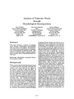
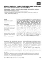
Concentrations of immunoglobulin isotypes and bovine serum albumin (BSA) in afferent and efferent lymph, milk and blood serum, before and after intramammary infusion of 50 μg of Escherichia coli endotoxinFigure 1
Concentrations of immunoglobulin isotypes and bovine serum albumin (BSA) in afferent and efferent lymph,
milk and blood serum, before and after intramammary infusion of 50 μg of Escherichia coli endotoxin. Data are
expressed as LS-mean ± SEM. Time 0 refers to samples collected before the endotoxin infusion.
0
10
20
30
40
50
024
BS
A
(mg/ml)
Serum Afferent lymph Ef f e r e n t ly mp h Milk
0
2
4
6
8
10
12
024
IgG1 (mg/ml)
Serum Afferent lymph Ef f e r e n t l y mph Milk
0
2
4
6
8
10
024
Time after endotoxin infusion (hrs)
IgG
2
(mg/ml
)
Ser um Afferent lymph Ef ferent lymph Milk
0
0,5
1
1,5
2
2,5
3
3,5
024
IgM (mg/ml
)
Serum Afferent lymph Efferent lymph Mi l k
0
0,04
0,08
0,12
0,16
0,2
024
Time after endotoxin infusion (hrs)
Ig
A
(mg/ml)
Serum Afferent lymph Ef f e r en t l y mph Milk
Acta Veterinaria Scandinavica 2008, 50:26 />Page 6 of 10
(page number not for citation purposes)
not to the surgical trauma [22]. In the samples taken
immediately before the endotoxin infusion (0 h) neu-
trophils comprised 7 ± 1% of leukocytes in milk, 6 ± 1%
of leukocytes in afferent lymph and 0 ± 0% of leukocytes
in efferent lymph. These values are in accordance with
neutrophil concentrations in bovine milk and afferent
and efferent lymph in the absence of inflammation [23].
Also the concentrations of different Ig isotypes and
cytokines in milk and lymph before the challenge in our
study [13] were on a whole in accordance with what has
previously been observed in normal milk from cows
[4,5,24-26] and in peripheral lymph under non-inflam-
matory conditions [5,18,27-29]. Thus, there was no indi-
cation of an inflammatory reaction being present in the
udder tissue before the endotoxin was infused and it is
likely that the observed inflammatory response was due to
the endotoxin infusion and not to surgical trauma.
BSA is the only parameter that was slightly higher before
challenge in both milk and lymph compared to most pre-
vious studies referred to. The higher milk BSA concentra-
tion was due to higher content in afferent lymph and not
to increased relative transfer through the epithelium. This
transfer was similar to previous observations in cows [5].
Inactivity has been shown to cause elevated albumin con-
centration in peripheral lymph [19] and is the most plau-
sible explanation for this finding before challenge.
Although the relative transfer of BSA through the endothe-
lium before inflammation was slightly higher than previ-
ously shown this was, interestingly, not the case for
transfer of the Igs through the same barrier, further indi-
cating that the Ig response was not triggered before the
endotoxin infusion.
The general differences between milk, and each of the two
lymph fluids under normal conditions, with lower con-
tents of BSA and Ig in milk than in afferent lymph and
highest in efferent lymph agree on a whole with previ-
ously recorded data [10]. However, the information about
cow mammary lymph is limited and Ig in efferent mam-
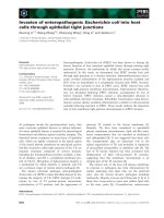
Relative concentrations of immunoglobulin isotypes and bovine serum albumin (BSA) before and after intramammary infusion of 50 μg of Escherichia coli endotoxinFigure 2
Relative concentrations of immunoglobulin isotypes and bovine serum albumin (BSA) before and after
intramammary infusion of 50 μg of Escherichia coli endotoxin. Afferent lymph:serum (A), efferent lymph:serum (B) and
milk:afferent lymph (C) concentration ratios express the percentage of the blood serum concentration that is simultaneously
found in afferent (A) and efferent (B) lymph, respectively, depicting the transfer through the endothelium; and the percentage
of the afferent lymph concentration that is simultaneously found in milk (C), depicting the transfer through the mammary
endothelium. Each value represents the LS-mean. Samples were collected before endotoxin infusion (0 h) and at postinfusion
hours (PIH) 2 and 4.
A
0
20
40
60
80
100
BSA IgG1 IgG2 IgM IgA
Relative concentration in afferent lymph
0 h PIH 2 PIH 4
Serum concentration
%
B
0
20
40
60
80
100
BSA IgG1 IgG2 IgM IgA
Relative concentration in efferent lymph
0 h PIH 2 PIH 4
Serum concentration
%
C
0
20
40
60
80
100
BSA IgG1 IgG2 IgM IgA
Relative concentration in milk
0 h PIH 2 PIH 4
A
fferent lymph concentration
%
Acta Veterinaria Scandinavica 2008, 50:26 />Page 7 of 10
(page number not for citation purposes)
mary lymph of cows has, to our knowledge, not been
investigated previously.
Immunoglobulin concentrations
After endotoxin infusion the Ig concentrations in both
lymph fluids increased rapidly with significantly
increased concentrations generally recorded at PIH 2. At 0
h and PIH 4, respectively, the Ig content was similar in the
two lymph fluids. The most rapid increase was observed
in afferent lymph, leading to that at PIH 2 the content of
IgM and IgA in afferent lymph was significantly higher
(IgM p < 0.001; IgA p < 0.05) and IgG1 tended to be
higher (p = 0.055), than in efferent lymph (Fig. 1). If the
source of these Igs had been blood only, the increase we
observed should have occurred equally rapid in the two
lymph fluids, similar to the pattern of the BSA concentra-
tions (Fig. 1). However, increased concentrations of IgA,
IgM and IgG1 were observed earlier in afferent than in
efferent lymph. This indicates a local increase of these Igs
in tissue that was measurable in afferent lymph at PIH 2
but in efferent lymph not until PIH 4, after the Igs had
been brought to the lymph node from the tissue by the
afferent lymph fluid. Thus, the content of IgA, IgM and
IgG1 in efferent lymph appeared to be significantly influ-
enced by that of afferent lymph during the inflammation,
in the present study. This is further discussed later under
"Modulation of immunoglobulins in the lymph node".
The concentration of the different Igs in tissue might to
various extents have been influenced by active transport,
selectively operating at the endothelium in the gland but
not in the lymph node, or by local synthesis. It is, how-
ever, most likely that the increased tissue concentrations
of IgA, IgM and IgG1 observed at PIH 2 were due to accu-
mulation in the tissue, since the transfer further through
the mammary epithelium of these Igs was hampered at
this particular time, as shown in decreased epithelial CRs
(Fig. 2C). It could, of course, also be speculated that the
lower Ig concentrations in efferent lymph compared to
those in afferent lymph at PIH 2 were due to suppressed
endothelial transfer, selectively in the lymph node –
rather than to increased concentrations in tissue. This is,
however, not likely considering the particularly high per-
meability in the high endothelial venules in the lymph
nodes.
Ig concentrations in milk increased more slowly than in
any of the lymph fluids post-infusion, the highest values
not being observed until PIH 4. In principal, the Igs in
milk are substantially a result of transfer from the tissue
fluid and it is therefore natural that the concentrations
increase later in milk than in afferent lymph.
The relative transfer of immunoglobulins
General comments
The results from this study show that, in general, the trans-
fer of Ig through the endothelium is merely a result of dif-
fusion while the transfer through the epithelium and the
concentrations in milk is more influenced by selective
mechanisms and local synthesis. Before endotoxin infu-
sion the endothelial CR of each Ig was to various extents
lower than that of BSA. The most plausible explanation is
that the Ig molecules did not diffuse as easy through the
tight junctions as BSA, under normal permeability condi-
tions. Although there is a big variation in molecular size,
each Ig is larger than the BSA molecule. IgM, being the
largest molecule of the Igs, showed the lowest relative
transfer through the endothelium which supports the
speculation that the molecular size influences the transfer.
During the inflammatory reaction the endothelial CR of
each Ig increased to values almost equal to each other,
however, still slightly lower than that of BSA.
The transfer of Igs through the mammary epithelium
before as well as after the endotoxin infusion appeared to
be highly affected by selective mechanisms or local pro-
duction, IgG2 being the exception. The milk:afferent lymph
CR for IgG1, IgM and IgA were notably higher than that of
BSA at 0 h and for IgA the difference was huge. Addition-
ally, during inflammation, the alterations of milk:afferent
lymph CR for each of the three Igs were strikingly different
from those of BSA.
IgG1
Previous studies, investigating blood and milk [3,32],
have indicated that the concentration of IgG1 in milk is
influenced by selective transport. The studies have, how-
ever, not made it possible to distinguish between the
transfer through the endothelium and epithelium, respec-
tively. Our results show that the selective mechanism is
operating at the mammary epithelium, before as well as
after the challenge. The epithelial CR of IgG1 in milk was
higher than that of BSA, most pronounced at 0 h (Fig. 2C),
while the endothelial CR of IgG1 and BSA were similar
(Fig. 2A). IgG1 specific receptors located on the surface of
alveolar epithelial cells have been identified in tissues
from cows producing colostrum but never from cows in
lactation [33-35]. Our observations show a selective
mechanism being present in the transfer of IgG1 through
the epithelium, also in lactating glands. The epithelial
transfer of IgG1 appears to be more influenced by passive
diffusion than that of IgA and IgM, since the relative IgG1
transfer increased from 0 h to PIH 4, along with increased
epithelial permeability in the gland, in contrast to the
transfer of IgA and IgM. However, a drop in milk:afferent
lymph CR at PIH 2, similar to that of IgA and IgM, was
observed also for IgG1 indicating a temporary suppres-
sion of the selective transport of IgG1 from tissue to milk
Acta Veterinaria Scandinavica 2008, 50:26 />Page 8 of 10
(page number not for citation purposes)
at this time. A reduction of free IgG1 due to e.g. enhanced
binding to leukocytes [36,37] is not a likely explanation
since the cellular response in milk was barely detected, at
this time.
IgA
In milk, the concentration of IgA was high, almost equal to
that in afferent lymph before endotoxin infusion with a
milk:afferent lymph CR of 0.97 to be compared to that of
BSA of 0.02. These observations are in accordance with
our previous studies where IgA concentration in milk was
even higher than that in afferent lymph in the non-chal-
lenged mammary gland [5]. The results indicate a local
synthesis of IgA in the mammary tissue, in addition to the
amount of IgA diffusing from tissue fluid, in agreement
with results from several previous scientific studies
[25,38,39]. A local synthesis is further supported by the
almost linear increase of IgA concentration in milk post-
infusion (Fig. 1) in contrast to that of the other Igs which
mainly occurred between PIH 2 and PIH 4. IgA producing
plasmacells have been found in both infected and non-
infected mammary parenchyma of lactating cows but
mainly in the interalveolar stroma and only a few adjacent
to the epithelial cells [25]. According to our results, IgA is
released primarily into milk, not into tissue fluid, which
suggests that the synthesis occur close to the epithelium
rather than in deeper sub-epithelial tissues of the udder.
Interestingly, the relative concentration of IgA in milk
compared to that in afferent lymph (the milk:afferent
lymph CR) decreased from 0 h to PIH 4 (Fig. 2C) in spite
of the gradually increasing epithelial permeability, diffu-
sion of BSA and neutrophil influx to milk. The lowest CR
was observed at PIH 2 indicating an inhibition mecha-
nism at this time point. A similar pattern was observed for
IgM (and IgG1). According to these observations the con-
centrations of IgA and IgM in milk during the inflamma-
tory reaction were, on a whole, not substantially
dependent on permeability conditions.
IgM
At 0 h, the mammary endothelial CR of IgM was notably
lower than that of BSA (Fig. 2A). A similar relationship
was observed between the CRs of IgM and BSA also at the
endothelium in the lymph node (Fig. 2B). This indicates a
factor that is limiting the diffusion of IgM compared to
that of BSA. A plausible explanation is that the large size
of the Ig M molecule makes passage through the capillary
endothelium difficult under physiological permeability
conditions. This is supported by the rapidly increased
afferent lymph:serum and efferent lymph:serum CRs of IgM
observed when the vascular permeability increased during
inflammation. It is however, puzzling that the increase in
concentration and CR of IgM was delayed in efferent
lymph compared to afferent lymph since the passage of
IgM through the endothelium of the highly permeable
high endothelial venules in the lymph node should rea-
sonably have occurred even more easily than in the mam-
mary tissue capillaries. Apparently, the content of efferent
lymph was strongly influenced by IgM brought there by
the afferent lymph. This indicates a local source of IgM, in
addition to blood, and/or an accumulation of IgM in the
tissue due to hampered transfer over the mammary epi-
thelium. In previous studies, IgM producing plasma cells
have been observed in the mammary gland tissue but
rarely close to the epithelial cells [25].
The highest CR of IgM at the mammary epithelium was,
surprisingly, recorded at 0 h. In contrast to the endothelial
CR, the epithelial CR of IgM tended to decrease after the
endotoxin infusion. Even if the CR may be influenced by
additional factors, it is notable that the increased epithe-
lial permeability between tissue and milk, as shown in ele-
vated BSA CR, was not reflected in the CR of IgM. The
findings further indicate that the IgM transfer through the
mammary epithelium was somehow hampered during
this time of the inflammatory reaction. The underlying
mechanism remains to be explained.
IgG2
Before endotoxin infusion the relative transfer of IgG2
through the endothelium to afferent lymph, as reflected in
the afferent lymph:serum CR, was fairly similar to that of
BSA, suggesting that the content of IgG2 in non-chal-
lenged afferent lymph is, in principal, a result of diffusion
from blood only. This is in accordance with previous stud-
ies of IgG2 in milk compared to blood [3]. At PIH 2 the
endothelial CR of IgG2 had increased, however, not at the
same rate as that of BSA. Considering that the large influx
of neutrophils to afferent lymph was observed at this time
a possible explanation is that the amounts of free IgG2
was reduced due to enhanced binding to IgG2 specific sur-
face receptors of the neutrophils [40].
The relative transfer of IgG2 from tissue into milk was
almost identical to that of BSA before as well as during
inflammation. Thus, the IgG2 transfer through the epithe-
lium appears to be an effect of passive diffusion only.
Modulation of immunoglobulins in the lymph node
It has been discussed to what extent the contents of effer-
ent lymph reflect that of afferent lymph and how much
the efferent lymph content is influenced by fluid and pro-
tein coming from blood through the postcapillary venules
in the node. Igs in the local lymph node and efferent
lymph are to some extent transferred from blood [8,30]
and previous studies, focusing on protein in lymph, have
reported that the blood derived contents may contribute
30–50% of the protein output in efferent lymph from un-
stimulated lymph nodes [30,31]. However, this may vary
Acta Veterinaria Scandinavica 2008, 50:26 />Page 9 of 10
(page number not for citation purposes)
between nodes in different regions. Since a significant
function of the lymph node is to provide a meeting place
for antigen-cell interactions in the initiation of the
immune defence, it is quite obvious that the lymph
formed also may be modulated within the node by tar-
geted addition or trapping of Igs and Ig-producing cells
[7,41] and additionally influenced by the content of affer-
ent lymph flowing into the lymph node [8,30,31]. Thus,
the degree of influence from different sources can be
expected to vary dependent on whether the node is anti-
gen stimulated or not.
In the present study concentration of each Ig isotype and
BSA, respectively, was similar in the two lymph fluids,
before the endotoxin challenge. This suggests, in agree-
ment with the previous studies [30,31], that the content in
both fluids were mainly blood derived at this time point.
After challenge, Ig isotype concentrations in efferent
lymph, particularly regarding IgA, IgM and IgG1,
increased more slowly than in afferent lymph, in contrast
to BSA, which increased equally rapid in both lymph flu-
ids. These observations indicate that after the endotoxin
challenge, Ig concentrations in efferent lymph were
mainly influenced by the contents of the afferent lymph,
flowing into the node and to a less extent dependent on
transfer from blood.
Conclusion
The most rapid increase of Igs was observed in afferent
lymph, resulting in significantly higher concentration of
each Ig isotype, except for IgG2, in afferent than in efferent
lymph at PIH 2, contrary to before challenge. Ig concen-
trations in milk were in general lower than in lymph and
they increased later. The transfer of Igs through the
endothelium appeared to be merely a result of diffusion
while the transfer through the epithelium and the Ig con-
centrations in milk seemed to be more influenced by
selective mechanisms and local sources, respectively. In
addition, the molecular size of the Igs appeared to nega-
tively affect their transfer through the endothelium, par-
ticularly under normal permeability conditions when the
CR of each Ig isotype, except for IgG2, was lower than that
of BSA. However, at the mammary epithelium the opposite
was observed; the CR of each Ig isotype, except for IgG2,
was higher than that of BSA, before challenge. Addition-
ally, the alterations in the epithelial CR of the Igs (IgG1,
IgM and IgA) during inflammation were strikingly differ-
ent from those of BSA. Our observations indicate a selec-
tive mechanism being present in the transfer of IgG1
through the epithelium, also in lactating glands which has
not been previously shown. The results also indicate a
local synthesis in the tissue of IgA and possibly also of
IgM, released primarily into milk, not into tissue fluid sug-
gesting that the synthesis occurs close to the epithelium.
The IgG2 transfer through endothelium as well as epithe-
lium appeared to be a result of passive diffusion only. In
the lymph node, the content of efferent lymph was
strongly influenced by IgG1, IgM and IgA brought to the
node by the afferent lymph, from the mammary tissue
and less dependent on transfer from blood.
Competing interests
The authors declare that they have no competing interests.
Authors' contributions
KÖ designed and planned the study and methods used,
performed the surgery, oversaw and participated in the
practical work and prepared the major part of the final
manuscript, SL performed the sample collection and lab-
oratory analyses, prepared a first draft of the manuscript
and participated in preparing the final manuscript. Both
authors read and approved the final manuscript.
Acknowledgements
This study was supported by Swedish Council for Forestry and Agricultural
Research. Dr. Lun received funds from the Swedish Foundation for Inter-
national Cooperation in Research and Higher Education (STINT). The
authors want to express their sincere gratitude to Dr Nils Lundeheim,
Department of Animal Breeding and Genetics, SLU for excellent help with
the statistical analyses.
References
1. Gretz JE, Norbury CC, Anderson AO, Proudfoot AE, Shaw S:
Lymph-borne chemokines and other low molecular weight
molecules reach high endothelial venules via specialized con-
duits while a functional barrier limits access to the lym-
phocyte microenvironments in lymph node cortex. J Exp Med
2000, 192:1425-1440.
2. Itano AA, McSorley SJ, Reinhardt RL, Ehst BD, Ingulli E, Rudensky AY,
Jenkins MK: Distinct dendritic cell populations sequentially
present antigen to CD4 T cells and stimulate different
aspects of cell-mediated immunity. Immunity 2003, 19:47-57.
3. Guidry AJ, Ost M, Mather IH, Shainline WE, Weinland BT: Sequen-
tial response of milk leukocytes, albumin, immunoglobulins,
monovalent ions, citrate, and lactose in cows given infusion
of Escherichia coli endotoxin into the mammary gland. Am J
Vet Res 1983, 44:2262-2267.
4. Anderson KL, Smith AR, Shanks RD, Whitmore HL, Davis LE, Gus-
tafsson BK: Endotoxin-induced bovine mastitis: immunoglob-
ulins, phagocytosis, and effect of flunixin meglumine. Am J Vet
Res 1986, 47:2405-2410.
5. Östensson K: Trafficking of leukocytes and immunoglobulin
isotypes in the bovine udder. In PhD thesis Swedish University of
Agricultural Sciences, Department of Obstetrics and Gynaecology,
Uppsala; 1993.
6. Pape KA, Catron DM, Itano AA, Jenkins MK: The humoral
immune response is initiated in lymph nodes by B cells that
acquire soluble antigen directly in the follicles. Immunity 2007,
26:491-502.
7. McKeever D, Reid HW: The response of the supramammary
lymph node of the sheep to orf virus infection. Vet Microbiol
1987, 14:3-13.
8. Tizard IR: Organs of the immune system. In Veterinary Immunol-
ogy: An introduction 7th edition. Edited by: Tizard IR. Philadelphia: Saun-
ders, Elsevier; 2004:78-91.
9. Guidry AJ, Paape MJ, Pearson RE: Effect of udder inflammation
on milk immunoglobulins and phagocytosis. Am J Vet Res 1980,
41:751-753.
10. Guyton AC, Hall JE: The microcirculation and the lymphatic
system: Capillary fluid exchange, interstitial fluid and lymph
flow. In Medical Physiology 10th edition. Edited by: Guyton AC. Phil-
adelphia: WB Saunders Company; 2000:162-174.
Publish with Bio Med Central and every
scientist can read your work free of charge
"BioMed Central will be the most significant development for
disseminating the results of biomedical research in our lifetime."
Sir Paul Nurse, Cancer Research UK
Your research papers will be:
available free of charge to the entire biomedical community
peer reviewed and published immediately upon acceptance
cited in PubMed and archived on PubMed Central
yours — you keep the copyright
Submit your manuscript here:
/>BioMedcentral
Acta Veterinaria Scandinavica 2008, 50:26 />Page 10 of 10
(page number not for citation purposes)
11. Shuster DE, Kehrli ME Jr: Administration of recombinant
human interleukin 1 receptor antagonist during endotoxin-
induced mastitis in cows. Am J Vet Res 1995, 56:313-320.
12. Lun S, Åström G, Magnusson U, Östensson K: Total and differen-
tial leukocyte counts and lymphocyte subpopulations in
lymph, afferent and efferent to the supramammary lymph
node, during endotoxin-induced mastitis in cows. Reprod Dom
Anim 2007, 42:126-134.
13. Persson Waller K, Colditz IG, Lun S, Östensson K: Cytokines in
mammary lymph and milk during endotoxin-induced bovine
mastitis. Res Vet Sci 2003, 74:31-36.
14. Schalm OW, Carrol EJ, Jain NC: Physical and chemical tests for
detection of mastitis. In Bovine mastitis Edited by: Schalm OW,
Carrol EJ, Jain NC. Philadelphia: Lea and Febiger Inc; 1971:139.
15. Obel N, Östensson K, Åström G: Sampling of lymph from ves-
sels afferent to the supramammary lymph gland in the cow.
Zentralbl Veterinarmed A 1989, 36:490-493.
16. Kottman J, Hampl A, Pravda D: The collection of lymph from
efferent vessel of supramammary lymphatic gland of the
cow in a long-term experiment. Acta Vet Brno 1970, 39:197-203.
17. Östensson K: Total and differential leukocyte counts, N-
acetyl-β-D-glucosaminidase activity, and serum albumin
content in foremilk and residual milk during endotoxin-
induced mastitis in cows. Am J Vet Res 1993, 54:231-238.
18. Kennedy JW, Watson DL, Bennell MA: The early phase of the
immune response to live and killed Staphylococcus aureus
vaccines in sheep – cellular and immunoglobulin parameters.
Vet Immunol Immunopathol 1981, 2:367-380.
19. Olszewski WL: The lymphatic system in body homeostasis:
physiological conditions. Lymph Res Biol 2003, 1:11-21.
20. Saad AM, Östensson K: Flow cytofluorometric studies on the
alteration of leukocyte populations in blood and milk during
endotoxin-induced mastitis in cows. American Journal of Veteri-
nary Research 1990, 51:1603-1607.
21. Prin-Mathieu C, Le Roux Y, Faure GC, Laurent F, Béné MC, Mous-
saoui F: Enzymatic activities of bovine peripheral blood leuko-
cytes and milk polymorphonuclear neutrophils during
intramammary inflammation caused by lipopolysaccharide.
Clin Diagn Lab Immunol 2002, 9:812-817.
22. Haig D, Deane D, Percival A, Myatt N, Thomson J, Inglis L, Rothel J,
Seow H-F, Wood P, Miller HRP, Reid HW: The cytokine response
of afferent lymph following orf virus reinfection of sheep. Vet
Dermatol 1996, 7:11-20.
23. Colditz IG, Kerlin RL, Watson DL: Migration of neutrophils, and
their role in elaboration of host defence. In Migration and hom-
ing of lymphoid cells Volume 1. Edited by: Husband AJ. Cleveland: CRC
Press; 1988:135-165.
24. Guidry AJ, Miller RH: Immunoglobulin isotype concentrations
in milk as affected by stage of lactation and parity. J Dairy Sci
1986, 69:1799-1805.
25. Doymaz MZ, Sordillo LM, Oliver SP, Guidry AJ: Effects of Staphy-
lococcus aureus mastitis on bovine mammary gland plasma
cell populations and immunoglobulin concentrations in milk.
Vet Immunol Immunopathol 1988, 20:87-93.
26. Shuster DE, Kehrli ME Jr, Baumrucker CR: Relationship of inflam-
matory cytokines, growth hormone, and insulin-like growth
factor-I to reduced performance during infectious disease.
Proc Soc Exp Biol Med 1995, 210:140-149.
27. Beh KJ, Watson DL, Lascelles AK: Concentrations of immu-
noglobulins and albumin in lymph collection from various
regions of the body of the sheep. Aus J Exp Bio Med Sci 1974,
52:81-86.
28. Quin JW, Shannon AD: The effect of anaesthesia and surgery on
lymph flow, protein and leucocyte concentration in lymph of
the sheep. Lymphology 1975, 8:126-135.
29. Haig DM, Hopkins J, Miller HRP: Local immune responses in
afferent and efferent lymph. Immunology 1999, 96:155-163.
30. Quin JW, Shannon AD: The influence of the lymph node on the
protein concentration of efferent lymph leaving the node. J
Physiol 1977, 264:307-321.
31. Maron MB: Modification of lymph during passage through the
lymph node: effect of histamine. Am J Physiol 1983,
245(4):H553-9.
32. Guidry AJ, Pearson RE, Paape MJ, Williams WF: Relationship
among leukocyte phagocytosis, milk immunoglobulins, and
susceptibility to intramammary infection. Am J Vet Res 1980,
41:997-1001.
33. Kemler R, Mossmann H, Strohmaier U, Kickhöfen B, Hammer DK: In
vitro studies on the selective binding of IgG from different
species to tissue sections of the bovine mammary gland. Eur
J Immunol 1975, 5:603-608.
34. Barrington GM, Besser TE, Davis WC, Gay CC, Reeves JJ, McFadden
TB: Expression of immunoglobulin G1 receptors by bovine
mammary epithelial cells and mammary leukocytes. J Dairy
Sci 1997, 80:86-93.
35. Barrington GM, Besser TE, Gay CC, Davis WC, Reeves JJ, McFadden
TB, Akers RM: Regulation of the immunoglobulin G1 receptor:
effect of prolactin on in vivo expression of the bovine mam-
mary immunoglobulin G1 receptor. J Endocrinol 1999,
163:25-31.
36. Norcross NL: Specific defense mechanisms of the udder. Flem-
ish Vet J 1991, 62:129-133.
37. Worku M, Paape MJ, Marquardt WW: Modulation of Fc receptors
for IgG on bovine polymorphonuclear neutrophils by inter-
feron-gamma through de novo RNA transcription and pro-
tein synthesis. Am J Vet Res 1994, 55:234-238.
38. Guidry AJ, Butler JE, Pearson RE, Weinland BT: IgA, IgG1, IgG2,
IgM, and BSA in serum and mammary secretion throughout
lactation. Vet Immunol Immunopathol 1980, 1:329-341.
39. Leitner G, Yadlin B, Glickman A, Chaffer M, Saran A: Systemic and
local immune response of cows to intramammary infection
with Staphylococcus aureus. Res Vet Sci 2000, 69:181-184.
40. Targowski SP, Niemialtowski M: Appearance of Fc receptors on
polymorphonuclear leukocytes after migration and their
role in phagocytosis. Infect Immun 1986, 52:798-802.
41. Huang H-S, Buxton D, Burrells C: Immune responses of the
ovine lymph node to Chlamydia psittaci. A cellular study of
popliteal efferent lymph. J Comp Path 1991, 105:191-203.