Báo cáo thú y: "Application of a pig ligated intestinal loop model for early Lawsonia intracellularis infection" doc
Bạn đang xem bản rút gọn của tài liệu. Xem và tải ngay bản đầy đủ của tài liệu tại đây (794.99 KB, 7 trang )
RESEARC H Open Access
Application of a pig ligated intestinal loop model
for early Lawsonia intracellularis infection
Torsten S Boutrup
1,2
, Kirsten Schauser
3
, Jørgen S Agerholm
2
, Tim K Jensen
1*
Abstract
Background: Porcine proliferative enteropathy in pigs is caused by the obligate, intracellular bacterium Lawsonia
intracellularis. In vitro studies have shown close bacterium-cell interaction followed by cellular uptake of the
bacterium within 3 h post inoculation (PI). However, knowledge of the initial in vivo interaction between porcine
intestinal epithelium and the bacterium is limited. The aims of the present study were to evaluate the usefulness of
a ligated small intestinal loop model to study L. intracellularis infections and to obtain information on the very early
L. intracellularis-enterocyte interactions.
Methods: A ligated small intestinal loop model using three different L. intracellularis inocula was applied to 10-11-
week-old pigs. The inocula were 1) wild type bacteria derived from overnight incubation of L. intracellularis bacteria
from spontaneous disease, 2) crude vaccine bacteria (Enterisol
®
Ileitis Vet), and 3) vaccine bacteria propagated in
cell culture. The bacteria-enterocyte interaction was visualised using immunohistochemistry on specimens derived
1, 3 and 6 h PI respectively.
Results: Although at a low level, close contact between bacteria and the enterocyte brush border including
intracellular uptake of bacteria in mature enterocytes was seen at 3 and 6 h PI for the vaccine and the propagated
vaccine inocula. Interaction between the wild-type bacteria and villus enterocytes was scarce and only seen at 6 h
PI, where a few bacteria were found in close contact with the brush border.
Conclusions: The ligated intestinal loop model was useful with respect to maintaining an intact intestinal
morphology for up to 6 h. Furthermore, the study demonstrated that L. intracellularis interacts with villus
enterocytes within 3 to 6 h after inoculation into intestinal loops and that the bacterium, as shown for the vaccine
bacteria, propagated as well as non-propagated, was able to invade mature enterocytes. Thus, the study
demonstrates the early intestinal invasion of L. intracellularis in vivo.
Introduction
The bacterium Lawsonia intracellularis is the infectio us
cause of proliferative enteropathy (PE) in pigs and a
range of other animal species [1,2]. The bacterium is
Gram negative, rod-shaped and belonging to the delta
division of the Proteobacteria. Bacterial growth requires
an intracellular environment and in v itr o isolation and
cultivation depends on cell culture [3]. The s uccessful
isolation and growth of the bacterium in vitro has estab-
lished the basis for v accine development [4,5]. Knowl-
edge on the initial host-pathogen interaction in vivo is
limited. However in vitro studies have shown close bac-
terium-cell interaction followed by cellular uptake of the
bacterium within 3 h post inoculation (PI) [6]. Recently
experimental infection of pigs has demonstrated entero-
cyte-bacterium interaction as early as 12 h PI [7].
Intestinal loop models have previously demonstrated
their usefulness in studies of Brachyspira hyodysenteriae
and Salmonella Typhimurium [8-11]. McOrist et al.
[12] used ligated intestinal loops to investigate events
between L. intracellularis and enterocytes at 1 h PI but
found no intracellular uptake of L. intracellularis or bac-
teria-enterocyte interactions. The aims of the present
study were to evaluate the usefulness of an intestinal
loop model to investigate L. intracellularis infections
and to ob tain information o n very ear ly L. intracellu-
laris-enterocyte interactions. Compared to the study
performed by McOrist et al. [12] the exposure time
between L. intracellularis and the intestinal epithelium
* Correspondence:
1
National Veterinary Institute, Technical University of Denmark, Bülowsvej 27,
DK-1790 Copenhagen V, Denmark
Boutrup et al. Acta Veterinaria Scandinavica 2010, 52:17
/>© 2010 Boutrup et a l; licensee BioMed Central Ltd. This is an Open Access a rticle distributed under the te rms of the Creative Commons
Attribution License ( which pe rmits unrestricted use, distribution, and reproduction in
any medium, provided the original work is properly cited.
in the loops were extended to 1, 3 and 6 h. Moreover
three different preparations of L. intracellularis inocu-
lums were used at each point.
Materials and met hods
Experimental animals
Four pigs were purchased from a high health (specific
pathogen free (SPF)) herd considered to be free of L.
intracellu laris infection after a medicated eradication
program. Twenty blood samples and 10 faecal samples
from pigs with body weights (BW) of 3 0 to 60 kg were
sampled twice from the herd and tested by ELISA and
PCR methods as described elsewhere [13,14] to ensure
herd status regarding L. intracellularis infectio n. All
samples tested negative.
The p igs were acclimatised for 2 weeks before enter-
ing the study. Clinical signs of disease were not
observed during this period. As a precaution, all pigs
were medicated with tiamulin at arrival (Tiamutin
®
vet.
200 mg/ml, Novartis, Copenhagen, Denmark), given at a
dosage of 20 mg/kg BW intramuscularly for 4 consecu-
tive days. Faecal samples taken before and after medica-
tion were all found negative for L. intracellularis by
PCR. To avoid adverse effect of the antibiotic treatment
on the study, treatment with tiamulin was ceased at
least 7 days before inoculation.
Thefourpigswerehousedtogetherandfedastan-
dard diet ad libitum (DLG, +10, Aarhus, Denmark) wit h
free access to water and straw. The animals were fasted
from the day before experimentation with free access to
water with glucose added. The pigs were 10-11-week-
old (BW 26 to 31 kg) at the time of surgery. The experi-
mental study wa s approved by the Da nish Animal
Experiments Inspectorate under the Ministry of Justice.
Inoculum
Infectious materials derived from spontaneously diseased
pigs
Prior to the trial, porcine small intestines having PE
were collected from a herd that had previously delivered
infectious materials for succ essful experimental infec-
tions [7,15]. The presence of L. intracellularis associated
with PE in the material was confirmed by immunofluor-
escense (IF) using an anti-L. intracellularis monoclonal
ant ibo dy (Law1-DK) [16,17]. The intestines were froz en
at -80°C in portions o f 100 g. The day before inocula-
tion, a portion w as thawed in a water bath at 37°C and
epithelial cells were isolated by immersing the material
into100mlofHank’ s balanced salts solution (HBSS)
without CaCl
2
and MgCl
2
(Invitrogen, 14180-046,
Taastrup, Denmark) diluted 1:10 in Milli Q water, with
5 mM EDTA (Merck, 15498, Albertslund, Denmark)
and incubated at 37°C for 80 min with occasiona l stir-
ring. Detached epithelial cells and L. intra cellularis
bacteria were harvested by centrifugation at 5000 g for
30 min. The cells were resuspended in 100 ml Dulbec-
co’ s Modified Eagle medium (DMEM) (Invitrogen,
41965) with 5% fetal bovine serum (FBS) (Sigma, F9665,
Vallensbaek, Denm ark), 1% L-glutamine (Invitrogen,
25030), 2% amphotericin B (Sigma, A2942), gentamycine
50 μg/ml (Sigma, G3632) and vancomycine 100 μg/ml
(Sigma, V2002) and incubated overnight at 37°C, in an
atmosphere of 8.8% CO
2
and 8.0% O
2
. Next day the
inocula were centrifuged at 5000 g for 30 min a nd
resuspendedin50mlofDMEMwith5%FBSandthe
epithelial cells were lysed by forcing the suspension
through a 3.5 inch 22 Gauge spinal syringe (Becton
Dickinson, 405256, Madrid, Spain). In vitro cell culture
inoculations have shown a n initial intracellular replica-
tion of similar level using this method compared to
crude mucosal scraping (data not shown). Compared to
crude mucosal scraping, the described method provides
rather homogenous inoculum.
Infectious materials derived from commercial L.
intracellularis live vaccine
AcommercialL. intracellularis live vaccine (Enterisol
®
Ileitis Vet., No. 024390, Batch no 30496-00) was pur-
chased and held at 5°C until use. Immediately before
inoculation into intestinal loops 0.8 g of freeze dried
vaccine were dissolved in 5 ml o f DMEM with 5% FBS.
This corresponds to four doses according to
manufacturer.
Infectious materials derived from commercial L.
intracellularis live vaccine propagated in cell culture
Infected cell cultures based on the L. intracell ularis vac-
cine were produced by suspending 0.4 g of freeze dried
vaccine in DMEM with 5% FBS and 1% L-glutamine
and inoculating the suspension into a McCoy cell cul-
ture (ATCC number: CRL-1696), T-80 bottles with 15
ml medium seeded with 2 × 10
5
cells per ml from the
day before. The infected cell cultures were incubated at
37°C, in 8.8% CO
2
and 8.0% O
2
. Passage of infection
was done by scraping of McCoy cells, which were lysed
by forcing the suspension through a 3.5 inch 22 Gauge
spinal syringe. Cell debris were removed by centrifuga-
tion at 150 g for 5 min, bacteria were harvested by cen-
trifugation at 5000 g for 20 min. The bacterial pellet was
re-suspended in 3 ml of medium and re-inoculated onto
new cell cultures as described above. At the day of
inoculation, two cell culture bottles with massive growth
of L. intracellularis were used. The cells were scraped
from the bottom and lysed as described above. Cells and
bacteria were centrifuged at 5000 g for 20 min, where
after the pellet was re-suspended in 10 ml of medium.
The concentration of L. intracellularis in the different
inocula was determined by serial 1:10 dilutions in
sucrose potassium glutamate (SPG) with 5% FBS. Ten μl
of each dilution were added to each well in a six-well
Boutrup et al. Acta Veterinaria Scandinavica 2010, 52:17
/>Page 2 of 7
glass slide and examined by indirect IF [16]. The num-
ber of L. intracellularis bacteria was counted at 40×
objective magnification in 10 view fields corresponding
to 1/25 of a well. The concentrations in the different
types of inocula are shown in Table 1. Five ml of eac h
inoculum was injected into the lumen of intestinal loops
via an 18 G syringe.
Anaesthetic and surgical procedure
Isoflurane inhalation anaesthesia and surgical proce-
duresweredoneasdescribedbyGrøndahlet al. [18]
and modified by Shauser et al. [10].Isotonicsalinewas
administered intravenously throughout the procedure.
Pulse, blood pressure, rectal temperature and blood gas
pressure were monitored. A midline abdominal incision
was made and ten loops were produced in the upper
jejunum and lower jej unum, resp ectively (Table 1). The
first loop in the lower jejunum was made 10 cm oral to
the ileocaecal orifice with additional nine loops ligated
in oral direction. The first upper jejunal loop was made
1 m oral to the confluent ileal Peyer’s patch with addi-
tional nine loops ligated in oral direction. Each loop was
approximately 5 cm long followed by an inte r-loop seg-
men t of around 2 cm. Ligation was done by a intestinal
circumferential ligature through the mesentery without
damaging grossly visible mesenteric vascular arcades
thus maintaining full blood supply for both loops and
inter- loop segments. The overall anaesthetic period was
7 to 8 h where after pigs were euthanised by an over-
dose of sodium pentobarbital while still anaesthetised.
Loops were inoculated for 1, 3 and 6 h for each inocu-
lum. Initially four lower jejunal and f our upper jejunal
loops were made (Table 1). One loop served as negative
control a nd were inoculated with DMEM with 5% FBS,
one loop was inoculated with the wild-type bacterial
suspension, one with vaccine suspension and one with
the suspension of cell culture propagated vaccine. This
procedure was repeated afte r 3 h and again after 5 h,
but without control loops (Table 1). Inter-loop segments
served as non-inoculated controls at 3 and 5 h.
Tissue processing
The loops were sampled at euthanasia by cutting the
mesentery and immediately cooled on thawing ice. The
ends of each loop were cut off, the lumen was rinsed
with isotonic saline and the tissues were fixed in 10%
neutral buffered formalin for 2 4 to 48 h. The tissue was
cut into transverse sectio ns, expos ed to graded series of
alcohol succeeded by xylene and embedded in paraffin.
Immunohistochemistry
The loop specimens, each consisting of two full cross
sections, were cut in 5 μm thick sections and mounted
on Super Frost*/plus slides (Menzel-Gläser, Braunsch-
weig, Germany). Mounted slides were heated to 60°C,
deparaf finised and rehydrated in xylene, graded series of
alcohol and finally in water. Endogenous peroxidase
activity was inhibited by incubation with 0.6% H
2
O
2
in
tris buffered saline (TBS) (50 mM Tris, 150 mM NaCl,
pH 7.6) for 20 min followed by washing in TBS 3 × 5
min. Slides were incubated with 0.05% protease (Sigma,
type XXIV, 8038) in TBS for 10 min followed by wash-
ing in TBS 3 × 5 min. Slides were incubated 1 h with
polyclonal rabbit anti L. intracellularis antibody [7]
diluted 1:10000 in TBS, washed for 3 × 5 min in TBS
and incubated with Envisio n
+
goat anti-rabbit conjungate
(DAKO, K4002, Glostrup, Denmark). After w ashing for
3×5mininTBS,reactionwasdevelopedfor15min
with a solution of 3-am ino-9-ethylcarbozole (AEC)
(Kementec, 4190, Copenhagen, Denmark) followed by
washing in TBS 3 × 5 min, counterstained by Mayer’ s
haematoxylin and mount ed with glycergel (DAKO,
C563). All procedures were undertaken at a room tem-
perature around 20°C.
Microscopic evaluation
Slides were evaluated by light microscopy using 40× and
63× objectives. In tissue from mock inoculated loops
and inter-loop s egments, absence of L. intracellula ris
antigens were evaluated for both intestinal lumen and
mucosa.
In inoculated loops the presence of intracellular bac-
teria was evaluated, including a specific search for bac-
teria in the brush border w ith no free space in between
ent erocytes and the bacterium (Figure 1C and 1D). The
presence of L. intrac ellularis antigen in the intestinal
Table 1 Overview of types- and concentrations of inocula
used in each ligated intestinal loops.
Loop
No.
Inoculation
time
Inoculation type Inoculum
concentration
1 Wild-type 4-6 × 10
8
bacteria/ml
2 Live vaccine 3-5 × 10
6
bacteria/ml
3 Propagated live
vaccine
2-8 × 10
7
bacteria/ml
4 Negative control Mock inoculum
5 Wild-type 4-6 × 10
8
bacteria/ml
6 Live vaccine 3- 5 × 10
6
bacteria/ml
7 Propagated live
vaccine
2-8 × 10
7
bacteria/ml
8 Wild-type 4-6 × 10
8
bacteria/ml
9 Live vaccine 3-5 × 10
6
bacteria/ml
10 Propagated live
vaccine
2-8 × 10
7
bacteria/ml
Ligation of ten loops (1-10) was done in the ileum and the jejunum,
respectively. All three types of inocula applied were exposed for 1, 3 and 6 h,
while only at 6 h a negative control was included (loop No. 4). The
concentration of Lawsonia intracellularis in the inocula is shown in the table; 5
ml of inoculum were used for each loop.
Boutrup et al. Acta Veterinaria Scandinavica 2010, 52:17
/>Page 3 of 7
lumen and mucus overlying villus epithelium and in the
crypts was noted but considered as a passive presence
due to inoculation.
Results
Ligation was found to induce grossly visible local
mesenteric oedema and decreased intestinal wall tonus.
Pallor of the intestinal wall indicating inadequate blood
supply did not occur, congestion of mesentery and
intestinal vessels remained at a low level and mild stro-
mal oedema was the only histologically circulatory asso-
ciated lesion (Figure 1A). Together these findings
indicate a limited negative impact on the intestinal
blood supply due to the p rocedures applied. In general
the rectal temperature was slowly decreased from
around 37.5°C to 36.2°C, although one pig had a term-
inal rectal temperat ure of 35.8°C. One pig had a mild
local chronic adhesi ve fibrous peritonitis. L. intracellu-
laris antigen was not foun d by IHC in the negative con-
trol loops or in the inter-loop segments.
Although only a few bacteria were seen in direct con-
tact with enterocytes or the brush border during the
first 6 h PI for all types of inocula, differences were
observed as bacteria of the vaccine inoculum and vac-
cine propagated inoculum seemed to be in di rect con-
tactwiththemucosamorefrequentlythanthewild
type.Bacteriawereseenassingledistinctorganisms
within in the brush border of the villus enterocytes 3 h
and 6 h PI (Figures 1C and 1D). The number of bacteria
in direct contact with the brush border varied but
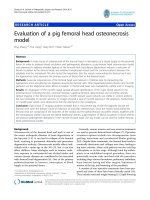
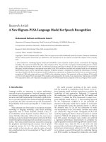
Figure 1 Visualisation of Lawsonia intracellularis in tissue of inoculated intestinal loops. Immunohistochemistry/haematoxy lin stain of
Lawsonia intracellularis in intestinal tissue; arrows point at immunopositive red stained L. intracellularis. A and B: Bacteria overlying ileal
epithelium 6 h post inoculation (PI). A) Vaccine derived inoculum. B) Wild-type derived inoculum. In both (A) and (B) close interactions between
bacteria and enterocytes is not found. Low level oedema seen as distended central lacteal (A) (asterisk). C and D: Solitary L. intracellularis bacteria
in intimate contact with the brush border of enterocytes 6 h PI. C) Vaccine derived inoculum in jejunal loop. D) Cell culture propagated vaccine
in ileal loop. E and F: Solitary intracellular L. intracellularis bacteria in villus enterocytes 6 h PI. E) Vaccine derived inoculum in jejunal loop. F) Cell
culture propagated vaccine in ileal loop. Insert in (E) shows a higher magnification of the area with the intracellular bacterium. Bars: 10 μm.
Boutrup et al. Acta Veterinaria Scandinavica 2010, 52:17
/>Page 4 of 7
mostly 10-25 organisms per full transverse intestinal
section were seen. In addition, single intracellular
L. intracellularis bacteria (1-5 organisms per intestinal
cross section) were found in villus enterocytes 6 h PI
(Figures 1E and 1F) indicating a low level infection. By
contrast, only 5-10 L. intracellularis bacteria of the wild
type were seen in close p roximity to the brus h border
for loops inoculated for 6 h but not for loops inoculated
for 1 or 3 h. Wild type intracellular bacteria were not
observed at all.
Interaction between bacteria and crypt epithelium was
not observed irrespectively of type of inoculum. How-
ever, IHC demonstrated that the inoculated material
had remained in the lumen.
Discussion
The study demonstrates that mature enterocytes are
infected by L. intracellularis thus, confirming previous
studies examining the bacterium-enterocyte interaction
during later stages of i nfection. In a recent study by
Boutrup et al. [7]L. intracellularis was demonstrated in
villus enterocytes 12 h PI in pigs inoculated by stomach
tube with a mucosal scrapingobtainedfrompigsnatu-
rally affected by PE. Whether L. intracellularis is able to
propagate in the mature amitotic enterocytes is however
not known. Interestingly, invasion was only shown for
vaccine derived L. intracellularis, cell culture propagated
as well as non-propagated. Interaction between bacteria
and mucosa was observed at 3 and 6 h PI. Similar to
the study by McOrist et al. [12] based on a modified
intestinal loop model inoculated with a laboratory atte-
nuated strain of L. intracellularis, we did not observe
interaction between bacteria and enteroc ytes 1 h PI. It
could be postulated that the observed interaction
occurred just by chance, i.e. that some bacteria pa ssively
adhered to the brush border. However, if that had been
the case we would have expected such a phenomenon
to occur randomly i n all lo ops. We did not see close
interaction at a ll 1 h PI despite the type of inoculum.
Furthermore, differences were observed among inocula
as the wild type showed less interaction than the vaccine
regarding both the number of interacting bacteria and
interaction 3 h PI. This indicates that interaction was
not an accidental event.
Direct evidence for specific target cells during the
initial exposure of the intestinal epithelium to L. intra-
cellularis has not been shown. However, data from
experimental studies [19-22] on the location and events
of L. intracellularis infectionfrom24hto3wksin
hamsters and pigs report the presence of intracellular
bacteria and the development of hyperplastic lesions as
taking place from infected crypt cells. Also some authors
propose the crypt cells to be the target cell population
for L. intracellularis [23,24]. Bacterial invasion of crypt
enterocytes was not observed in t his study. However,
this may be due to retention of the inoculum above the
crypt-villus junction.
The ligated intestinal loop model has previously
shown its usefulness in studies of intestinal bacterial
infections [8-11]. The validity of the model highly
depends on conservation of a normal intestinal funct ion
and environment. Our study shows that the model
seems useful with respect to maintaining an intact
intestinal morphology as the only histomorphological
change in the intestinal mucosa seen after ligation of
intestinal loops for up to 6 h was a slight stromal
oedema. As lethal or sublethal changes, as e.g. hydrophic
degeneration or enhanced exfoliat ion of enterocytes, did
not occur, we suggest that the intestinal barrier
remained intact and mimicked the epithelium of a non-
ligated intestine. However, we can not exclude the pre-
sence of ultrastructural changes of e.g. the cytoskeleton,
which might play a role for uptake of bacteria and intra-
cellular replication [25]. However, the model may have
several pitfalls. The uneven distribution of the inoculum
may indicate an impaired intestinal motility. Also the
intestinal microenvironmentmayhavebeeninfluenced
as a 5 ml inoculum was injected into ligated segments
thus arresting normally occurring bacteria and their
metabolic products in a confined space. Although not
being associated with significant lesions, the ligation
may have affected vasculature and nerves causing a
change in e.g., pH and oxygen tension in the microen-
vironment. It cannot be excluded that such physical
and/or chemical changes may have affected the proper-
ties of L. intracellularis. The low level of infection is
however surprising, especially because a well established
infection is esta blished no later than 12 h PI of infec-
tious material by stomach tube [7] and because the bac-
teria were in active growth a s observed b y direct
microscopy of cell cultures . The causes remain specula-
tive. The microenvironment may have been unfavour-
able for both bacteria and enterocytes as discussed
previously e.g. the course of an infection with L. intra-
cellularis depends on feeding strategies [15,26,27] indi-
cating an importance of intestinal microenvironment on
the bacteria. Also the bypassing of the stomach may
have influenced the pathogenic potential of the bacteria.
The observed patterns of local isation for the wild-type
and vaccine derived L. intracellularis differed as the
wild-type seemed less infective than the vaccine . This is
surprising as the wild-type was supposed to be more
virulent. The difference may be due to the proced ures
used for isolation of the wild-type bacteria. For example,
HBBS/EDTA treatment or the addition of antibiotics to
the growth medium may have impeded the wild-type.
Therefore, this study can not be used for comparison of
virulence but only to study the early pathogenesis.
Boutrup et al. Acta Veterinaria Scandinavica 2010, 52:17
/>Page 5 of 7
Based on several experiments, it is our experience that
induction of clinical disease (diarrhoea, loss of weight
and extensive proliferative lesions) following oral inocu-
lation with L. intracellularis in pigs older than 6-8
weeks is difficult. This observation is supported by Map-
other et al. [28], which produced severe gross lesio ns in
pigs weighing around 7 kg but only mild lesions in lar-
ger pigs weighing around 55 and 90 kg. The pigs used
in the present study were 10-11-week-old at the time of
the surgical procedure. Even though others have
reporte d the induction of experimental infection in pigs
being 10- week-old [29] or older [30], we believe that an
additional study using younger pigs should be per-
formed to evaluate whether this could increase the mag-
nitude of bacteria-enterocyte interaction, and thereby
the usefulness of the model.
Conclusions
The study shows that as early as 3 to 6 h after inocula-
tion into intestinal loops, L. intracellularis interacts
with villus epithelium resulting in subsequent uptake
in mature enterocytes. Furthermore, this study shows
the usefulness of a pig ligated intestinal loop model as
an alternative to in vitro models in investigating early
bacteria-host cells interactions in L. intracellulari s
infections. However the limited number of bacteria
seen in close association with or intracellular in enter-
ocytes limits the models usefulness with regard to
investigating factors enhanci ng or blocking cellular
uptake.
Acknowledgements
The excellent technical assistance of Annie Ravn Pedersen, Dennis Schultz
Jensen and Hanne Hornemann Møller is gratefully appreciated.
Author details
1
National Veterinary Institute, Technical University of Denmark, Bülowsvej 27,
DK-1790 Copenhagen V, Denmark.
2
Department of Veterinary Disease
Biology, Faculty of Life Sciences, University of Copenhagen, Ridebanevej 3,
DK-1870 Frederiksberg C, Denmark.
3
Department of Basic Animal and
Veterinary Sciences, Faculty of Life Sciences, University of Copenhagen,
Grønnegårdsvej 7, DK-1870 Frederiksberg C, Denmark.
Authors’ contributions
TSB designed the study, prepared the inoculum, performed the surgical
procedures, sampled materials, did the initial histopathological and
immunohistochemical evaluations, participated in interpretation of results
and drafted the manuscript. KS participated in designing the study and
participated in the surgical procedures and drafting of the manuscript. JSA
and TKJ participated in designing the study, interpretation of results and
drafting of the manuscript. All authors read and approved the final
manuscript.
Competing interests
The authors declare that they have no competing interests.
Received: 3 November 2009 Accepted: 24 February 2010
Published: 24 February 2010
References
1. McOrist S, Gebhart CJ, Boid R, Barns SM: Characterization of Lawsonia
intracellularis gen. nov., sp. nov., the obligately intracellular bacterium of
porcine proliferative enteropathy. Int J Syst Bacteriol 1995, 45:820-825.
2. Lawson GHK, Gebhart CJ: Proliferative enteropathy. J Comp Pathol 2000,
122:77-100.
3. Lawson GHK, McOrist S, Jasni S, Mackie RA: Intracellular bacteria of
porcine proliferative enteropathy: cultivation and maintenance in vitro. J
Clin Microbiol 1993, 31:1136-1142.
4. Guedes RBC, Gebhart CJ: Onset and duration of fecal shedding, cell-
mediated and humoral immune responses in pigs after challenge with a
pathogenic isolate or attenuated vaccine strain of Lawsonia
intracellularis . Vet Micobiol 2003, 91:135-145.
5. Kroll JJ, Roof MB, McOrist S: Evaluation of protective immunity in pigs
following oral administration of an avirulent live vaccine of Lawsonia
intracellularis . Am J Vet Res 2004, 65:559-565.
6. McOrist S, Jasni S, Mackie RA: Entry of the bacterium ileal symbiont
itracellularis into cultured enterocytes and its subsequent release. Res Vet
Sci 1995, 59:255-60.
7. Boutrup TS, Boesen HT, Boye M, Agerholm JS, Jensen TK: Early
pathogenesis in porcine proliferative enteropathy caused by Lawsonia
intracellularis . J Comp Pathol .
8. Hughes R, Olander HJ, Williams CB: Swine dysentery: Pathogenecity of
Treponema hyodysenteriae . Am J Vet Res 1975, 36:971-977.
9. Whipp SC, Harris DL, Kinyon JM, Songer JG, Glock RD: Enteropathogenicity
testing of Treponema hyodysenteriae in ligated colonic loops of swine.
Am J Vet Res 1978, 39:1293-1296.
10. Schauser K, Olsen JE, Larsson L: Immunocytochemical studies of
Salmonella Typhimurium invasion of porcine jejunal epithelial cells. J
Med Microbiol 2004, 53:691-695.
11. Schauser K, Olsen JE, Larsson L: Salmonella Typhimurium infection in the
porcine intestine: evidence for caspase-3-dependent and -independent
programmed cell death. Histo Chem Cell Biol 2005, 123:43-50.
12. McOrist S, Gebhardt CJ, Bosworth BT: Evaluation of porcine ileum models
of enterocyte infection by Lawsonia intracellularis. Can J Vet Res
2006,
70:155-159.
13. Boesen HT, Jensen TK, Møller K, Nielsen LH, Jungersen G: Evaluation of a
novel enzyme-linked immunosorbent assay for serological diagnosis of
porcine proliferative enteropathy. Vet Microbiol 2005, 109:105-112.
14. Lindecrona RH, Jensen TK, Andersen PH, Møller K: Application of a 5’
nuclease assay for detection of Lawsonia intracellularis in fecal samples
from pigs. J Clin Microbiol 2002, 40:984-987.
15. Boesen HT, Jensen TK, Schmidt AS, Jensen BB, Jensen SM, Møller K: The
influence of diet on Lawsonia intracellularis colonization in pigs upon
experimental challenge. Vet Microbiol 2004, 103:35-45.
16. Jensen TK, Møller K, Leser TD, Jorsal SE: Comparison of histology,
immunohistochemistry and polymerase chain reaction for detection of
Lawsonia intracellularis in natural porcine proliferative enteropathy. Eur J
Vet Pathol 1997, 3:115-123.
17. Boesen HT, Jensen TK, Jungersen G, Riber U, Boye M, Møller K:
Development, characterization and diagnostic application of a
monoclonal antibody specific for a proteinase K resistant Lawsonia
intracellularis antigen. Vet Microbiol 2005, 105:199-206.
18. Grøndahl ML, Jensen GM, Nielsen CG, Skadhauge E, Olsen JE, Hansen MB:
Secretory pathways in Salmonella Typhimurium-induced fluid
accumulated in the porcine small intestine. J Med Microbiol 1998,
47:151-157.
19. Frisk CS, Wagner JE: Experimental hamster enteritis: An electron
microscopic study. Am J Vet Res 1977, 38:1861-1868.
20. Jasni S, McOrist S, Lawson GHK: Experimentally induced proliferative
enteritis in hamsters: an ultrastructural study. Res Vet Sci 1994, 56:186-192.
21. Johnson EA, Jacoby RO: Transmissible ileal hyperplasia of hamsters II.
Ultrastructure. Am J Pathol 1978, 91:451-468.
22. McOrist S, Lawson GHK, Rowland AC, MacIntyre N: Early lesions of
proliferative enteritis in pigs and hamsters. Vet Pathol 1989, 26:260-264.
23. Smith DGE, Lawson GHK: Lawsonia intracellularis : getting inside the
pathogenesis of proliferative enteropathy. Vet Microbiol 2001, 82:331-345.
24. Lawson GHK, Gebhart CJ: Proliferative enteropathy. J Comp Path 2000,
122
:77-100.
Boutrup et al. Acta Veterinaria Scandinavica 2010, 52:17
/>Page 6 of 7
25. Lawson GHK, Mackie RA, Smith DGE, McOrist S: Infection of cultured rat
enterocytes by Ileal symbiont intracellularis depends on host cell
function and actin polymerisation. Vet Microbiol 1995, 45:339-350.
26. Stege H, Jensen TK, Møller K, Bækbo P, Jorsal SE: Risk factors for intestinal
pathogens in Danish finishing pig herds. Prev Vet Med 2001, 50:153-164.
27. Mølbak L, Johnsen K, Boye M, Jensen TK, Johansen M, Møller K, Leser TD:
The microbiota of pigs influenced by diet texture and severity of
Lawsonia intracellularis infection. Vet Microbiol 2008, 128:96-107.
28. Mapother ME, Joens LA, Glock RD: Experimental reproduction of porcine
proliferative enteritis. Vet Rec 1987, 121:533-36.
29. Collins AM, Love RJ: Re-challenge of pigs following recovery from
proliferative enteropathy. Vet Microbiol 2007, 120:381-386.
30. Rowland AC, Rowntree PGM: A haemorrhagic bowel syndrome associated
with intestinal adenomatosis in the pig. Vet Rec 1972, 91:235-41.
doi:10.1186/1751-0147-52-17
Cite this article as: Boutrup et al.: Application of a pig ligated intestinal
loop model for early Lawsonia intracellularis infection. Acta Veterinaria
Scandinavica 2010 52:17.
Submit your next manuscript to BioMed Central
and take full advantage of:
• Convenient online submission
• Thorough peer review
• No space constraints or color figure charges
• Immediate publication on acceptance
• Inclusion in PubMed, CAS, Scopus and Google Scholar
• Research which is freely available for redistribution
Submit your manuscript at
www.biomedcentral.com/submit
Boutrup et al. Acta Veterinaria Scandinavica 2010, 52:17
/>Page 7 of 7