Báo cáo thú y: " DNA methylation patterns detected by the Amplified Methylation Polymorphism Polymerase Chain Reaction (AMP PCR) technique among various cell types of bulls" pot
Bạn đang xem bản rút gọn của tài liệu. Xem và tải ngay bản đầy đủ của tài liệu tại đây (996.12 KB, 9 trang )
RESEARC H Open Access
Different DNA methylation patterns detected by
the Amplified Methylation Polymorphism
Polymerase Chain Reaction (AMP PCR) technique
among various cell types of bulls
Nawapen Phutikanit
1*
, Junpen Suwimonteerabutr
1
, Dion Harrison
2
, Michael D’Occhio
3
, Bernie Carroll
2
,
Mongkol Techakumphu
1
Abstract
Background: The purpose of this study was to apply an arbitrarily primed methylation sensitive polymerase chain
reaction (PCR) assay called Amplified Methylation Polymorphism Polymerase Chain Reaction (AMP PCR) to
investigate the methylation profiles of somatic and germ cells obtained from Holstein bulls.
Methods: Genomic DNA was extracted from sperm, leukocytes and fibroblasts obtained from three bulls and
digested with a methylation sensitive endonuclease (HpaII). The native genomic and enzyme treated DNA samples
were used as templates in an arbitrarily primed-PCR assay with 30 sets of single short oligonucleotide primer. The
PCR products were separated on silver stained denaturing polyacrylamide gels. Three types of PCR markers;
digestion resistant-, digestion sensitive-, and digestion dependent markers, were analyzed based on the presence/
absence polymorphism of the markers between the two templates.
Results: Approximately 1,000 PCR markers per sample were produced from 27 sets of primer and most of them
(>90%) were digestion resistant markers. The highest percentage of digestion resistant markers was found in
leukocytic DNA (94.8%) and the lowest in fibroblastic DNA (92.3%, P ≤ 0.05). Sp ermatozoa contained a higher
number of digestion sensitive markers when compared with the others (3.6% vs. 2.2% and 2.6% in leukocytes and
fibroblasts respectively, P ≤ 0.05).
Conclusions: The powerfulness of the AMP PCR assay was the generation of methylation-associated markers
without any prior knowledge of the genomic sequence. The data obtained from different primers provided an
overview of genome wide DNA methylation content in different cell types. By using this technique, we found that
DNA methylation profile is tissue-specific. Male germ cells were hypomethylated at the HpaII locations when
compared with somatic cells, while the chromatin of the well-characterized somatic cells was heavily methylated
when compared with that of the versatile somatic cells.
Background
Methylation of genomic DNA plays an important role in
genomic imprintin g, X-chromosome inacti vation, tissue-
specific gene expression and silencing of retrotransposable
elements [1]. In mammalian genome, DNA methylation
occurs mainly at the cytosine residues [2] and its pattern
changes according to different gene activities during cellu-
lar development [3-5]. Different genome-wide methylation
content between different cell types has been investigated
in murine and bovine tissues for more than 20 years [6-8].
However, the results were mostly qualitative and did not
provide any possibility for further investigation of the dif-
ferentially methylated locations in the samples.
Study of DNA methylation can be carried out in
various ways. The digestion of genomic DNA with
methylation sensitive restrictio n endonuclease e nzymes
* Correspondence:
1
Department of Obstetrics Gynaecology and Reproduction, Faculty of
Veterinary Science, Chulalongkorn University, Henri Dunant Rd, Bangkok
10330, Thailand
Phutikanit et al. Acta Veterinaria Scandinavica 2010, 52:18
/>© 2010 Phutikanit et al; licensee BioMed Central Ltd. This is an Open Access article distributed under the terms of the Creative
Commons Attribution License ( g/licenses/by/2.0), which permits unrestricted use, distribution, and
reproduction in any medium, provided the original work is properly cited.
combined with southe rn blot analysis is a classical
method to give an overview of whole-genome DNA
methylation profile, while location specific investigation
canbearchivedbybisulfitesequencing [9]. Recently,
arbitrarily-primed polymerase chain reaction (PCR), in
combination with a technique called Random Amplifica-
tion of Polymorphic DNA (RAPD), has been applied to
study the genomic alterations in tissues, especially
between cancerous and normal samples [10,11]. The
powerfulness of this technique is the possibility to evalu-
atemanygenomiclocationssimultaneouslybycompar-
ing PCR product alterations between tissue samples.
Moreover, the PCR products can be retrieved for further
investigation [12]. Therefore, in terms of DNA methyla-
tion investigation, comparison between genomic DNA
and DNA digested with methylation sensitive enzymes
could possibly provide some useful information about
different methylation status at the same genomic loca-
tion between two templates.
In this present study, we applied the technique called
Amplified Methylation Polymorphism Polymerase Chain
Reaction (AMP PCR) to compare methylation patterns
between genomic- and enzym e digested DNA templates
from various types of tissues. The main objective of this
studywastoapplyAMPPCRassaytoinvestigatethe
degree of difference of methylation content between
somatic and germ cells by comparing the number of
markers produced by the technique.
Methods
All chemicals used in this experiment were purchased
from BDH AnalaR
®
(VWR International Ltd., Poole,
England), unless stated elsewhere.
Cell samples and DNA extraction
Samples used in this study were obtained from three
Holstein bulls between 2 to 3 years old. Three types of
cell samples were selected for this experiment. Sperm
cells were selected as germ cell lineage, fibroblasts as
versatile- and leukocytes as well-characterized somatic
cell lineages. Spermatozoa were collected from fresh
ejaculates and were separated from seminal plasma by
centrifugation at 3000 rpm for 10 min at room tempera-
ture. Leukocytes were separated from fresh whole blood
samples by centrifugation at 3000 rpm for 10 min at
room temperature. Fibroblast cells were collected from
monolayer cell culture originated from ear tissue
explants.
Genomic DNA was extracted from leukocytes and
fibroblasts using a commercial DNA extraction kit
(QIAamp
®
DNA mini kit, Qiagen, Hilden, Germany).
DNA from sperm cells was extracted by treating the
samples with l ysis buffer containing 1% (v/v) Triton X-
100 (Sigma, Steinhelm, Germany), 1 mM Defero xamine
mesylate (Sigma, Germany), 5 mM MgCl
2
,0.32M
Sucrose and 10 mM Tris. Sperm DNA was then
released from protamines by 5 M NaCl and 1 M Dithio-
threitol (DTT, Roche, Mannheim, Germany) and was
separated from the solution by alcohol precipitation.
The genomic DNA samples were kept in TE buffer, and
the concentration was adjusted to 10 to 20 ng/μl.
Restriction enzyme digestion of the genomic DNA
The genomic DNA samples were treated with a methy-
lation-sensitive enzyme, HpaII (Invitrogen
®
,Hong
Kong). The digestion solution consisted of autoclaved
de-ionized water and buffer solution plus bovine serum
albumin provided with the enzyme by the manufacturer.
Theamountofenzymeusedtodigestthegenomic
DNA, time and temperature applied to the digestion
reaction were in accordance with the recommendation
provided with the product. Digested DNA samples were
ethanol precipitated and separat ed from digestion buffer
by centrifugation at 13500 rpm for 30 min at 4°C. DNA
pellet was re-suspended with autoclaved de -ionized
water and kept at 4°C.
Amplified Methylation Polymorphism Polymerase Chain
Reaction (AMP PCR)
The PCR reaction consisted of DNA sample (genomic
or digested template), Taq polymerase enzyme (Ampli-
Taq
®
Stoffel fragment, Applied Biosystems, Branchburg ,
New Jersey, USA), 10 mM dNTPs mix (Invitrogen
®
), 10
μM custom-designed oligonucleotide primers (Invitro-
gen Custom Primers), Dimethyl sulpho xide (DMSO),
PCR buffer (10 mM Tris, 10 mM KCl, 5 mM MgCl
2
)
and autoclaved de-ionized water.
Thirty sets of custom-design ed oligonucleotide primer
were used. Each primer contained 10 base pairs: four of
which were the Hpa II recognition site (5 ’ -CCGG-3’) and
the other six bases were randomly designed (Table 1).
The PCR reaction was started at 94°C for 2 min and
each cycle was as follows: 94°C 30 sec, 57°C 1 min, 56°C
1 min, 55°C 1 min, 54°C 1 min, 53°C 1 min. The cycle
was repeated for 30 times plus a final extension at 72°C
for 5 min. The PCR products were separated on 4%
denaturing polyacrylamide gels by electrophoresis and
silver stained.
PCR marker classification and statistical analysis
The comparison of markers was made based on the pre-
sence-absence manner of the PCR products between
genomic and HpaII digested templates. There were 3
types of markers:
(1) Digestion-resistant (R) marker appears both in
genomic and digested DNA template (Fig. 1a), indicat-
ing that the location is resistant to the enzymatic diges-
tion by the protection of the methylation.
Phutikanit et al. Acta Veterinaria Scandinavica 2010, 52:18
/>Page 2 of 9
(2) Digestion-sensitive (S) marker appears only in the
genomic DNA template but not in the digested one
(Fig. 1b), indicating that the enzyme can break the DNA
at this location. Therefore, this location is non-methy-
lated, and
(3) Digestion-dependent (D) marker appears only in
the digested DNA template (Fig. 1c). The formation of
this marker is still under investigation.
The observation of marker pattern was done by pla-
cing the dried silver-stained gel attached on the glass
plate on a light box designed for X-ray film examina-
tion. Only clear and reproducible marker bands were
counted, and the comparison of bands between genomic
and digested templates was done according to the
appearance of the marker mentioned above.
Number and percentage of each marker were reported
in the individual bull and the average and standard
deviation were calculated from the pooled data. The dif-
ference between each marker type in somatic and germ
cells was evaluated by C hi-square test using a SAS sta-
tistical program (SAS 2002, SAS/Stat
®
, Cary, NC, USA).
Results
Cell samples from three bulls showed similar methyla-
tion profiles (Fig. 2). Approximately 1,000 PCR markers
per sample per animal were produced from 27 sets of
primer (Tables 2, 3 and 4) or, in average, 30-40 markers
per primer. The o ther 3 primers gave poor marker pat-
terns (smear or faint bands) and were excluded from
the study.
When the data from thre e bulls was pooled together
and comparison was made between each cell lineage.
More than 90% of markers were digestion-resistant (R)
markers and the average percentage of this marker
found in each sample is reported, with the error bars, in
Fig. 3. Within the somatic cell lineage, a higher number
of R markers were found in leukocytic DNA when com-
pared with fibroblastic DNA (94.8% vs 92.3%, P <0.05),
while the amount of this marker in germ cells was in
between (93.4%).
The average percentage of S marker found in each
sample is shown in Fig. 4. Sperm DNA significantly con-
tained more S marker (3.6%, P < 0.05) than fibroblastic
DNA (2.6%), and leukocytic DNA showed the lowest
percentage of this marker (2.2%).
ThehighestnumberoftheDmarkerwasfoundin
fibroblastic DNA (5.1%, P < 0.05) when compared with
that in leukocytic (3.0%) and sperm DNA (3.0%) (Fig. 5).
When considered the data obtained from each bull,
variations among individualwereapparent.BullNo.1
had only one significant difference in the percentage of
D marker between fibroblasts and the others. Bull No. 2
showed significant differences in the percentage of R
marker between leukocytes and fibroblasts and in the
percentage of D marker between fibroblasts and the
others. While Bull No. 3 exhibited significant differences
in all markers: R marker between leukocytes and fibro-
blasts, S marker between leukocytes and the others and
D marker between fibr oblasts and the others. The sum-
mary of the individual variations mentioned above is
shown in Fig. 6.
Discussion
The AMP PCR is a PCR based technique that we
applied to study DNA methylation profiles in different
cell types. Like other DNA fingerprinting techniques,
such as RAPD or DNA Amplification Fingerprinting
(DAF), AMP PCR can generate DNA markers by mean
of arbitrary amplification w ith single short oligonucleo-
tide primers and the resulting markers can be evaluated
by electrophoresis separation on polyacrylamide sequen-
cing gels. The genomic DNA digestion with methylation
sensitiveendonucleaseandtheuseofprimerscontain-
ing recognition sequence of the applied enzyme that
allows assessing of DNA methylati on status of the parti-
cular locations throughout the genome. The alterations
of PCR products in the digested DNA template provide
the impression that the locations are intact or destroyed
after the enzymatic digestion. The absence of PCR mar-
ker in the digested template referred to the loss of the
particular genomic location due to enzymatic treatment.
Therefore, this particular location is unmethylated. On
the other hand, no change in PCR marker between the
two templates indicates that the amplified locations are
protected from the digestion by DNA methylation.
The results showed that AMP PCR assay could pro-
duce DNA marker patterns from genomic and digested
Table 1 Sequences of primers designed for AMP PCR in
combination with HpaII restriction enzyme treatment
Primer Sequence (5’-3’) Primer Sequence (5’-3’)
1 TGGACCGGTG 16 AAGACCGGGA
2ACCCGGTCAC 17 TCCCGGTGAG
3 AACCCGGGAA 18 GAATCCGGCA
4 TTCCCGGGTT 19 ACCCGGAAAC
5 TTTGCCCGGT20TGCCGGTTCA
6CCCGGCATAA 21 AGCCGGGTAA
7 CACCCGGATG 22 CCCGGAAGAG
8 TCAGTCCGGG 23 CTACCGGCAC
9TGCCGGCTTG 24 ACCTCCGGTC
10 CCCCGGTAAC 25 CTCCGGATCA
11 CAGTGCCGGT 26 TTTCCGGGAG
12 ACCGGCTTGT 27 AGGCCGGTCA
13 GTCCGGAGTG 28 CAACCGGTCT
14 ACACCGGAAC 29 CCGCCGGTAA
15 CCCGGATGGT 30 TCCGGGACTC
Four bases in bold letters are the HpaII enzyme recognition sequence
Phutikanit et al. Acta Veterinaria Scandinavica 2010, 52:18
/>Page 3 of 9
a
b
c
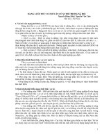
G-S G-L G-F D-S D-L D-F
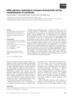
Figure 1 Examples of PCR markers generated by the AMP PCR technique.(a) Digestion-resistant ( R) markers from primer No.21, (b)
Digestion-sensitive (S) markers in sperm DNA produced from primer No.30 (white arrows indicate the PCR markers in the genomic samples and
black arrows indicate the lost of the markers in the digested samples) and (c) Digestion-dependent (D) markers in fibroblastic DNA produced
from primer No.1 (white arrows indicate no PCR markers in the genomic samples and black arrows indicate the PCR markers appear in the
digested samples). G-S = Genomic sperm DNA, G-L = Genomic leukocytic DNA, G-F = Genomic fibroblastic DNA, D-S = Digested sperm DNA, D-
L = Digested leukocytic DNA, D-F = Digested fibroblastic DNA
Phutikanit et al. Acta Veterinaria Scandinavica 2010, 52:18
/>Page 4 of 9
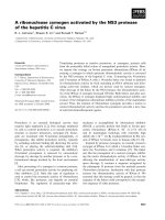
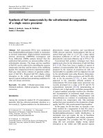
Figure 2 Example of the AMP PCR profile generated by the AMP PCR technique. This profile belonged to primer No.15. S = Sperm, L =
Leukocyte, F = Fibroblast. Long arrow indicates the digestion dependent marker appeared only in bull number 2. Short arrows indicate the
digestion resistant marker found in every cell sample from bull number 2 and in sperm DNA sample from bull number 3.
Table 2 Summary of the AMP PCR markers found in
sperm DNA
Bull 1 Bull 2 Bull 3 Ave ± SD
Sperm DNA R marker n 990 997 994 993.7 ± 3.5
% 92.8 92.9 94.4 93.4 ± 0.9
S marker n 37 44 34 38.3 ± 5.1
% 3.5 4.1 3.2 3.6 ± 0.5
D marker n 40 32 25 32.3 ± 7.5
% 3.7 3.0 2.4 3.0 ± 0.7
Total marker 1067 1073 1053 1064.3 ± 10.3
Table 3 Summary of the AMP PCR markers found in
fibroblastic DNA
Bull 1 Bull 2 Bull 3 Ave ± SD
Fibroblastic DNA R marker n 994 1006 1000 1000.0 ± 0.6
% 92.3 91.3 93.3 92.3 ± 1.0
S marker n 27 35 24 28.7 ± 5.7
% 2.5 3.2 2.2 2.6 ± 0.5
D marker n 56 61 48 55.0 ± 5.6
% 5.2 5.5 4.5 5.1 ± 0.5
Total marker 1077 1102 1072 1083.7 ± 16.1
Phutikanit et al. Acta Veterinaria Scandinavica 2010, 52:18
/>Page 5 of 9
DNA templates. The number of markers gained by this
technique was, in average, 30-40 markers per primer,
which was comparable with other studies [13,14]. In this
study, we applied a high concentration of oligonucleo-
tide primers (10 μmol) and used DNA polymeras e Stof-
fel fragment as some reports suggested that more PCR
markers could be obtained via this condition [13,15].
However, there are other factors affecting the marker
production. The sequence of primer, for instance, might
play an important role in this assay. From 30 sets of pri-
mer, we could summarize the results from only 27 sets,
while the other three primers gave poor patterns that
could not be scored. The annealing temperature in the
PCR step is also crucial [16]. In the present study, we
employed a high annealing temperature (53-57°C) to
prevent spurious amplification. The same condition has
been used in arbitrarily primed PCR technique with
good marker patterns [13,17,18]. However, the amount
of DNA markers gained per primer in this study was
slightly low when compared with other reports. This
might be due to differ ent marker detection methods.
We used acidic silver staining which has less sensitivity
than radioactive or fluorescent detection.
The similar AMP PCR profiles generated from three
bulls indicated that bull genome is highly conserved
with approximately 1.6% variations among individuals.
When the comparison of D NA methylation profil es was
made between germ- and somatic cells, we found that
germ cells contained less methylated HpaII locations in
their genome. This finding was in accordance with other
repo rts [7,19]. The hypomethylation status of spermato-
zoa might be associated with a special genome structure
designed for meiosis division, and possibly be involved
in specific gene expression at early stage of embryo
development [20].
Furthermo re, when we compared the methylation pat-
terns obtained from leukocytic and fibroblastic DNA,
the results showed that leukocytes had the highest
amount of DNA methylation in their genome. This
result was i n agreement with the knowledge that well-
characterized differentiated cells need only a small num-
ber of genes to be actively expressed to maintain their
functions, and the rest are suppressed by DNA methyla-
tion or other gene regulation processes [21]. On the
other hand, somatic cells possessing the a bility to
change their morphology and cell functions like fibro-
blasts exhibited differently. Our results showed that
Table 4 Summary of the AMP PCR markers found in
leukocytic DNA
Bull 1 Bull 2 Bull 3 Ave ± SD
Leukocytic DNA R marker n 1000 1012 1004 1005.3 ± 6.1
% 94.9 94.2 95.2 94.8 ± 0.5
S marker n 21 29 20 23.3 ± 4.9
% 2.0 2.7 1.9 2.2 ± 0.4
D marker n 33 33 30 32.0 ± 1.7
% 3.1 3.1 2.8 3.0 ± 0.2
Total marker 1054 1074 1054 1060.7 ± 11.5
Sperm
Fibroblast
Leukocyte
90
91
92
93
94
95
96
97
*
*
,
**
**
Percentage (%)
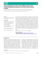
Figure 3 Percentage of the digestion resistant (R) markers
calculated from the pooled data. The box represents the average
percentage and the error bars standard deviations. Samples with
different number of asterisk (*) are statistically different.
Sperm
Fibroblast
Leukocyte
0
1
2
3
4
5
*
**
**
Percentage (%)
Figure 4 Percentage of the digestion sensitive (S) markers
calculated from the pooled data. The box represents the average
percentage and the error bars standard deviations. Samples with
different number of asterisk (*) are statistically different.
Sperm
Fibroblast
Leukocyte
0
1
2
3
4
5
6
*
**
**
Percentage (%)
Figure 5 Percentage of the digestion dependent (D) markers
calculated from the pooled data. The box represents the average
percentage and the error bars standard deviations. Samples with
different number of asterisk (*) are statistically different.
Phutikanit et al. Acta Veterinaria Scandinavica 2010, 52:18
/>Page 6 of 9
A
90
91
92
93
94
95
96
Bull 1 Bull 2 Bull 3
a
b
ab
c
c
d
Per centage of R mar ker
0
1
2
3
4
5
Bull 1Bull 2Bull 3
a
a
b
Percentage of S marker
B
0
1
2
3
4
5
6
Bull 1Bull 2Bull 3
a
a
b
c
c
d
e
e
f
Per centage of D mar ker
C
Sperm Leukocyte Fibroblast
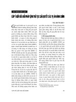
Figure 6 Individual variations of markers among bulls. Percentages of the R markers (Fig. 6-A), S markers (Fig. 6-B) and D markers (Fig. 6-C)
in sperm, leukocytic and fibroblastic DNA found in each bull. Different letters between cell samples within the same bull indicate that the
difference is statistic significance (P < 0.05).
Phutikanit et al. Acta Veterinaria Scandinavica 2010, 52:18
/>Page 7 of 9
fibroblast DNA was somehow hypomethylated when
compared with leukocyte and sperm DNA. Moreover,
we found a high percentage of digestion dependent mar-
kers in this cell type. The formation of this marker by
AMP PCR technique is not clearly understood, but we
hypothesized that the enzyme digestion might remove
some secondary s tructures of the genome, and this
allowed the binding of primers to their intact recogni-
tion locations hidden inside those complex structures.
In this case we surmised that fibroblast cells possibly
had special genomic architectures owing to their versati-
lity. It is challenging to figure out the origin of the
digestion dependent marker and the hypothesis of the
complex structures could be elucidated.
From this work, we proved that the AMP PCR techni-
que could generate methylation-associated fingerprints
from different cells and tissues obtained from Holstein
bulls. The technique could be used as a screening test
for the DNA methylation pattern of the animal. The dif-
ference of the AMP PCR patterns between each cell
type, though at a very low degree and could not be used
as an individual identification tool, could possibly facili-
tate the discovery of some differentially methylated loca-
tions in the genome. However, the results of this
present study were from the HpaII enzyme recognition
locations only. These particular locations are abundant
in the mammalian genome and many may not closely
associate with gene regulatory domains. To enhance the
ability of the AMP PCR in the study of the gene-specific
methylation profile in different tissues, other methyla-
tion sensitive restriction endonuclease enzymes recog-
nizing the methylation locations within gen es or gene
promoter regions could provide valuable information in
terms of methylation-associate gene expression. Radio-
labeling or fluorescent deoxynucleoside triphospate
could also be used in the PCR to increase the sensitivity
of marker detection.
Conclusions
We applied an arbitrarily primed PCR-based technique,
Amplified Methylation Polymorphism Polymerase Chain
Reaction (AMP PCR), to investigate DNA methylation
profiles in three different cell types obtained from Hol-
stein bulls. The methylation status of approximately
1,000 HpaII locations throughout the genome could be
identified by this present technique. We found that the
HpaII DNA methylation profile is tissue-specific. Male
germ cells were hypomethylated at the HpaII locations
when compa red with somatic cells, whil e the chromatin
of the well-characterized somatic cells was heavily
methylated when compared with that of the versatile
somatic cells.
Acknowledgements
This work was supported by Royal Golden Jubilee PhD Program, Thailand
Research Fund, The RTA 5080010 TRF grant and The RU
Rachadapiseksompoj endownment fund, Chulalongkorn University. We
acknowledge Associate Professor Dr. Padet Tummaruk for his help in
statistical analysis. We also would like to thank Dr. Masahiro Kaneda,
Associate Professor Dr. Kaywalee Chatdarong and Assistant Professor Dr.
Theerawat Tharasanit for the critical review of the manuscript.
Author details
1
Department of Obstetrics Gynaecology and Reproduction, Faculty of
Veterinary Science, Chulalongkorn University, Henri Dunant Rd, Bangkok
10330, Thailand.
2
School of Chemistry and Molecular Bioscience, Faculty of
Science, The University of Queensland, Brisbane, QLD 4072, Australia.
3
School
of Animal Studies, Faculty of Natural Resources, Agriculture and Veterinary
Science, The University of Queensland, Gatton, QLD 4343, Australia.
Authors’ contributions
NP carried out the AMP PCR assays and marker analysis. JS contributed in
preparing the chemicals used in the experiment. DH and BC contributed in
the experimental designs and techniques. MO and MT provided the concept
of the experiment and helped to draft the manuscript. All authors read and
approved the final manuscript.
Competing interests
The authors declare that they have no competing interests.
Received: 13 September 2009 Accepted: 5 March 2010
Published: 5 March 2010
References
1. Reik W, Santos F, Dean W: Mammalian epigenomics: reprogramming the
genome for development and therapy. Theriogenology 2003, 59:21-32.
2. Ehrlich M, Gama-sosa MA, Huang LH, Midgett RM, Kuo KC, McCune RA,
Gehrke C: Amount and distribution of 5-methylcytosine in human DNA
from different types of tissues of cells. Nucleic Acids Res 1982,
10:2709-2721.
3. Rougier N, Bourc’his D, Gomes DM, Niveleau A, Plachot M, Paldi A, Viegas-
Pequignot E: Chromosome methylation patterns during mammalian
preimplantation development. Genes Dev 1998, 12:2108-2113.
4. Shiota K, Kogo Y, Ohgane J, Imamura T, Urano A, Nishino K, Tanaka S,
Hattori N: Epigenetic marks by DNA methylation specific to stem, germ
and somatic cells in mice. Genes Cells 2002, 7:961-969.
5. Song F, Mahmood S, Ghosh S, Liang P, Smiraglia DJ, Nagase H, Held WA:
Tissue specific differentially methylated regions (TDMR): Changes in
DNA methylation during development. Genomics 2008, 93:130-139.
6. Kaput J, Sneider TW: Methylation of somatic vs germ cell DNAs analyzed
by restriction endonuclease digestion. Nucleic Acids Res 1979, 7:2303-2322.
7. Sturm KS, Taylor JH: Distribution of 5-methylcytosine in the DNA of
somatic and germline cells from bovine tissues. Nucleic Acids Res 1981,
9:4537-4546.
8. Ponzetto-Zimmerman C, Wolgemuth DJ: Methylation of satellite
sequences in mouse spermatogenic and somatic DNAs. Nucleic Acids Res
1984, 12:2807-2822.
9. Liu ZJ, Maekawa M: Polymerase chain reaction-based methods of DNA
methylation analysis. Anal Biochem 2003, 317:259-265.
10. Papadopoulos S, Benter T, Anastassiou G, Pape M, Gerhard S, Bornfeld N,
Ludwig WD, Dorken B: Assessment of genomic instability in breast
cancer and uveal melanoma by random amplified polymorphic DNA
analysis. Int J Cancer 2002, 99:193-200.
11. Ribeiro GRH, Francisco G, Teixeira LVS, Romao-Correia RF, Sanches JA Jr,
Neto CF, Ruiz IRG: Repetitive DNA alterations in human skin cancers. J
Dermatol Sci 2004, 36:79-86.
12. Gu W, Post CM, Aguirre GD, Ray K: Individual DNA bands obtained by
RAPD analysis of canine genomic DNA often contain multiple DNA
sequences. J Hered 1999, 90:96-98.
13. Waldron J, Peace CP, Searle IR, Furtado A, Wade N, Finlay I, Graham MW,
Carroll BJ: Randomly amplified DNA fingerprinting: A culmination of DNA
Phutikanit et al. Acta Veterinaria Scandinavica 2010, 52:18
/>Page 8 of 9
marker technologies based on arbitrarily-primed PCR amplification. J
Biomed Biotech 2002, 2:141-150.
14. DeLaat DM, Carvalko MRS, Lovato MB, Acedo MDP, da Fonseca CG:
Applicability of RAPD markers on silver-stained polyacrylamide gels to
ascertain genetic diversity in Peripatus acacioi (Peripatidae;
Onychophora). Genet Mol Res 2005, 4:716-725.
15. McClelland M, Welsh J: DNA fingerprinting by arbitrarily primed PCR.
Genome Res 1994, 4:S59-S65.
16. Atienzar FA, Jha AN: The random amplified polymorphic DNA (RAPD)
assay and related techniques applied to genotoxicity and carcinogenesis
studies: A critical review. Mutat Res 2006, 613:76-102.
17. Kohno T, Kawanishi M, Inazawa J, Yokoto J: Identification of CpG islands
hypermethylated in human lung cancer by the arbitrarily primed-PCR
method. Hum Genet 1998, 102:258-264.
18. Liang G, Gonzalgo ML, Salem C, Jones PA: Identification of DNA
methylation differences during tumorigenesis by methylation-sensitive
arbitrarily primed polymerase chain reaction. Methods 2002, 27:150-155.
19. Oakes CC, La Salle S, Smiraglia DJ, Robaire B, Trasler JM: A unique
configuration of genome-wide DNA methylation patterns in the testis.
Proc Natl Acad Sci USA 2006, 104:228-233.
20. Yamagata K, Yamazaki T, Miki H, Ogonuki N, Inoue K, Ogura A, Baba T:
Centromeric DNA hypomethylation as an epigenetic signature
discriminates between germ and somatic cell lineages. Dev Biol 2007,
312:419-426.
21. Reik W, Dean W: DNA methylation and mammalian epigenetics.
Electrophoresis 2001, 22:2838-2843.
doi:10.1186/1751-0147-52-18
Cite this article as: Phutikanit et al.: Different DNA methylation patterns
detected by the Amplified Methylation Polymorphism Polymerase
Chain Reaction (AMP PCR) technique among various cell types of bulls.
Acta Veterinaria Scandinavica 2010 52:18.
Submit your next manuscript to BioMed Central
and take full advantage of:
• Convenient online submission
• Thorough peer review
• No space constraints or color figure charges
• Immediate publication on acceptance
• Inclusion in PubMed, CAS, Scopus and Google Scholar
• Research which is freely available for redistribution
Submit your manuscript at
www.biomedcentral.com/submit
Phutikanit et al. Acta Veterinaria Scandinavica 2010, 52:18
/>Page 9 of 9