Báo cáo thú y: " Dynamics of serum antibodies to and load of porcine circovirus type 2 (PCV2) in pigs in three finishing herds, affected or not by postweaning multisystemic wasting syndrome" pdf
Bạn đang xem bản rút gọn của tài liệu. Xem và tải ngay bản đầy đủ của tài liệu tại đây (472.46 KB, 10 trang )
RESEARC H Open Access
Dynamics of serum antibodies to and load of
porcine circovirus type 2 (PCV2) in pigs in three
finishing herds, affected or not by postweaning
multisystemic wasting syndrome
Inger M Brunborg
1*
, Caroline Fossum
2
, Bjørn Lium
1,5
, Gunilla Blomqvist
3
, Elodie Merlot
2,4
, Anne Jørgensen
5
,
Lena Eliasson-Selling
6
, Espen Rimstad
7
, Christine M Jonassen
1
, Per Wallgren
3,8
Abstract
Background: Despite that PMWS commonly affects pigs aged eight to sixteen weeks; most studies of PMWS have
been conducted during the period before transfer to finishing herds. This study focused on PCV2 load and
antibody dynamics in finishing herds with different PMWS status.
Methods: Sequentially collected blood samples from 40 pigs in each of two Swedish (A and B) and one
Norwegian (C) finishing herds were analysed for serum PCV2-load and -antibodies and saliva cortisol. The two
Swedish herds differed in PMWS status, despite receiving animals from the same sow pool (multi-site production).
However, the PMWS-deemed herd (A) had previously also received pigs from the spot market. ResultsThe initial
serum PCV2 load was similar in the two Swedish herds. In herd A, it peaked after two weeks in the finishing herd
and a high number of the pigs had serum PCV2 levels above 10
7
per ml. The antibody titres increased continually
with exception for the pigs that developed PMWS, that had initially low and then declining antibody levels. Pigs in
the healthy herd B also expressed high titres of antibodies to PCV2 on arrival but remained at that level
throughout the study whereas the viral load steadily decreased. No PCV2 antibodies and only low amounts of
PCV2 DNA were detected in serum collected during the first five weeks in the PMWS-free herd C. Thereafter a peak
in serum PCV2 load accompanied by an antibody response was recorded. PCV2 from the two Swedish herds
grouped into genotype PCV2b whereas the Norwegian isolate grouped into PCV2a. Cortisol levels were lower in
herd C than in herds A and B.
Conclusions: The most obvious difference between the Swedish finishing herds and the Norwegian herd was the
time of infection with PCV2 in relation to the time of allocation, as well as the genotype of PCV2. Clinical PMWS
was preceded by low levels of serum antibodies and a high load of PCV2 but did not develop in all such animals.
It is notable that herd A became affected by PMWS after errors in management routine, emphasising the
importance of proper hygiene and general disease-preventing measures.
Background
A role of porcine circovirus type 2 (PCV2) in the etiology
of postweaning multisystemic wasting syndrome (PMWS)
was first observed in Canada in 1991, and described in the
late 1990s [1]. Since then, PMWS has been diagnosed
globally [2], but no single factor that triggers PMWS in
PCV2-infected pigs has been identified. Attempts to relate
the occurrence of PMWS to infection with PCV2 of a cer-
tain genotype have not been conclusive and the spread of
PMWS is still enigmatic [3]. PCV2 seems to be ubiquitous
in pigs [2], and the ambiguity of PMWS is evident in
multi-site sow pool systems which can include both
healthy and PMWS-affected satellites, despite that the
sows are mixed at a common sow hold during the dry per-
iod, and alter between farrowing sites [4].
PMWS appeared comparatively late at the Scandina-
vian Peninsula and wa s not diagnosed in Sweden or
* Correspondence:
1
National Veterinary Institute, PO Box 750 Sentrum, N-0106 Oslo, Norway
Brunborg et al. Acta Veterinaria Scandinavica 2010, 52:22
/>© 2010 Brunborg et al; licensee BioMed Central Ltd. This is an Open Access article distributed under the terms of the Creative
Commons Attribution License ( which permits unrestricted use, distribut ion, a nd
reproduction in any medi um, provided the original work is properly cited.
Norway until 2003 when two Norwegian herds were
affected by PMWS [5]. These herds were stamped out
during the spring/summer of 2004, and until February
2008 no new case of PMWS was diagnosed in Norway
as also demonstrated by screening programs performing
necropsies on runt pigs [6]. In Sweden, PMWS was
diagnosed for the first time in December 2003 [7].
Three years later, 124 herds had been diagnosed with
PMWS and the disease was regarded as endemic in the
country [8]. Thus, the spread of PMWS was interrupted
inNorwaybutprevailedinSweden,andin2007,when
the present study was conducted, PCV2 was present in
pigs from both countries but PMWS was only diagnosed
in Swedish herds.
Pigs can be affected by PMWS up to 16 weeks of
age [2,9,10], which includes at least the first month in the
finishing unit. As the mean economical loss for each dead
finishing pig exceeds that of a dead weaner by 50% [11],
and because the mortality figures due to PMWS in Swe-
den have been fairly equal in all categories of herds [8],
the economic impact of PMWS is like ly to be higher in
finishing herds than in piglet producing herds. Despite
this, most studies of PMWS have focused on the period
from weaning until transfer to finishing herds. In a recent
field study conducted in Denmark and Spain it was shown
that the majority of cases with PMWS in Denmark
occurred in the nurseries whereas the incidence of PMWS
in Spain was highest in the finishing facilities [12].
The primary objective of the present field study was to
investigate the relation between PCV2 load and levels of
antibodies to the virus in serum collected from finishing
pigs housed in herds with and without PMWS. As stress
level has been suggested to contribute to the d evelop-
ments of PMWS [13], saliva was collected for the assess-
ment of cortisol levels. Two Swedish herds, o ne affected
with PMWS (A) and one n ot affected (B), were investi-
gated. These herds had equally sized finishing units and
recruited growers from different herds within the same
Swedish sow pool (a multi site productio n system where
piglet producing herds lease pregnant sows from a shared
central unit). For compar ison a Norwegian finishing herd
(C) recruiting growers from a Norwegian sow pool free
from PMWS was included. The study was conducted in
2007 when PMWS was endemic in Sweden, but no clini-
cal case of PMWS was diagnosed in Norway.
Materials and methods
General health status and description of herds
Both Sweden and Norway are free from diseases listed
by the Office Inter national des Epizo oties (OIE), includ-
ing Aujeszky’s disease (AD) and porcine reproductive
and respiratory syndrome (PRRS), as well as from por-
cine endemic diarrhoea (PED) and transmissible gastro-
enteritis (TGE).
The three herds (A, B and C) included in the study,
were selected in order to match in size, type and man-
agement. The sows were not vaccinated against PCV2,
no vaccinations of the grovers were performed. and the
feed was free from antibiotics. All three herds effectu-
ated all in-all out production in cycles of 16 weeks in
unit s with 350 to 400 pigs, and recruited growers at the
weight of about 30 kg from piglet producing satellite
herds in sow pools. The trade with pigs within the
Swedish sow pool is illustrated in Figure 1 and a brief
description of the herds is given below.
Herd A was a specialised Swedish finishing herd with
4 units, recruiting 400 growers to one of the units every
4
th
week. The herd used to recruit every second batch
of growers from herd B until September 2006, and the
batches in-between these from the open market. In
order to receive all growers from the same source, herd
A contracted herd Z that was a specialised piglet produ-
cing satellite within the same sow pool as herd B. The
first batch from herd Z arrived in June 2006, and from
October 2006 all growers emanated from that herd.
Herd A generally cleaned and washed every unit
between consecutive batches, but during the process of
changing piglet supplier (from herd B and open market
to herd Z), occasionally market weight finishing pigs left
a unit in the morning and new growers arrived in the
afternoon, leaving little or no time for hygienic mea-
sures. In accordance with the EU-definition [14], herd
A was diagnosed with PMWS in February 2007.
Herd B was an integrated Swedish farrow to finish
herd with two finishing units, recrui ting 400 growers to
one of the units every 8
th
week. The herd had four far-
rowing units and farrowing took place every 4
th
week.
At every second farrowing, herd B recruited own pigs to
one of the two finishing units that were located less
than 100 m from the farrowing units. Herd B cleaned
and washed every unit between consecutive batches, and
the empty time between batches had been 5.7 ± 0.6
days for the last 15 batches (120 weeks). Herd B was,
and by September 2009 still is, free from PMWS.
Herd C was a recently established Norwegian finishing
herd with two identical units each with 350 pigs,
recruiting pigs to both units every 16
th
week. The he rd
recruited growers from piglet producing satellites in a
Norwegian sow pool. It cleaned and washed each unit
between consecutive batches, and the empty time
between the seven first batches was 4.3+1.5 days. By
September 2009 this herd is still free from signs of
PMWS.
General study design
This study was approved by the ethical committee in
Uppsala, Sweden (License C120/7). The study was car-
ried out during the spring of 2007, one month after
Brunborg et al. Acta Veterinaria Scandinavica 2010, 52:22
/>Page 2 of 10
herd A had been diagnosed with PMWS. In each herd,
40 pigs in one batch were scrutinised. One week after
arrival to the finishing unit, 4 randomly s elected pigs
from each of 10 pens were given an identity by ear tag-
ging. Blood samples without additive were collected
weekly from each of these pigs by jugular vein p uncture
during weeks 1 to 5 after arrival i n all herds, and the
serum samples were stored at -20°C until analys ed. Two
additional samplings were carried out in herd C at
weeks 9 and 11 after arrival. Clinical signs of disease
were recorded weekly for the 40 pigs. Clinical signs that
could indicate PMWS were examined and the pigs were
accordingly referred to as “ healthy” , “thin” (under
weight) and/or “hairy” (having a rough appearance). The
chest perimeter was measured to estimate the individual
growth rate and every pig suspected for PMWS was
culled and the clinical diagnosis was either confirmed or
rejected by necropsy. To measure chronic stress, saliva
samples were collected a t week five from ten pigs
housed in pens adjacent to the experimental pigs t o
measure cortisol levels. The saliva samples were col-
lected by letting the pigs chew on cotton swabs
(Salivette, Sarstedt AG, Nümbrecht, Germany) until
moistened. The cotton buds were kept on ice until cen-
trifuged for 15 minutes at 300 g, 4°C, and the recovered
liquid was stored at -20°C until analysed. All saliva sam-
ples were collected at mid-day to avoid differences due
to the normal diurnal variation in cortisol levels.
Measurement of saliva cortisol levels
The cortisol was measured using a luminescence immu-
noassay kit (LIA, IBL, D-22335 Hamburg, Germany).
The assay sensitivity was 0.15 ng per ml. The inter- and
intra-assay coefficients of variation were 7.8 % and 6.1 %,
respectively, at 2.1 ng/ml.
Nucleotide sequencing of isolates
The vi rus isolates from the three herds were determined
by nucleotide sequencing of the entire genome by two
overlapping PCR products. Sequences were acquired
from three pigs from each herd and a consensus
sequence was created. Primers used for amplification
were PCV2-ORF1-1673 towards PCV-F-1319L21, and
PCV2-Cap-sense towards PCV-C-1256U21 (Table 1).
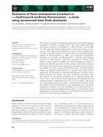
Figure 1 The supply of growers to herd A. A change of pig supplier was initiated because Herd Z could supply herd A with all finishing pigs.
During this process the empty time between batches was decreased giving little or no time for hygienic measures. Herd A was diagnosed with
PMWS in February 2007, while finishing pigs in herd B remained free from PMWS. Herd B and herd Z were neighbours and received pregnant
sows from the same sow pool. The distance to herd A was 115 km for both herds. (FU = farrowing units, FiU = finishing units).
Brunborg et al. Acta Veterinaria Scandinavica 2010, 52:22
/>Page 3 of 10
Briefly, a 50 μl PCR reaction (0.3 mM dNTP, 0.5 μMof
each primer, 1.5 U HotStar Taq DNA polymerase in a
1× PCR buffer provided with the kit) (HotStar Taq
DNA Polymeras e, Qiagen, Germantown, MD, USA) was
run with the follo wing program (95°C for 15 min fol-
lowed by 41 cycles of 94°C for 50 sec, 55°C for 60 sec
and 72°C for 45 sec (PCV-C-1256U21) or 95 sec (PCV-
F-13119L21), with a final elongation step of 5 min at
72°C). Inner primers used for sequencing are displayed
in Table 1. Nucleotide sequencing was run on the Avant
3100 (Applied Biosystems, Foster City, CA, USA) and
sequence analysis was performed using Sequencing Ana-
lysis 5.2 Patch 2 (Applied Biosystems), Sequencher 4.5
(Gene Codes Corporation, Ann Arbor, MI, USA) and
MEGA 3.1 . The sequences
were compared pair-wi se at both the nuclotide and
amino acid levels using Lasergen and MegAlign Soft-
ware, version 1.13 (DNASTAR). Multiple alignments
were performed using the CLUSTAL W program.
Real-time PCR for quantification of PCV2
DNA was isolated, and a quantitative real-time PCR was
run on all serum samples. Briefly, n ucleic acids were
isolated from 200 μl ser um using a NucliSEN S® easy-
MAG™ nucleic acids extractor (bioMérieux, Durham,
NC, USA), and elut ed in 55 μl elution buffer. Following
sequencing of the viruses found in each herd, tailored
primers and probe based on a previously described pro-
tocol [15], were used f or unbiased a mplification and
absolute quantification of PCV2 DNA. In brief, forward
primer PCV-E-1319L21 and reverse primer PCV-
A-1256U21 in combination with TaqMan2-PCV2 were
used for the Swedish samples (herds A and B). The
Norwegian samples (herd C) were analy sed using for-
ward primer PCV-D-1 319L21, reverse primer PCV2-
84-1256U21 and TaqMan-1286-1314 as probe. For each
sample, 2.5 μl of the eluate was run in a 25 μl reaction
with an annealing step at 60°C, on an MxPro 3005 PCR
machine (Stratagene, Agilent Technologies, Inc., Santa
Clara, CA, USA). Results are given as number of DNA
copies per ml serum.
Detection of PCV2 specific serum antibodies
Antibodies to PCV2 wer e measured in indiv idual serum
samples using an immunoperoxidase monolayer assay
(IPMA) technique previously described [16] with slight
modifications [17]. The serum samples were diluted in
serial two-fold st eps (from 1:10 to 1:20,480) in PBS con-
taining 0.05 % Tween and 5% fat-free milk powder. The
results are presented as 10 log values of the highest
dilution with positive reaction in the IPMA. Titres less
than 1/40 (10
1.6
) were considered as negative.
Statistical analysis
Quantitative real-time PCR-samples below the d etection
limit of 1.1 × 10
3
copies per ml serum were set to 550
(0.55 × 10
3
) copies per ml serum, representing the mean
of the values, and likewise, the samples calculated to be
between the detection limit and the quantification limit
of 1.1 × 10
4
copies per ml serum [15], were set to 6.05
×10
3
copies per ml serum. Fisher test was used for
comparison of number of animals with v iral load above
10
7
PCV2 DNA copies per ml serum. To evaluate differ-
ences in PCV2 load, levels of antibodies to PCV2, and
production data of the pigs in the three herds, groups
were compared pair wise usi ng double sided t-tests (two
sample tests with unequal variation).
Results
General health status and performance
Moderate lameness and coughing were o bserved in a
few pigs in each herd, but the general health status and
performance were high in all herds. During the early
rearing period, this was demonstrated by steadily
increasing c hest perimeters of the 40 principals in each
herd. From weeks 1 to 5 the chest perimeters increased
with 16.1 ± 5.4 cm, 14.3 ± 2.7 cm and 12.7 ± 1.8 cm in
herd A, B and C, respectively (A and B vs. C; p < 0.01,
A vs. B; p = 0.07).
All three herds had a h igh daily weight gain and the
mean daily weight gain of pigs that reached market
weight in herd A was not affected during the period
when the herd was diagnosed with PMWS (Table 2).
However, the mortality during the rearing period
increased from 1.8 ± 0.5% to 2.9 ± 1.3% (p < 0.01), and
the prevalence of pigs slaughtered at underweight
increased from 1.7 ± 1.0% to 3.6 ± 2.5% (p < 0.05). The
mean mortality in herds B and C was l ess than 1%
throughout the study.
An increased frequency of runts, wasting pigs and
mortality was observed during the period when herd A
changed piglet supplier from herd B and the open mar-
ket to herd Z during June to October in 2006. Due to a
14 day discrepancy between farrowing periods in these
herds, less than 24 hours were allowed between batches
at several occasions (Table 2). In February 2007, the
Table 1 Primers used for amplification of PCV2 DNA and
nucleotide sequencing.
Primer designation Primer sequence
PCV-C-1256U21 3’-ATA GCG GGA GTG GTA AGA GAA-5’
PCV-F-1319L21 3’-GCA ACA GCC CTA ACC TAT GAC-5’
PCV2-Cap-sense 5’-ATG ACG TAT CCA AGG AGG CG-5’
PCV2-ORF1-415 3’-CTG TGA GTA CCT TGC TGG AGA-5’
PCV2-ORF1-501 3’-GCT CAC TTT CAA AAG TTC AGC-5’
PCV2-ORF1-804 3’-CTG ATT ACC AGC AAT CAG ACC-3’
PCV2-ORF1-881 3’-CCT CCG ATA GAG AGC TTC TAC-3’
PCV2-ORF1-1673 3’-TGG CCA AGA TGG CTG CGG-5’
Brunborg et al. Acta Veterinaria Scandinavica 2010, 52:22
/>Page 4 of 10
mortality in a batch reached 4.3% and herd A was then
officially diagnosed with PMWS based on cl inical and
laboratory findings. At that time pigs in the elde st batch
had arrived at the herd in November 2006. However,
batches with increased mortality had been observed ear-
lier,peakingat3.6%inagroupthatarrivedbytheend
of November 2005. Therefore, batches arriving from
that time until the herd was officially diagnosed with
PMWS are referred to “suspected” for PMWS in Table 2.
Herd A was officially declared free from PMWS at the
end of February 2008, and batches arriving from March
2008 are again referred to as healthy (Table 2).
Clinical signs
One week after arrival, two pigs in herd A expressed
clinical signs resembling PMWS (under weighted =
“thin” or having a rough appearance = “hairy”). At the
following observations such signs were observed in 2-6
pigs. Five percent (2/40) of the pigs in herd A developed
clinical PMWS (pig number 13 at day 18, and pig num-
ber 6 at day 35). Both pigs expressed an acute wasting
that was also mirrored by a reduced chest perimeter
(from 67 to 58 cm within 4 days in pig 13, and from 65
to 61 cm in pig 6 during the last week), and enlarged
inguinal lymph nodes. Both pigs were euthanized during
wasting and PMWS was confirmed by necropsy by ful-
filling the criteria demanded, including enlarged lymph
nodes with lymphocyte depletion, presence of giant cells
and a massive quantity o f PCV2 detected by immun os-
taining [14].
In herd B signs resembling PMWS ("thin ” and/or
“hairy”) were observed in two pigs, but no pig in this
herd developed clinical PMWS. In herd C, no clinical
signs PMWS were observed in any pig.
Nucleotide sequence typing
A high similarity (99.7%) was found at t he nucleotide
level when compari ng the full genome sequence of PCV2
obtained fro m the tw o Swedis h herds ( A and B), des pite
that they o riginated from a pig diagnosed with PMWS
(herd A), and from a healthy pig (herd B). The similarity
between these two Swedish sequences and that obtained
from the Norwegian (herd C) was 95.5%. According to
the proposed nomenclature for definition of PCV2 geno-
types [18], the Norwegian isolate grou ped into PCV2a
whereas the two Swedish isolates grouped into PCV2b.
PCV2 load in serum
The PCV 2 DNA copy number was determined by quan-
titative real-time PCR as an estimate of PCV2 viral load
Table 2 Production data for a Swedish finishing herd (A) during the course of PMWS.
Herd A Herd B Herd C
Health status regarding PMWS Healthy Suspected Deemed Healthy Healthy Healthy
Arrival of first and last batch in category Jan05 – Oct05 Nov05 – Oct06 Nov06-Feb08 Mar08 – Sept08
Number of batches 11 13 18 6 11 7
Source of finishing pigs:
Herd B (number of batches) 6/11 6/13 0/18 0/6 11
1
/11 Norwegian
Open market (number of batches) 5/11 4/13 0/18 0/6 0/11 sow
Herd Z (number of batches) 0/11 3/13 18/18 6/6 0
1
/11 pool
Batches not preceded by empty days (n) 3 5 4 0 0 0
Mean empty time between batches (days) 4.0 ± 2.8 3.8 ± 3.6 4.4 ± 2-9 5.8 ± 1.2 5.7 ± 0.6 4.3 ± 1.5
Pigs/batch (n) 385 ± 1 385 ± 1 385 ± 1 385 ± 1 389 ± 17 704.7 ± 4.3
Arrival weight (kg) 32.0 ± 3.4
a
** 31.9 ± 4.0
a
** 28.0 ± 3.6
b
27.4 ± 2.5
b
31.1 ± 1.8 29.0 ± 0.9
Slaughter weight carcas (kg) 87.9 ± 1.6 87.5 ± 2.3 89.3 ± 2.1 87.4 ± 1.8 86.1 ± 2.1 80.3 ± 2.6
Rearing period (days) 104.8 ± 3.5 102.5 ± 5.4 106.3 ± 4.6 103.8 ± 5.6 104.4 ± 5.8 98.4 ± 4.7
Percentage meat of carcas (%) 57.5 ± 0.8 57.8 ± 0.3 57.8 ± 0.7 57.3 ± 0.4 57.8 ± 0.7 56.3 ± 0.6
Mortality, mean (%) 1.8 ± 0.5
a
** 2.2 ± 0.9
a
* 2.9 ± 1.3
b
2.4 ± 1.0
b
0.5 ± 0.6 0.6 ± 0.5
Mortaliy, range (%) 1.1 – 2.6 1.0 – 3.6 1.0 – 6.2 0.8 – 3.4 0.0 – 2.2 0.1 – 1.4
Condemned at slaughter (%) 0.5 ± 0.5 0.5 ± 0.6 0.4 ± 0.5 0.6 ± 0-6 0.2 ± 0.2 0.5 ± 0.5
Daily weight gain (g) 910 ± 30 911 ± 35 914 ± 35 898 ± 30 886 ± 21 924.7 ± 32.3
Slaughter weight < 73 kg (%) 1.7 ± 1.0
a
** 2.4 ± 1.5 3.6 ± 2.5
b
5.3 ± 3.4 3.5 ± 1.6 No records
Slaughter weight < 73 kg, range 0.5 - 3.5 0.3 – 4.9 0.1 - 10.3 1.6 – 9.9 0.5 – 5.9 No records
Results on the same line with different letters differ significantly from each other; p < 0.05 (*) or p < 0.01(**)
For comparison, corresponding data are given for a herd (B) that received animals from the same sow pool as herd A but remained free from PMWS, and for a
healthy Norwegian finishing herd (C). The count ry of Norway was free from PMWS when the study was conducted. Herd B and herd Z received sows from the
same sow pool, i.e. from the same source.
Brunborg et al. Acta Veterinaria Scandinavica 2010, 52:22
/>Page 5 of 10
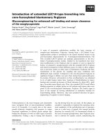
in serum (Figure 2). One week after arrival, the mean
DNA copy number was similar (10
6
per ml serum) for
pigs in herds A and B, but as seen in Table 3, p igs in
herd A tended to express either high or low viral load
(13 pigs above 10
7
DNA copies per ml serum and 7 pigs
with less than 10
4
DNA copies per ml serum). The aver-
age viral load for pigs in herd A peaked at 10
6.5
per ml
serum two weeks after arrival to the finishing unit, and
then declined to 10
5.4
per ml in week five. In herd B,
theaverageviralloaddecreasedcontinuouslyfrom10
6
per ml to 10
5
per ml serum in week 5. In contrast, no
PCV2 DNA was detectable in serum of any pig in herd
C during the fir st week after arrival. After five weeks in
herd C the avera ge viral load was 10
3.5
per ml s erum,
but values up to 10
6.4
per ml serum were recorded in
individual pigs. During the extended period of sampling
in herd C, the highest mean viral load (10
4.3
per ml
serum ) was recorded nine weeks after arrival. The high-
est incidence of pigs with a high viral load (exceeding
10
7
per ml serum) was found in the PMWS affected
herd (A), predominantly during the early fattening per-
iod(Table3).TheloadofPCV2inthetwopigsthat
0,0
1,0
2,0
3,0
4,0
5,0
6,0
7,0
8,0
9,0
12345911
Log PCV2 per ml
0
0,5
1
1,5
2
2,5
3
3,5
4
4,5
5
12345911
Week after arrival
Log titre
Figure 2 Mean l og levels of PCV2 DNA copy number per ml serum (upper) and log titre of antibodies to PCV2 (lower) in herds A
(PMWS; filled diamonds), B (healthy, open squares) and C (healthy, open circles).
Table 3 Number of pigs with a serum load of PCV2
exceeding 10
7
per ml serum.
Herd Week 1 Week 2 Week 3 Week 4 Week 5
A 13/40 11/40 7/39 4/39 2/39
B 1/40 2/40 0/40 0/40 0/40
C 0/40 0/40 1/40 0/40 0/40
The animals were sampled during the first five weeks in two Swedish (Herds
A and B) and one Norwegian (Herd C) finishing unit. Two of the forty pigs in
herd A were diagnosed with PMWS, at 18 and 35 days after arrival,
respectively. Based on fisher tests the number of pigs with virus load above
10
7
DNA copies per ml serum was significantly higher in herd A than in the
other two herds in week 1, 2 and 3 (p < 0.05).
Brunborg et al. Acta Veterinaria Scandinavica 2010, 52:22
/>Page 6 of 10
developed PMWS increased to 10
10
per ml serum at the
last occasion of sampling (day 18 and day 35, respectively).
Antibody titres to PCV2 in serum
In herd A, the mean antibody titre to PCV2 was 10
2.52
on arrival, and had increased to 10
3.52
five weeks later
(Figure 2). Pigs in herd B had in general a higher level
of antibodies to PCV2 (10
3.88
) than those in herd A
when arrivi ng at the finishing unit and remained at that
level during the five weeks of sampling. Pigs in herd C
were seronegative to PCV2 on arrival and remained
negative during the first five weeks in the herd. By week
nine these pigs had seroconverted to PCV2 and had a
mean antibody titre of 10
3.24
at the l ast sampling o cca-
sion (11 weeks after arrival).
The two pigs in herd A that developed PMWS were
both seronegative to PCV2 in the IPMA test (titre
<10
1.6
) at the last occasion of sampling. In pig 6 that
was s till alive at the last sampling occasion, no indica-
tion of a serological antibody response to the increasing
viral load was seen.
Cortisol levels in saliva
Cortisol levels in saliva were lower in herd C (1.06 ±
0.14 ng per ml) than in herds A and B (1.80 ± 0. 24 and
1.87 ± 0.19 ng per ml, respectively, p < 0.05). However,
the cortisol levels were within the normal range in all
herds.
Discussion
In addition to different genotype of PCV2 in the Norwe-
gian herd compared to the Swedish herds, the most
remarkable differences betw een pigs from the three fin-
ishing herds were the levels and kinetics o f their anti-
body response to PCV2, indicat ive of differe nt starting
situations at the time of allocation to the finishing herd.
The highest levels of antibo dies to PCV2 were recorded
in serum f rom pigs in the healthy Swedish herd (B). In
contrast, pigs in the healthy Norwegian herd (C) were
seronegative to PCV2 at arrival and remained so during
the first observation period of five weeks. The sampling
period was therefore prolonged in this herd and a sero-
conversion to PCV2 took place between 5 and 9 weeks
after arrival. Most pigs (29/40) in the Swedish PMWS-
affected herd (A) were seropositive to PCV2 at arrival,
but had lower titres than animals in herd B (p < 0.01).
The antibody ti tres increased continuously in herd A,
with exception for the two pigs that developed PMWS.
These two pigs had initially low, declining antibody
levels to P CV2 and were regarded as seronega tive when
displaying clinical symptoms of PMWS.
The observed serological responses to PCV2 are well
in line with prev ious studies [12,17,19-21] supporting
the relationship between PCV2 and PMWS also in
finishing pigs. The lack of a proper antibody response in
the two pigs that developed PMWS in herd A, further
support earlier studies pointing out that neutralizing
antibodies to PCV2 are protective against PMWS
[22-25]. The IPMA-method used in this study does not
discriminate between neutralizing and non-neutralizing
antibodies, but a positive correlation between neutraliz-
ing antibodies and total amount of antibodies has pre-
viously been reported [22]. Indeed, the mean antibody
titres to PCV2 increased steadily for the majority of pigs
in the PMWS affected herd (A), indicating an ongoing
infection with PCV2 on herd level.
The quantification of PCV2 DNA copies in serum
revealed a similar viral load in pigs when entering the
two Swedish finishing herds. A discrepancy was, how-
ever, that the mean serum viral load i ncreased during
the two first week s for pigs that were allocated to the
finishing unit affected by PMWS, whereas this load stea-
dily decreased in serum samples collected from pigs in
the healthy Swedish finishing herd. In clear contrast, no
PCV2 DNA was detected in any serum sample collected
during the first week in the Norwegian herd. Instead,
low levels of PCV2 DNA could be detected in serum of
a handful of these pigs after th ree weeks in the fin ishing
unit, coinciding in time with seroconversion. Thus, most
of these pigs were exposed to PCV2 at an age of 16 - 21
weeks, i.e. when pigs are regarded less likely to develop
PMWS [2,9,10]. This discrepancy in age at the time of
infection was also observed by Grau-Roma and others,
as pigs in Spain were infected a t a higher age tha n the
Danish pig s [12]. Epidemiological studies of risk factors
in PMWS dynamics have also shown that early infection
increases the risk of PMWS [26-28].
It is notable that the viral load of PCV2 was higher in
herd A than in the other herds, and that the number of
pigs with serum viral levels above a proposed cut off at
107 per ml serum [15] as also supported by others [29]
differed between the three herds. Herd A had a signifi-
cantly higher number of pigs with serum PCV2 levels
above 107 per ml during the first three weeks after arri-
val (p < 0.05), corresponding to the period of risk for
PMWS in finishing herds [2,9,10]. This shows that
although it is a crude tool, serum virus level may be
used as an indicator of PMWS status on herd level, pro-
vided that the pigs are sampled at an appropriate time,
i.e. during the first weeks in the finishing herd. It should
however be noted that pigs with high viral load of PCV2
maymountaprotectiveimmuneresponsetotheinfec-
tion, and do not necessarily develop PMWS [12,17]. In
the present study, 18 of 20 pigs with a viral load above
107 per ml serum did not develop clinical PMWS or
other PCV2 associated clinical signs.
Several external factors, including increa sed stress
levels, have been suggested to contribute to the
Brunborg et al. Acta Veterinaria Scandinavica 2010, 52:22
/>Page 7 of 10
developments of PMWS as reviewed [13]. Social stress
of pigs is associated with a negative effect on the anti-
viral immunity [30] and experimental studies have indi-
cated that dexamethasone treatment can influence the
pathogenic effect of PCV2, suggesting a role of stress
and glucocorticoids in the P MWS aetiology [31]. Herd
A distinguished from the two other herds by a higher
mortality even du ring the periods f ree from PMWS.
Furthermore, herd A became affected by PMWS after
intensified r outines with no empty time between some
of the batches. Cor tisol secretion was det ermined in
order to test whether the more intensive management
practices of herd A could have generated higher stress
levels. The levels of cortisol determined in saliva col-
lected from pigs in adjacent pens to those examined
were similar in the two Swedish herds. Although these
mean values were somewhat higher than those recorded
for t he Norwegian pigs, the cortisol levels for the three
herds were all within the normal range [32] and no
extreme stress-re lated behaviour suc h as tail-biting were
recorded in any of the herds. Thus, long-term stress
was unlikely to have caused the outbreak of PMWS in
herd A.
Another factor that differed between the investigated
herds was the predominating genotype of PCV2.
Sequencing rev ealed that according to the nomenclature
proposed by Segalés et al (2008), PCV2a was present in
the Norwegian samples, wherea s PCV2b wa s found in
the two Swedish he rds. In Sweden, PCV2b has be en
found in s amples from herds diagnosed with PMWS as
well as from healthy herds, whereas PCV2a has not yet
been demonstrated in herds diagnosed with PMWS
[33]. Currentl y there is a controversy regarding the pos-
sible influence of PCV2 genotype on the development of
PMWS, and during experimental conditions PCV2a
readily induces PMWS [34-36]. Furthermore, in a survey
on the island of Ireland, both genotypes of PCV2 were
demonstrated in a longitudinally stud y of a herd before
and after it was affected by PMWS at farm level [37]. In
Norway, sequencing of PCV2 from pigs in a bout 30
non-PMWS herds has revealed PVC2a in all herds.
From February 2008, more than six months after termi-
nating the sample collection of this trial, new cases of
PMWS have been identified in Norway, and sequencing
of PCV2 from pigs in these herds has demonstrated
genotype PCV2b in all the 11 affected herds examined
so far (ongoing project, unpublished data). This corre-
lates well with the shift in predominant genotype from
PCV2a to PCV2b observed during the PMWS epizooty
in Switzerland [38].
Herd A was not officially deemed for PMWS on herd
level until herd Z was the so le deliverer of growers, and
herd Z itself was soon thereafter diagnose d with PMWS
at a herd level. Nevertheless the historical data clearly
indicate turbulence in herd A before the shift in source
of growers. The problem occurred when herd A for the
first time reduced the empty time between delivering
slaughter pigs/introducing new finishing pigs to less
than 24 hours (“instant repopulation”) and the problem
then accelerated as this error in management routine
was repeated during the switch of piglet supplier. Inter-
estingly, the growth of pigs that reached market weight
was not affected by PMWS, but the herd suffered eco-
nomically from an increased mortality and an incr eased
incidence of underweighted pigs at slaughter.
Neither shedding of, nor seroconversion to PCV2, was
seen during the first five weeks in t he Norwegian finish-
ing herd (C), and this comparatively late infection with
PCV2 appears likel y to contribute to why pigs in this
herd were not affected by PMWS. Obviously, pigs origi-
nating from the Swedish sow pool that delivered animal s
to both herd A and B had a potential risk to develop
PMWS. Yet, herd B remained free from PMWS, confirm-
ing the earlier observation that only occasional sow pool
satellites will be affected by PMWS despite that the sows
alter between the satellites [4]. The differences between
affected and non-affected satellites have been linked to
the intensity of the rearing strategies [8], and it is striking
that logistics had forced herd A to exclude empty days
between batches prior to the PMWS diagnosis and dur-
ing the early course of the disease. The all in-al l out con-
cept was kept, b ut not the ti me for cleaning, disi nfection
or spontaneous microbial mort ality. Furt hermore, Herd
A distinguished from herds B and C by a higher mortality
even during the periods free from PMWS. The manage-
ment practices in herd A might have been more intensive
than in the two other herds and might have generated
higher animal stress levels. Stress has been suggested,
among other external factors, to contribute to the devel-
opments of PMWS [13]. Social stre ss of pigs is associated
with a negative effect on the antiviral immunity [30] and
experimental studies have indicated that dexamethasone
treatment can influence the pathogenic effect of PCV2,
suggesting a role of stress and glucocorticoids in the
PMWS aetiology [31]. However, the levels of cortisol
determined in saliva collected fro m pigs in adjacent pens
to those examined were similar in the two Swedish herds.
Although these mean values were somewhat higher than
those recorded for the Norwegian pigs, the cortisol levels
for the thre e herds were all withi n the normal range [32]
and no extreme stress-related b ehaviour such as tail-bit-
ing were reco rded in any of the herds. Thus, long-term
stress was unlikel y to have caused the outbreak of
PMWS in herd A. Another thing that could be discussed
in preventing PMWS is age at allocation. Pigs are still not
mature when allocated to fattening enterprises, and the
effect of one or two additional weeks before allocation
may well be beneficial for prevention of PMWS, and
Brunborg et al. Acta Veterinaria Scandinavica 2010, 52:22
/>Page 8 of 10
indeed a correlation between immaturity of the immune
system and PMWS has been suggested [39].
Conclusions
In the present study, cortisol measurements excluded
the presence of chronic stress in all herds. The most
obvious difference between the two Swedish finishing
herds and the Norwegian herd was the time of infecti on
with PCV2 in relation to time of allocation, as well as
thegenotypeofPCV2.TheSwedishherdsdifferedin
PMWS status, and the herd that remained healthy had a
higher serum antibo dy level to PCV2 when entering the
finishing herd. It is also notable that the Swe dish finish-
ing herd that was affected by PMWS became so after
errors in management routine, emphasising the impor-
tant role of proper hygiene and general disease-prev ent-
ing measures, whereas stress levels did not appear to
play a major role. There was also a significant difference
in the number of animals with viral titers above the cut-
off at 10
7
copies/ml serum in the PMWS affected herd
compared to the two other herds.
Acknowledgements
This study was supported by FORMAS, SLF, EU (FOOD-CT-2004-513928), by
Grant
No. 143286 from the Norwegian Research Council and by MERIAL. The
authors would also like to thank Anja Bråthen Kristoffersen for assistance
with statistical analysis.
Author details
1
National Veterinary Institute, PO Box 750 Sentrum, N-0106 Oslo, Norway.
2
Section of Immunology, BVF, Swedish University of Agricultural Sciences
(SLU), PO Box 588, SE-751 23 Uppsala, Sweden.
3
National Veterinary Institute,
SVA, SE-751 89 Uppsala, Sweden.
4
INRA, UMR1079, F-35000 Rennes, France.
5
Norwegian Pig Health Service, PO Box 396 Økern, N-0513 Oslo, Norway.
6
Swedish Animal Health Service, Kungsängens gård hus 6B, 753 23 Uppsala,
Sweden.
7
Norwegian School of Veterinary Science, PO Box 8146 Dep, N-0033
Oslo, Norway.
8
Department of Clinical Sciences, Swedish University of
Agricultural Sciences (SLU), PO Box 7070, SE-750 07 Uppsala, Sweden.
Authors’ contributions
IMB, CF, BL, GB, EM, ER, CMJ and PW initiated the study. They participated in
its design and coordination and helped to draft the manuscript. Samplings
and clinical evaluations were carried out by AL and BL in the Norwegian
herd and by PW, CF, BG, EM and LES in the Swedish herds. GB performed
the serological analysis, EM the cortisol analyses and PW the statistical
analyses. IMB carried out the quant itative PCR, nucleotide sequencing, and
sequence alignment, and drafted the manuscript. All authors read and
approved the final manuscript.
Competing interests
The authors declare that they have no competing interests.
Received: 12 October 2009 Accepted: 19 March 2010
Published: 19 March 2010
References
1. Ellis J, Hassard L, Clark E, Harding J, Allan G, Willson P, Strokappe J, Martin K,
McNeilly F, Meehan B, Todd D, Haines D: Isolation of circovirus from
lesions of pigs with postweaning multisystemic wasting syndrome. Can
Vet J 1998, 39:44-51.
2. Segalés J, Domingo M: Postweaning multisystemic wasting syndrome
(PMWS) in pigs. A review. Vet Q 2002, 24:109-124.
3. Ramamoorthy S, Meng XJ: Porcine circoviruses: a minuscule yet
mammoth paradox. Anim Health Res Rev 2009, 10:1-20.
4. Wallgren P, Ehlorsson CJ: Diverging signs of PMWS in sattelites belonging
to a sow pool affeced by PMWS.Nielsen JP, Jorsal SE Denmark 2006, 171.
5. Brunborg IM, Moldal T, Jonassen CM, Gudmundsson S, Lium B, Bratberg B:
Evidence of postweaning multisystemic wasting syndrome (PMWS) in
Norway.Blaha TB, Pahlitzch C Hamburg 2004, 48.
6. Moldal T, Hofmo PO, Jonassen CM, Ylving S, Lium B: Examination of
Norwegian nucleus herds with reference to occurrence of postweaning
multisystemic wasting syndrome (PMWS).Jonker H Durban 2008, 52.
7. Wallgren P, Hasslung F, Bergström G, Linder A, Belak K, Hård af Segerstad C,
Stampe M, Molander B, Björnberg KT, Norregard E, Ehlorson CJ,
Tornquist M, Fossum C, Allan GM, Robertsson JA: Postweaning
multisystemic wasting syndrome - PMWS. The first year with the disease
in Sweden. Vet Q 2004, 26:170-187.
8. Wallgren P, Belak K, Ehlorsson CJ, Bergström G, Lindberg M, Fossum C,
Allan GM, Robertsson JA: Postweaning multisystemic wasting syndrome
(PMWS) in Sweden from an exotic to an endemic disease. Vet Q 2007,
29:122-137.
9. Allan GM, Ellis JA: Porcine circoviruses: a review. J Vet Diagn Invest 2000,
12:3-14.
10. Opriessnig T, Meng XJ, Halbur PG: Porcine circovirus type 2 associated
disease: update on current terminology, clinical manifestations,
pathogenesis, diagnosis, and intervention strategies. J Vet Diagn Invest
2007, 19:591-615.
11. Wallgren P: Ethical, ecological and economical aspects on diseases
among pigs in Sweden. Svensk Veterinärtidning 2000, 52:685-694.
12. Grau-Roma L, Hjulsager CK, Sibila M, Kristensen CS, López-Soria S, Enoe C,
Casal J, Bøtner A, Nofrarias M, Bille-Hansen V, Fraile L, Bækbo P, Ségales J,
Larsen LE: Infection, excretion and seroconversion dynamics of porcine
circovirus type 2 (PCV2) in pigs from post-weaning multisystemic
wasting syndrome (PMWS) affected farms in Spain and Denmark. Vet
Microbiol 2009, 135:272-282.
13. Madec F, Rose N, Grasland B, Cariolet R, Jestin A: Post-weaning
multisystemic wasting syndrome and other PCV2-related problems in
pigs: a 12-year experience. Transbound Emerg Dis 2008, 55:273-283.
14. The control of porcine circovirus diseases (PCVD): Towards improved
food quality and safety. [].
15. Brunborg IM, Moldal T, Jonassen CM: Quantitation of porcine circovirus
type 2 isolated from serum/plasma and tissue samples of healthy pigs
and pigs with postweaning multisystemic wasting syndrome using a
TaqMan-based real-time PCR. J Virol Methods 2004, 122:171-178.
16. Ladekjær-Mikkelsen AS, Nielsen J, Stadejek T, Storgaard T, Krakowka S, Ellis J,
McNeilly F, Allan G, Bøtner A: Reproduction of postweaning multisystemic
wasting syndrome (PMWS) in immunostimulated and non-
immunostimulated 3-week-old piglets experimentally infected with
porcine circovirus type 2 (PCV2). Vet Microbiol 2002, 89:97-114.
17. Wallgren P, Brunborg IM, Blomqvist G, Bergström G, Wikström F, Allan G,
Fossum C, Jonassen CM: The index herd with PMWS in Sweden: presence
of serum amyloid A, circovirus 2 viral load and antibody levels in
healthy and PMWS-affected pigs. Acta Vet Scand 2009, 51:13.
18. Segalés J, Olvera A, Grau-Roma L, Charreyre C, Nauwynck H, Larsen L,
Dupont K, McCullough K, Ellis J, Krakowka S, Mankertz A, Fredholm M,
Fossum C, Timmusk S, Stockhofe-Zurwieden N, Beattie W, Armstrong D,
Grassland B, Bækbo P, Allan G: PCV-2 genotype definition and
nomenclature. Vet Rec 2008, 162:867-868.
19. Carasova P, Celer V, Takacova K, Trundova M, Molinkova D, Lobova D,
Smola J: The levels of PCV2 specific antibodies and viremia in pigs. Res
Vet Sci 2007, 83:274-278.
20. Larochelle R, Magar R, D’Allaire S: Comparative serologic and virologic
study of commercial swine herds with and without postweaning
multisystemic wasting syndrome. Can J Vet Res 2003, 67:114-120.
21. McIntosh KA, Harding JC, Ellis JA, Appleyard GD: Detection of Porcine
circovirus type 2 viremia and seroconversion in naturally infected pigs
in a farrow-to-finish barn. Can J Vet Res 2006, 70:58-61.
22. Blanchard P, Mahé D, Cariolet R, Keranflec’h A, Baudouard MA, Cordioli P,
Albina E, Jestin A: Protection of swine against post-weaning
multisystemic wasting syndrome (PMWS) by porcine circovirus type 2
(PCV2) proteins. Vaccine 2003, 21:4565-4575.
Brunborg et al. Acta Veterinaria Scandinavica 2010, 52:22
/>Page 9 of 10
23. Fort M, Olvera A, Sibila M, Segalés J, Mateu E: Detection of neutralizing
antibodies in postweaning multisystemic wasting syndrome (PMWS)-
affected and non-PMWS-affected pigs. Vet Microbiol 2007, 125:244-255.
24. McKeown NE, Opriessnig T, Thomas P, Guenette DK, Elvinger F, Fenaux M,
Halbur PG, Meng XJ: Effects of porcine circovirus type 2 (PCV2) maternal
antibodies on experimental infection of piglets with PCV2. Clin Diagn
Lab Immunol 2005, 12:1347-1351.
25. Meerts P, Misinzo G, Lefebvre D, Nielsen J, Bøtner A, Kristensen CS,
Nauwynck HJ: Correlation between the presence of neutralizing
antibodies against porcine circovirus 2 (PCV2) and protection against
replication of the virus and development of PCV2-associated disease.
BMC Vet Res 2006, 2:6.
26. López-Soria S, Segalés J, Rose N, Vinas MJ, Blanchard P, Madec F, Jestin A,
Casal J, Domingo M: An exploratory study on risk factors for
postweaning multisystemic wasting syndrome (PMWS) in Spain. Prev Vet
Med 2005, 69:97-107.
27. Rose N, Larour G, Le DG, Eveno E, Jolly JP, Blanchard P, Oger A, Le
Dimna M, Jestin A, Madec F: Risk factors for porcine post-weaning
multisystemic wasting syndrome (PMWS) in 149 French farrow-to-finish
herds. Prev Vet Med 2003, 61:209-225.
28. Rose N, Eveno E, Grasland B, Nignol AC, Oger A, Jestin A, Madec F:
Individual risk factors for Post-weaning Multisystemic Wasting Syndrome
(PMWS) in pigs: a hierarchical Bayesian survival analysis. Prev Vet Med
2009, 90:168-179.
29. Olvera A, Sibila M, Calsamiglia M, Segalés J, Domingo M: Comparison of
porcine circovirus type 2 load in serum quantified by a real time PCR in
postweaning multisystemic wasting syndrome and porcine dermatitis
and nephropathy syndrome naturally affected pigs. J Virol Methods 2004,
117:75-80.
30. de Groot J, Ruis MA, Scholten JW, Koolhaas JM, Boersma WJ: Long-term
effects of social stress on antiviral immunity in pigs. Physiol Behav 2001,
73:145-158.
31. Kawashima K, Tsunemitsu H, Horino R, Katsuda K, Onodera T, Shoji T,
Kubo M, Haritani M, Murakami Y: Effects of dexamethasone on the
pathogenesis of porcine circovirus type 2 infection in piglets. J Comp
Pathol 2003, 129:294-302.
32. Couret D, Otten W, Puppe B, Prunier A, Merlot E: Behavioral, endocrine
and immune responses to repeated social stress in pregnant gilts.
Animal 2008, 3:118-127.
33. Timmusk S, Wallgren P, Brunborg IM, Wikström FH, Allan G, Meehan B,
McMenamy M, McNeilly F, Fuxler L, Belak K, Podersoo D, Saar T, Berg M,
Fossum C: Phylogenetic analysis of porcine circovirus type 2 (PCV2) pre-
and post-epizootic postweaning multisystemic wasting syndrome
(PMWS). Virus Genes 2008, 36:509-520.
34. Allan GM, McNeilly F, Meehan B, Kennedy S, Johnston D, Ellis J, Krakowka S,
Fossum C, Wattrang E, Wallgren P: Reproduction of PMWS with a 1993
Swedish isolate of PCV-2. Vet Rec 2002, 150:255-256.
35. Allan G, McNeilly F, Meehan B, McNair I, Ellis J, Krakowka S, Fossum C,
Wattrang E, Wallgren P, Adair B: Reproduction of postweaning
multisystemic wasting syndrome in pigs experimentally inoculated with
a Swedish porcine circovirus 2 isolate. J Vet Diagn Invest 2003,
15:553-560.
36. Hasslung F, Wallgren P, Ladekjær-Hansen AS, Bøtner A, Nielsen J,
Wattrang E, Allan GM, McNeilly F, Ellis J, Timmusk S, Belak K, Segall T,
Melin L, Berg M, Fossum C: Experimental reproduction of postweaning
multisystemic wasting syndrome (PMWS) in pigs in Sweden and
Denmark with a Swedish isolate of porcine circovirus type 2. Vet
Microbiol 2005, 106:49-60.
37. Allan GM, McNeilly F, McMenamy M, McNair I, Krakowka SG, Timmusk S,
Walls D, Donnelly M, Minahin D, Ellis J, Wallgren P, Fossum C: Temporal
distribution of porcine circovirus 2 genogroups recovered from
postweaning multisystemic wasting syndrome affected and nonaffected
farms in Ireland and Northern Ireland. J Vet Diagn Invest 2007, 19:668-673.
38. Wiederkehr DD, Sydler T, Buergi E, Haessig M, Zimmermann D, Pospischil A,
Brugnera E, Sidler X: A new emerging genotype subgroup within PCV-2b
dominates the PMWS epizooty in Switzerland. Vet Microbiol 2009,
136:27-35.
39. Grierson SS, King DP, Tucker AW, Donadeu M, Mellencamp MA, Haverson K,
Banks M, Bailey M: Ontogeny of systemic cellular immunity in the
neonatal pig: correlation with the development of post-weaning
multisystemic wasting syndrome. Vet Immunol Immunopathol 2007,
119:254-268.
doi:10.1186/1751-0147-52-22
Cite this article as: Brunborg et al.: Dynamics of serum antibodies to
and load of porcine circovirus type 2 (PCV2) in pigs in three finishing
herds, affected or not by postweaning multisystemic wasting
syndrome. Acta Veterinaria Scandinavica 2010 52:22.
Submit your next manuscript to BioMed Central
and take full advantage of:
• Convenient online submission
• Thorough peer review
• No space constraints or color figure charges
• Immediate publication on acceptance
• Inclusion in PubMed, CAS, Scopus and Google Scholar
• Research which is freely available for redistribution
Submit your manuscript at
www.biomedcentral.com/submit
Brunborg et al. Acta Veterinaria Scandinavica 2010, 52:22
/>Page 10 of 10