Báo cáo khoa học: " Detection of Babesia divergens in southern Norway by using an immunofluorescence antibody test in cow sera" docx
Bạn đang xem bản rút gọn của tài liệu. Xem và tải ngay bản đầy đủ của tài liệu tại đây (745.45 KB, 9 trang )
RESEARC H Open Access
Detection of Babesia divergens in southern
Norway by using an immunofluorescence
antibody test in cow sera
Gunnar Hasle
1,2*
, Gunnar A Bjune
2
, Dan Christensson
3
, Knut H Røed
4
, Anne C Whist
5
, Hans P Leinaas
1
Abstract
Background: The incidence of bovine babesiosis, caused by Babesia divergens (Apicomplexa: Piroplasmida) has
decreased markedly since the 1930 s, but may re-emerge as a consequence of climate change and changes in
legislation and pasturing practices. This is a potentially serious disease, with both economical and animal welfare
consequences. Therefore, there is a need to survey the distribution of B. divergens.
Methods: We tested sera from 306 healthy pastured cows from 24 farms along the souther n Norwegian coast by
using an indirect immunofluorescence IgG antibody test (IFAT). Fractions of seropositive cows were compared by
calculating 95% CI.
Results: The results of this test showed that 27% of the sera were positive for B. divergens antibodies. The fraction
of antibody-positive sera that we detected showed a two-humped distribution, with a high fraction of positives
being found in municipalities in the western and eastern parts of the study area, while the municipalities between
these areas had few or no positive serum samples.
Conclusions: Neither the farmers’ observations nor the Norwegian Dairy Herd Recording System give an adequate
picture of the distribution of bovine babesiosis. Serological testing of cows by using IFAT is a convenient way of
screening for the presence of B. divergens in an area.
Background
Though the incidence of bovine babesiosis is low in
Norway, these pathogens have immense economic
importance throughout the world, with the highest pre-
valence being found in the tropics [1]. The costs asso-
ciated with this infection are associated with mortality,
ill-thrift, abortions, loss of milk and meat production as
well as with measures taken to control its spread [2].
Babesia di vergens is the main cause of bovine babesiosis
in northern Europe [3], although B. major, occurs in
southeast England, Holland and the Friesian Islands in
Germany [4]. Babesia species are intraerythrocytic pro-
tozoa that cause fever, haemoglobinuria (redwater) and
anaemia in cattle that are exposed to the parasite as
adults. Calves are relatively resistant to B. divergens [5,6]
and exhibit mild or no effects of the disease, while
infected adults may have a high mortality [7,8]. Babesia
spp. can cause serious infections in humans who do not
have a functioning spleen or who are immunocompro-
mised as a result of immunosuppressive drugs, malig-
nancyorHIV-infection[9].Theonlycaseofhuman
B. divergens diagnosed in Norway is a splenectomised
veterinarian in Western Norway in 2007 (personal com-
munication, Kristine Mørch, Haukeland University
Hospital).
Cattle are the only natural vertebrate host for B. diver-
gens. Reindeer and gerbils, and splenectomised indivi-
duals of other species may be infected experimentally.
Sheep, wild cervids and rodents that occur in the area
where it is distributed are all considered to be resistant
to B. divergens [3]. However, this issue is controversial,
as new studies indicate that roe deer and red deer may
be infected by B. divergens [10,11]. The vector of
B. diver gens in Western Europe is Ixode s ricinus (Acari:
Ixodidae) [3], which can parasitise a wide range of verte-
brates [12]. Vertebrate hosts may act as vehicles for
* Correspondence:
1
Department of Biology, University of Oslo, P.O. Box 1050 Blindern, N-0316
Oslo, Norway
Full list of author information is available at the end of the article
Hasle et al. Acta Veterinaria Scandinavica 2010, 52:55
/>© 2010 Hasle et al; licensee BioMed Central Ltd. This is an Open Access article distributed un der t he t erms of the Creative Commons
Attribution License ( which permits unrestricted use, distribution, and reproductio n in
any medium, provid ed the original wor k is properly cited.
spreading Babesia-infected ticks, though only adult
females of I. ricinus can become infected with B. diver-
gens from cattle [13]. Transovarial and transstadial
transmission of B. divergens occur in I. ricinus [14], and
the infection can last for at least two generations [13].
Thus, these tick s may also represent a reservoir of the
parasites, though only a small percentage of the larvae
from the infected females usually carry the pathogen
[13]. Each female of I. ricinus produces approximately
2,000 eggs [15], so there will be a correspondingly high
mortalityfromonestagetothenextinastabletick
populati on. Supposing a maximum 3 years generation
time of I. ricinus and a maximum of t hree generations
of parasite survival through transovarial transmission,
the pathogen would, therefore, be expected to gradually
disappear within a decade in areas where there are no
vertebrate hosts present to transmit the infection to the
ticks. After recovering from acute babesiosis, cattle may
sustain a low level of parasitaemia for at least two years,
which may be followed by the development of immunity
to the parasite, without any detectable parasites in the
blood [16]. Opsonising antibodies play an important
role in protecting hosts against B. diverg ens infection,
but the acquired immunity is not dependent on circulat-
ing antibodies, and in vitro tests have d emonstrated a
role of T-lymphocytes in protection against the disease.
Antibody levels generally fall below the level of detec-
tion within six months after treatment [2]. The long-
last ing host-parasit e interaction results in the cattle act-
ing as an effective reservoir of the parasites [17].
In Norway, the law does not mandate obligat ory noti-
fication of bovine babesiosis, and no systematic study on
the distribution of this parasite has been undertaken
since the work of Thambs-Lyche from 1933-1940 where
1388 cases per year were reported [18]. One way of esti-
mating the number of c ases of this infection that exist
today is by looking at sales of imidocarb, a veterinary
medicine used to treat bovine babesiosis. Approximately
300 vials of 1200 mg imidocarb are sold per year in
Norway ( Bjørn Loe, Schering-Plough, personal co mmu-
nication), and this amount would be suffici ent for treat-
ment of a maximum of 600 individuals. Alternatively,
data recorded at the Norwegian Dairy Herd Recording
System (NDHRS) can be examined, since eve ry cow in
Norway is assigne d an in dividual Cow H ealth Card on
which all diseases are recorded by veterinarians or farm-
ers and then reported to the NDHRS. This system has
been in operation nationally since 1975 [19], and the
health code and date of all disease treatment events are
maintained in a central database. From 1996-2008, 121
cases of bovine babesiosis were reported in the NDHRS
per year. Thus, both of these estimation methods indi-
cate that the incidence of bovine babesiosis in Norway
has fallen markedly since the 1930 s. This decrease
coincides with, and may be explained by, a marked
decrease in pasturing of cattle. In 1938, almost all of the
1.3 million cattle population in Norway were pastured
regularly, whereas only 220,000 of the present 920,000
cattle population are pastured during the summer
[20,21]. A decrease in bovine babesiosis has also been
documented in Ireland. Gray et al. suggested that this
might be due to a combination of several facto rs, such
as an increase in average farm size and destruction of
ticks’ habitat by increased sheep pasturing. On the other
hand, they suggeste d that the rate of clinical disease is
low in western Ireland because of enzootic stability, i.e.,
the herds are naturally immune [22].
Bovine babesiosis is regarded as a limited problem in
Norway, being confined to coastal areas north to south-
ern Nordland county [23]. However, there may be a
locally elevated risk of contracting babesiosis, w hich
might be an argument against importing adult cows from
inland localities where redwater does not occur and that,
therefore, will not harbour any acquired immunity to the
disease. In addition, chang es in climate and pasturing
practices could also lead to an increase in the incidence
and distribution of bovine babesiosis. As the distribu-
tional range of ticks in Scandinavia expands [24], bovine
babesiosis may be introduced into areas where livestock
do not ha ve a natural immunity to infection. We have no
sound scientific data in support of an expansion of tick
distribution in Norway, although this has been documen-
ted in Sweden [24]. Moreover, since 2004 all tie-stalled
cattle in Norway have been required to be pastured for a
minimum of 8 weeks during the summer [25], and this
same legislation will also apply to cows in free-stalls by
2013, which could lead to an increase in bovine babesi o-
sis. Because of these changes an updated map of the dis-
tribution of this parasite is needed for the purpose of
better management . The distribution of B. divergens
could be mapped by testing for the presence of the
pathogen in ticks using PCR. Lundsett [26] tested 439
flagged ticks along the southern Norwegian coast and
found only one tick that was positive for B. divergens
using this method. Radzijevskaja [27] found no B. diver-
gens in 91 ticks (16 adults, 75 nymphs) collected in Jom-
fruland, where we found that redwater is perceived to be
a problem by farmers. Thus, testing ticks for B. divergens
directly is both laborious and costly, and would require
analysis of a very large number of ticks.
The aim of this study was to use a well-established
indirect immunofluorescent antibody test (IFAT) to
detect the presence of B. divergens antibodies in blood
sera [28], and to evaluate this method as a means of
mapping the distribution of the pathogen by comparing
our results with information obtained either through
reporting through the NDHRS or by interviewing the
farmers.
Hasle et al. Acta Veterinaria Scandinavica 2010, 52:55
/>Page 2 of 9
Materials and methods
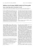
The study area consisted of farms with cows that were
pastured in wooded areas within the previously estab-
lished distribution of bovine babesiosis [29]. Twenty-
four farms scattered along most of th e southern Norwe-
gian coast west of the Oslo Fjord (Figure 1) were
included in the study. Farms using hillside or wooded
areas for pasturing were identified with the help of local
agricultural authorities. None of the farmers who were
asked to participate in the study refused. All the farmers
confirmed that ticks occur on their farms, and the cattle
were past ured on the prop erty. All of the 3 06 cows
included in the study were healthy and at least one year
old when tested. On one farm (Farm 23), all the cows
had been purchased one year prior to the study from
various inland localitie s and had been pastured for just
one season at this farm. I. ricinus is distributed mainly
near the coast in this part of Norway. The study
Figure 1 Map of Vest-Agder, Aust-Agder and Telemark counties, with study localities numbered from west to east (Table 1).Farm
number 24 is in Vestfold County. (Copyright, map basis: Cappelen Damm as.).
Hasle et al. Acta Veterinaria Scandinavica 2010, 52:55
/>Page 3 of 9
included one inland farm approximately 30 kilome tres
from the sea (Farm 7) that was included because human
Lyme borreliosis had been reported in this municipality,
thus indicating the presence of ticks, according to the
Norwegian Surveillance System for Communicable Dis-
eases (MSIS) [30]. Blood samples were collected in May
2004 on farms 20 and 21, and samples were collected
from all other sites in October and November 2005.
The blood samples were stored at 4°C within a few
hours after collection, and the serum portion of the
samples was separated and frozen within 72 hours.
All of the sera were tested using an indirect immuno-
fluorescent antibody test (IFAT) [28] for IgG as
described by Christensson [31,32], and Christensson and
Moren [33] w ith the fol lowing modifications: Antigen
was prepared in 2002 from blood of a calf infected with
Babesia divergens with approx. 10% infected erythro-
cytes as described by Christensson [32]. The antiserum
used was was FITC conjugated rabbit anti bovine IgG,
produced by ICN Cappel, code 55280, lot 03683, diluted
at 1/200 to give comparable readings with control sera
used by Christensson and Morén [33]. Control sera
were obtained from calves used for vaccine production
in the year 2001 drawn bef ore infection and four weeks
aft er having showed acute parsitaemia. Negative control
serum showed no or uncertain reaction at a dilution of
1/20 or higher. The positive control sera had an end-
point titre of 1/1 280-1/2560. For each day of reading
IFAT-slides a negative control at 1/40 and a positive
control at 1/40, 1/160 and 1/1280 were included. As the
purpose of the test was to identify seropositive/serone-
gative animals sera were read a t dilutions at1/40 and 1/
160. Slides were read blindly and scored by Christensson
as having uncertain (+), positive (++) or strongly posi-
tive immunofluorescence (+++), at dilutions of 1:40 and
1:160. To minimise the risk of false positives, only sera
with a minimum +++ score at a dilution of 1:40 were
counted as positive.
Farmers were interview ed to determine if there had
been cases of re dwater on their farms and if they had
experienced redwater in cows that were imported to the
farm. Data on the cases of babesiosis in the included
farms were obtained from the NDHRS.
To test the suitability of using PCR on full blood, we
chose samples for a pilot study from four farms where
redwater was common, according t o the local farmers,
and DNA from 100 μl from 20 samples of frozen
EDTA-blood, and 25 samples of 100 μlbloodclot,fro-
zen after spinning and removal of the serum, were iso-
lated in a spin-column, using DNeasy Blood & Tissue
Kit (Qiagen), and eluated to 200 μl, according to the
manufacturer’ sprotocol.TheisolationofDNAcon-
tained a lysis step and washing. Five μl of the eluate was
run in B. divergens real-time PCR for 40 cycles with
primers BdiF, BdiR and BdiT. The PCR was perfo rmed
by Telelab (Skien, Norway), using an in-house method,
as described by Lundsett [26]. The laboratory used a
synthetic amplicon with the sequence of B. divergens,
serially diluted in human DNA as a positive control.
The reaction mix and human DNA was used as a nega-
tive control. The observed cutoff for detection was 30
B. divergens DNA copies, i.e. 15 to 30 individual cells,
depending of whether they are asexual, diploid cells or
sexual, haploid cells.
Exact confidence intervals for binomial proportions
were calculated using the principles introduced by Clop-
per and Pearson [34] and implemented in R (R Develop-
ment Core Team, 2008).
Results
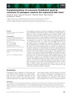
Of the 306 sera that we tested, 84 (27%) had positive
IFAT results. A high percentage of these positive results
were found in the western and eastern ranges of the
study area, and a much lower rate of positive test results
was found in the middle range of the study area (Table
1; Figure 2). Farm 23 had 3 positive test results among
Table 1 Municipality of the test localities in Figure 1 and
test results of indirect immunofluorescence antibody
tests (IFAT) for Babesia divergens.
Farm Municipality Neg Pos
1
N % pos
1 Farsund 6 5 11 45
2 Farsund 6 9 15 60
3 Lyngdal 6 2 8 25
4 Mandal 0 9 9 100
5 Mandal 5 7 12 58
6 Søgne 2 7 9 78
7 Songdalen 13 1 14 7
8 Søgne 3 1 4 25
9 Kristiansand 9 0 9 0
10 Kristiansand 3 0 3 0
11 Lillesand 16 2 18 11
12 Lillesand 8 0 8 0
13 Grimstad 29 2 31 6
14 Grimstad 19 0 19 0
15 Arendal 10 0 10 0
16 Arendal 14 1 15 7
17 Arendal 6 4 10 40
18 Risør 4 13 17 76
19 Kragerø 11 1 12 8
20 Kragerø 6 12 18 67
21 Kragerø 2 5 7 71
22 Bamble 12 0 12 0
23 Porsgrunn 13 3 16 19
24 Larvik 19 0 19 0
Total 222 84 306 27
1. IFAT IgG titres scored as 1:40 (+++) or higher are defined as positive.
Hasle et al. Acta Veterinaria Scandinavica 2010, 52:55
/>Page 4 of 9
the 16 cows t hat had been imported from inland local-
ities one year before the study, indicating that there is a
substantial risk o f babesiosis in their present locality.
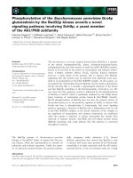
The presence of B. divergens was c onfirmed by IFAT in
a total of 17 of the 24 farms we test ed. Farmers had
observed redwater in only ten of the farms where B.
divergens was detected, and only four of these cases of
redwater had been recorded by the NDHRS (Figure 3).
All of the cows on one of the farms in the study were
B. divergens-antibody positive, though the owner had
never seen any cases of redwater. We detected B. diver-
gens antibodies in 17 of the 25 cows that we tested on
Jomfruland, where Radzijevskaja [27] found no infected
ticks.
The PCR pilot study gave no positive results.
Discussion
In Norway and Sweden the only cattle Babesia reported
is B. divergens [35,36]. With regard to this and the
strong reaction to the antigen used we assume that the
seropositive animals were/had been infected with the
species Babesia divergens. Our results demonstrate that
testing of cattle for seropositivity to B. divergens is a far
better method for mapp ing the distribution of this
pathogen than using indirect methods, such as inter-
viewing farmers or relying on the NDHRS. When it pre-
sents clinically, redwater is easily recognizable by
farmers and veterinarians, and because prompt treat-
ment is usually required to prevent deleterious effects of
the disease, veterinarians often treat the disease without
performing any laboratory tests. There are few data
available on the attack rate of bovine B. divergens infec-
tions. Our data indicate that there are many subclinical
cases of B. divergens infection, which is in agreement
with previous studies on outbreaks [7,37] and in stable
infected herds [38]. An extensive study of B. divergens
seroprevalence was conducted in Northern Ireland,
showing an overall seroprevalence of 31,8%[39], i.e.,
close t o the overall seroprevalence in our limited mate-
rial. A second study carried out in Northern Ireland
[40] found consistent estimates when compa ring results
from a farm survey, a veterinary practise survey and ser-
oprevalence data, with an estimated clinical i ncidence of
0,26% per year. The number of cases in the Agder coun-
ties, according to the NHDRS, is 18.4 cases per year in a
population of ca. 10400 dairy cows (Statistics Norway,
/>03-07.html, Jordbrukstelling 1999), which would give an
incidence of 0.18% per year. Our results indicate an
incomplete registration of cases of redwater in the
NHDRS, possibly because veterinarians are not always
consulted e. g. during the dry period, in mild cases of
redwater, or that the farmers fail to observe redwater
while the cows are out at pasture. The farms that we
included in our study were not randomly selected,
but were cho sen because the pastures were in wooded
areas, and were situated near the coast in the distribu-
tion area of I. ricinus in Norway. They would therefore
be expected to have more babesiosis than average farms
in the same counties.
Figure 2 Fraction of cows positive for Babesia divergens IFAT IgG antibodies at a titre of 1:40 (+++) or higher in 24 different farm s
along the southern Norwegian coast, arranged form west to east. Error bars: 95% confidence intervals.
Hasle et al. Acta Veterinaria Scandinavica 2010, 52:55
/>Page 5 of 9
Because cows are parasitised by hundreds of ticks in
thecourseofaseason,andasinglebitefroman
infected tick is sufficient for transmission of Babesia,
[41] cows are likely to contract B. divergens if it is pre-
sent in their pasturing areas. The screening of cows for
B. divergens infection would therefore be expected to be
a sensitive method for detecting the presence of the
parasite in a locality, if testing is performed at a time of
the year when Babesia-antibodies are at the highest.
Serum samples that we collected on Jomfruland in May
were not directly comparable to those that we collected
in October and Novemb er, as the May s amples could
either contain persistent antibodies f rom the previous
year, or there might be early infections from the same
year. The mean temperature April 1
st
-15
th
was 5.3°C,
and no temperatures of below 0°C were recorded (The
Norwegian Meteorological Institute), which means that
tick questing may well have occurred during this period.
With an incubation time of 1-3 weeks [3], seroconver-
sions during May 2004 would be expected to occur. As
we tested only once for each locality we did not demon-
strate the seasonal and yearly variation of antibodies
described by l’ Hostis et al. [38]. Further studies are
needed to decide which month would be optimal for
detecting the presence of B. divergens in a locality along
the Southern Norwegian coast. However, ticks are still
parasitizing the cows in October and November and
these months would therefore be expected to be a good
choice for detecting B. divergens antibodies.
The sensitivity of serologic testing for detecting B.
divergens will depend on the cut-off level that is set for
apositivescoreonthetest.Atacut-offlevelof1:40(+
+) the sensitivity and specificity of an individual anti-
body test are reported to be 100% and 97%, respectively
[32]. Setting the cut-off value at this l evel would, there-
fore, likely resul t in the detection of a few false positives
due to non-specific cross reactivity. This problem is illu-
stratedbyourresultsonFarm24,whereonlyonecow
was found to be positive at the detection level of 1:40
(++), and there were no positive tests at more stringent
detection levels. This result could represent either a
false positive or a low titre in a cow that was infected a
long time ago. Because the aim of this study was to be
able to detect the present occurrence of B. divergens at
a particular l ocality, a high sensitivity for detecting the
pathogen on a given farm is desirable, and the number
of cows tested is crucial. By testing a median of twelve
cows per locality, we were able to achieve a much
higher sensitivity for detecting B. divergens on a given
farm than farmers’ observations and the existing
NDHRS can provide. At all the fa rms where samples
with 1/40(+++) were detected there were also samples
positive at 1/160, indicating that these are real positives.
Therefore, by setting a cut-off level of 1:40 (+++) for
defining a case of seropositivity for B. divergens,anti-
body testing should result in a specificity of nearly
100%, unless cross-reacting Babesia spp. are occurring
and, consequently, the risk of falsely concluding that B.
divergens occurs on a farm will be small. The related
species B. capreoli cause babesiosis in roe deer and red
deer [42], and roe deer ma y also be infected by the
newly discovered Babesia sp. EU1 [43]. These parasites
cannot be serologically distinguished from B. divergens.
They cannot give clinical infection in cattle, but there is
a possibility that a sub clinical infe ction may cause sero-
conversion [44], although Sch mid et al. [45] found no
seropositive cows in an area in which ticks positive for
Farm
IFAT IgG
positive
Noticed
Notified
1
2
3
4
5
6
7
8
9
10
11
12
13
14
15
16
17
18
19
20
21
22
23
24
Figure 3 Compari son of three sources of i nfor mati on for the
occurrence of babesiosis on the farms in this study. IFAT IgG
positive: At least one cow positive for IFAT Babesia divergens IgG.
Noticed: Farmers’ statement that redwater occurs in cows on the
farm or is detected when adult cattle are imported to the farm.
Notified: Clinical cases registered on the diary cow health cards,
compiled by the Norwegian Dairy Herd Recording System from
1996-2008.
Hasle et al. Acta Veterinaria Scandinavica 2010, 52:55
/>Page 6 of 9
these two non-bovine Babesia species were found. It is
therefore unlikely that these Babesia species would
influence the number of seropositive cows in this study
significantly. There are no published studies on these
Babesia species in Norway, but a Swedish study sug-
gested that babesiosis ca used by B. capreoli is very rare
in Sweden [46].
An alternative to antibody testing is to test directly for
the presence of the pathogen in cattle blood samples.
Calder et al. [47] found an approximately 80% sensitivity
for detecting Babesia bovis by PCR in steers, up to 300
days after experimental infection. The method these
investigators used required a concen tration step invol-
ving ultracentrifugation of haemolysed blood. We con-
sidered this to be too laborious a method to be useful as
a field assay. We did attempt direct PCR to detect B.
divergens without performing the concentration step in
30 samples from areas where we found the highest inci-
dence of B. divergens by IFAT, bu t none of these sam-
ples were fo und to be B. divergens-positive by thi s
method. Cultivation of Babesia in cell culture, which
enables detecti on of Babesia at a level of 10 pa rasites
per 1 ml of blood [48], is another possibility for map-
ping the distribution of this parasite, but it is not feasi-
ble to use this method when sampling is being carried
out in scattered locations. For our purposes, therefore,
we found that antibody screening was a much more
convenient method for assessment of the occurrence of
B. divergens in a locality than any of the other methods
that are available for detecting this pathogen. Gerbil-
derived antigen is found to be equally specific to B.
divergens obtained from cattle [49], and co uld be a
cheaper alternative in future studies.
In the communities on the coast of souther n Norway
where cows are pastured, the animals are confined to
the farms on which they are kept. Consequently, testing
cows for the presence of B. divergens infection should
provide results that are specific to a given locality, as
opposed to performing serological testing on other hosts
of tick-borne pat hogens, such as wildlife, dogs or
humans. Because B. divergence is unlikely to survive for
more than a decade in regions where cattle are not pas-
turing and cattle is the only host for B . divergens at the
Southern coast o f Norway, testing cow sera appears to
be an effective method for mapping B. divergens over
the area of distribution of I. ricinus. The same is not the
case if using cattle as sentinel animals for serological
testing for other tick-borne pathogens, such as Ana-
plasma, Borrelia or the TBE virus, that infect a wider
range of hosts.
Malandrin et al. [48] found a drop in IFAT antibody
titre from 320, 320 and 1280 to 80, 80 and 320 respec-
tively in samples from three cows taken 6 and 9 months
after acute babesiosis, indicating an antibody duration of
more than a year, but much shorter than the cows’ life-
span. Sahibi et al. [50] found no significant cumulative
effect of cow age on the presence of Babesia-antibodies.
This is consistent with a short duration of antibod ies in
the bloodstream after infection, meaning that detection
of antibodies indicates a recent infection, as is illustrated
by the seasonal variation of B. div ergens-antibodies that
was found by l’Hostis et al. [38], indicating repeated
infections during the season. This implies that the life-
time risk of acquiring bovine babesiosis is higher than
the current rate of infection that was determined in the
study we present here.
Our IFAT data indicate that there are two areas along
the southern Norwegian coast in which bovine babesio-
sis is highly endemic, consisting of one western area
(Lista-Mandal) and one eastern area (Kroger-Risør) (Fig-
ure 1, Table 1). This uneven distribution was not
reported by Thambs-Lyche in a study carried out along
the same part of coastal Norway [29]. For other Babesia
species, it has been shown that reduction of the inci-
dence of tick bites can bring the reproduction rate of
the parasite below 1, indicating that it could be possible
to eradicate the parasite [41,51]. Our results indicate
that, in the area from Sandaled to Arundel, which is
within the distribution area of I. ricinus and is an are a
where cattle are pastured in a natural setting, B. diver-
gens occurs at very low frequencies or not at all. In fact,
the disease associated with this pathogen has virtually
disappeared since the 1930 s, when Thambs-Lyche
reported babesiosis in this area. This seems promising
with regard to the possibility of eradicat ing this disease.
An attempt to eradicate the disease would require the
implementation of control m easures over its entire dis-
tribution because wild hosts can spread infected ticks.
Cervid animals are the most important hosts for adult
ticks [52]. Red deer, roe deer and moose have yearly
migratoryrangesof200,100and50-60kilometres
respectively [53], and Cervid animals, therefore, have the
potential for transporting large numbers of ticks over
long distances. Furthermore, birds can transport ticks
across geographical barriers. In a recent study, 7.3% of
northward migratory passerine birds were found to
carry one or more ticks [54], so, in a situation where
cows are pastured in an area that is free of B. divergens,
or where there is an unstable population of the patho-
gen, B. divergens could conceivably be introduced by
birds.
Conclusions
At present, bovine babesiosis is a limited animal health
problem in Norway. The most obvious possible cause of
the decline i n incidence since the 1930 s is changes in
the use of pastures. Changes in legislation leading to
increased use of wood pasturing may reverse the decline
Hasle et al. Acta Veterinaria Scandinavica 2010, 52:55
/>Page 7 of 9
in incidence, and we may also see a climate-related
increase. An increased incidence of B. divergens in cattle
could have important economic and animal welfare con-
sequences, and further studies are needed to evaluate
whether it would be cost effective to implement preven-
tive measures against the spread of this pathogen. Anti-
bodytestingofpasturedcowsisasimplewayof
mapping the distribution of the pathogen.
Acknowledgements
Thanks to Ph. D. student Lise Heyer, Department of Biology, University of
Oslo, Norway, for help with statistical analyses.
Author details
1
Department of Biology, University of Oslo, P.O. Box 1050 Blindern, N-0316
Oslo, Norway.
2
Institute for Health and Society, Faculty of Medicine,
University of Oslo, Norway.
3
Department of Virology, Immunobiology and
Parasitology. National Veterinary Institute, Uppsala, Sweden.
4
Department of
Basic Sciences and Aquatic Medicine, Norwegian School of Veterinary
Science, Norway.
5
Department of Cattle Health Services, TINE Norwegian
Dairy Association, Norway.
Authors’ contributions
GH prepared the fieldwork, interviewed the farmers, performed all the blood
sampling and wrote the main part of the paper. GB, KHR and HPL provided
valuable and significant contributions to the writing of the paper. DC
headed the laboratory work, and performed all the microscopy of the slides
in the immunofluorescence antibody test. Furthermore, he contributed
significantly to the writing of the paper. ACW contributed with data from
the Norwegian Dairy Herd Recording System, and also contributed
significantly to the writing of the paper. All authors read and approved the
final manuscript
Competing interests
The authors declare that they have no competing interests.
Received: 12 April 2010 Accepted: 6 October 2010
Published: 6 October 2010
References
1. Acha P, Szyfres B: Parasitoses. Zoonoses and Communicable Diseases
Common to Man and Animals Washington DC: Pan American Health
Organization (PAHO), Third 2003, 2.
2. Bock R, Jackson L, De Vos A, Jorgensen W: Babesiosis of cattle. Parasitology
2005, 129:247-269.
3. Zintl A, Mulcahy G, Skerrett HE, Taylor SM, Gray JS: Babesia divergens,a
bovine blood parasite of veterinary and zoonotic importance. Clin
Microbiol Rev 2003, 16:622-636.
4. Purnell RE: Bovine babesiosis in the European Community. Veterinary
Science Communications 1977, 1:289-296.
5. Christensson DA: Inverse age resistance to experimental Babesia
divergens infection in cattle. Acta Vet Scand 1989, 30:453-464.
6. Zintl A, Gray JS, Skerrett HE, Mulcahy G: Possible mechanisms underlying
age-related resistance to bovine babesiosis. Parasite Immunol 2005,
27:115-120.
7. Donnelly J, Joyner LP, Crossman PJ: Incidence of Babesia divergens
infection in a herd of cattle as measured by the indirect
immunofluorescent antibody test. Res Vet Sci 1972, 13:511-514.
8. Taylor SM: Assessment of prevalence of clinical babesiosis in cattle in
Northern Ireland. Vet Rec 1983, 112:247-250.
9. Gorenflot A, Moubri K, Precigout E, Carcy B, Schetters TP: Human
babesiosis. Annals of Tropical Medicine and Parasitology 1998, 92:489-501.
10. García-Sanmartín J, Aurtenetxe O, Barral M, Marco I, Lavin S, García-Pérez AL,
Hurtado A: Molecular detection and characterization of piroplasms
infecting cervids and chamois in Northern Spain. Parasitology 2007,
134:391-398.
11. Darja Duh, Miroslav Petrovec, Andrej Bidovec, Avsic-Zupanc T: Cervids as
Babesiae Hosts, Slovenia. Emerg Infect Dis 2005, 11:1121-1123.
12. Jaenson TGT, Talleklint L, Lundqvist L, Olsen B, Chirico J, Mejlon H:
Geographical distribution, host associations, and vector roles of ticks
(Acari: Ixodidae, Argasidae) in Sweden. J Med Entomol 1994, 31:240-256.
13. Donnelly J, Peirce MA: Experiments on the transmission of Babesia
divergens to cattle by the tick Ixodes ricinus. Int J Parasitol 1975, 5:363-367.
14. Bonnet S, Jouglin M, Malandrin L, Becker C, Agoulon A, L
’Hostis M,
Chauvin A: Transstadial and transovarial persistence of Babesia divergens
DNA in Ixodes ricinus ticks fed on infected blood in a new skin-feeding
technique. Parasitology 2007, 134:197-207.
15. Randolph SE: Ticks are not insects: Consequences of contrasting vector
biology for transmission potential. Parasitol Today 1998, 14:186-192.
16. Joyner LP, Davies SFM: Acquired resistance to Babesia divergens in
experimental calves. J Protozool 1967, 14:260.
17. Chauvin A, Moreau E, Bonnet S, Plantard O, Malandrin L: Babesia and its
hosts: adaptation to long-lasting interactions as a way to achieve
efficient transmission. Vet Res 2009, 40:37.
18. Thambs-Lyche H: Ixodes ricinus og piroplasmosen I Norge. Norsk
Veterinærtidsskrift 1943, 60:337-366.
19. Solbu H: Disease recording in Norwegian dairy cattle. I. Disease
incidences and non-genetic effects on mastitis, ketosis and milk fever. Z
Tierzuecht Zuechtungsbiol 1983, 100:139-157.
20. Husdyr på utmarksbeite 2006. [ />jordbruk_miljo/tabeller4.2/beitedyr_2006_00.html].
21. Talet på storfe og sau per 1. januar, etter fylke 2010. [ />emner/10/04/10/jordhus/tab-2010-04-08-02.html].
22. Gray J, Harte L, Talty P: Decline of Bovine Babesiosis in Ireland. Irish
Veterinary Journal 1996, 49:157-159.
23. Wesenberg GR: In Terapianbefaling: Antiparasittærbehandling av
produksjonsdyr. Edited by: legemiddelverk S. Statens legemiddelverk: Oslo;
2001:.
24. Lindgren E, Talleklint L, Polfeldt T: Impact of climatic change on the
northern latitude limit and population density of the disease-
transmitting European tick Ixodes ricinus. Environ Health Perspect 2000,
108:119-123.
25. Forskrift om hold av storfe. [ />sf/sf-20040422-0665.html#10].
26. Lundsett AL: Flåtten Ixodes ricinus som sykdomsvektor i Sør-Norge Telemark
University College 2004.
27. Radzijevskaja J, Paulauskas A, Rosef O: Prevalence of Anaplasma
phagocytophilum and Babesia divergens in Ixodes ricinus ticks from
Lithuania and Norway. Int J Med Microbiol 2008, 298
:218-221.
28. Ross JPJ, Lohr KF: Serological diagnosis of Babesia bigemina infection in
cattle by the indirect fluorescent antibody test. Res Vet Sci 1968, 9:557.
29. Thambs-Lyche H: Ixodes ricinus og piroplasmosen i Norge. Norsk
Veterinærtidsskrift 1943, 60:401-441.
30. The Norwegian Surveillance System for Communicable Diseases (MSIS).
[ />31. Christensson DA: Improvement of the teflonized slide used in the
immunoflourescent antibody technique. Acta Vet Scand 1986, 27:296-297.
32. Christensson DA: A modified IF-test to demonstrate IgM antibodies to
Babesia divergens of cattle. Acta Vet Scand 1987, 28:361-371.
33. Christensson DA, Moren T: Seroresponse (IgG) after vaccination and
natural infection of cattle with Babesia divergens. Acta Vet Scand 1987,
28:393-402.
34. Clopper CJ, Pearson ES: The use of confidence or fiducial limits illustrated
in the case of the binomial. Biometrika 1934, 26:404-413.
35. Christensson DA: Babesia of cattle and sheep in Sweden. 1989.
36. Mørk T, Sviland S: Flåttbårne sykdommer hos storfe: Babesiose. 2009.
37. Christensson D, Enfors E: An outbreak of babesiosis (B. divergens)ina
dairy herd comprising different age groups of cattle. Acta Vet Scand
1987, 28:125-126.
38. l’Hostis M, Chauvin A, Valentin A, Precigout E, Gorenflot A: Survey of
Babesia divergens antibody kinetics in cattle in western France. Vet Res
1997, 28:481-488.
39. Taylor SM, Kenny J, Strain A: The distribution of Babesia divergens
infection within the cattle population of Northern Ireland. British
Veterinary Journal 1982, 138:384-392.
Hasle et al. Acta Veterinaria Scandinavica 2010, 52:55
/>Page 8 of 9
40. Gray JS, Harte LN: An estimation of the prevalence and economic
importance of clinical bovine babesiosis in the Republic of Ireland. Irish
Veterinary Journal 1985, 39:75-78.
41. Mahoney DF, Ross DR: Epizootiological factors in the control of bovine
babesiosis. Aust Vet J 1972, 48:292.
42. Gray JS, Murphy TM, Taylor SM, Blewett DA, Harrington R: Comparative
morphological and cross transmission studies with bovine and deer
babesias in Ireland. Preventive Veterinary Medicine, 1990, 9:185-193.
43. Bonnet S, Jouglin M, L’Hostis M, Chauvin A: Babesia sp. EU1 from Roe
Deer and Transmission within Ixodes ricinus. Emerging Infectious Diseases
2007, 13.
44. Adam KMG, Blewett DA: The isolation and characterization of a Babesia
from red deer (Cervus elaphus). Parasitology 1976, 73:1-11.
45. Schmid N, Deplazes P, Hoby S, Ryser-Degiorgis M-P, Edelhofer R, Mathis A:
Babesia divergens-like organisms from free-ranging chamois (Rupicapra
r. rupicapra) and roe deer (Capreolus c. capreolus) are distinct from B.
divergens of cattle origin - An epidemiological and molecular genetic
investigation. Vet Parasitol 2008, 154:14-20.
46. Aguirre AA, Bröjer C, Mörner T: Descriptive epidemiology of roe deer
mortality in Sweden. Journal of Wildlife Diseases 1999, 35:753-762.
47. Calder JAM, Reddy GR, Chieves L, Courtney CH, Littell R, Livengood JR,
Norval RAI, Smith C, Dame JB: Monitoring Babesia bovis infections in
cattle by using PCR-based tests. J Clin Microbiol 1996, 34:2748-2755.
48. Malandrin L, L’Hostis M, Chauvin A: Isolation of Babesia divergens from
carrier cattle blood using in vitro culture. Vet Res 2004, 35:131-139.
49. Gray JS, Kaye B: Studies on the use of gerbil-derived Babesia divergens
antigen for diagnosis of bovine babesiosis. Vet Parasitol 1991, 39:215-224.
50. Sahibi H, Rhalem A, Berrag B, Goff WL: Bovine babesiosis. Seroprevalence
and ticks associated with cattle from two different regions of Morocco.
Ann N Y Acad Sci 1998, 849:213-8.
51. Bowman D: Successful and currently ongoing parasite eradication
programs. Vet Parasitol 2006, 139:293-307.
52. Ostfeld R, Canham C, Oggenfuss K, Winchcombe R, Keesing F: Climate,
deer, rodents, and acorns as determinants of variation in Lyme-disease
risk. PLoS Biol 2006, 4:1058-1068.
53. Reimers E: Hjortedyr. In Norges dyr. Edited by: Semb-Johansson A. Oslo:
Cappelens forlag AS; 1990:.
54. Hasle G, Bjune G, Edvardsen E, Jakobsen C, Linnehol B, Røer J, Mehl R,
Røed K, Pedersen J, Leinas H: Transport of ticks by migratory passerine
birds to Norway. J Parasitol 2009, 95:1342-1351.
doi:10.1186/1751-0147-52-55
Cite this article as: Hasle et al .: Detection of Babesia divergens in
southern Norway by using an immunofluorescence antibody test in
cow sera. Acta Veterinaria Scandinavica 2010 52:55.
Submit your next manuscript to BioMed Central
and take full advantage of:
• Convenient online submission
• Thorough peer review
• No space constraints or color figure charges
• Immediate publication on acceptance
• Inclusion in PubMed, CAS, Scopus and Google Scholar
• Research which is freely available for redistribution
Submit your manuscript at
www.biomedcentral.com/submit
Hasle et al. Acta Veterinaria Scandinavica 2010, 52:55
/>Page 9 of 9