Báo cáo y học: "Lung recruitment manoeuvres are effective in regaining lung volume and oxygenation after open endotracheal suctioning in acute respiratory distress syndrome" ppt
Bạn đang xem bản rút gọn của tài liệu. Xem và tải ngay bản đầy đủ của tài liệu tại đây (336.83 KB, 8 trang )
Available online />Research
Lung recruitment manoeuvres are effective in regaining lung
volume and oxygenation after open endotracheal suctioning in
acute respiratory distress syndrome
Thomas Dyhr
1
, Jan Bonde
2
and Anders Larsson
3
1
Research fellow, Section of Intensive Care Medicine, Department of Anaesthesiology, Gentofte University Hospital, Hellerup, Denmark
2
Director of the ICU, Department of Anaesthesiology and Intensive Care, Herlev University Hospital, Herlev, Denmark
3
Professor, Section of Intensive Care Medicine, Department of Anaesthesiology, Gentofte University Hospital, Hellerup, Denmark
Correspondence: Thomas Dyhr,
55
ALI = acute lung injury; ARDS = acute respiratory distress syndrome; EELV = end-expiratory lung volume; ETS = endotracheal suctioning; FiO
2
=
fractional inspired oxygen; LIP = lower inflection point; LR = lung recruitment; PaO
2
= partial arterial oxygen tension; PEEP = positive end-expiratory
pressure; SF
6
= sulphur hexafluoride; SpO
2
= blood oxygen saturation.
Abstract
Introduction Lung collapse is a contributory factor in the hypoxaemia that is observed after open
endotracheal suctioning (ETS) in patients with acute lung injury and acute respiratory distress
syndrome. Lung recruitment (LR) manoeuvres may be effective in rapidly regaining lung volume and
improving oxygenation after ETS.
Materials and method A prospective, randomized, controlled study was conducted in a 15-bed
general intensive care unit at a university hospital. Eight consecutive mechanically ventilated patients
with acute lung injury or acute respiratory distress syndrome were included. One of two suctioning
procedures was performed in each patient. In the first procedure, ETS was performed followed by LR
manoeuvre and reconnection to the ventilator with positive end-expiratory pressure set at 1 cmH
2
O
above the lower inflexion point, and after 60 min another ETS (but without LR manoeuvre) was
performed followed by reconnection to the ventilator with similar positive end-expiratory pressure; the
second procedure was the same as the first but conducted in reverse order. Before (baseline) and
over 25 min following each ETS procedure, partial arterial oxygen tension (Pa
O
2
) and end-expiratory
lung volume were measured.
Results After ETS, Pa
O
2
decreased by 4.3 (0.9–9.7) kPa (median and range; P < 0.005). After LR
manoeuvre, Pa
O
2
recovered to baseline. Without LR manoeuvre, PaO
2
was reduced (P = 0.05) until
7 min after ETS. With LR manoeuvre end-expiratory lung volume was unchanged after ETS, whereas
without LR manoeuvre end-expiratory lung volume was still reduced (approximately 10%) at 5 and
15 min after ETS (P = 0.01).
Discussion A LR manoeuvre immediately following ETS was, as an adjunct to positive end-expiratory
pressure, effective in rapidly counteracting the deterioration in Pa
O
2
and lung volume caused by open
ETS in ventilator-treated patients with acute lung injury or acute respiratory distress syndrome.
Keywords acute respiratory distress syndrome, alveolar recruitment, atelectasis, hypoxaemia,
suction/instrumentation
Received: 15 July 2002
Revisions requested: 31 July 2002
Revisions received: 3 September 2002
Accepted: 9 October 2002
Published: 31 October 2002
Critical Care 2003, 7:55-62 (DOI 10.1186/cc1844)
This article is online at />© 2003 Dyhr et al., licensee BioMed Central Ltd
(Print ISSN 1364-8535; Online ISSN 1466-609X). This article is
published in Open Access: verbatim copying and redistribution of this
article are permitted in all media for any non-commercial purpose,
provided this notice is preserved along with the article's original URL.
Open Access
56
Critical Care February 2003 Vol 7 No 1 Dyhr et al.
Introduction
After discontinuation of positive end-expiratory pressure
(PEEP), lung collapse occurs rapidly in ventilator-treated
patients with acute respiratory distress syndrome (ARDS) [1].
Endotracheal suctioning (ETS), which is a common procedure
in patients with acute lung injury (ALI) and ARDS, abolishes
the positive airway pressure and even may generate negative
pressure, promoting de-recruitment and hypoxaemia [2].
The most common method used to mitigate the reduction in
oxygenation induced by suctioning is to increase the frac-
tional inspired oxygen (Fi
O
2
) [3–5]. This strategy is often
effective in patients with less severe lung diseases, but is less
efficacious in patients with ARDS with high shunt fractions
[6]. In addition, a high Fi
O
2
may augment lung collapse by
causing absorption atelectasis [7–9]. It has recently been
suggested that a closed suction system may be effective in
preventing suctioning-induced decreases in lung volume and
oxygenation. In fact, Pesenti and coworkers [10] found no
reduction in end-expiratory lung volume (EELV) or arterial
oxygen saturation in patients with ALI and ARDS after suc-
tioning with such a system. Significant drawbacks with
closed suction systems include risk for producing high nega-
tive pressures and reduced efficacy in removing thick secre-
tions from the airways [2,11,12]. Brochard and coworkers
[13] showed that lung volume and arterial oxygenation could
be maintained during open suctioning by using constant flow
insufflation. This method appears to be effective but necessi-
tates use of a special endotracheal tube. Another measure to
counteract suctioning-induced hypoxaemia is hyperinflation of
the lungs. This is usually performed by administering large
breaths using an anaesthetic balloon, without attention to
monitoring of levels, duration or maintenance of the end-inspi-
ratory and end-expiratory pressures [14,15].
Because de-recruitment occurs during and after suctioning, a
plausible method to mitigate hypoxaemia is to re-recruit col-
lapsed lung using a lung recruitment (LR) manoeuvre. Indeed,
using computed tomography, Lu and coworkers [16] showed
that LR manoeuvres are effective in resolving atelectasis and
improving oxygenation after ETS in sheep with normal lungs.
However, as far as we know, this has not been verified in
patients with ALI and ARDS. The present study was therefore
conducted to examine the additive effect of LR manoeuvre to
adequate PEEP on lung volumes and oxygenation after a
standardized open ETS procedure in eight mechanically ven-
tilated patients with ALI or ARDS.
Materials and method
Patients
The present study was approved by the local Human Ethics
Committee and informed consent was obtained from next of
kin. After a power analysis (see Statistical analysis, below),
eight patients with ALI or ARDS requiring mechanical ventila-
tion were enrolled [17]. Exclusion criteria were pneumo-
thorax, documented history of chronic obstructive lung
disease, haemodynamic instability and a contraindication to
deep sedation. The patients (Table 1) were studied in the
supine position, with the upper part of the body slightly higher
than the lower, and ventilated via an endotracheal tube (size
7.5–8; Mallincrodt, Hazelwood, MO, USA) either in the
volume-controlled or pressure-controlled mode (Servo Venti-
lator 900C; Siemens-Elema, Solna, Sweden; Table 2). Seda-
tion was performed with continuous intravenous infusion of
Table 1
Patient characteristics at inclusion and patient outcome
PaO
2
/ Cause of
Patient Age Sex Weight EELV PEEP FiO
2
acute lung MV
number (years) (F/M) (kg) (ml) (cmH
2
O) (kPa) LIS Underlying disease injury (days) Outcome
1 75 M 69 2284 10 36.8 2.3 Secondary lung cancer Pneumonia 2 S
2 69 F 75 1272 11 20.0 2.5 Liver cirrhosis, colectomy Sepsis, pneumonia 7 D
3 76 M 96 1513 8 9.0 2.7 AAA, secondary bowel ischaemia Sepsis 2 S
4 67 M 85 1309 9 19.4 3.3 Fasciitis Aspiration pneumonia 5 D
5 66 M 95 2245 10 19.6 2.7 – Pneumonia 2 S
6 81 M 109 1639 13 23.3 2.5 AAA SIRS 3 S
7 65 F 72 949 11 17.5 3.0 TAA Sepsis 3 S
8 58 F 63 1233 15 15.4 3.3 CABG Pneumonia 5 S
Mean 70 3/5 83 1550 11 20.1 2.8 – – 3.6 –
± SD ± 7 ± 16 ± 480 ± 2 ± 8 ± 0.4 ± 1.8
The two patients who died did so 22 and 31 days after the study in multiple organ dysfunction syndrome after discontinuation of active life support.
AAA, abdominal aortic aneurysm; CABG, coronary artery bypass graft surgery; D, died; EELV, end-expiratory lung volume; LIS, lung injury score;
MV, days of mechanical ventilation before measurements; S, survived; SIRS, systemic inflammatory response syndrome; TAA, thoracic aortic
aneurysm.
57
propofol (50–150 mg/h) and intermittent intravenous mor-
phine (1–5 mg). The infusion rate of propofol was adjusted
so that the patient exhibited no spontaneous breathing
efforts during the study. If signs of arousal appeared, then
an intravenous bolus of 20–50 mg propofol was adminis-
tered. Approximately 5 min before start of the study the
patient was given an intravenous bolus of 30–50 mg propo-
fol. Muscle relaxants were not used. If the physician in
charge considered them necessary, fluids and blood prod-
ucts were administered. The patients were monitored by
electrocardiography, continuous invasive blood pressure
monitoring and pulse oximetry (HP model 68S, Viridia CMS;
Hewlett-Packard, Boeblingen, Germany). We considered a
transient decrease in arterial saturation estimated by pulse
oximetry (Sp
O
2
) to 80% to be acceptable, without constitut-
ing a breach of protocol.
Protocol
A crossover design was employed. Before the start of the
study, the patients were randomized to one of two sequences
of two open ETS procedures. In the first sequence patients
were first subjected to ETS followed by an immediate LR
manoeuvre (ETS+LR), and then after 60 min they were sub-
jected to another ETS procedure but without a LR manoeuvre
(ETS–LR). In the second sequence patients were first sub-
jected to ETS without a LR manoeuvre, and then after 60 min
they were subjected to another ETS procedure but immedi-
ately followed by a LR manoeuvre (i.e. the same as the first
sequence but in reverse order). Following each ETS proce-
dure, measurements were taken over a period of 25 min.
After a 30 min standardization period (see below), ETS+LR
consisted of disconnection of the tube from the ventilator,
then ETS, followed by reconnection to the ventilator with the
set PEEP and an immediate LR manoeuvre. After a 30 min
standardization period (see below), ETS–LR consisted of dis-
connection of the endotracheal tube from the ventilator, then
ETS, followed by reconnection to the ventilator with the set
PEEP (Fig. 1).
ETS was performed by inserting the tip of a suction catheter
(size 14, Oppo-cath I; Pennine Healthcare, Derby, UK) 2 cm
below the distal end of the endotracheal tube. In order to
mimic the suctioning routines in our intensive care unit, the
Available online />Table 2
Ventilatory parameters at baseline of the two different suction
procedures
Parameter ETS+LR ETS–LR
PaO
2
/FiO
2
(kPa) 20 (11–36) 23 (12–48)
Set PEEP (cmH
2
O) 12 (9–16) 12 (9–16))
Intrinsic PEEP (cmH
2
O) 1 (0–3) 1 (0–3)
Vt/kg (ml/kg) 6 (5–9) 6 (5–9)
Note that the baseline values are after the standardization procedure
(see text). ETS+LR, endotracheal suctioning followed by a lung
recruitment manoeuvre; ETS–LR, endotracheal suctioning without a
following lung recruitment manoeuvre; FiO
2
, fractional inspired oxygen;
Set PEEP; set positive end-expiratory pressure; Intrinsic PEEP, PEEP
above set PEEP after an expiratory hold; PaO
2
, partial oxygen tension;
Vt, tidal volume.
Figure 1
Timeline of the study in minutes. The order of the two suctioning procedures α and β was randomized. The vertical lines above the timeline indicate
blood gas samplings. ETS+LR, endotracheal suctioning followed by a lung recruitment manoeuvre; ETS–LR, endotracheal suctioning without a
following lung recruitment manoeuvre; EELV, end-expiratory lung volume; LIP, lower inflection point; LR, lung recruitment manoeuvre; PEEP,
positive end-expiratory pressure; PV, inspiratory pressure–volume curve.
58
suctioning pressure at the wall inlet was set to generate a
peak pressure of 400 mmHg when the catheter was totally
occluded. The trachea was suctioned three times for 5 s with
an interval of 10 s between each suctioning, during which the
catheter was changed. This resulted in a 35 s period of dis-
connection from the ventilator.
The LR manoeuvre consisted of two hyperinflations using the
continuous positive airways pressure function of the ventilator
to an airway pressure of 45 cmH
2
O for 20 s, with an interval
of 1 min in between [18].
After randomization the EELV was measured, an airway pres-
sure–lung volume curve was obtained (from zero end-expira-
tory pressure in order to identify the lower inflexion point
[LIP]) and blood gases were sampled and analyzed using a
blood gas analyzer (ABL 725; Radiometer, Copenhagen,
Denmark). Fi
O
2
was adjusted if partial arterial oxygen tension
(Pa
O
2
) was below 10 kPa, and was then kept unchanged
during the study. EELV was measured by a wash-in/washout
method using sulphur hexafluoride (SF
6
) as the tracer gas
[19]. This measurement technique can be used without dis-
connecting the patient from the ventilator, with pressure-con-
trolled or volume-controlled ventilation, and with Fi
O
2
up to
0.995. The measurement system consists of a ventilator
(Servo Ventilator 900C; Siemens-Elema), a tracer gas dis-
pensing valve, an in-line infrared SF
6
transducer/analyzer, and
a computer that governs the dispensing valve and uses the
SF
6
signal from the transducer/analyzer and the flow signals
from the ventilator for calculations of lung volume. The tracer
gas is insufflated via the dispensing valve in proportion to the
inspiratory flow, so that a uniform concentration of 0.5% is
achieved. When the alveolar concentration is stable, as
assessed by a constant expiratory plateau concentration from
breath to breath, wash-in is stopped and washout is started.
Washout is considered complete when the mean end-tidal
concentration is less than 0.005% in the last five breaths. The
SF
6
flow in the expired breath is integrated and the accumu-
lated amount of SF
6
during washout is calculated (ΣSF
6
) and
EELV is obtained from the following equation: EELV =
ΣSF
6
/%SF
6
at the end of wash-in.
The inspiratory pressure–volume curves were obtained using
the computerized method described by Jonson and coworkers
[20,21]. After a prolonged (6 s) expiratory pause, an inspiratory
pressure–volume curve is recorded during slow insufflation.
The flow is integrated to obtain the volume, after correcting for
the compliance of the ventilator tubing. The pressure drop
caused by the resistance of the tracheal tube (which is mea-
sured in vitro) is subtracted from the pressure measured in the
ventilator. The pressure–volume curve is then mathematically
described according to the principle of Newton–Raphson to a
three-segment model: a lower nonlinear segment, over which
compliance increases linearly with volume; a middle linear
segment, with a constant compliance; and an upper nonlinear
segment, over which compliance falls linearly with volume. The
transition between the lower and middle segment is defined as
the LIP. Before measurement of EELV and the pressure–
volume curves in each patient, the measurement equipment
was calibrated. The volumes presented are converted from
ambient temperature pressure saturated (ATPS) to body tem-
perature pressure saturated (BTPS).
With regard to the standardization period, before each ETS,
lung volume was standardized via a LR manoeuvre, after
which the patient was ventilated with PEEP set at 1 cmH
2
O
above LIP for 30 min. This PEEP level was used during the
whole study. Also, the end-inspiratory plateau pressures, tidal
volumes and rates were kept constant during the study
(Table 2). During the standardization period blood gases
were sampled and EELV was measured at 5 and 20 min
(baseline values) after the LR manoeuvre. In addition, a pres-
sure–volume curve (with the starting pressure at PEEP, in
order to prevent de-recruitment during the procedure) was
obtained at 5 min after the LR manoeuvre (Fig. 1).
After each ETS, measurements of EELV were taken at 5, 15
and 25 min, and blood gases were sampled at 0, 1, 2, 3, 4, 5,
6, 7, 15 and 25 min. However, because of logistical factors,
after ETS+LR blood gases were not sampled during the LR
manoeuvre (i.e. at 1 and 2 min). A pressure–volume curve was
obtained (from the PEEP) at 25 min after ETS. The blood gas
samples taken during the first 7 min after ETS were stored on
ice and analyzed after approximately 10 min. The blood gas
samples taken at 15 and 25 min were analyzed immediately.
Statistical analysis
A power analysis (assuming that the difference in change in
Pa
O
2
at 5 min would be 30 ± 15%) indicated that eight
patients needed to be included (with α = 0.05 and 1 – β =
0.80, using a crossover design). Because we were interested
in changes in oxygenation and lung volume during the proce-
dure, the values obtained were normalized to the value just
before each ETS procedure (baseline values). Changes
within the procedures were assessed using analysis of vari-
ance and a post hoc analysis (PLSD), and changes between
the procedures at similar points in time were assessed using
the Wilcoxon signed rank test. P < 0.05 was considered sta-
tistically significant. The differences in volume at similar pres-
sures on the pressure–volume curves were compared using
the Kruskall–Wallis test. Data are presented as mean ± SD, if
not otherwise indicated.
Results
The demographic data for the patients, and their underlying con-
ditions and initial respiratory parameters are presented in
Table 1. Seven patients had ARDS and one had ALI at inclusion
[17]. Lung injury score (median [range]) [22] was 2.7 (2.3–3.3).
Haemodynamics
During LR manoeuvres, the decrease in blood pressure never
exceeded 14 mmHg and mean arterial pressure was always
Critical Care February 2003 Vol 7 No 1 Dyhr et al.
59
greater than 50 mmHg (Table 3). No arrhythmias were
observed. Also, when ETS was performed, no arrhythmias
and no major changes in pulse rate or blood pressure
occurred in any patient (Table 3).
Oxygenation
In all but one patient, Sp
O
2
was above 80% throughout the
period of study. The patient (no. 3) was randomly assigned to
start with ETS+LR. Arterial oxygen tension and Fi
O
2
were
11 kPa and 1.0, respectively. Immediately after the second
intervention (i.e. ETS–LR), a Sp
O
2
of 75% was observed. The
saturation increased within 1 min to above 80% without inter-
vention, and during the subsequent 4 min to 90%. Pa
O
2
obtained immediately after ETS was 5.1 kPa. Because the
blood gases were not analyzed until 8–10 mins after ETS, we
were not aware of this low value. At the time when the blood
gases were analyzed, Pa
O
2
had recovered well (PaO
2
obtained at 7 min was 8.9 kPa). However, PaO
2
was 6.4, 6.7,
6.7 and 7.0 kPa at 1, 2, 3 and 4 min after ETS.
At inclusion Pa
O
2
was 11.4 ± 3.1 kPa, and immediately
before suctioning (baseline Pa
O
2
) it was about 2 kPa higher.
The changes, presented as percentage of baseline Pa
O
2
,
during the study are shown in Fig. 2. Immediately after suc-
tioning, Pa
O
2
decreased by 31.7 ± 13.3% (P < 0.05) and
43.0 ± 9.4% (P = 0.0001) of baseline with ETS+LR and
ETS–LR, respectively (difference between the two interven-
tions not significant). This corresponds to a median (range)
decrease in Pa
O
2
by 4.3 (0.9–9.7) kPa (P < 0.005) with both
procedures. The lowest Pa
O
2
was 5.1 kPa (see above). With
the LR manoeuvre (ETS+LR), Pa
O
2
returned to baseline
(100.3 ± 40%) at the first blood gas sample taken after LR
manoeuvre (at 3 min) and increased to 121.8 ± 23% at
7 min. Without a LR manoeuvre (ETS–LR), Pa
O
2
did not
return to baseline until 7 min after ETS. At 7, 15 and 25 min
Pa
O
2
was 88 ± 13%, 91.5 ± 14.3% and 97.2 ± 16.8% of
baseline, respectively. After the LR manoeuvre there was a
significant difference between the two procedures until 7 min
after ETS.
End-expiratory lung volume
EELV was 1550 ± 480 ml on 8–15 cmH
2
O PEEP (Table 1)
at inclusion, and increased after lung volume standardization
(see above) to 1677 ± 618 ml and 1719 ± 571 ml for
ETS+LR and ETS–LR, respectively (difference between the
two procedures not significant). Because of the wash-
in/washout SF
6
technique, EELV could not be measured until
5 min after the ETS procedure. With the LR manoeuvre
(ETS+LR) EELV was similar to baseline at all measurement
time points (5, 15 and 25 min after ETS; Fig. 3). Without a LR
manoeuvre (ETS–LR) EELV was reduced by 11 ± 10% at
5 min (P < 0.001) and 9 ± 6% at 15 min (P < 0.02). After
suctioning, EELV was significantly different between ETS+LR
and ETS–LR at all measurement points.
Available online />Table 3
Haemodynamic parameters at baseline and 2 min after the two different suction procedures
ETS+LR ETS–LR
Parameter Baseline 2 min after ETS+LR Baseline 2 min after ETS–LR
HR (beats/min) 90 ± 8 96 ± 9* 90 ± 7 91 ± 7
CVP (mmHg) 15 ± 4 18 ± 5 15 ± 4 15 ± 4
MAP (mmHg) 76 ± 8 68 ± 8 76 ± 9 72 ± 10
Values are expressed as mean ± SD. *P < 0.05 between the two procedures. CVP, central venous pressure; ETS+LR, endotracheal suctioning
followed by a lung recruitment manoeuvre; ETS–LR, endotracheal suctioning without a following lung recruitment manoeuvre; HR: heart rate; MAP,
mean systemic arterial pressure.
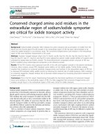
Figure 2
Arterial partial oxygen tension (PaO
2
), expressed as percentage of
baseline, at time points before (baseline) and after the two
endotracheal suctioning procedures. Endotracheal suctioning (᭹) with
and (᭺) without a following lung recruitment manoeuvre. Values are
expressed as means ± SEM (bars). *P < 0.05 between the two
procedures,
§
P < 0.05, within the procedures between the
measurements.
60
Mechanics of the respiratory system
At inclusion, the maximal slope of the pressure–volume curves
(maximal compliance of the respiratory system) was
32 ml/cmH
2
O. LIP could be identified in all patients and was
located at (median [range]) 11 (8–15) cmH
2
0 airway pressure.
The pressure–volume curves shown in Fig. 4 were normalized
to absolute lung volume at 17.5 cmH
2
O of airway pressure, in
order to integrate all patients, including those with high PEEP,
in the calculation of the curves. The pressure–volume curves
for ETS+LR and ETS–LR obtained during the standardization
period (baseline curves) were almost identical. The
pressure–volume curve obtained at 25 min after ETS with the
LR manoeuvre (ETS+LR) was similar to the baseline curve but
had a tendency to shift upward at high pressures. The maximal
slope was 33.3 ± 14.6 ml/cmH
2
O and 35.9 ± 14.7 ml/cmH
2
O
for the baseline and the 25 min curve, respectively (not signifi-
cant). The pressure–volume curve obtained at 25 min after ETS
without a LR manoeuvre (ETS–LR) was located at lower lung
volumes (P < 0.05), and tended to shift downward as com-
pared with the baseline curve. The maximal slope was
35.7 ± 14.6 ml/cmH
2
O and 32.3 ± 14.7 ml/cmH
2
O (P < 0.05)
for the baseline and 25 min curves, respectively.
Discussion
In the present study we demonstrated that a LR manoeuvre,
as an additive measure to PEEP after open ETS in mechani-
cally ventilated patients with ALI and ARDS, was well toler-
ated and produced a rapid recovery in EELV, compliance of
the respiratory system and Pa
O
2
. In addition, we confirmed
that open ETS per se may result in a significant drop in oxy-
genation and in lung volume.
We studied the effect of the open suctioning procedure used
clinically in our unit. The suction pressure was –400 mmHg,
which is more than is recommended in some guidelines, but
is not uncommon clinical practice in Scandinavia. In the study
we checked that the endotracheal tube was not occluded by
secretions around the suction catheter, and therefore a
marked pressure drop could not occur in the airways. In a
lung model test, similar suction pressure gave negative pres-
sures of –17 and –14 cmH
2
O at the ‘tracheal level’ for endo-
tracheal tubes with internal diameters of 7.5 and 8 mm,
respectively (Fig. 5). Although we cannot exclude the possi-
bility that the level of suction pressure might have con-
tributed, we believe that disconnection from positive airway
pressure was the major reason for the decrease in lung
volume and Pa
O
2
found in the study. This notion is in accor-
dance with the study conducted by Pesenti and coworkers
[10], who found a marked immediate decrease in lung volume
(as measured using respiratory inductive plethysmography)
when the endotracheal tube was disconnected, followed by a
less pronounced decrease at the start of suctioning [10]. In
fact, in an experimental ARDS model using computed tomo-
graphy, Neumann and coworkers [1] showed that major lung
collapse occurred within 0.6 s after opening the endotracheal
tube to the atmosphere. Moreover, in other studies using
lesser suction pressures and duration of suctioning proce-
dure [13,23,24], the reductions in Pa
O
2
and saturation are
similar to those reported here.
Critical Care February 2003 Vol 7 No 1 Dyhr et al.
Figure 3
End-expiratory lung volume (EELV) expressed as percentage of
baseline, at time points before (baseline) and after the two
endotracheal suctioning procedures. Endotracheal suctioning (᭹) with
and (᭺) without a following lung recruitment manoeuvre. Values are
expressed as means ± SEM (bars). *P < 0.05 between the two
procedures,
§
P < 0.01, within the procedures between the
measurements.
Figure 4
Pressure volume–curves at baseline and at 25 min after the two
procedures of endotracheal suctioning. The volumes are normalized to
the absolute lung volume at an airway pressure of 17.5 cmH
2
O at
baseline. Values are expressed as means ± SEM (bars).
#
P < 0.05
between ETS–LR baseline curve and ETS–LR 25 min curve. ETS+LR,
endotracheal suctioning followed by a lung recruitment manoeuvre;
ETS–LR, endotracheal suctioning without a following lung recruitment
manoeuvre.
61
In order to standardize lung volume, we performed a LR
manoeuvre and ventilated the patients with PEEP set at
1 cmH
2
O above LIP for 30 min before the ETS. This pro-
duced mean increases in lung volume and Pa
O
2
of approxi-
mately 150 ml and 3 kPa, respectively. Interestingly, one
patient (no. 2) changed lung injury category after the first
standardization period from ARDS to ALI, and after the
second standardization period that patient did not fulfil either
the ARDS nor the ALI criteria of Pa
O
2
:FiO
2
ratio [17]. Both
lung volumes and compliance values may appear high for this
type of patient but agree well with values found by Brochard
and coworkers [13], who used computed tomography in
patients with acute respiratory failure. Few studies have
examined the reduction in lung volume caused by ETS.
Brochard and coworkers [13] found that ETS caused an
immediate reduction in EELV by about 400 ml in acute respi-
ratory failure, and in patients with ALI and ARDS Pesenti and
coworkers [10] identified a reduction in EELV by about
1200 ml. The different results might be due to differences in
measurement techniques and patient populations, but not to
differences in suction procedures because the pressures
were similar (–80 and –100 mmHg, respectively), and the
duration of suctioning was longer in the study by Brochard
and coworkers. With our lung volume measurement tech-
nique it was not possible to measure EELV immediately but
only after 5 min, at which time the reduction in EELV without
a LR manoeuvre was about 200 ml, which is in accordance
with the study by Brochard and coworkers [13].
We found that a LR manoeuvre was effective as an additive
measure to PEEP in rapidly regaining lung volume, compli-
ance of the respiratory system and Pa
O
2
after open ETS.
However, just ventilation with PEEP did slowly increase both
lung volume and Pa
O
2
. Because LR is an inspiratory phenom-
enon, the major effect of PEEP is prevention of de-recruit-
ment of the lung regions recruited by the increased airway, or
rather transpulmonary, pressure during inspiration. Even if we
ventilated with small tidal volumes (i.e. about 7 ml/kg), this
resulted in end-inspiratory airway pressures up to 35 cmH
2
O,
which could very well have recruited some collapsed lung
regions and improved oxygenation. However, higher end-
inspiratory pressures are needed to recruit more manifest
lung collapse [25,26]. This was indicated in the present study
by the fact that, without a LR manoeuvre, the maximal compli-
ance of the respiratory system obtained from the pressure–
volume curves had not recovered at 25 min, and lung volume
at all measurement points was lower as compared with ETS
followed by a LR manoeuvre.
It is important to emphasize that a LR manoeuvre is not pre-
ventive, but rather is a therapeutic measure to regain lung
volume and oxygenation rapidly after ETS. Four main methods
have been suggested to prevent hypoxaemia in connection
with ETS: administration of oxygen; hyperinflation of the lungs;
closed-suction systems; and continuous flow insufflation. The
former two methods are not very effective in patients with
lungs that are prone to collapse and with high intrapulmonary
shunt fractions, but may be used in less severe lung disease
[6]. In addition, by using high inspired oxygen concentration,
absorption atelectasis may develop [7]. The two latter
methods might be effective in preventing hypoxaemia and lung
collapse in ARDS [10,13,27,28]. However, with the closed
suction system it is important that the ventilator is set at pres-
sure-controlled or assisted mode, the trigger level is set low,
and suction flow is less or similar to that delivered by the venti-
lator. Otherwise, a highly negative pressure may be generated,
which is counterproductive [2,12]. Also, with continuous flow,
the delivered flow should be higher than the flow in the suction
catheter and sufficiently high to compensate for loss of air via
the open endotracheal tube. In this context, it is also important
to recognize the purpose of suctioning (i.e. removing secre-
tions), and with a high bias flow this effect may be reduced. In
lung-lavaged pigs, Lindgren and coworkers [11] showed that
closed suctioning was less effective than open suctioning.
Nevertheless, the present study confirmed that ETS was
associated with lung volume loss and hypoxaemia, and there-
fore we believe that preventive measures are important, and
the most important measure is to avoid ETS at all if possible.
In the present study, this is emphasized by the fact that, in
one patient ventilated with Fi
O
2
at 1.0, PaO
2
decreased from
11 to 5 kPa during suctioning.
Available online />Figure 5
Setup for the lung model for measurement of suction pressure. The
test device consisted of a Plexiglas bottle with a connected water
manometer. The endotracheal (ET) tube was inserted in the bottle
through an opening at the top and the opening was sealed airtight
thereafter. The tip of the suctioning catheter was introduced through
the ET tube to 2 cm below the distal end of the ET tube. The catheter
was then connected to a suction pressure of –400 mmHg at the wall
inlet. The pressure generated in the bottle was measured as the
difference between the water levels ‘A’ and ‘B’.
62
The study has some inherent limitations. First, the number of
patients studied was low, although the number of patients was
enough to ensure adequate significance between the proce-
dures. Second, we studied only one kind of open suctioning
procedure, and other procedures may give different results.
Third, the patients were studied early in the disease process,
and LR manoeuvres might have different effects in late ARDS.
Fourth, the patients were haemodynamically stable and deeply
sedated, and tolerated both ETS and the LR manoeuvres well
and without circulatory compromise. In haemodynamically
unstable or less deeply sedated patients, the results might be
different. Finally, the LR manoeuvre was the same in all
patients, and should preferably be individualized.
In conclusion, the present study confirms that open suction-
ing is associated with a substantial risk for hypoxaemia in
patients with ARDS, stressing that ETS should be avoided
unless absolutely necessary in such patients. Preferably,
suction methods that prevent hypoxaemia should be used,
but when open suctioning is indicated the present study sug-
gests that a LR manoeuvre as an additive measure to PEEP
causes a rapid recovery in EELV and Pa
O
2
.
Competing interests
None declared.
References
1. Neumann P, Berglund JE, Mondejar EF, Magnusson A, Heden-
stierna G: Dynamics of lung collapse and recruitment during
prolonged breathing in porcine lung injury. J Appl Physiol
1998, 85:1533-1543.
2. Stenqvist O, Lindgren S, Karason S, Sondergaard S, Lundin S:
Warning! Suctioning. A lung model evaluation of closed suc-
tioning systems. Acta Anaesthesiol Scand 2001, 45:167-172.
3. Clark AP, Winslow EH, Tyler DO, White KM: Effects of endotra-
cheal suctioning on mixed venous oxygen saturation and
heart rate in critically ill adults. Heart Lung 1990, 19:552-557.
4. Grap MJ, Glass C, Corley M, Parks T: Endotracheal suctioning:
ventilator vs manual delivery of hyperoxygenation breaths. Am
J Crit Care 1996, 5:192-197.
5. Preusser BA, Stone KS, Gonyon DS, Winningham ML, Groch KF,
Karl JE: Effects of two methods of preoxygenation on mean
arterial pressure, cardiac output, peak airway pressure, and
postsuctioning hypoxemia. Heart Lung 1988, 17:290-299.
6. Lumb A: Distribution of pulmonary ventilation and perfusion. In
Nunn’s Applied Respiratory Physiology, 5th ed. Edited by Lumb
A. Oxford: Butterworth-Heinemann, 2000:163-199.
7. Santos C, Ferrer M, Roca J, Torres A, Hernandez C, Rodriguez-
Roisin R: Pulmonary gas exchange response to oxygen
breathing in acute lung injury. Am J Respir Crit Care Med
2000, 161:26-31.
8. Rothen HU, Sporre B, Engberg G, Wegenius G, Reber A, Heden-
stierna G: Prevention of atelectasis during general anaesthe-
sia. Lancet 1995, 345:1387-1391.
9. Rothen HU, Sporre B, Engberg G, Wegenius G, Hogman M,
Hedenstierna G: Influence of gas composition on recurrence
of atelectasis after a reexpansion maneuver during general
anesthesia. Anesthesiology 1995, 82:832-842.
10. Cereda M, Villa F, Colombo E, Greco G, Nacoti M, Pesenti A:
Closed system endotracheal suctioning maintains lung
volume during volume-controlled mechanical ventilation.
Intensive Care Med 2001, 27:648-654.
11. Lindgren S, Almgren B, Högman M, Lethvall S, Lundin S, Sten-
qvist O: Closed system suctioning has little suctioning effect
and little side-effects [abstract]. Intensive Care Med 2001, 27
(suppl 2):S246.
12. Taggart JA, Dorinsky NL, Sheahan JS: Airway pressures during
closed system suctioning. Heart Lung 1988, 17:536-542.
13. Brochard L, Mion G, Isabey D, Bertrand C, Messadi AA, Mancebo
J, Boussignac G, Vasile N, Lemaire F, Harf A: Constant-flow
insufflation prevents arterial oxygen desaturation during
endotracheal suctioning. Am Rev Respir Dis 1991, 144:395-
400.
14. Goodnough SK: The effects of oxygen and hyperinflation on
arterial oxygen tension after endotracheal suctioning. Heart
Lung 1985, 14:11-17.
15. Stone KS, Vorst EC, Lanham B, Zahn S: Effects of lung hyperin-
flation on mean arterial pressure and postsuctioning hypox-
emia. Heart Lung 1989, 18:377-385.
16. Lu Q, Capderou A, Cluzel P, Mourgeon E, Abdennour L, Law-
Koune JD, Straus C, Grenier P, Zelter M, Rouby JJ: A computed
tomographic scan assessment of endotracheal suctioning-
induced bronchoconstriction in ventilated sheep. Am J Respir
Crit Care Med 2000, 162:1898-1904.
17. Bernard GR, Artigas A, Brigham KL; Carlet J; Falke K; Hudson L;
Lamy M; Legall JR; Morris A; Spragg R: Report of the American-
European consensus conference on ARDS: definitions, mech-
anisms, relevant outcomes and clinical trial coordination. The
Consensus Committee. Intensive Care Med 1994; 20:225-232.
18. Lapinsky SE, Aubin M, Mehta S, Boiteau P, Slutsky AS: Safety
and efficacy of a sustained inflation for alveolar recruitment in
adults with respiratory failure. Intensive Care Med 1999, 25:
1297-1301.
19. Larsson A, Linnarsson D, Jonmarker C, Jonson B, Larsson H,
Werner O: Measurement of lung volume by sulfur hexafluo-
ride washout during spontaneous and controlled ventilation:
further development of a method. Anesthesiology 1987, 67:
543-550.
20. Svantesson C, Drefeldt B, Sigurdsson S, Larsson A, Brochard L,
Jonson B: A single computer-controlled mechanical insuffla-
tion allows determination of the pressure-volume relationship
of the respiratory system. J Clin Monit Comput 1999, 15:9-16.
21. Jonson B, Richard JC, Straus C, Mancebo J, Lemaire F, Brochard
L: Pressure–volume curves and compliance in acute lung
injury: evidence of recruitment above the lower inflection
point. Am J Respir Crit Care Med 1999, 159:1172-1178.
22. Murray JF, Matthay MA, Luce JM, Flick MR: An expanded defini-
tion of the adult respiratory distress syndrome. Am Rev Respir
Dis 1988, 138:720-723.
23. Berman IR, Stahl WM: Prevention of hypoxic complications
during endotracheal suctioning. Surgery 1968, 63:586-587.
24. Bodai BI: A means of suctioning without cardiopulmonary
depression. Heart Lung 1982, 11:172-176.
25. Medoff BD, Harris RS, Kesselman H, Venegas J, Amato MB, Hess
D: Use of recruitment maneuvers and high-positive end-expi-
ratory pressure in a patient with acute respiratory distress
syndrome. Crit Care Med 2000, 28:1210-1216.
26. Pelosi P, Cadringher P, Bottino N, Panigada M, Carrieri F, Riva E,
Lissoni A, Gattinoni L: Sigh in acute respiratory distress syn-
drome. Am J Respir Crit Care Med 1999, 159:872-880.
27. Kelly RE, Yao FS, Artusio JF Jr: Prevention of suction-induced
hypoxemia by simultaneous oxygen insufflation. Crit Care
Med 1987, 15:874-875.
28. Bodai BI, Walton CB, Briggs S, Goldstein M: A clinical evalua-
tion of an oxygen insufflation/suction catheter. Heart Lung
1987, 16:39-46.
Critical Care February 2003 Vol 7 No 1 Dyhr et al.
Key message
• LR manoeuvres, as additive measures to PEEP, are
effective in reversing the reductions in EELV and Pa
O
2
caused by ETS