Báo cáo y học: "Bench-to-bedside review: Functional relationships between coagulation and the innate immune response and their respective roles in the pathogenesis of sepsis" ppsx
Bạn đang xem bản rút gọn của tài liệu. Xem và tải ngay bản đầy đủ của tài liệu tại đây (316.4 KB, 16 trang )
Available online />
Review
Bench-to-bedside review: Functional relationships between
coagulation and the innate immune response and their
respective roles in the pathogenesis of sepsis
Steven M Opal1 and Charles T Esmon2
1Professor
of Medicine, Infectious Disease Division, Brown University School of Medicine, Providence, Rhode Island, USA
Howard Hughes Medical Institute and Head and Member of the Cardiovascular Biology Research Program, Oklahoma Medical Research
Foundation, Oklahoma City, Oklahoma, USA
2Investigator,
Correspondence: Steven M Opal,
Published online: 20 December 2002
Critical Care 2003, 7:23-38 (DOI 10.1186/cc1854)
This article is online at />© 2003 BioMed Central Ltd (Print ISSN 1364-8535; Online ISSN 1466-609X)
Abstract
The innate immune response system is designed to alert the host rapidly to the presence of an invasive
microbial pathogen that has breached the integument of multicellular eukaryotic organisms. Microbial
invasion poses an immediate threat to survival, and a vigorous defense response ensues in an effort to
clear the pathogen from the internal milieu of the host. The innate immune system is able to eradicate
many microbial pathogens directly, or innate immunity may indirectly facilitate the removal of pathogens
by activation of specific elements of the adaptive immune response (cell-mediated and humoral
immunity by T cells and B cells). The coagulation system has traditionally been viewed as an entirely
separate system that has arisen to prevent or limit loss of blood volume and blood components
following mechanical injury to the circulatory system. It is becoming increasingly clear that coagulation
and innate immunity have coevolved from a common ancestral substrate early in eukaryotic
development, and that these systems continue to function as a highly integrated unit for survival
defense following tissue injury. The mechanisms by which these highly complex and coregulated
defense strategies are linked together are the focus of the present review.
Keywords coagulation, disseminated intravascular coagulation, inflammation, sepsis, septic shock
Humoral and cellular elements of the innate immune system
(complement components, mannose binding lectins, soluble
CD14, defensins, antimicrobial peptides, neutrophils, monocyte/macrophage cell lines, natural killer cells) are recognized
as the principal early responders following microbial infection.
Perturbations of the clotting system frequently accompany
systemic inflammatory states, and at least some of the elements of the coagulation system are almost invariably activated in patients with septic shock [1–3]. The simultaneous
activation of the inflammatory response and the clotting
cascade following tissue injury is a phylogenetically ancient
survival strategy. The linkage between coagulation and inflammation can be traced back to the earliest events in eukaryotic
evolution before the separation of plants and invertebrate
animals from the evolutionary pathway that led toward vertebrate animal development.
Homologous structures of the Toll and IL-1 receptor domain
are found in plants, where they function to activate antimicrobial peptides within plant cells in response to microbial invasion (for review [4,5]). The evolutionary linkage between
coagulation and inflammation is perhaps best exemplified by
the study of host defenses of the horseshoe crab (Limulus
polyphemus). This ubiquitous crab commonly inhabits coastal
marine waters in the temperate regions of the Northern Hemisphere. This animal has been invaluable in the study of ancestry of the coagulation cascades and antimicrobial defense
mechanisms of the innate immune response.
APC = activated protein C; CRP = C-reactive protein; EPCR = endothelial protein C receptor; IL = interleukin; LBP = lipopolysaccharide-binding
protein; LPS = lipopolysaccharide; MAPK = mitogen-activated protein kinase; NF-κB = nuclear factor-κB; PAI = plasminogen activator inhibitor;
PAR = protease-activated receptor; TAFI = thrombin activatable fibrinolysis inhibitor; TF = tissue factor; TFPI = tissue factor pathway inhibitor;
TLR = Toll-like receptor; TNF = tumor necrosis factor.
23
Critical Care
February 2003 Vol 7 No 1
Opal and Esmon
This invertebrate species possesses an open circulatory
system (the hemolymph) and lacks differentiated, bloodforming elements such as neutrophils, erythrocytes and
platelets. They have evolved a relatively simple but remarkably
successful mechanism for defending the host after a breach
of their integument (exoskeleton) by either trauma or infection
[6,7]. The horseshoe crab has probably inhabited the earth
largely unchanged from its current form for over 250 million
years. Remarkably similar horseshoe crab ancestors can be
found in the fossil record dating back to almost 1 billion years
ago. There is suggestive biochemical evidence that endotoxin
evolved and was expressed in cyanobacteria that existed on
earth at least 2 billion years ago [8].
‘gold standard’ for detection of endotoxin within biologic
fluids [13].
The innate immune system evolved to recognize highly conserved, simple but essential structures that are widely
expressed within members of the Archea and Bacteria kingdoms, but are not found in multicellular, eukaryotic organisms
[5]. Molecules such as lipopolysaccharide (LPS; also known
as endotoxin), bacterial flagellin, peptidoglycan, and unmethylated CpG motifs of bacterial DNA are unique and essential
structural elements of prokaryotic organisms [4]. The ability to
discriminate rapidly between these non-self and self-molecules has an obvious survival advantage and forms the fundamental molecular basis for the innate immune defense
strategy against microbial pathogens [7,9–12].
The clotting proteins of the crab provide an additional
defense against microbial invasion. The Limulus coagulation
cascade contains a regulatory protein known as Limulus antiLPS factor, which recognizes and neutralizes bacterial LPS
[14]. Limulus anti-LPS factor has antibacterial properties
against Gram-negative bacteria, and forms the basic elements of a rudimentary innate immune defense within this
phylogenetically preserved but remarkably successful invertebrate species [7,15].
Any injury to the exoskeleton of the crab immediately jeopardizes the integrity of the internal milieu of the organism. Not
only is there a real threat of loss of internal contents of the
crab to the external environment, but there is also an
omnipresent risk for entry of potentially pathogenic microorganisms from the marine environment through the damaged
protective crab shell. Both the loss of internal milieu through
the crab’s open circulatory system and contamination of its
vital structures by microbial invaders threaten the survival of
the entire arthropod organism.
24
In response to this threat, the horseshoe crab has evolved a
rapid response system that begins with activation and
degranulation of its sole circulating blood element, known as
the hemocyte or amebocyte, at the site of local injury. The
amebocyte simultaneously performs the dual functions of
both platelets and phagocytic cells. The amebocyte will recognize the presence of bacterial LPS via its Toll receptors
and engage micro-organisms by phagocytosis in an attempt
to clear microbes from the site of injury. The primary molecular alarm signal that initiates this cellular host response is bacterial endotoxin [6]. Endotoxin induces degranulation and
release of a complex series of soluble proteins from intracellular granules from amebocytes. These proteins work as a
cascade system that terminates in the formation of an insoluble extracellular clot. This reaction occurs with such speed
and reliability that it forms the basis for the widely used
Limulus amebocyte lysate gelation reaction for endotoxin
detection. The Limulus amebocyte lysate test remains the
This coagulation reaction serves two critical functions for the
crab following tissue injury: it mechanically plugs up the physical damage to the integument of the animal, and it seals off the
injured area from the remainder of the crab’s open circulatory
system. The animal will often clot off and sacrifice an entire
extremity (they have seven more appendages to spare and
they will grow back) and thereby avoid a potentially lethal systemic infection. The clot reaction walls off the injured site and
contains the inevitable microbial contamination that occurs following a breach in the integument of this marine animal.
The basic elements of clotting and inflammation have
diverged in vertebrates into the platelets, neutrophils,
macrophages, and other antigen-presenting cells, but the
essential co-operation and interactions between clotting and
inflammation are well preserved and readily demonstrable in
human physiology today. Most of the inflammatory signals
responsible for immune activation will also precipitate procoagulant signals to the coagulation system. As is discussed
in considerable detail in the following sections, elements of
the coagulation system feed back and rapidly upregulate
innate immune responses. Many of the coagulation molecules
and inflammatory molecules of the human innate immune
system share structural homologies suggesting a common
ancestral origin (e.g. CD40 ligand from platelets and the
tumor necrosis factor [TNF] superfamily of proteins, and
tissue factor [TF] homologies with cytokine receptors). Coagulation directly contributes to the systemic inflammation that
characterizes severe sepsis [15–23].
It was recently shown that administration of a recombinant
form of the endogenous anticoagulant activated protein C
(APC) improves the outcome of patients with severe sepsis
[24], confirming the therapeutic value of coagulation inhibitors
in human sepsis. Importantly, this anticoagulant also significantly reduced circulating levels of IL-6 – a commonly measured inflammatory biomarker in septic patients. This verifies
the intricate linkage between coagulation and inflammation in
human sepsis, and indicates that the systemic inflammation of
sepsis can be limited by the use of this natural anticoagulant.
Evolutionary biologists have observed that cascades of proteins that serve as precursor molecules with active and
Available online />
inactive forms have evolutionary advantages in eukaryotic
development [25]. These protein cascades provide sufficient
flexibility and redundancy that mutations in these regulatory
pathways may alter the expression of multiple enzyme
systems. These mutations in cascades of regulatory proteins
could be tolerated without the loss of the entire organism’s
viability. This permits phenotypic variation in enzyme systems
within populations of metazoan life forms. These systems
provide a substrate for what is termed ‘evolvability’ within
complex multicellular organisms [25]. The more complex and
the greater the need for longevity in vertebrate evolution, the
more common and multifunctional these protein cascades
become. Such protein signaling cascades are replete in the
human coagulation system and in the innate immune response
to invasive microbial pathogens. These evolutionary adaptations gave rise to the highly integrated clotting and inflammatory pathways in the human host response to tissue injury.
Functional interrelationships between
clotting and the innate immune response
The close ancestral and functional linkage between clotting
and inflammation is readily appreciated in study of human
physiologic responses to a variety of potentially injurious
stimuli. Many of the same proinflammatory stimuli that activate
the contact system of the human clotting cascade also activate the phagocytic immune effector cells [2,3].
Pattern recognition molecules of the innate immune system
function in a manner that is remarkably similar to that of
contact factors of the intrinsic clotting system. The pattern
recognition molecules of the innate immune defense system
recognize surface features of microbial pathogens that differ
from human cell membranes [4,5]. This results in the generation of a network of early host response signals that alerts the
host to the presence of a potential microbial threat [12].
The contact factors of the intrinsic clotting system recognize
damaged host cell membranes, foreign substances (particularly negatively charged molecules, including lipids such as
bacterial LPS), and abnormalities along endothelial surfaces
[1,2,26]. TF initiates the extrinsic pathway of blood coagulation, the primary pathway that is responsible for normal hemostasis. TF is expressed at high levels on vascular cells
surrounding the endothelium [27]. Vascular damage leads to
contact with these extravascular cells, with resultant rapid
clot formation and cessation of blood loss. As is the case
with the innate immune response, localized activation of the
coagulation system and clot formation serves an important
survival function to the host in the presence of a discrete traumatic injury.
De novo TF expression is responsible for triggering blood
coagulation in response to systemic microbial invasion [1,28].
In this case, TF expression is induced on monocytes/
macrophage systemically, but is rarely seen on endothelium
[29]. Perhaps with localized infection the localized
fibrin/platelet deposition might aid in walling off the infection,
as seen with Limulus crabs. However, generalized intravascular coagulation (as is seen in the presence of invasive bloodstream infections) is clearly disadvantageous to the host, with
consumption of clotting factors and widespread deposition of
fibrin clots throughout the microcirculation [2,19–21].
The actions of the human coagulation system and the innate
immune system are strikingly homologous in septic shock.
Localized and controlled immune responses to discrete, focal
infectious processes are clearly advantageous to the host.
This contains the infection, eliminates the microbial
pathogens, and initiates the tissue repair process. The survival advantage afforded by the immune response to localized
infection becomes disadvantageous in the presence of systemic infection. The generalized systemic immune activation
that follows bloodstream invasion participates in the genesis
of widespread endothelial injury and diffuse tissue damage,
culminating in lethal septic shock [30–32].
Humans are considered to be among the most sensitive of all
mammalian species to the pathophysiologic effects to bacterial endotoxin [33], and the human clotting system is extremely
efficient in responding to any form of vascular injury [19,20].
Selection pressures placed on the human genome over hundreds of thousands of years of early hominid evolution appear
to have favored a vigorous early response to tissue injury
and/or an infectious challenge. Our primate ancestors’ relatively thin skin, in concert with the adoption of a predatory
lifestyle only a few million years ago, would inevitably have led
to an accumulation of frequent minor injuries and infections.
This must have placed great demands on our innate immune
defenses and clotting potential.
The recent acquisition of antimicrobial agents, immunizations,
public sanitation, improved surgical techniques, and critical
care units has changed the survival advantage that accrues
from a potent immune defense system. We now find human
populations in developed countries suffering from a dramatic
increase in the incidence of chronic inflammatory (i.e. Crohn’s
disease, asthma) and immune-mediated disorders (i.e. type 1
diabetes mellitus, multiple sclerosis) as infectious diseases
become less prevalent in modern societies [34,35].
It is now feasible to stabilize and successfully resuscitate
patients with severe and very extensive injuries. Such patients
would have had no chance for survival even a few generations ago. Systemic infections or major traumatic injuries that
are routinely managed in modern critical care units would
have represented a death sentence a century ago.
Currently, the same endogenous clotting and potent inflammatory processes that were so advantageous to our hominid
ancestors often prove to be a liability in the intensive care unit
patient with severe sepsis. Therapeutic approaches that limit
the deleterious effects of excess clotting and inflammation
25
Critical Care
February 2003 Vol 7 No 1
Opal and Esmon
have become major research priorities in critical care medicine. The key element in this type of research is to limit the
systemic inflammatory and coagulopathic damage, while
retaining the benefits of controlled antimicrobial clearance
capacity and localized clot formation [1,15,18].
tol-linked tail, which lacks a membrane-spanning domain and
is incapable of directly transmitting an intracellular signal. The
molecular nature of the actual signal-transducing surface
receptor was not discovered until 1998, with the identification of the TLRs [40].
Major elements of the human innate immune
response
The TLRs exist as a rather large family of type 1 transmembrane receptors. A total of 10 TLR open reading frames exist
in the human genome. The gene products exhibit a number of
structural and functional similarities. All TLRs express a series
of leucine-rich repeats in their ectodomain, a transmembrane
domain, and an intracellular domain that bears striking homology to the intracellular domain of the IL-1 type 1 receptor.
This region of homology is known as the TIR (TLR IL-1 receptor) domain. When macrophages are activated by LPS, a
complex of membrane CD14, an adapter protein known as
MD2, and a TLR4 homodimer cluster on the cell surface in
close proximity in ‘raft’-like arrays [41].
The innate immune system (monocyte/macrophage cell lines,
neutrophils, natural killer cells, the alternative complement
pathway, and other humoral elements of innate immunity) has
evolved as an early, rapid response system to microbial invasion. Actions against the invading pathogens are either direct
(e.g. phagocytosis and killing) or indirect, through release of
cytokines or other stimulatory molecules that trigger the
adaptive immune system by activating B and T cells.
The identification of infectious agents by means of conserved
structural features through pattern recognition receptors is
the central unifying concept of innate immunity [4,8]. The
conserved components expressed by microbial pathogens
that trigger the immune response are termed ‘pathogen-associated molecular patterns’. The Toll-like receptors (TLRs),
along with CD14 and its accessory molecules, are the major
pattern recognition receptors that detect these pathogenassociated molecular patterns. The major microbial elements
that are recognized by innate immune cells and their respective pattern recognition receptors are listed in Table 1.
Interleukin-1 receptor/Toll-like receptor superfamily
and innate immunity
Several key molecules are involved in self/non-self recognition in the innate immune system. These include the serum
complement [11], C-reactive protein (CRP) [10], mannose
binding lectin [9], LPS-binding protein (LBP), and soluble
CD14 [12]. The cell surface receptors include membrane
CD14, the CD11–CD18 complex, and the TLRs on the
surface of neutrophils and monocyte/macrophage cell types.
According to our current understanding, the process of LPS
recognition is initiated by binding of fragments of bacterial
cell walls, and even whole bacteria, to LBP (a serum acute
phase protein). Complexes of LBP and bacterial constituents
may then easily bind to membrane CD14, a process that
occurs much less effectively in the absence of LBP, although
alternative mechanisms of LPS transfer to membrane CD14
may also exist [36].
26
Membrane-bound CD14, previously termed ‘the endotoxin
receptor’, is a glycosyl phosphatidylinositol-anchored surface
protein on myeloid cells. CD14 binds a wide array of microbial constituents in addition to bacterial endotoxin, such as
peptidoglycan, lipoteichoic acid, and even fungal antigens
[37,38]. CD14 is therefore a prototypical pattern recognition
receptor [39]. Soon after the discovery of CD14 it became
evident that the molecule is anchored to the cell membrane
by a single covalent bound via its glycosyl phosphatidylinosi-
In contrast to CD14, TLRs have greater ligand specificity for
the microbial structures and they can detect and discriminate
between types of bacteria and other microbial components.
Gram-positive bacterial components such as peptidoglycan
and lipopeptides are recognized by heterodimers consisting
of TLR2 in combination with TLR6 (for peptidoglycan) or
TLR1 (for bacterial lipopeptides). Most forms of Gram-negative bacterial LPS are specifically recognized by TLR4. TLRs
may detect additional microbial structures in a CD14-independent manner, including the following: flagellin (TLR5);
prokaryotic unmethylated CpG motifs in bacterial DNA
(TLR9); mycobacterial lipoarabinomannan (TLR2); fungal
constituents (TLR6 and TLR2 heterodimers); and even
double-stranded viral RNA (TLR3). These ligand–receptor
interactions are summarized in Table 1. The intracellular
events that follow engagement of each TLR by its cognate
natural ligand are increasingly being recognized and consist
of release of a specific series of tyrosine kinases and
mitogen-activated protein kinases (MAPKs) that result in activation of transcriptional activators such as nuclear factor-κB
(NF-κB) and activator protein-1 (for detailed review [5,42]).
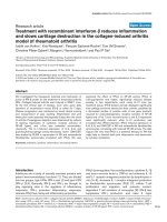
The principal elements of the signaling events that follow
engagement of the human TLRs are shown in Fig. 1.
Major elements of the human coagulation
system
This subject was reviewed in considerable detail recently
[1–3,43,44], and the major clotting parameters that interact
in sepsis are shown in Fig. 2. The extrinsic pathway (TF
pathway) is the primary mechanism by which thrombin is generated in sepsis, hemostasis and thrombosis. The intrinsic
cascade (contact factor pathway) primarily serves an accessory role in amplifying the prothrombotic events that are initiated in sepsis. Thrombin, factor Xa, and the TF–factor VIIa
complex directly activate endothelial cells, platelets and white
blood cells, and induce a proinflammatory response. The
inflammatory reaction to tissue injury activates the clotting
Available online />
Table 1
Pattern recognition receptors of the innate immune response and their major known natural ligands
Receptor or related
structure
Cell type or soluble factor
CD14
Myeloid cells and soluble forms PAMPs from bacterial, fungal, and mycobacterial antigens
Mannose binding lectin
Soluble factor
Binds to mannosides found on bacteria and fungi; activates complement and
opsonin for neutrophils
C-reactive protein
Soluble protein
Opsonin for Gram-positive bacteria
LPS-binding protein
Soluble protein
Binds to LPS in Gram-negative bacteria and lipoteichoic acid
Gram-positive bacteria
C′3, alternative complement
Examples of known natural microbial ligands
Soluble proteins
Polysaccharide capsules of bacteria, fungi
MD1
B cells
Coreceptor for LPS on cell surface of B cells
MD2
Myeloid cells
Coreceptor for LPS on macrophages, neutrophils
TLR1
Myeloid cells
Lipopeptide, lipoteichoic acid, LPS of leptospirosis
TLR2
Myeloid cells
Peptidoglycan, lipopeptide, lipoarabinomannan, fungal cell wall components,
LPS of leptospirosis
TLR3
Myeloid cells
Double-stranded viral RNA
TLR4
Myeloid cells
LPS, respiratory syncytial virus proteins
TLR5
Myeloid cells
Flagellin from Gram-positive or Gram-negative bacteria
TLR6
Myeloid cells
Zymosan (fungal constituents) along with TLR2
TLR9
Dendritric cells, B cells,
epithelial cells
Unmethylated CpG motifs in prokaryotic DNA
LPS, lipopolysaccharide; PAMP, pathogen associated molecular pattern; TLR, Toll-like receptor.
system, inhibits the endogenous anticoagulants, and attenuates the fibrinolytic response. The net effect within the microcirculation is a procoagulant state, which has major
therapeutic implications [16,17,24,45].
It was traditionally thought that the contact factors, factor XII,
factor XI, prekallikrein, and high-molecular-weight kininogen
were the primary activators of the clotting cascade in sepsis.
It is clear that contact factors can be activated by cell wall
components found on both Gram-positive and Gram-negative
bacteria. The negatively charged bacterial molecule LPS is
the prototypical microbial inducer of the coagulation cascade.
Activation of these coagulation factors generates a factor X
converting complex consisting of factor IXa and the acceleration factor VIIIa, resulting in activation of the common clotting
pathway at the level of factor X activation. The cascade then
follows via conversion of prothrombin to thrombin by activated factor X in the presence of the accelerating cofactors,
namely factor Va, negatively charged phospholipid, and
calcium. Thrombin generation is immediately followed by
fibrin monomer production through degradation of fibrinogen
with subsequent polymerization to fibrin clots and stabilization
of fibrin by the action of factor XIIIa, an enzyme that is generated by thrombin activation of factor XIII.
It is now recognized that this classical view of the coagulation
activation via the contact factor system is not the primary
pathway of thrombin generation and fibrin generation in most
patients with sepsis. The extrinsic pathway of coagulation is
the essential pathway of clot formation in sepsis (see below).
Studies in which sublethal doses of endotoxin were administered to human volunteers [1,11,46] revealed no evidence of
contact factor activation, despite thrombin generation as
measured by thrombin–antithrombin complexes and prothrombin fragment 1.2 generation. Prothrombin fragment 1.2
is a reliable measure of ongoing thrombin generation because
this peptide is released from prothrombin during active thrombin generation. Moreover, the antibodies that specifically
block the intrinsic clotting system do not diminish the frequency of thrombin generation in experimental animal studies
of sepsis [46]. These antibodies did, however, prevent the
contact pathway generation of bradykinin. Thus, although the
contact pathway is activated in experimental sepsis and contributes to vasodilatation, it does not contribute significantly
to thrombin generation [2,26].
It should be noted that, during actual human septic shock,
contact factor activation might in fact occur, as indicated by
systemic release of bradykinin from high-molecular-weight
kininogen. Bradykinin is a potent vasoactive substance that
may contribute to the hypotension and diffuse capillary leak
that typifies septic shock [26]. The intrinsic pathway can also
be activated by thrombin itself, and this system may function
as an amplification pathway in sepsis-induced disseminated
27
Critical Care
February 2003 Vol 7 No 1
Opal and Esmon
Figure 1
Figure 2
Major Coagulation Parameters in Septic Shock
Bacterium
Amplification
pathway
Tissue Factor
Pathway
TF expression
F VII
F IXa
+F VIIIa
TF:F VIIa
TIR
TIR NFκB
TIR
κ
TIR
FX
IKK
NIK
ECSIT
P TRAF6
IRAK
TIR
MyD88
The human Toll-like receptors (TLRs) and their known ligands. CpG,
cytosine-phosphoryl-quanine; ECSIT, evolutionarily conserved
signaling intermediate of Toll; IκB, inhibitory kappaB; IKK, IκB inducing
kinase; IRAK, IL-1 receptor associated kinase; LBP-lipopolysaccharidebinding protein; LPS, lipopolysaccharide; MyD88, myeloid
differentiation factor 88; NF-κB, nuclear factor-κ for B cells; NIK, NFκB inducing kinase; PG, peptidoglycan; TIR, Toll IL-1 receptor domain;
TRAF6, tumor necrosis factor receptor associated factor-6.
intravascular coagulation [47]. Recent evidence indicates
that the intrinsic clotting system is frequently activated in
experimental and perhaps clinical streptococcal toxic shock
[48].
28
F XIa
Current evidence indicates a dominant role of the TF pathway
(extrinsic pathway) for coagulation activation in sepsis
(Fig. 2). TF is not normally expressed within the endovascular
system, and resides on vascular smooth muscle cells and
fibroblasts within the adventitia around blood vessels. This
arrangement is ideal under physiologic conditions because
TF only becomes exposed to other clotting components after
injury to the vessel wall when blood is extravasated into the
interstitium [47]. TF expression is upregulated on monocytes/macrophages and to a limited extent, if at all, on
endothelial cells [27] following exposure to proinflammatory
mediators such as endotoxin, CRP, IL-1, and IL-6 [1,2,49,50].
Soluble TF, defined as TF activity resident in plasma, may
also be found in the circulation in patients with sepsis. Much
of the soluble TF may be resident in microparticles released
from activated or damaged mononuclear cells [51]. TF
expressed within the intravascular space will bind to circulating factor VII, resulting in a TF–factor VIIa complex [52].
TF–factor VIIa complexes can directly activate the common
pathway of coagulation by converting factor X to factor Xa.
Factor Xa in the presence of factor Va forms a prothrombinconverting complex, resulting in thrombin formation and subsequent generation of a fibrin clot [2,53]. TF–factor VIIa
complexes may also activate the intrinsic clotting system by
converting factor IX to IXa, which, in the presence of factor
F Xa
+F Va
Prothrombin
Common
pathway
Thrombin
Fibrinogen
+F XIII
Fibrin
Clot
The major coagulation factors and the pathways of coagulation
activation in sepsis. TF, tissue factor; t-PA, tissue-type plasminogen
activator.
VIIIa, can also activate factor X and result in the generation of
a fibrin clot. This latter pathway appears to be important
because the TF–factor VIIa complex is rapidly inactivated by
tissue factor pathway inhibitor (TFPI) once traces of factor Xa
are formed (See the section Tissue factor pathway inhibitor,
below).
The contact factors of the intrinsic pathway play an important
accessory role as an amplification loop in sepsis once the TF
pathway activates coagulation. Thrombin generation feeds
back at the level of factor XI and, to a lesser degree, factor VIII
and factor V, to promote factor X conversion and thrombin
generation via the contact factor system. The contact factor
pathway also functions to activate the fibrinolytic system,
along with the proinflammatory cytokine TNF [46,54].
Endogenous mechanisms to prevent
thrombus formation
There are four major systems that minimize thrombus formation in humans (Fig. 3). These include the fibrinolytic system,
antithrombin, TFPI, and the protein C–protein S–thrombomodulin pathway. Depletion of these systems contributes to
the consumptive coagulopathy and/or microvascular thrombosis of sepsis [1,2].
Fibrinolytic system
The fibrinolytic system is rapidly activated by proinflammatory
cytokines, particularly TNF, in the early phases of sepsis
[16,18]. This essential activity is initiated when plasminogen
is converted into the potent, broad-spectrum protease
plasmin. Plasmin degrades fibrin, fibrinogen, the acceleration
factors V and VIII, and probably other substrates as well [55].
In most septic patients generation of plasmin is abruptly
downregulated by simultaneous increase in the levels of
Available online />
Figure 3
Endogenous Inhibitors of Coagulation in Sepsis
TF expression
Activated
Protein C
F VII
F XIa
F IXa
+F VIIIa
TF:F VIIa
FX
Tissue Factor
Pathway Inhibitor
F Xa
+F Va
Prothrombin
Antithrombin
Plasminogen
Thrombin
Plasmin
Fibrinogen
Fibrin
t-PA
Fibrinolytic
System
The principal coagulation regulatory pathways and their sites of action.
TF, tissue factor; t-PA, tissue-type plasminogen activator.
inhibitors of fibrinolysis [54], including plasminogen activator
inhibitor (PAI)-1 and PAI-2 [16,54,56–58]. Intravascular
fibrinolysis is principally mediated by the actions of tissuetype plasminogen activator on plasminogen, and its primary
inhibitor is PAI-1. Extravascular clots (e.g. fibrin deposition
within the alveoli in acute respiratory distress syndrome, or
inflammatory foci in tissues) are primarily degraded when
urokinase-type plasminogen activator induces plasmin formation. PAI-2 inhibits the activity of urokinase-type plasminogen
activator in the extravascular space [55].
Thrombin activatable fibrinolysis inhibitor (TAFI) is a procarboxypeptidase B that is rapidly activated by the
thrombin–thrombomodulin complex. The TAFIa that is generated removes basic lysine and arginine residues from the carboxyl terminus of peptides and proteins. In the case of fibrin,
removal of carboxyl-terminal lysine residues renders the fibrin
less sensitive to lysis by decreasing the ability of plasminogen
and tissue-type plasminogen activator to bind to the fibrin, a
step that facilitates clot lysis. Inhibition of thrombin generation
by anticoagulants such as the APC–protein S complex prevents TAFI generation from its inactive precursor [56–58].
Genetic polymorphisms that lead to excess expression of
PAI-1 [59] or TAFI [60] may place certain septic patients at
greater risk for diffuse thrombosis and mortality because
excess levels of these fibrinolysis inhibitors attenuate the fibrinolytic system. Although TAFI was named for its unquestionable ability to inhibit fibrinolysis, recent studies have
suggested that a comparable if not more important function
of TAFIa may be in the control of vasoactive substances (see
the section on Activated protein C, below).
Tissue factor pathway inhibitor
TFPI is a 42-kDa protein that consists of three closely linked
Kunitz domains [61,62]. These domains allow TFPI to func-
tion by a unique mechanism. Factor Xa generated by the
TF–factor VIIa complex binds very tightly to and inactivates
factor Xa. By virtue of the ability of factor Xa to bind to negatively charged phospholipids, the resultant complex interacts
with damaged cells, raising the local concentration of TFPI.
The TFPI–factor Xa complex then binds to the TF–factor Vlla
complex. The latter interaction is of lower affinity and occurs
poorly in the absence of the concentrating effects that result
from the formation of the TFPI complex with factor Xa.
Because this inhibitor rapidly inhibits factor VIIa bound to TF
once the first factor Xa molecules are formed, the alternative
activation of factor IX by the TF–factor VIIa complex becomes
critical to thrombin generation and hemostasis. At therapeutic
levels, TFPI anticoagulates blood both by direct inhibition of
factor Xa and by the factor Xa dependent inhibition of the
TF–factor VIIa complex.
The dynamics of TFPI activity in the microcirculation are
rather complex and vary according to the amount of TFPI
bound to endothelium or stored in endothelial vacuoles [63].
TFPI levels bound to lipoprotein and in platelets, and circulating TFPI that may be present in active form or less active
cleaved TFPI [61,64]. The less active cleaved form of TFPI
results from cleavage by neutrophil elastase or other serum
proteases that are generated in severe sepsis. Most clinical
assay systems are not able to discriminate between inactive
cleaved TFPI and fully active TFPI [61]. These technical problems, along with the very low (nanogram range) quantities
that are measurable in the circulation, have rendered TFPI
levels in human sepsis difficult to measure and interpret
[61,65]. Perhaps more important is that only a small amount
of the total TFPI circulates in the blood. Heparin administration can elevate plasma levels of TFPI by about 10-fold, presumably reflecting release of bound or stored endothelial cell
TFPI [62]. Because the vast majority of the endothelium is in
the microcirculation, these findings indicate that the highest
levels of TFPI are also found in the microvasculature and
suggest that this inhibitor plays a key role in the regulation of
microvascular thrombosis.
Consistent with a critical role played by TFPI in regulating
microvascular thrombosis, genetic deletion of the TFPI gene
in mice results in early embryonic lethality and microvascular
thrombosis [66]. Furthermore, inhibiting TFPI with antibodies
exacerbates the response to endotoxin infusion in experimental rabbit models of sepsis [67].
Certainly, however, both experimental and clinical evidence
indicates that functionally active TFPI levels are inadequate
within the microcirculation to prevent ongoing coagulation
and organ dysfunction in sepsis. Exogenously added TFPI
has been shown to reduce inflammatory [65,68] and coagulation activities [69,70] in experimental models of sepsis, and
to improve outcomes in septic animals. These experimental
findings form the therapeutic rationale for recombinant TFPI
therapy, which is a logical strategy in clinical sepsis. Regret-
29
Critical Care
February 2003 Vol 7 No 1
Opal and Esmon
tably, a recently completed, large, phase 3 international
sepsis trial with TFPI treatment was apparently unable to
demonstrate a benefit from treatment. The details of that
study are not available at present, pending the publication of
the final study findings in the near future.
Antithrombin
The anticoagulant actions of antithrombin (formerly referred
to as antithrombin III) are well known and relate to its ability to
function as a potent endogenous serine protease inhibitor.
Antithrombin is a hepatically synthesized plasma protein that
is activated by the process of allosteric activation by heparin
and related heparans. Specific polysulfated pentasaccharides, which are found in repeating units in glycosaminoglycans and mucopolysaccharides, are necessary to bind to a
highly basic, central domain in antithrombin. A conformational
change takes place in antithrombin following interactions with
these acidic pentasaccharide moieties, bringing a critical
arginine residue at position 393 to link covalently within the
active site of serine proteases, thereby accelerating the inactivation of these proteases [71,72]. The conformational
change in antithrombin induced by heparin is only part of the
heparin mechanism, however. For heparin to function as an
efficient stimulator of thrombin inhibition, higher molecular
weight forms of heparin are needed. A higher molecular
weight would allow heparin to form a bridge between
antithrombin and thrombin. Heparins that are too small to form
this bridge have almost no effect on thrombin inhibition by
antithrombin but retain the ability to inactivate factor Xa [73].
Many of the clotting factors and regulators of the coagulation
system are serine proteases, including thrombin, factor X,
components of the contact system, and TF–factor
VIIa–heparin complexes [74,75]. The broad substrate enzymatic activity of this plasma protease inhibitor allows
antithrombin to play a central role in the regulation of coagulation. Antithrombin is rapidly consumed in sepsis by covalent
linkage and clearance, along with the activated clotting
factors [72]. Antithrombin levels are further diminished by
enzymatic cleavage by neutrophil elastase production [76]
and by diminished hepatic synthesis during sepsis [77]. Loss
of anticoagulant activity as a result of reduced antithrombin
levels participates in the generation of the prothrombotic
state that characterizes septic shock [72,78,79].
30
In the absence of heparin, antithrombin binds to specific pentaccharide-bearing glycosaminoglycans on the cell surface of
endothelial cells, such as heparan sulfate. When in contact
with endothelial cells, antithrombin exerts both local anticoagulant and anti-inflammatory activities [80–82]. This is mediated in part by antithrombin-mediated induction of
prostacyclin synthesis by endothelial cells. Prostacyclin is a
potent antiplatelet agent that inhibits platelet aggregation and
attachment. Prostacyclin also inhibits neutrophil–endothelial
cell attachment and attenuates IL-6, IL-8, and TNF release by
endothelial cells [82–84].
It has recently been demonstrated that antithrombin has additional anti-inflammatory effects via direct binding to neutrophil, lymphocyte, and monocyte cell surface receptors
such as syndecan-4 [85–88]. Antithrombin reduces expression of IL-6 and TF, and inhibits of activation of the transcription factor NF-κB in LPS-stimulated monocytes and
endothelial cells [89,90].
Antithrombin reduces chemokine (IL-8)-induced chemotaxis
of neutrophils and monocytes in experimental systems. This
may be mediated by a reduction in chemokine receptor
density on leukocyte cell surfaces after binging to antithrombin. This direct inhibitory effect is blocked by heparin and synthetic pentasaccharides via competitive inhibition against
antithrombin binding to sydecan-4 [85,86].
These anti-inflammatory activities are observed in vivo in a
number of animal systems in which attenuation of white
cell–endothelial cell interactions have been demonstrated by
intravital microscopy [71,91,92]. The administration of
antithrombin to LPS-challenged animals significantly reduced
the interaction of inflammatory cells with the vessel wall (characterized by rolling, sticking, and transmigration events),
thereby limiting capillary leakage and subsequent organ
damage. Recently, Hoffman and coworkers [92] have confirmed these anti-inflammatory activities in a hamster model
that quantifies functional capillary density in vivo. White cell
adherence and loss of functional capillary density was rapidly
induced by LPS, and this loss of microcirculatory surface was
inhibited by therapeutic doses of antithrombin. Antithrombinmediated preservation of functional capillary density is completely prevented by unfractionated or low-molecular-weight
heparin. These anti-inflammatory actions have been demonstrated in a number of experimental systems [80,83,84,
92–97] and are presumably physiologically important within
the microcirculation in human sepsis as well [72].
This may provide a partial explanation for the results of a
recent phase 3 clinical trial with high-dose antithrombin in
severe sepsis [98]. No overall benefit was found by administration of 30 000 IU of plasma-derived antithrombin over
4 days in that large international trial conducted in 2314
patients (38.9% antithrombin versus 38.7% placebo; not significant). It was observed that a prespecified subgroup of
patients who received no heparin (30% of the overall study
population) appeared to derive some modest benefit from
antithrombin (15% relative risk reduction in mortality after
90 days; P < 0.05). These patients might have derived longterm benefits with respect to morbidity and quality of life
indices as well [99].
The subgroup of patients who received heparin (up to 10 000
units/day, as allowed by the study protocol) experienced no
improvement in outcome with antithrombin therapy but exhibited a significantly greater risk for hemorrhage than did
placebo-treated patients (10.9% with antithrombin versus
Available online />
6.2% in the control group; P < 0.01). The use of concomitant
heparin with antithrombin in that study might have blocked
any potential, salutary, anti-inflammatory effects of antithrombin within the microcirculation, and this combination clearly
exacerbated the risk for bleeding in severely septic patients
[98].
Activated protein C
The APC pathway of anticoagulation is a classic negative
feedback loop initiated by thrombin-dependent generation of
the anticoagulant APC. The vitamin K-dependent protein C
zymogen is transformed into APC by the proteolytic cleavage
of 12 amino acids from the amino terminus of the heavy chain
of protein C. This activation step is catalyzed very slowly by
thrombin itself. Rapid activation of protein C occurs along the
luminal surface of capillary endothelial cells. Thrombin is first
complexed with its specific, membrane-bound, protein receptor, thrombomodulin. Once bound to thrombomodulin, thrombin is incapable of binding to fibrinogen for conversion to
fibrin, can no longer activate platelets, and loses its pro-coagulant activity [21,22]. The thrombin–thrombomodulin complex
retains a capacity to bind to its other substrate, protein C,
and the rate of protein C activation relative to thrombin alone
is increased about 1000-fold. Thrombin now becomes an
anticoagulant enzyme converting the inactive precursor
protein C to APC.
APC is a potent serine protease that, in comparison to other
serine proteases (which usually have a half-life of seconds),
has a relatively long elimination half-life from the plasma of
approximately 15–20 min [18,22]. Feedback inhibition of new
thrombin generation by APC is mediated by proteolytic
degradation of the acceleration coagulation factors Va and
VIIIa. APC activity is facilitated several fold by reversible
binding to another hepatically synthesized, vitamin K-dependent protein known as protein S. This ‘accessory’ protein
associates with APC only in its free circulating form; protein S
bound to C4b-binding protein from the complement system
cannot bind to APC [2,22,23].
In addition to inhibition of fibrin formation, APC also promotes
fibrinolysis in vitro by inhibiting two important inhibitors of
plasmin generation, namely PAI-1 [18,54] and TAFI [56–58].
This profibrinolytic activity of APC is not shared by antithrombin [1,2]. APC actually binds to the active site of PAI-1 and
as such blocks the serine protease inhibitor actions of PAI-1
[2,100,101]. The reaction of APC with PAI-1 is relatively
slow, but the rate is enhanced dramatically by vitronectin
[102], raising the possibility that the profibrinolytic effects of
APC might center around cells such as platelets that can
release vitronectin. It has been speculated that these
profibrinolytic activities of APC might have significantly contributed to the therapeutic efficacy observed in the recent
phase 3 trial with recombinant human APC (drotrecogin alfa
[activated]) in human sepsis [24]. The clinical relevance of
this activity of APC remains to be convincingly demonstrated.
As discussed previously, TAFI is activated by the
thrombin–thrombomodulin complex and this activation probably occurs in the microcirculation. TAFIa has been shown to
inhibit fibrinolysis [57]. Inhibitors of thrombin formation would
therefore inhibit TAFI activation and presumably facilitate clot
lysis. TAFIa, however, is a carboxypeptidase with broad substrate specificity and a preference for removal of carboxyl-terminal arginine residues [103]. Removal of carboxyl-terminal
arginine residues is a major mechanism for inactivation of
vasoactive peptides. It was recently proposed that TAFIa is
the major inhibitor of complement anaphylatoxin C5a.
Because both prothrombin activation and complement activation occur in severe sepsis, it is not surprising that key regulatory mechanisms that are involved in controlling coagulation
might also control complement. Inactivation of C5a would be
expected to decrease neutrophil chemotaxis and systemic
vasodilatation [103]. This is another example of the close
interrelationship between clotting regulators and innate
immune reactions. A summary of inflammatory reactions to
the procoagulant and loss of anticoagulant activity found in
sepsis is provided in Table 2.
APC has direct anti-inflammatory effects in experimental
studies that are independent of the antithrombotic actions of
this endogenous anticoagulant (for review [104]). APC binds
to specific receptors on endothelial cells and white cells. The
only receptor isolated and characterized to date is known as
endothelial protein C receptor (EPCR) [18,105]. This
APC–EPCR complex can translocate from the plasma membrane to the nucleus, where it presumably alters gene expression profiles. Other evidence suggests that APC cleaves a
receptor on the cell surface [106,107], and in some cases
this appears to be EPCR dependent [108]. APC bound to
EPCR has also been shown to cleave protease-activated
receptor (PAR)-1 and PAR-2 [109], but how this facilitates
the in vivo anti-inflammatory effects observed with APC [104]
remains to be determined. In vivo, the anti-inflammatory
effects of APC that are independent of its anticoagulant
effects include inhibition of neutrophil adhesion, decreased
TNF elaboration, and decreased drops in blood pressure (for
review [104]). APC has multiple effects in tissue culture
systems, including limitation in NF-κB-mediated proinflammatory activity [110], attenuation of inflammatory cytokine and
chemokine generation [23], and upregulation of antiapoptotic
genes of the Bcl-2 family of homologs [111].
Endothelial cells are relatively resistant to apoptosis as a result
of the constitutive synthesis of a number of antiapoptotic proteins [112]. Microbial mediators, such as bacterial LPS, can
overcome the inhibition of apoptosis within endothelial cells.
APC protects endothelial cells from apoptosis in experimental
systems. It remains to be demonstrated whether this activity is
relevant to the protective effects of APC in human sepsis.
In experimental studies and in human sepsis circulating blood
levels of protein C rapidly decline, with loss of this important
31
Critical Care
February 2003 Vol 7 No 1
Opal and Esmon
Table 2
The inflammatory effects of coagulation and loss of anticoagulants
Coagulation parameter
Proinflammatory effects
Thrombin generation
Promotes cytokine and chemokine synthesis (IL-6, IL-8) via PARs, P-selectin, E-selectin
and PAF expression, which facilitates neutrophil–endothelial cell interactions,
bradykinin and histamine release
Factor Xa and TF–factor VIIa complex generation
Promotes cytokine and chemokine synthesis (IL-6, IL-8) via PAR-1 and PAR-2
Reduced antithrombin
Results in the loss of prostacyclin synthesis by endothelial cells, increased cytokine
synthesis, increased leukocyte adherence and chemotaxis
Reduced protein C/protein S activity
Results in increased E-selectin expression, increased cytokine generation and neutrophil
adherence; promotes apoptosis of endothelial cells
Reduced TFPI activity
Results in loss of regulation of cytokine synthesis within microcirculation
Platelet activation
Platelet derived P-selectin promotes neutrophil adherence, neutrophil–endothelial cell
interactions; platelet CD40 ligand promotes endothelial cell chemokine and adhesion
molecule expression; activated platelets secrete chemokines and IL-1β
Intravascular fibrin deposition
Neutrophil and monocyte adherence
Reduced TM expression on
endothelial cells
Loss of TM lectin domain activity that inhibits neutrophil–endothelial cell adherence may
promote neutrophil binding
IL, interleukin; PAF, platelet-activating factor; PAR, protease activated receptor; TF, tissue factor; TFPI, tissue factor pathway inhibitor;
TM, thrombomodulin.
coagulation inhibitor function [113,114]. Protein S functional
levels also decrease. There is evidence that peripheral conversion of protein C to APC is impaired as a result of diminished expression or cleavage of EPCR [115–117] and
thrombomodulin [118] in the microcirculation. Soluble thrombomodulin is readily measurable in the circulation of septic
patients [119,120], and biopsies of blood vessels in patients
with meningococcal disease confirm the loss of thrombomodulin and EPCR expression along endothelial surfaces during
severe sepsis [121]. The extent to which these protein C activators are downregulated in severe sepsis appears to vary
widely [122]. These findings provide the therapeutic rationale
for the administration of APC in severely septic patients [24].
In the phase 3 clinical trial [24], recombinant human APC
(drotrecogin alfa [activated]), administered by continuous
infusion at a dose of 24 µg/kg per hour for 4 days, reduced
the mortality rate from 30.8% in the placebo group (n = 840)
to 24.7% in the recombinant human APC group (n = 850;
P = 0.005). This indicates an absolute reduction in mortality
rate of 6.1% and a relative risk reduction of 19.4% associated with treatment with drotrecogin alfa (activated).
32
In experimental models of sepsis, soluble thrombomodulin
has been shown to have both anticoagulant and anti-inflammatory activity. Much of the anti-inflammatory activity was
believed to be mediated by protein C activation. Recently, the
lectin domain of thrombomodulin was shown to have direct
anti-inflammatory activity by reducing adhesion molecule
expression and inhibiting MAPK and NF-κB pathways,
thereby inhibiting the ability of leukocytes to bind to activated
endothelium in vivo [123]. As mentioned above, infusion of
thrombomodulin would be expected to inhibit the activities of
vasoactive substances and to inhibit thrombin clotting activity
directly. These newly identified functions of thrombomodulin
suggest that it might be a good therapeutic target in severe
sepsis, but one that might require protein C supplementation
to be effective.
EPCR, the other receptor that is involved in protein C activation, appears to have direct anti-inflammatory activity also.
Soluble EPCR, which is released in response to thrombin
activation of the endothelium [124], binds to proteinase-3, a
serine protease released from activated neutrophils. This
complex in turn binds to Mac-1 [125], which is an important
integrin involved in tight neutrophil adhesion. Of interest, proteinase-3 is the autoantigen in Wegener’s granulomatosis. It
appears that soluble EPCR binding to this complex results in
inhibition of tight neutrophil adhesion.
In considering therapy with protein C pathway components,
protein C supplementation is an obvious possibility, especially because protein C levels are decreased, sometimes
severely, in severe sepsis. There are several anecdotal
reports of success in treating patients with severe sepsis with
protein C [126–128]. The disadvantage of this approach is
that the protein C activation complex may be downregulated
severely in some patients with severe sepsis. The advantage,
however, is that protein C activation is tightly regulated and
ceases locally as soon as thrombin formation is controlled.
Available online />
Table 3
Procoagulant effects of inflammatory mediators
Inflammatory mediator
Procoagulant effects
Proinflammatory cytokines
Increased TF expression on endothelium, monocytes; decreased TM and endothelial protein C receptor;
increased PAI-1; release of TFPI from endothelium with loss of activity
Complement components
Decreased C1-esterase inhibitor leads to loss of contact factor regulation; damaged cell membranes promote
procoagulant activity on cell surfaces of endothelial cells
Acute phase proteins
Increase in clotting factor synthesis; decrease in synthesis of antithrombin; α1-antitrypsin decreases APC and
cleaves TFPI; CRP promotes TF expression; C4b-binding protein binds to protein S and limits protein C activity
Neutrophils
Elastase destroys antithrombin, C1-inhibitor, thrombomodulin, and cleaves TFPI; intravascular neutrophil–platelet
aggregates occlude capillary beds
Activated monocytes
Upregulation of TF expression; IL-6 and TNF synthesis promote acute phase proteins with procoagulant activities;
release of microvesicles with TF in circulation
Activated endothelium
P-selectin promotes platelet aggregation, procoagulant surface upregulation of TF; PAF expression stimulates
platelets; shedding of glycosaminoglycans limits antithrombin binding; loss of TM and EPCR expression limits
APC synthesis
APC, activated protein C; CRP, C-reactive protein; EPCR, endothelial protein C receptor; PAF, platelet activating factor; PAI, plasminogen
activator inhibitor; TF, tissue factor; TFPI, tissue factor pathway inhibitor; TM, thrombomodulin; TNF, tumor necrosis factor.
Thus, protein C supplementation has the advantage of generating high levels of APC locally. In addition, because both
protein C and APC bind to EPCR, activation of endogenous
protein C allows selective loading of EPCR with APC, which
should serve to amplify APC-dependent, receptor-mediated
anti-inflammatory activities of APC.
The mechanisms by which inflammatory
responses promote coagulation
Inflammation promotes coagulation via a large number of molecular and cellular mechanisms (Table 3). Perhaps the most
direct mechanism responsible for the procoagulant activity of
the inflammatory response is through the generation of proinflammatory cytokines [1,2]. A number of cytokines, particularly IL-6, increase the expression of TF on endothelial
surfaces and monocytes [1,129,130]. The TF pathway, also
referred to as the extrinsic clotting cascade, is the principal
activator of clotting in the presence of systemic inflammation
and generalized infection.
TNF and IL-1β, which are major cytokines in the pathogenesis
of septic shock, also inhibit the expression of EPCR [117]
and thrombomodulin on endothelial cells [118]. Thrombomodulin is primarily located on the endothelial surfaces of
capillaries within the microcirculation, whereas EPCR is principally located on endothelial surfaces of larger vessels on
small arteries and arterioles [119]. Together, these molecules
facilitate the generation of APC by bringing thrombin into
direct contact with protein C. Loss of thrombomodulin and
EPCR via proinflammatory cytokine production impairs conversion of protein C to APC [121]. The reduced levels of
protein C and APC contribute toward the procoagulant state
that typifies severe sepsis [1,2,24,131,132]. TNF is a potent
inducer of fibrinolysis in sepsis through the synthesis of
tissue-type plasminogen activator; however, fibrinolytic activity is rapidly inhibited by the almost simultaneous production
of increased levels of PAI-1 [54]. This culminates in a pathophysiologic state of cytokine-induced activation of coagulation, diminished anticoagulant activity, and suppressed
fibrinolysis, with widespread thrombin generation and
intravascular fibrin deposition.
Activated neutrophils along endothelial surfaces release the
broadly reactive proteolytic enzyme elastase that destroys
antithrombin and C1-esterase inhibitor and releases thrombomodulin in a less active form [94,128]. Both of these regulatory proteins are important endogenous inhibitors of the
coagulation system. C1-esterase inhibitor is the major regulator of the intrinsic or contact factor pathway of coagulation
[1,2,131]. This pathway remains of significance in sepsis
despite the fact that the TF pathway is the initiator of clotting
in systemic inflammatory states. Factor IX of the intrinsic
system can be activated by the TF–factor VII complex, and
thrombin can activate factor XI and to some degree
factors VIII and V. This accessory system serves to amplify
and maintain coagulation in sepsis [132].
The acute phase protein CRP upregulates TF, whereas
another acute phase protein, namely α1-antitrypsin, inhibits
APC. Both of these actions result in a procoagulant state
[43,50,53,120]. CRP synthesis has activities other than the
promotion of TF expression and coagulation activation. It also
promotes complement activation via the classical complement pathway [10]. Complement activation in response to
inflammatory stimuli depletes several control elements of the
coagulation system. Complement components activate neutrophils, promote neutrophil chemotaxis, stimulate cytokine
synthesis, and contribute to increased capillary permeability
33
Critical Care
February 2003 Vol 7 No 1
Opal and Esmon
and systemic hypotension [2,11,43]. Contact factor activation generates systemic synthesis of bradykinin, resulting in
systemic hypotension and tissue hypoperfusion. Moreover,
focal regions of tissue ischemia as a result of intravascular
clot formation stimulate an intense inflammatory response
[1,19,20].
The acute phase complement component C4b-binding
protein is upregulated in inflammation, and binds and inactivates protein S. Protein S is an endogenous coagulation
inhibitor that enhances the activity of APC as an inhibitor of
factors Va and VIIIa [2,100]. A summary of some of the procoagulant effects that accompany the acute inflammatory
response is provided in Table 3.
Role of the coagulation pathways in systemic
inflammation
Intravascular thrombin generation is highly inflammatory
within the microcirculation via interaction with specific receptors on platelets, endothelial cells and white blood cells
known as PARs [133,134]. The PARs are typical seven-transmembrane, G-protein-linked receptors that differ from other
receptors in that their ectodomain possesses a sequestered
internal ligand that is tethered to the amino-terminus of its
extracellular domain. Thrombin cleaves the amino-terminus of
the PAR, allowing the internal ligand to autoactivate the
receptor.
There are four known PARs in human biology named PAR1–4. PAR-2 is responsive to trypsin. The TF–factor VIIa
complex may also activate PAR receptors [135], which may
be mediated by PAR-2. Factor Xa can also directly activate
cells via PAR-1-mediated cell activation [135]. Activation of
cells mediated by thrombin and other coagulation factors
increases proinflammatory cytokine synthesis and calcium
flux, alters intracellular signaling cascades such as the MAPK
pathway, and induces nitric oxide synthesis [17,18]. Thrombin also stimulates production of platelet-activating factor
[136]. Platelet-activating factor is a potent neutrophil-activating substance, especially when the neutrophils are tethered
to P-selectin, a molecule that is expressed on endothelium
and platelets in response to thrombin [137,138].
34
Activated platelets contribute to local inflammatory processes
at the site of clot formation by a number of mechanisms.
Platelets can secrete chemokines and IL-1, which activate
white cells and promote neutrophil and monocyte adherence
[139]. Platelets express P-selectin (see below) and promote
neutrophil–platelet–endothelial cell interactions. It was
recently demonstrated that platelets are a major source of
soluble CD40 ligand (also known as CD154) [140]. CD40
ligand belongs to the TNF superfamily of molecules and has
multiple actions that may be of significance within the microcirculation in sepsis [140]. These actions include upregulation of cytokine chemokine expression on vascular smooth
muscle cells and endothelial cells; increased expression of
surface adhesion molecules on endothelial cells; and upregulation of TF synthesis on macrophages [141].
P-selectin expression on endothelium and platelets is mediated by thrombin. It is a major surface adhesin that promotes
the initial rolling and tethering interactions between circulating granulocytes, monocytes, and lymphocytes to endothelial
cells at sites of tissue injury [138,142]. P-selectin glycoprotein ligand-1 is expressed on neutrophils, monocytes and
some lymphocytes, and specifically binds to P-selectin on
endothelial and platelet surfaces [142]. Thrombin-induced
P-selectin expression on the endothelium promotes white cell
adherence and cellular activation within capillaries and postcapillary venules. Activated neutrophils, in turn, release elastase that destroys antithrombin and cleaves TFPI. The loss of
antithrombin and TFPI further disrupts the endogenous
control mechanisms for thrombin generation, leading to a
potentially lethal state of systemic activation of coagulation
and inflammation.
E-selectin also facilitates neutrophil–endothelial cell interactions in the tissues. The E-selectin binding between neutrophils and endothelial cells is attenuated in vitro by
protein C [143]. It has been speculated that the deficiency in
protein C that accompanies severe sepsis contributes to the
observed exaggerated neutrophil-mediated endothelial injury
[18,23,143]. By these mechanisms, thrombin generation
itself is now recognized as a potent inducer of proinflammatory reactions within the microcirculation.
Thrombin-initiated endothelial IL-6 production stimulates TF
expression and further perpetuates ongoing coagulation
[18,136]. This positive feedback loop between clotting and
inflammation terminates in disseminated intravascular coagulation DIC and septic shock (the ‘vicious cycle’ of clotting and
inflammation sepsis). All of these actions promote neutrophil,
lymphocyte, and platelet interactions with the capillary
endothelium, and this results in diffuse endothelial injury,
increased vascular permeability, and cellular apoptosis.
The realization that thrombin activation not only initiates fibrin
deposition but also activates a proinflammatory reaction has
prompted efforts to inhibit thrombin generation in patients
with severe sepsis [10,30]. The hypothesis that has been
generated indicates that a potent inhibitor of thrombin activation should both prevent intravascular fibrin deposition and
microcirculatory failure and provide an anti-inflammatory
message to attenuate the proinflammatory state that typifies
septic shock. However, at least with respect to the protein C
system, inhibition of thrombin would diminish APC generation and thus suppress the anti-inflammatory and profibrinolytic activities of APC. Indeed, in comparative baboon
models, natural anticoagulants have worked effectively in
prophylaxis against E. coli infusion, but a very potent and
specific anticoagulant, namely active site blocked factor Xa,
failed [144].
Available online />
Conclusion
The coagulation system is integrally related to the innate
immune response, and its activation and regulation is dependent on local and systemic immune responses. The simultaneous activation of clotting and the innate immune response
is a phylogenetically ancient host response to tissue injury,
and has become the primary survival strategy throughout the
long history of vertebrate evolution. This close linkage
between clotting and inflammation has proven to be a survival
advantage in response to the vicissitudes of life on earth, in
which multicellular animals must constantly compete with well
equipped microbial pathogens.
The molecular events that control coagulation are increasingly understood and the genetic elements that regulate the
clotting and immune systems are being defined by human
genome studies. It is evident from detailed experimental study
that the dysregulated coagulation system that typifies the
pathophysiology of septic shock contributes to systemic
inflammation and lethality in sepsis.
The results of the initial clinical trials with recombinant human
APC verify that it is possible to reverse the pathologic events
that follow the onset of human sepsis and significantly
improve the outcome of critically ill patients. It is hoped that
the knowledge gained in unraveling the pathophysiology of
coagulation and inflammation will result in further refinements
and improved therapies for patients with severe systemic
injuries and septic shock.
Competing interests
None declared.
References
1.
Levi M, ten Cate H: Disseminated intravascular coagulation. N
Engl J Med 1999, 341:586-592.
2. Vervloet MG, Thijs LG, Hack CE: Derangements of coagulation
and fibrinolysis in critically ill patients with sepsis and septic
shock. Semin Thromb Hemost 1998, 24:33-44.
3. Lorente JA, García-Frade LJ, Landín L, dePablo R, Torrado C,
Renes E, Garcí-Avello A: Time course of hemostatic abnormalities in sepsis and its relation to outcome. Chest 1993, 103:
1536-1542.
4. O’Neill LA, Greene C: Signal transduction pathway is activated
by the IL-1 receptor family: ancient signaling machinery in
mammals, insects, and plants. J Leukoc Biol 1998, 63:650657.
5. Opal SM, Huber CE: The Toll-like receptors and their role in
septic shock. Crit Care 2002, 6:125-136.
6. Levin J, Bang FB: The role of endotoxin in the extracellular
coagulation of Limulus blood. Bull Johns Hopkins Hosp 1964,
115:265-274.
7. Kawabata S-I: Molecular basis of non-self recognition by horeshoe crab lectins. J Endotoxin Res 2002, 8:175-176.
8. Rietschel ET, Westphal O: Endotoxin: historical perspectives.
In Endotoxin in Health and Disease. Edited by Brade H, Opal SM,
Vogel SN, Morrison D. New York: Marcel Dekker Inc.; 1999:1-30.
9. Turner MW: Mannose-binding lectin: the pluripotent molecule
of the innate immune system. Immunol Today 1996, 17:532540.
10. Wolbink G-J, Bossink AWJ, Groeneveld ABJ, DeGroot MCM,
Thijs LG, Hack CE: Complement activation in patients with
sepsis is in part mediated by C-reactive protein. J Infect Dis
1998, 177:81-87.
11. Dries DJ: Activation of the clotting system and complement
after trauma. New Horizons 1996, 4:278-288.
12. Pugin J, Heumann ID, Tomasz A, Kravchenko VV, Akamatsu Y,
Nishijima M, Glauser MP, Tobias PS, Ulevitch RJ: CD14 is a
pattern recognition receptor. Immunity 1994, 1:509-516.
13. Novitsky TJ: Limulus amebocyte lysate (LAL) detection of
endotoxin in human blood. J Endotoxin Res 1994, 1:253-263.
14. Hoess A, Watson S, Siber GR, Liddington R: Crystal structure of
an endotoxin-neutralizing protein from the horseshoe crab,
Limulus anti-LPS factor, at 1.5 Å resolution. EMBO J 1993, 12:
3351-3356.
15. Opal SM: The phylogenetic relationships between the coagulation system and the inflammatory networks. Crit Care Med
2000, 28(suppl):S77-S82.
16. van Gorp EC, Suharti C, ten Cate H, Dolmans WM, van der Meer
JW, ten Cate JW, Brandjes DP: Review: infectious diseases
and coagulation disorders. J Infect Dis 1999, 180:176-186.
17. Mavrommatis AC, Theodoridis T, Orfanidou A, Roussos C,
Christopoulou-Kokkinou V, Zakynthinos S: Coagulation system
and platelets are fully activated in uncomplicated sepsis. Crit
Care Med 2000, 28:451-457.
18. Esmon CT: Are natural anticoagulants candidates for modulating the inflammatory response to endotoxin? Blood 2000, 96:
1113-1116.
19. Carvalho AC, Freeman NJ: How coagulation defects alter the
outcome of sepsis:survival may depend on reversing the procoagulant conditions. J Crit Illness 1994, 9:51-75.
20. Hinshaw LB: Sepsis/septic shock: participation of the microcirculation: an abbreviated review. Crit Care Med 1996, 24:
1072-1078.
21. Taylor FB Jr, Stearns-Kurosawa DJ, Kurosawa S, Ferrell G, Chang
AC, Laszik Z, Kosanke S, Peer G, Esmon CT: The endothelial
cell protein C receptor aids in host defense against
Escherichia coli sepsis. Blood 2000, 95:1680-1686.
22. Esmon CT: The roles of protein C and thrombomodulin in the
regulation of blood coagulation. J Biol Chem 1989, 264:47434746.
23. Murakami K, Okajima K, Uchida M, Johno M, Nakagaki T, Okobe
H, Takatsuki K: Activated protein C prevents LPS-induced pulmonary vascular injury by inhibiting cytokine production. Am J
Physiol 1997, 272:L197-L202.
24. Bernard GR, Vincent JL, Laterre PF, LaRosa SP, Dhainaut JF,
Lopez-Rodriguez A, Steingrub JS, Garber GE, Helterbrand JD, Ely
EW, Fisher CJ Jr; Recombinant human protein C Worldwide Evaluation in Severe Sepsis (PROWESS) study group: Efficacy and
safety of recombinant human activated Protein C for severe
sepsis. N Engl J Med 2001, 344:699-709.
25. Kirschner M, Gerhart J: Evolvability. Proc Natl Acad Sci USA
1998, 95:8240-8427.
26. Pixley RA, De La Cadena R, Page JD, Kaufman N, Wyshock EG,
Chang A, Taylor FB, Colman RW: The contact system contributes to hypotension but not to disseminated intravascular
coagulation in lethal bacteremia. J Clin Invest 1993, 91:61-70.
27. Drake TA, Morrissey JH, Edgington TS: Selective cellular
expression of tissue factor in human tissues: Implications for
disorders of hemostasis and thrombosis. Am J Pathol 1989,
134:1087-1097.
28. Randolph MM, White GL, Kosanke SD, Bild G, Carr C, Galluppi
G, Hinshaw LB, Taylor FB Jr: Attenuation of tissue thrombosis
and hemorrhage by ala-TFPI does not account for its protection against E. coli: a comparative study of treated and
untreated non-surviving baboons challenged with LD100 E.
coli. Thromb Haemost 1998, 79:1048-1053.
29. Drake TA, Cheng J, Chang A, Taylor FB Jr: Expression of tissue
factor, thrombomodulin, and E-selectin in baboons with lethal
E. coli sepsis. Am J Pathol 1993, 142:1458-1470.
30. Wheeler AP, Bernard G: Treating patients with severe sepsis.
N Engl J Med 1999, 340:207-214.
31. Astiz ME, Rackow EC: Septic shock. Lancet 1998, 351:15011505.
32. Beutler B, Poltorak A: Sepsis and evolution of the innate
immune response. Crit Care Med 2001, 29(suppl):S2-S6.
33. Beutler B: Sepsis begins at the interface of pathogen and
host. Biochem Soc Trans 2001, 29:853-859.
34. Lauener RP, Birchler T, Adamski J, Braun-Fahrlander C, Bufe A,
Herz U, von Mutius E, Nowak D, Riedler J, Waser M, Sennhauser
FH; ALEX study group: Expression of CD14 and Toll-like
35
Critical Care
36
February 2003 Vol 7 No 1
Opal and Esmon
receptor 2 in farmers’ and non-farmers’ children. Lancet 2002,
360:465-466.
35. Bach J-F: Mechanisms of disease: the effect of infections on
the susceptibility to autoimmune and allergic diseases. N
Engl J Med 2002, 347:911-920.
36. Medzhitov R, Janeway C: Innate immunity. N Engl J Med 2000,
343:338-344.
37. Fenton MJ, Golenbock DT: LPS-binding proteins and receptors.
J Leukoc Biol 1998, 64:25-32.
38. Antal-Szalmas P: Evaluation of CD14 in host defense. Eur J
Clin Invest 2000, 30:167-179.
39. Landmann R, Müller B, Zimmerli W: CD14, new aspects of
ligand and signal diversity. Microb Infect 2000, 2:295-304.
40. Poltorak A, He X, Smirnova I, Liu MY, Huffel CV, Du X, Birdwell D,
Alejos E, Silva M, Galanos C, Freudenberg M, Ricciardi-Castagnoli P, Layton B, Beutler B: Defective signaling in C3H/HeJ and
C57BL/10ScCr mice: mutations in Tlr4 gene. Science 1998,
282:2085-2088.
41. Pfeiffer A, Bottcher A, Orso E, Kapinsky M, Nagy P, Bodnar A,
Spreitzer I, Liebisch G, Drobnik W, Gempel K, Horn M, Holmer S,
Hartung T, Multhoff G, Schutz G, Schindler H, Ulmer AJ, Heine H,
Stelter F, Schutt C, Rothe G, Szollosi J, Damjanovich S, Schmitz
G: Lipopolysaccharide and ceramide docking to CD14 provokes ligand specific receptor clustering in rafts. Eur J
Immunol 2001, 31:3153-3164.
42. Means TK, Golenbock DT, Fenton MJ: The biology of toll-like
receptors. Cytokine Growth Factor Rev 2000, 11:219-232.
43. ten Cate H: Pathophysiology of disseminated intravascular
coagulation in sepsis. Crit Care Med 2000, 28:S9-S11.
44. Abraham E: Coagulation abnormalities in acute lung injury and
sepsis. Am J Respir Cell Mol Biol 2000, 22:401-404.
45. Abraham E: Tissue factor inhibition and clinical trial results of
tissue factor pathway inhibitor in sepsis. Crit Care Med 2000,
28:S31-S33.
46. Levi M: Keep in contact: the role of the contact system in infection and sepsis. Crit Care Med 2000, 28:3765-3766.
47. Aird WC: Vascular bed-specific hemostasis: role of endothelium in sepsis pathogenesis. Crit Care Med 2001, 29:S28-S35.
48. Sriskandan S, Kemball-Cook G, Moyes D, Canvin J, Tuddenham
E, Cohen J: Contact activation in shock caused by invasive
group A Streptococcus pyogenes. Crit Care Med 2000, 28:
3684-3691.
49. Johnson K, Choi Y, DeGroot E, Samuels I, Creasey A, Aarden L:
Potential mechanisms for a proinflammatory vascular
cytokine response to coagulation activation. J Immunol 1998,
160:5130-5135.
50. Volk T, Kox WJ: Endothelium function in sepsis. Inflamm Res
2000, 49:185-198.
51. Mirlashari MR, Høiby EA, Holst J, Lyberg T: Outer membrane
vesicles from Neisseria meningitidis: effects on tissue factor
and plasminogen activator inhibitor-2 production in human
monocytes. Thrombosis Res 2001, 102:375-380.
52. ten Cate H, Bauer KA, Levi M, Edgington TS, Sublett RD, Barzegar S, Kass BL, Rosenberg RD: The activation of factor X and
prothrombin by recombinant factor VII in vivo is mediated by
tissue factor. J Clin Invest 1993, 92:1207-1212.
53. Tapper Hans, Herwald H: Modulation of hemostatic mechanisms
in bacterial infectious diseases. Blood 2000, 96:2329-2337.
54. Voss R, Matthias FR, Borkowski G, Reitz D: Activation and inhibition of fibrinolysis in septic patients in an internal medicine
intensive care unit. Br J Haematol 1990, 75:99-105.
55. Hajjar K. Fibrinolysis. In: Hematology of Infancy and Childhood.
Edited by Nathan D, George JN. Philadelphia: Lippincott, Williams
& Wilkins; 1997:1553-1573.
56. Bajzar L, Nesheim M, Tracy PB: The profibrinolytic effect of activated protein C in clots formed from plasma is TAFI-dependent. Blood 1996, 88:2093-2100.
57. Nesheim M, Wang W, Boffa M, Nagashima M, Morser J, Bajzar L:
Thrombin, thrombomodulin and TAFI in the molecular link
between coagulation and fibrinolysis. Thromb Haemost 1997,
78:386-391.
58. Bajzar L, Morser J, Nesheim M: TAFI, or plasma procarboxypeptidase B, couples the coagulation and fibrinolytic cascades
through the thrombin-thrombomodulin complex. J Biol Chem
1996, 271:16603-16608.
59. Hermans PWM, Hibberd ML, Booy R, Daramola D, Hazelzet JA,
de Groot R, Levin M, and the meningococcal research group: 4G-
60.
61.
62.
63.
64.
65.
66.
67.
68.
69.
70.
71.
72.
73.
74.
75.
76.
77.
78.
79.
5G promoter polymorphism in the plasminogen-activatorinhibitor-1 gene and outcome of meningococcal disease.
Lancet 1999, 354:556-560.
Henry M, Aubert H, Morange PE, Nanni I, Alessi MC, Tiret L,
Juhan-Vague I: Identification of polymorphisms in the promoter and the 3’ region of the TAFI gene: evidence that
plasma TAFI antigen levels are strongly genetically controlled.
Blood 2001, 97:2053-2058.
Hack CE: Tissue factor pathway of coagulation in sepsis. Crit
Care Med 2000, 28(suppl):S25-S30.
Faucette KJ, Parker CJ, McCluskey T, Bernshaw NJ, Rodgers GM:
Induction of tissue factor activity in endothelial cells and
monocytes by a modified form of albumin present in normal
human plasma. Blood 1992, 79:2888-2895.
Lupu C, Lupu F, Dennehy U, Kakkar VV, Scully MF: Thrombin
induces the redistribution and acute release of tissue factor
pathway inhibitor from specific granules within human
endothelial cells in culture. Arterioscler Thromb Vasc Biol 1995,
15:2055-2062.
Bajaj MS, Bajaj SP: Tissue factor pathway inhibitor: potential
therapeutic applications. Thromb Haemost 1997, 78:471-477.
Enkhbaatar P, Okajima K, Murakami K, Uchiba M, Okabe H,
Okabe K, Yamaguchi Y: Recombinant tissue factor pathway
inhibitor reduces lipopolysaccharide-induced pulmonary vascular injury by inhibiting leukocyte activation. Am J Respir Crit
Care Med 1997, 162:1752-1759.
Huang Z-F, Higuchi D, Lasky N, Broze GJ Jr: Tissue factor
pathway inhibitor gene disruption produces intrauterine
lethality in mice. Blood 1997, 90:944—951.
Sandset PM, Warn-Cramer BJ, Maki SL, Rapaport SI: Immunodepletion of extrinsic pathway inhibitor sensitizes rabbits to
endotoxin-induced intravascular coagulation and the generalized Shwartzman reaction. Blood 1991, 78:1496-1502.
Opal SM, Palardy JE, Parejo NA, Creasey AA: The activity of
tissue factor pathway inhibitor in experimental models of
superantigen-induced shock and polymicrobial intra-abdominal sepsis. Crit Care Med 2001, 29:13-17.
Yoshimura N, Kobayashi Y, Nakamura K, Yamagishi H, Oka T: The
effect of tissue factor pathway inhibitor on hepatic ischemic
reperfusion injury of the rat. Transplantation 1999, 67:43-53.
Creasey AA, Chang AC, Feigen L, Wun TC, Taylor FB Jr, Hinshaw
LB: Tissue factor pathway inhibitor reduces mortality from
Escherichia coli septic shock. J Clin Invest 1993, 91:28502856.
Dickneite G: Antithrombin III in animal models of sepsis and
organ failure. Semin Thromb Hemostas 1998, 24:61-69.
Eisele B, Lamy M: Clinical experience with antithrombin III concentrates in critically ill patients with sepsis and multiple
organ failure. Semin Thromb Hemost 1998, 24:71-80.
Gan Z-R, Li Y, Chen Z, Lewis SD, Shafer JA: Identification of
basic amino acid residues in thrombin essential for heparincatalyzed inactivation by antithrombin III. J Biol Chem 1994,
269:1301-1305.
Jesty J, Lorenz A, Rodriguez J, Wun TC: Initiation of tissue factor
pathway of coagulation in the presence of heparin: control by
antithrombin III and tissue factor pathway inhibitor. Blood
1996, 15:2301–2307.
Rao LVM, Nordfang O, Hoand AD, Pendurthi UR: Mechanism of
antithrombin III inhibition of factor VIIa/tissue factor activity
on cell surfaces. comparison with tissue factor pathway
inhibitor/factor Xa-induced inhibition of factor VIIa/tissue
factor activity. Blood 1995, 85:121-129.
Jordan RE, Kilpatrick J, Nelson RM: Heparin promotes the inactivation of antithrombin by elastase. Science 1987, 237:777779.
Niessen RW, Lamping RJ, Jansen PM, Prins MH, Peters M, Taylor
FB Jr, de Vijlder JJ, ten Cate JW, Hack CE, Sturk A: Antithrombin
acts as a negative acute phase protein as established with
studies on HepG2 cells and in baboons. Thromb Haemost
1997, 78:1088-1092.
Fourrier F, Chopin C, Goudemand J, Hendrycx S, Caron C, Rime
A, Marey A, Lestavel P: Septic shock, multiple organ failure,
and disseminated intravascular coagulation. Compared patterns of antithrombin III, protein C, and protein S deficiencies.
Chest 1992, 101:816-823.
Mesters RM, Mannucci PM, Coppola R, Keller T, Ostermann H,
Kienast J: Factor VII and antithrombin III activity during sepsis
Available online />
80.
81.
82.
83.
84.
85.
86.
87.
88.
89.
90.
91.
92.
93.
94.
95.
96.
97.
and septic shock in neutropenic patients. Blood 1996, 88:881886.
Uchida M, Okajima K, Murakami K: Effects of various doses of
antithrombin III and endotoxin-induced endothelial cell injury
and coagulation abnormalities in rats. Thromb Res 1998, 89:
233-241.
Yamauchi T, Umeda F, Inoguchi I, Nawata H: Antithrombin III
stimulates prostacyclin production by cultured aortic
endothelial cells. Biochem Biophys Res Commun 1989, 163:
1404-1411.
Horie S, Ichii H, Kazama M: Heparin-like glycosaminoglycan is
a receptor for antithrombin III-dependent but not thrombindependent prostacyclin production in human endothelial
cells. Thromb Res 1990, 59:899-904.
Uchiba M, Okajima K: Antithrombin III prevents LPS-induced
vascular injury: novel biological activity of antithrombin III.
Semin Thromb Hemost 1997, 23:583-590.
Ostrovsky L, Woodmann RC, Payne D, Teoh D, Kubes P:
Antithrombin III prevents and rapidly reverses leukocyte
recruitment in ischemia/reperfusion. Circulation 1997, 96:
2302-2310.
Dunzendorfer S, Kaneider N, Rabensteiner A, Meierhofer C,
Reinisch C, Romisch J, Wiedermann CJ: Cell-surface heparan
sulfate proteoglycan-mediated regulation of human neutrophil migration by the serpin antithrombin III. Blood 2001,
97:1079-1085.
Kaneider NC, Egger P, Dunzendorfer S, Weidermann CJ: Syndecan-4 as antithrombin receptor of human neutrophils.
Biochem Biophys Res Commun 2001, 287:42-46.
Souter PJ, Thomas S, Hubbard AR, Poole S, Romisch J, Gray E:
Antithrombin inhibits lipopolysaccharide-induced tissue
factor and interleukin-6 production by mononuclear cells,
human umbilical vein endothelial cells, and whole blood. Crit
Care Med 2001, 29:134-139.
Holschermann H, Romisch J, Leithauser B, Oelschläger C,
Staubiz J, Strauss H, Tillman H: Antithrombin prevents activaκ
tion of NF-κB in monocytes and endothelial cells [abstract
P2048]. In International Congress of Thrombosis and Hemostasis; July 8–12 2001; Paris, France.
Reinisch C, Kaneider N, Rabensteiner A, Dunzendorfer B,
Röuisch J, Weidermann CJ: Specific deactivation of monocyte
and lymphocyte migration by ATIII concentrate. Intensive Care
Med 2000, 26:S302, A341.
Hoffmann JN, Vollmar B, Inthorn D, Schildberg FW, Menger MD:
Antithrombin reduces leukocyte adhesion during chronic
endotoxemia by modulation of the cyclooxygenase pathway.
Am J Cell Physiol 2000, 279:C98-C107.
Nevière R, Tournoys A, Mordon S, Marechal X, Song FL, Jourdain
M, Fourrier F: Antithrombin reduces mesenteric venular leukocyte interactions and small intestine injury in endotoxemic
rats. Shock 2001, 15:220-225.
Hoffmann JN, Vollmar B, Laschke MW, Inthorn D, Kaneider NC,
Dunzendorfer S, Wiedermann CJ, Romisch J, Schildberg FW,
Menger MD: Adverse effect of heparin on antithrombin action
during endotoxemia: microhemodynamic and cellular mechanisms. Thromb Haemost 2002, 88:242-252.
Giebler R, Schmidt U, Koch S, Peters J, Scherer R: Combined
antithrombin III and C1-esterase inhibitor treatment
decreases intravascular fibrin deposition and attenuates cardiorespiratory impairment in rabbits exposed to Escherichia
coli endotoxin. Crit Care Med 1999, 27:597-604.
Taylor FB Jr, Emerson TE Jr, Jordan R, Chang AK, Blick KE:
Antithrombin III prevents the lethal effects of E. coli infusion
in the baboons. Circ Shock 1988, 26:227-235.
Kessler CM, Tang Z, Jacobs HM, Szymanski LM: The suprapharmacologic dosing of antithrombin concentrate for Staphylococcus
aureus-induced
disseminated
intravascular
coagulation in guinea pigs: substantial reduction of morbidity
and mortality. Blood 1997, 89:4393-4401.
Minnema MC, Chang AC, Jansen PM, Lubbers YT, Pratt BM,
Whittaker BG, Taylor FB, Hack CE, Friedman B: Recombinant
human antithrombin III improves survival and attenuates
inflammatory responses in baboons lethally challenged with
Escherichia coli. Blood 2000, 95:1117-1123.
Warren BL, Eid A, Singer P, Pillay SS, Carl P, Novak I, Chalupa P,
Atherstone A, Penzes I, Kubler A, Knaub S, Keinecke HO, Heinrichs H, Schindel F, Juers M, Bone RC, Opal SM; KyberSept Trial
Study Group: High-dose antithrombin III in severe sepsis: a
randomized controlled trial. JAMA 2001, 286:1869-1878.
98. Rublee D, Opal SM, Schramm W, Keinecke H-O, Knaub S:
Quality of life effects of AT III in sepsis survivors: results from
the Kybersept trial. Crit Care 2002, 6:249-256.
99. Hesselvik JF, Malm J, Dahlback B, Blomback M: Protein C,
protein S and C4b-binding protein in severe infection and
septic shock. Thromb Haemost 1991, 65:126-129.
100. Alcaraz A, Espana F, Sanchez-Cuenca J, Zuazu I, Estelles A, Aznar
J, Vicente V: Activation of the protein C pathway in acute
sepsis. Thromb Res 1995, 79:83-93.
101. Esmon CT: The endothelial cell protein C receptor. Thromb
Haemost 2000, 83:639-643.
102. Rezaie AR: Vitronectin functions as a cofactor for rapid inhibition of activated protein C by plasminogen activator inhibitor1. Implications for the mechanism of profibrinolytic action of
activated protein C. J Biol Chem 2001, 276:15567-15570.
103. Campbell WD, Lazoura E, Okada N, Okada H: Inactivation of
C3a and C5a octapeptides by carboxypeptidase R and carboxypeptidase N. Microbol Immunol 2002, 46:131-134.
104. Yuksel M, Okajima K, Uchiba M, Horiuchi S, Okabe H: Activated
protein C inhibits lipopolysaccharide-induced tumor necrosis
α
factor-α production by inhibiting activation of both nuclear
κ
factor-κB and activator protein-1 in human monocytes.
Thromb Haemost 2002, 88:267-273.
105. White B, Schmidt M, Murphy C, Livingstone W, O’Toole D, Lawler
M, O’Neill L, Kelleher D, Schwarz HP, Smith OP: Activated
protein C inhibits lipopolysaccharide-induced nuclear transloκ
cation of nuclear factor kappa B (NF-κB) and tumour necrosis
α
factor alpha (TNF-α) production in the THP-1 monocytic cell
line. Br J Haematol 2000, 110:130-134.
106. Grey S, Hau H, Salem HH, Hancock WW: Selective effects of
protein C on activation of human monocytes by lipopolysaccharide, interferon-gamma, or PMA: Modulation of effects on
CD11b and CD14 but not CD25 or CD54 induction. Transplant
Proc 1993, 25:2913-2914.
107. Hancock WW, Grey ST, Hau L, Akalin E, Orthner C, Sayegh MH,
Salem HH: Binding of activated protein C to a specific receptor on human mononuclear phagocytes inhibits intracellular
calcium signaling and monocyte-dependent proliferative
responses. Transplantation 1995, 60:1525-1532.
108. Shu F, Kobayashi H, Fukudome K, Tsuneyoshi N, Kimoto M, Terao
T: Activated protein C suppresses tissue factor expression on
U937 cells in the endothelial protein C receptor-dependent
manner. FEBS Lett 2000, 477:208-212.
109. Riewald M, Petrovan RJ, Donner A, Mueller BM, Ruf W: Activation of endothelial cell protease activated receptor 1 by the
protein C pathway. Science 2002, 296:1880-1882.
110. Joyce DE, Gelbert L, Ciaccia A, DeHoff B, Grinnell BW: Gene
expression profile of antithrombotic protein C defines new
mechanisms modulating inflammation and apoptosis. J Biol
Chem 2001, 276:11199-11203.
111. Bannerman DD, Tupper JC, Ricketts WA, Bennett CF, Winn RK,
Harlan JM: A constitutive cytoprotective pathway protects
endothelial cells from lipopolysaccharide-induced apoptosis.
J Biol Chem 2001, 276:14924-14932.
112. Mesters RM, Helterbrand J, Utterback BG, Yan B, Chao YB, Fernandez JA, Griffin JH, Hartman DL: Prognostic value of protein
C concentrations in neutropenic patients at high risk for
severe septic complications. Crit Care Med 2000, 28:22092216.
113. Fisher CJ, Yan SB: Protein C levels as a prognostic indicator of
outcome in sepsis and related diseases. Crit Care Med 2000,
28:S49-S56.
114. Taylor FB Jr, Peer GT, Lockhart MS, Ferrell G, Esmon CT:
Endothelial protein C receptor plays an important role in
protein C activation in vivo. Blood 2001, 97:1685-1688.
115. Xu J, Esmon NL, Esmon CT: Reconstitution of the human
endothelial cell protein C receptor with the thrombomodulin
in phosphatidylcholine vesicles enhances protein C activation.
J Biol Chem 1999, 274:6704-6710.
116. Fukudome K, Esmon CT: Identification, cloning and regulation
of a novel endothelial cell protein C/activated protein C
receptor. J Biol Chem 1994, 269:26486-26491.
117. Conway EM, Rosenberg RD: Tumor necrosis factor suppresses
transcription of the thrombomodulin gene in endothelial cells.
Mol Cell Biol 1988, 8:5588-5592.
37
Critical Care
38
February 2003 Vol 7 No 1
Opal and Esmon
118. Krafte-Jacobs B, Brilli R: Increased circulating thrombomodulin
in children with septic shock. Crit Care Med 1998, 26:933-938.
119. Gando S, Kameue T, Nanzaki S, Nakanishi Y: Cytokines, soluble
thrombomodulin and disseminated intravascular coagulation
in patients with systemic inflammatory response syndrome.
Thromb Res 1995, 80:519-526.
120. Faust SN, Levin M, Harrison OB, Goldin RD, Lockhart MS, Kondaveeti S, Laszik Z, Esmon CT, Heyderman RS: Dysfunction of
endothelial protein C activation in severe meningococcal
sepsis. N Engl J Med 2001, 345:408-416.
121. van der Poll T, Levi M, Hack CE, ten Cate H, van Deventer SJ,
Eerenberg AJ, de Groot ER, Jansen J, Gallati H, Buller HR, et al.:
Elimination of interleukin 6 attenuates coagulation activation
in experimental endotoxemia in chimpanzees. J Exp Med
1994, 179:1253-1259.
122. Liaw PCY, Ferrell GL, Loeb M, Foley R, Weitz JI, Esmon CT:
Patients with severe sepsis vary markedly in their ability to
generate activated protein C [abstract 1864]. Blood 2001,
98:445a.
123. Conway EM, Van de Wouwer M, Pollefeyt S, Jurk K, Van Aken H,
De Vriese A, Weitz JI, Weiler H, Hellings PW, Schaeffer P,
Herbert JM, Collen D, Theilmeier G: The lectin-like domain of
thrombomodulin confers protection from neutrophil-mediated
tissue damage by suppressing adhesion molecule expression via nuclear factor κB and mitogen-activated protein
kinase pathways. J Exp Med 2002, 196:565-577.
124. Gu J-M, Katsuura Y, Ferrell GL, Grammas P, Esmon CT: Endotoxin and thrombin elevate rodent endothelial cell protein C
receptor mRNA levels and increase receptor shedding in vivo.
Blood 2000, 95:1687-1693.
125. Kurosawa S, Esmon CT, Stearns-Kurosawa DJ: The soluble
endothelial protein C receptor binds to activated neutrophils:
Involvement of proteinase-3 and CDIIb/CDI8. J Immunol 2000,
165:4697-4703.
126. White B, Livingstone W, Murphy C, Hodgson A, Rafferty M, Smith
OP: An open-label study of the role of adjuvant hemostatic
support with protein C replacement therapy in purpura fulminans-associated meningococcemia. Blood 2000, 96:37193724.
127. Smith OP, White B: Infectious pupura fulminans: diagnosis
and treatment. Br J Haematol 1999, 104:202-207.
128. Takano S, Kimura S, Ohdama S, Aoki N: Plasma thrombomodulin in health and diseases. Blood 1990, 76:2024-2029.
129. Stouthard JM, Levi M, Hack CE, Veenhof CH, Romijn HA, Sauerwein HP, van der Poll T: Interleukin-6 stimulates coagulation,
not fibrinolysis, in humans. Thromb Haemost 1996, 76:738742.
130. McGilvray ID, Rotstein OD: Role of the coagulation system in
the local and systemic inflammatory response. World J Surg
1998, 22:179-186.
131. López-Aguirre Y, Páramo JA: Endothelial cell and hemostatic
activation in relation to cytokines in patients with sepsis.
Thromb Res 1999, 94:95-101.
132. Coughlin SR: Thrombin receptor function and cardiovascular
disease. Trends Cardiovasc Med 1994, 4:77-83.
133. Kahn ML, Nakanishi-Matsue M, Shapiro MJ, Ishihara H, Coughlin
SR: Protease-activated receptors 1 and 4 mediate activation
of human platelets by thrombin. J Clin Invest 1999, 103:879—
887.
134. Hansen CB, Pyke C, Peterson LC, Rao LV: Tissue factor-mediated endocytosis, recycling, and degradation of factor VIIa by
a clathrin-independent mechanism not requiring the cytoplasmic domain of tissue factor. Blood 2001, 97:1712-1720.
135. Riewald M, Kravchenko VV, Petrovan RJ, O’Brien PJ, Brass LF,
Ulevitch RJ, Ruf W: Gene induction by coagulation factor Xa is
mediated by activation of protease-activated receptor-1.
Blood 2001, 97:3109-3116.
136. Lorant DE, Patel KD, McIntyre TM, McEver RP, Prescott SM, Zimmerman GA: Coexpression of GMP-140 and PAF by endothelium stimulated by histamine or thrombin: a juxtacrine system
for adhesion and activation of neutrophils. J Cell Biol 1991,
115:223-234.
137. Coughlan AF, Hau H, Dunlop LC, Berndt MC, Hancock WW: Pselectin and platelet-activating factor mediate initial endotoxin-induced neutropenia. J Exp Med 1994, 179:329-334.
138. Palabrica T, Lobb R, Furie BC, Aronovitz M, Benjamin C, Hsu YM,
Sajer SA, Furie B: Leukocyte accumulation promoting fibrin
deposition is mediated in vivo by P-selectin on adherent
platelets. Nature 1992, 359:848-851.
139. Loppnow H, Bil R, Hirt S, Schonbeck U, Herzberg M, Werdan K,
Rietschel ET, Brandt E, Flad HD: Platelet-derived interleukin-1
induces cytokine production but nor proliferation of human
vascular smooth muscle cells. Blood 1998, 91:134-141.
140. Henn V, Slupsky JR, Grafe M, Anagnostopoulos I, Forster R,
Muller-Berghaus G, Kroczek RA: CD40 ligand on activated
platelets triggers an inflammatory reaction of endothelial
cells. Nature 1998, 391:591-594.
141. Aukrust P, Waehre T, Damas JK, Gullestad L, Solum NO: The
inflammatory roles of platelets in acute coronary syndromes.
Heart 2001, 86:605-606.
142. Lorant DE, Topham MK, Whatley RE, McEver RP, McIntyre TM,
Prescott SM, Zimmerman GA: Inflammatory roles of P-selectin.
J Clin Invest 1993, 92:339-570.
143. Grinnell BW, Hermann RB, Yan B: Human protein C inhibits
selectin-mediated cell adhesion: role of unique fucosylated
oligosaccharide. Glycobiology 1994, 4:221-226.
144. Taylor FB Jr, Chang ACK, Peer GT, Mather T, Blick K, Catlett R,
Lockhart MS, Esmon CT: DEGR-factor Xa blocks disseminated
intravascular coagulation initiated by Escherichia coli without
preventing shock or organ damage. Blood 1991, 78:364-368.