Báo cáo khoa học: " Early decompressive craniectomy and duraplasty for refractory intracranial hypertension in children: results of a pilot study" ppsx
Bạn đang xem bản rút gọn của tài liệu. Xem và tải ngay bản đầy đủ của tài liệu tại đây (208.65 KB, 6 trang )
Available online />Research
Early decompressive craniectomy and duraplasty for refractory
intracranial hypertension in children: results of a pilot study
Bettina Ruf
1
, Matthias Heckmann
1
, Ilona Schroth
2
, Monika Hügens-Penzel
3
, Irwin Reiss
1
,
Arndt Borkhardt
1
, Ludwig Gortner
4
and Andreas Jödicke
2
1
Department of Pediatrics, University Medical Centre, Justus-Liebig-University, Giessen, Germany
2
Department of Neurosurgery, University Medical Centre, Justus-Liebig-University, Giessen, Germany
3
Department of Neuroradiology, University Medical Centre, Justus-Liebig-University, Giessen, Germany
4
Professor, Department of Pediatrics, University Medical Centre, Justus-Liebig-University, Giessen, Germany
Correspondence: Bettina Ruf,
Introduction
Severe traumatic brain injury (TBI) (Glasgow Coma Scale
< 8) occurs in 60% of polytraumatized children after car acci-
dents or child abuse, and it is associated with a high mortality
and morbidity [1,2]. The primary therapeutic aim is to maintain
an adequate cerebral blood flow (estimated from cerebral
perfusion pressure) and brain oxygenation. Intensive care
management of severe head injury in cases of refractory
R133
CBF = cerebral blood flow; CEO
2
= cerebral extraction rate for oxygen; CT = computed tomography; ICP = intracranial pressure; SEP =
somatosensory evoked potentials; TBI = traumatic brain injury.
Abstract
Introduction Severe traumatic brain injury (TBI) in childhood is associated with a high mortality and
morbidity. Decompressive craniectomy has regained therapeutic interest during past years; however,
treatment guidelines consider it a last resort treatment strategy for use only after failure of conservative
therapy.
Patients We report on the clinical course of six children treated with decompressive craniectomy after
TBI at a pediatric intensive care unit. The standard protocol of intensive care treatment included
continuous intracranial pressure (ICP) monitoring, sedation and muscle relaxation, normothermia, mild
hyperventilation and catecholamines to maintain an adequate cerebral perfusion pressure.
Decompressive craniectomy including dura opening was initiated in cases of a sustained increase in
ICP > 20 mmHg for >30 min despite maximally intensified conservative therapy (optimized sedation
and ventilation, barbiturates or mannitol).
Results In all cases, the ICP normalized immediately after craniectomy. At discharge, three children
were without disability, two children had a mild arm-focused hemiparesis (one with a verbal
impairment), and one child had a spastic hemiparesis and verbal impairment. This spastic hemiparesis
improved within 6 months follow-up (no motor deficit, increased muscle tone), and all others remained
unchanged.
Conclusion These observational pilot data indicate feasibility and efficacy of decompressive
craniectomy in malignant ICP rise secondary to TBI. Further controlled trials are necessary to evaluate
the indication and standardization of early decompressive craniectomy as a ‘second tier’ standard
therapy in pediatric severe head injury.
Keywords craniectomy, intensive care, pediatric, severe head injury
Received: 19 June 2003
Revisions requested: 10 July 2003
Revisions received: 18 July 2003
Accepted: 22 July 2003
Published: 10 September 2003
Critical Care 2003, 7:R133-R138 (DOI 10.1186/cc2361)
This article is online at />© 2003 Ruf et al., licensee BioMed Central Ltd
(Print ISSN 1364-8535; Online ISSN 1466-609X). This is an Open
Access article: verbatim copying and redistribution of this article are
permitted in all media for any purpose, provided this notice is
preserved along with the article's original URL.
Open Access
R134
Critical Care December 2003 Vol 7 No 6 Ruf et al.
intracranial pressure (ICP) is not based on controlled, ran-
domized studies. Studies in adults report more side effects
than positive benefits [3].
Decompressive craniectomy has regained some therapeutic
interest during the past decade. However, treatment guide-
lines for traumatic brain injury from German, European (Euro-
pean Brain Injury Consortium [4]) North-American (Brain
Trauma Foundation [5]) and international (pediatric neuro-
surgery) [6] medical societies consider decompressive
craniectomy only as last resort treatment strategy after failure
of conservative therapy. In the pediatric population, a mere
handful of case reports, cohort studies and pilot studies
discuss the indication for decompressive craniectomy [7,8].
We report on the clinical course of six pediatric patients
enrolled in a pilot study secondary to decompressive craniec-
tomy after TBI.
Patients
All patients were immediately treated by the medical emer-
gency team at the accident site and transferred to the Pedi-
atric Intensive Care Unit (see Table 1 for details on diagnosis,
treatment and follow-up). Early parameters of treatment at
admission were transcutaneous oxygen saturation > 92% and
an estimated cerebral perfusion pressure of at least
50 mmHg.
Standard protocol of treatment
After emergent clinical evaluation with stabilization of ventila-
tion and hemodynamics, a computed tomography (CT) scan
was initiated. Significant traumatic masses were treated surgi-
cally on an emergency basis. In all other cases, an external
ventriculostomy was performed and/or an ICP monitor was
inserted. Insertion of an external ventriculostomy was per-
formed in cases with accessible ventricles on admission for
cerebrospinal fluid drainage as required by ICP monitoring.
The ICP was monitored continuously by an intraparenchymal
probe (MicroSensor; Codman, Johnson & Johnson, Raynham,
MA, USA) in all cases. The treatment protocol generally
applied was sedation and continuous muscle relaxation,
15–30° elevation of the upper part of the body, normothermia
(36–37°C), and mild hyperventilation (pCO
2
= 30–35 mmHg).
To maintain a sufficient cerebral perfusion pressure
(50–60 mmHg; see [9]), all patients received catecholamines
(epinephrine and norepinephrine) as needed.
Intensified treatment protocol
Standard therapy was intensified in cases of an ICP increase
> 20 mmHg for at least 30 min. Body position, body tempera-
ture, blood pressure, fluid management and ventilation as well
as analgosedation were evaluated and optimized in order to
lower the ICP. In each case of an unexpected and sustained
elevation of ICP, a current CT scan was evaluated to rule out
new space occupying intracranial lesions [10]. Continuing
and sustained deviation of the ICP > 20 mmHg for longer
than 30 min was treated by single doses of barbiturate
(2–5 mg/kg) and by infusion of mannitol (0.5 g/kg in 15 min).
No treatment response within 30 min or even a further
increase of ICP lead to immediate surgical treatment (decom-
pressive craniectomy).
Table 1
Basic clinical data and course in study infants
Timepoint of
Glasgow craniectomy Extubation
Age Type of Coma Scale Peak ICP Extent of (days post- (days
Patient (years) Sex trauma on admission (mmHg) Initial cranial CT craniectomy trauma) post-trauma)
1 5 Female Fall (3 m) 4 43 Bilateral skull fracture, infratentorial Bilateral 1 and 2 7
tSAH, DBS
2 5 Female Fall (5 m) 5 30 Right-sided frontal brain contusion Bilateral 3 and 5 8
and tSAH, secondary DBS
3 11 Female Child abuse 3 30 Right-sided acute subdural Right 1 6
hematoma, extensive DBS
4 6 Male Car accident 4 70 Unilateral skull fracture; brain Bilateral 6 11
contusion in frontal lobe, basal
ganglia and corpus callosum (DAI)
5 11 Male Car accident 3 41 Left-sided calvarial and skull base Left 2 9
fracture, tSAH, DBS
6 9 Female Kick by a horse 7 20 Left-sided temporal brain contusions, Suboccipital 2 7
traumatic ventricular bleeding,
infratentorial tSAH
CT, computed tomography; DAI, diffuse axonal injury; DBS, diffuse brain swelling; ICP, intracranial pressure; tSAH, traumatic subarachnoid
hemorrhage.
R135
Surgical procedure
A unilateral or bilateral fronto-temporo-parietal craniectomy
was performed depending on the extent and location of the
brain swelling. The removed bone flap was stored by kryo-
preservation until secondary cranioplasty. The dura was
opened and enlarged by an autologous galeal flap or by a
Goretex patch. In patient 1, dura enlargement was restricted
to one side despite bilateral decompression. In patient 6
(cerebellar contusion), a suboccipital craniectomy and
duraplasty was performed because of a cerebellar swelling
and altered somatosensory evoked potentials (SEP), in addi-
tion to a severe head trauma after blunt injury to the cranio-
cervical junction.
Results
Immediate postoperative course
In five out of six patients, the ICP normalized (< 12 mmHg)
immediately after craniectomy and no secondary elevation in
ICP was noticed. The continuous sedation and muscle relax-
ation could be tapered and stopped on day 5 or day 6 after
surgical decompression.
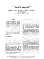
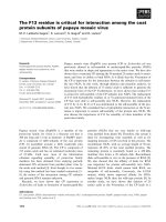
Special clinical courses
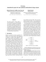
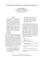
Patient 2 showed a secondary brain swelling with an increase
of ICP level intractable to intensified medical treatment on
day 4 after unilateral decompression. A craniectomy of the
contralateral side was therefore performed, with subsequent
normalization of the ICP. Figure 1 presents the CT scan
before and Figure 2 after bilateral craniectomy of patient 2.
Complications
There was neither infection nor disturbance of wound healing,
nor mortality.
One patient (patient 3) developed a late aseptic necrosis of
the replaced bone flap. In this case, a post-traumatic hydro-
cephalus led to subgaleal cerebrospinal fluid collection with
surgical revision and transient insertion of a ventriculo-peri-
toneal shunt. This might have caused insufficient fixation of
the bone flap and a lack of revascularization with subsequent
partial necrosis. The shunt was removed 3 months after
trauma and the bony defect was covered by an autologous
calvarial split graft.
Neurological outcome
The neurological outcomes at discharge and at 6 months
follow-up are presented in Table 2. Furthermore, SEP of the
median nerve before and after decompressive craniectomy
are described in Table 2.
Patient 1 suffered from a severe transitional syndrome after
discontinuation of sedation. The neurological status was
normal after recovery, in spite of pathological SEP of the
median nerve. At discharge from the intensive care unit,
patient 2 showed a hemiparesis, predominantly of the left
arm, which resolved to normal strength in the following
weeks.
None of the patients with severe head injury suffered from
post-traumatic seizures or received anticonvulsive medica-
tion. Based on findings for SEP of the median nerve, a favor-
able and stable long-term outcome could be predicted for all
of our patients suffering from TBI, confirming previously pub-
lished data [11]. The SEP 1 week after trauma correlated with
the neurological outcome 6 months after trauma, except for
patient 1. Mild disturbances of SEP were seen in patient 1,
but revealed no deterioration during follow-up.
Available online />Figure 1
CT scan of patient 2 before craniectomy.
Figure 2
CT scan of patient 2 after bilateral craniectomy.
R136
Discussion
After exclusion or surgical removal of traumatic hematomas
and other space occupying lesions, prevention of secondary
brain injury is the mainstay of intensive care treatment in pedi-
atric severe head injury. Diffuse brain swelling and multiple
cerebral contusions are the most common cause of morbidity
and death after severe head injury in pediatric patients [12].
Standardized treatment protocols have been suggested for
the management of severe head injury in children [13], includ-
ing drainage of cerebrospinal fluid, mild hyperventilation
(pCO
2
lower threshold of 30 mmHg) and mannitol bolus
(unless serum osmolality exceeds 320 mosmol/l) as generally
accepted baseline therapies for the pediatric population [6].
In cases of sustained elevated ICP (> 20 mmHg) and
reduced critically cerebral perfusion pressure (< 50 mmHg),
despite optimal medical therapy including controlled hyper-
ventilation, further management using ‘second tier’ therapy is
a matter of controversy [6] and has to follow the different
stages of postinjury cerebral insults.
Brain swelling and intracranial hypertension in the early post-
traumatic period has been proposed to induced by cerebral
hyperemia (i.e. increased cerebral blood flow [CBF]), espe-
cially in children [14,15]. However, the impact of hyperemia
on outcome has been rated controversially. Beneficial [16,17]
as well as detrimental effects have been discussed [18].
‘Second tier’ intensified conservative treatment will have to
rely on specific prognostic monitoring parameters. Therefore,
CBF-dependent therapy has been studied [19]. But, as cere-
bral blood flow is age dependent in the unaffected child
(normal range from 40 to > 100 ml/100 g/min [20)], absolute
cerebral hyperemia may only be defined within narrow age
ranges [21]. CBF thresholds cannot be taken from adult
studies for the initiation of therapeutic interventions in the
pediatric population.
Monitoring of cerebral metabolic parameters has been
reported for treatment in adult patients. In children, an early
decrease in the cerebral metabolic rate of oxygen and the
arterio-venous difference for oxygen has been reported to
occur 1–3 days after trauma [14]. Recently, Cruz and col-
leagues [15] predicted clinical outcome based on monitoring
of the ICP and the cerebral extraction rate for oxygen (CEO
2
)
in children. In their observational study of 45 children, an
increased ICP and a decreased CEO
2
indicated cerebral
hyperemia during the first 5 days after head injury. An unfavor-
able outcome occurred in children with higher ICP and lower
CEO
2
(< 17%). Monitoring of the CEO
2
(or oxygen saturation
at the jugular vein bulb for hemoglobin > 12 g/l) might there-
fore be used to direct ventilation and medical therapy in chil-
dren in the future. However, two out of 45 patients died prior
to intended decompressive surgery while being monitored for
CEO
2
, which points towards the need for shortened monitor-
ing intervals and early surgical decompression.
Prolonged barbiturate therapy inherits a high risk of unwanted
therapeutic effects, and revealed small benefits in the
outcome in children [22]. In a proven state of refractory
absolute hyperemia, selective reduction of the CBF by cere-
bral vasomodulation (dihydroergotamine, metoprolol and
Critical Care December 2003 Vol 7 No 6 Ruf et al.
Table 2
Neurological outcome of patients with decompressive craniectomy at discharge and after 6 months compared with somatosensory
evoked potentials of the median nerve (M-SEP) before and after craniectomy
Neurological status Neurological status M-SEP
Patient (on demission) (6 months post-trauma) M-SEP (prior to craniectomy) (first week after craniectomy)
1 Normal Normal Not tested Moderate impairment (right)
2 Normal Normal Severe impairment (right) Normal
3 Left-sided hemiparesis, Distinct improvement of Not tested Severe impairment (right)
VP shunt (PTH) hemiparesis predominantly of
the left arm, VP shunt removed
4 Central impairment of Residual spasticity but not Moderate impairment (right), Mild impairment (left)
coordination with tremor and impaired in motor skills; severe impairment (left)
ataxia; predominantly right- visits a normal school
sided spasticity; speech
retardation
5 Normal Normal Not tested Normal
6 Hemiparesis predominatly of the Rehabilitation Mild impairment (right), Severe impairment (left)
right arm; left-sided abducent moderate impairment (left)
nerve paresis; impairment of
swallowing and speech
PTH, post-traumatic hydrocephalus; VP shunt, ventriculo–peritoneal shunt.
R137
clonidin [22], or a monotherapy dihydroergotamine respec-
tively [23]) might be considered, but these treatment options
are still not for routine application and require very intensive
multimodal monitoring.
Brain edema associated with cerebral ischemia requires opti-
mized cerebral perfusion and fluid management. Experimental
medical treatment is proposed to lower the ICP and to re-
establish sufficient CBF after failure of mannitol and vaso-
pressors to support sufficient CBF. Hypertonic saline (7.2%)
as a bolus or an infusion decreased the ICP in adults and
children, and may therefore be indicated preferably in hypo-
volemia [24–26].
As a surgical ‘second tier’ option, controlled lumbar drainage
of cerebrospinal fluid has been proposed. This regimen
necessitates an external ventriculostomy and discernible
basal cisterns on CT with careful control of both external
drainage systems. In a study cohort of 16 pediatric head
injury patients, Levy and colleagues [27] reported good
control of refractory intracranial hypertension without
drainage-related mortality.
Surgical decompression using craniectomy is largely seen as
a last resort therapeutic option. This may be due to disap-
pointment from previous anecdotic results based on late
intervention. Encouraging results have been reported from
studies in adolescent and adult patients indicating an early
time point of decompression as extremely important to
achieve a favorable outcome [3,8,28].
In addition to the ‘optimal’ time point for decompression, the
extent of brain decompression seems to be important [3].
Restoration of cerebral perfusion by surgical enlargement of
the intracranial space is the primary goal of decompression
[3]. This may necessitate a large craniotomy with duraplasty.
Prospective controlled, randomized studies on the effect of
surgical decompression in TBI in childhood are missing. A
pilot study by Taylor and colleagues [8] demonstrated an
improved neurological outcome of patients who were treated
with an early decompressive craniectomy in a cohort of
27 children compared with historical controls. In contrast to
our patients, only a small temporal craniectomy without
opening the dura was performed. The risk of transtentorial
herniation can be lowered in this way, but restoration of the
cerebral perfusion can hardly be achieved. However, a
benefit from temporal craniectomy without duraplasty has
been shown by Taylor and colleagues, which underlines the
potential of a larger decompression. Studies in adults demon-
strated a greater decrease of the ICP after duraplasty than in
cases with craniectomy only [3,29].
Neither in these studies nor in our cohort was a higher rate of
complications such as infections or hygroma noted due to
duraplasty. Immediate normalization of the ICP after supraten-
torial surgical decompression was achieved in all patients from
our study cohort. A good neurological outcome was achieved
in all our patients suffering from TBI treated with decompres-
sive craniectomy and duraplasty. Due to the early timepoint of
decompression after failure of first-line treatment options,
unwanted effects of prolonged medical therapy (e.g. barbitu-
rate coma) or brain herniation with secondary brain stem
compromise could be prevented, and all children survived.
There currently seems to be no specific treatment regimen in
children compared with adults in severe head injury [21], and
there is no preference for a special ‘second tier’ treatment
strategy in pediatric head injury [6]. The presented pilot trial
adds an additional argument for surgical decompression at
an early stage in case of treatment-refractory intracranial
hypertension, and calls for a controlled trial that includes this
treatment option in pediatric severe head injury patients.
Conclusion
This pilot trial and the favorable results from the study by
Taylor and colleagues [8] demonstrate the necessity of a mul-
ticenter, controlled, randomized study to evaluate the indica-
tion and standardization of early decompressive craniectomy
as a ‘second tier’ standard therapy in children with severe
head injury.
Competing interests
None declared.
References
1. Berger MS, Pitts LH, Lovely M, Edwards MS, Bartkowski HM:
Outcome from severe head injury in children and adoles-
cents. J Neurosurg 1985, 62:194-199.
2. Marshall LF, Gautille T, Klauber MR, Eisenberg HM, Jane JA,
Luerssen TG, Marmarou A, Foulkes MA: The outcome of severe
closed head injury. J Neurosurg 1991, 75:528-536.
3. Guerra WK, Gaab MR, Dietz H, Mueller JU, Piek J, Fritsch MJ:
Surgical decompression for traumatic brain swelling: indica-
tion and results. J Neurosurg 1999, 90:187-196.
4. Maas AI, Dearden M, Teasdale GM, Braakman R, Cohadon F, Jan-
notti F, Karimi A, Lapierre F, Murray G, Ohmann J, Persson L, Ser-
vadei F, Stochetti N, Unterberg A: EBIC-guidelines for
management of severe head injury in adults. European Brain
Injury consortium. Acta Neurochir 1997, 139:286-294.
5. Brain Trauma Foundation, American Association of Neurological
Surgeons, Joint Section on Neurotrauma and Critical Care:
Guidelines for the management of severe head injury.
J Neurotrauma 1996, 13:641-734.
6. Rekate HL: Head injuries: management of primary injuries and
prevention of secondary damage. Childs Nerv Syst 2000, 17:
632-634.
7. Dam Hieu P, Sizun J, Person H, Besson G: The place of decom-
pressive surgery in the treatment of uncontrollable post-
traumatic intracranial hypertension in children. Childs Nerv
Syst 1996, 12:270-275.
Available online />Key messages
• In case of sustained increase in ICP (>20 mmHg)
under intensified conservative therapy conditions and
early decompressive craniectomy including duraplasty
has to be considered
R138
8. Taylor A, Butt W, Rosenfeld J, Shann F, Ditchfield M, Lewis E,
Klug G, Wallace D, Henning R, Tibballs J: A randomized trial of
very early decompressive craniectomy in children with trau-
matic brain injury and sustained intracranial hypertension.
Childs Nerv Syst 2001, 17:154-162.
9. Downard C, Hulka F, Mullins RJ, Piatt J, Chesnut R, Quint P, Mann
N: Relationship of cerebral perfusion pressure and survival in
pediatric brain-injured patients. J Trauma 2000, 49:654-658.
10. Bruce DA: Imaging after head trauma: why, when and which.
Childs Nerv Syst 2000, 16:755-759.
11. Carter BG, Taylor A, Butt W: Severe brain injury in children: long-
term outcome and its prediction using somatosensory evoked
potentials (SEPs). Intensive Care Med 1999, 25:722-728.
12. Marshall LF, Toole BM, Bowers SA: The national traumatic
coma data bank. Part 2: patients who talk and deteriorate:
implications for treatment. J Neurosurg 1983: 59:285-288.
13. Chiaretti A, Piastra M, Pulitanò S, Pietrini D, De Rosa G, Barbaro
R, Di Rocco C: Prognostic factors and outcome of children
with severe head injury: an 8-year experience. Childs Nerv
Syst 2002, 18:129-136.
14. Sharples PM, Stuart AG, Mathews DSF, Synsley-Green A, Eyre
JA: Cerebral blood flow and metabolism in severely head-
injured children: Part I — relationship to age, Glascow coma
score, outcome, intracranial pressure and time after surgery.
J Neurol Neurosurg Psychiatry 1995, 58:145-152.
15. Cruz J, Nakayama P, Imamura JH, Rosenfeld KGW, De Souza HS,
Giorgetti GVF: Cerebral extraction of oxygen and intracranial
hypertension in severe, acute, pediatric brain trauma: prelimi-
nary novel management strategies. Neurosurgery 2002, 50:
774-780.
16. Lang DA, Teasdale GM, Macpherson P, Lawrence A: Diffuse
brain swelling after head injury: more often malignant in
adults than in children? J Neurosurg 1994, 80:675-680.
17. Sakas DE, Bullock MR, Patterson J, Hadley D, Wyper DJ, Teas-
dale GM: Focal cerebral hyperemia after focal head injury in
humans: a benign phenomenon? J Neurosurg 1995, 83:277-
284.
18. Feickert HJ, Drommer S, Heyer R: Severe head injury in chil-
dren: impact of risk factors on outcome. J Trauma 1999, 47:
33-38.
19. Siotos PJ, Orozco JA, Carter LP, Weinand ME, Hamilton AJ,
Williams FC: Continuous regional cerebral cortical blood flow
monitoring in head-injured patients. Neurosurgery 1995, 36:
943-950.
20. Susuki K: The changes of regional cerebral blood flow with
advancing age in normal children. Nagoya Med J 1990, 34:
159-170.
21. Zwienenberg M, Muizelaar JP: Severe pediatric head injury: the
role of hyperemia revisited. J Neurotrauma 1999, 16:937-943.
22. Ecker C, Asgeirsson B, Grände PO, Schalen W, Nordström CH:
Improved outcome after severe injury with a new therapy
based on principles for brain volume regulation and pre-
served microcirculation. Crit Care Med 1998, 26:1881-1886.
23. Orliaguet GA, Meyer PG, Renier D, Blanot S, Carli PA: Success-
ful treatment of uncontrollable posttraumatic intracranial
hypertension with dihydroergotamine in a child. Anesth Analg
1997, 85:1218-1220.
24. Simma B, Burger R, Falk M, Sacher P, Fanconi S: A prospective,
randomized, and controlled study of fluid management in chil-
dren with severe head injury. Crit Care Med 1998, 26:1265-
1270.
25. Horn P, Munch E, Vajkoczy P, Herrmann P, Qintel M, Schilling L,
Schmiedek P, Schurer L: Hypertonic saline solution for control
of elevated intracranial pressure in patients with exhausted
response to mannitol and barbiturates. Neurol Res 1999, 21:
758-764.
26. Munar F, Ferrer AM, de Nadal M, Poca MA, Pedraza S, Sahuquillo
J, Garnacho A: Cerebral hemodynamic effects of 7.2% hyper-
tonic saline in patients with head injury and raised intracranial
pressure. J Neurotrauma 2000, 17:41-51.
27. Levy DI, Rekate HL, Cherny B, Manwaring K, Moss D, Baldwin
HZ: Controlled lumbar drainage in pediatric head injury.
J Neurosurg 1995, 83:453-460.
28. Polin RS, Shaffrey ME, Bogaev CA, Tisdale N, Germanson T,
Bocchi Jane JA: Decompressive bifrontal craniectomy in the
treatment of severe refractory posttraumatic cerebral edema.
Neurosurgery 1997, 41:84-92.
29. Yoo DS, Kim DS, Cho KS, Huh PW, Park CK, Kang JK: Ventriku-
lar pressure monitoring during bilateral decompression with
dural expansion. J Neurosurg 1999, 91:953-959.
Critical Care December 2003 Vol 7 No 6 Ruf et al.