Báo cáo khoa học: "Difference in end-tidal CO2 between asphyxia cardiac arrest and ventricular fibrillation/pulseless ventricular tachycardia cardiac arrest in the prehospital setting" pps
Bạn đang xem bản rút gọn của tài liệu. Xem và tải ngay bản đầy đủ của tài liệu tại đây (70.16 KB, 6 trang )
Available online />Research
Difference in end-tidal CO
2
between asphyxia cardiac arrest and
ventricular fibrillation/pulseless ventricular tachycardia cardiac
arrest in the prehospital setting
Štefek Grmec, Katja Lah and Ksenija Tušek-Bunc
Center of Emergency Medicine, Prehospital Unit Maribor, Maribor, Slovenia
Correspondence: Katja Lah,
R139
CPR = cardiopulmonary resuscitation; PetCO
2
= partial pressure of end-tidal carbon dioxide; ROSC = return of spontaneous circulation; VF =
ventricular fibrillation; VT = ventricular tachycardia.
Abstract
Introduction There has been increased interest in the use of capnometry in recent years. During
cardiopulmonary resuscitation (CPR), the partial pressure of end-tidal carbon dioxide (PetCO
2
)
correlates with cardiac output and, consequently, it has a prognostic value in CPR. This study was
undertaken to compare the initial PetCO
2
and the PetCO
2
after 1 min during CPR in asphyxial cardiac
arrest versus primary cardiac arrest.
Methods The prospective observational study included two groups of patients: cardiac arrest due to
asphyxia with initial rhythm asystole or pulseless electrical activity, and cardiac arrest due to acute
myocardial infarction or malignant arrhythmias with initial rhythm ventricular fibrillation (VF) or pulseless
ventricular tachycardia (VT). The PetCO
2
was measured for both groups immediately after intubation
and then repeatedly every minute, both for patients with and without return of spontaneous circulation
(ROSC).
Results We analyzed 44 patients with asphyxial cardiac arrest and 141 patients with primary cardiac
arrest. The first group showed no significant difference in the initial value of the PetCO
2
, even when we
compared those with and without ROSC. There was a significant difference in the PetCO
2
after 1 min
of CPR between those patients with ROSC and those without ROSC. The mean value for all patients
was significantly higher in the group with asphyxial arrest. In the group with VF/VT arrest there was a
significant difference in the initial PetCO
2
between patients without and with ROSC. In all patients with
ROSC the initial PetCO
2
was higher than 10mmHg.
Conclusions The initial PetCO
2
is significantly higher in asphyxial arrest than in VT/VF cardiac arrest.
Regarding asphyxial arrest there is also no difference in values of initial PetCO
2
between patients with
and without ROSC. On the contrary, there is a significant difference in values of the initial PetCO
2
in
the VF/VT cardiac arrest between patients with and without ROSC. This difference could prove to be
useful as one of the methods in prehospital diagnostic procedures and attendance of cardiac arrest.
For this reason we should always include other clinical and laboratory tests.
Keywords asphyxial cardiac arrest, end-tidal CO
2
, prognosis
Received: 14 May 2003
Revisions requested: 13 June 2003
Revisions received: 29 July 2003
Accepted: 8 August 2003
Published: 24 September 2003
Critical Care 2003, 7:R139-R144 (DOI 10.1186/cc2369)
This article is online at />© 2003 Grmec et al., licensee BioMed Central Ltd
(Print ISSN 1364-8535; Online ISSN 1466-609X). This is an Open
Access article: verbatim copying and redistribution of this article are
permitted in all media for any purpose, provided this notice is
preserved along with the article's original URL.
Open Access
Introduction
Monitoring of end-tidal CO
2
has become a standard in the
prehospital setting to ensure proper placement and function
of the endotracheal tube and to help monitor the adequacy of
ventilation [1]. In addition, it has been noted that cardiac
arrest causes an abrupt fall in end-tidal CO
2
levels to values
R140
Critical Care December 2003 Vol 7 No 6 Grmec et al.
near zero [2,3]. During cardiac arrest the partial pressure of
end-tidal carbon dioxide (PetCO
2
) falls to very low levels,
reflecting the very low cardiac output achieved with car-
diopulmonary resuscitation (CPR). It has been shown that the
PetCO
2
achieved during advanced cardiac life support reli-
ably predicts an outcome of cardiac arrest [2–12]. Higher
levels of the PetCO
2
indicate better cardiac output, higher
coronary perfusion pressure and a greater likelihood of suc-
cessful resuscitation [13,14]. After the onset of cardiac arrest
caused by ventricular fibrillation (VF), the PetCO
2
abruptly
decreases to nearly zero and then begins to increase after
the onset of effective CPR. Further increase is detected upon
return of spontaneous circulation (ROSC) to normal or
above-normal levels [2,3,9,12].
In an experimental animal model of asphyxial arrest during
CPR, PetCO
2
levels were initially high (after the onset of
arrest), then decreased to subnormal levels and then
increased again to near-normal levels [15,16]. During a respi-
ratory arrest, the cardiac output of pulmonary blood flow con-
tinues for some period of time prior to cardiac standstill. The
CO
2
produced in the tissue during this period will continue to
be delivered to the lungs, thereby increasing alveolar CO
2
(two-compartment hydraulic model of CO
2
kinetics).
However, it is also important to recognize that it is not only
the cessation of cardiac output alone that causes the fall of
PetCO
2
, but the cessation in conjuction with the washout of
alveolar gas. This means that, in the absence of alveolar gas
washout, CO
2
will remain in the lungs and probably that, as
alveolar oxygen is being utilized, more CO
2
will be delivered.
On the basis of such a concept we built a hypothesis main-
taining that the initial PetCO
2
should be higher in an asphyxial
arrest model than in a VF/pulseless ventricular tachycardia
(VT) cardiac arrest model. In the asphyxial cardiac arrest
model there should also be no difference in patients with and
without ROSC regarding the initial PetCO
2
, since the initial
PetCO
2
in this case reflects CO
2
cumulated in the alveolar
compartment. This would suggest that the initial values of
end-tidal carbon dioxide in asphyxial arrest do not have a
prognostic value for ROSC as they do in VF/VT cardiac
arrest.
If our results confirm both hypotheses, then this difference
could be helpful in determining the mechanism of arrest in the
prehospital setting.
Methods
This prospective observational study was conducted at the
Center of Emergency Medicine, Maribor. The study included
two groups of patients. The first group represented patients
who suffered from heart arrest due to asphyxia. The causes of
asphyxia included a foreign body in the airway, aspiration,
suicide by hanging, drowning, edema or tumor of the airway,
intoxication and acute asthma attack. The definitive cause of
arrest has been confirmed in the hospital with further diag-
nostic and/or pathological report (autopsy). The initial rhythm
was either asystole or pulseless electrical activity (all patients
from this group with VT/VF as the initial rhythm were
excluded). Patients with severe hypothermia (core tempera-
ture < 30°C) were also excluded.
The second group included the patients with primary cardiac
arrest (acute myocardial infarction or malignant arrhythmias).
The initial rhythm was VF/VT (all patients from this group pre-
senting with asystole or pulseless electrical activity were
excluded). The definitive diagnosis (cause of arrest) was con-
firmed in the hospital (further diagnostic and/or pathologi-
cal/autopsy report). The inclusion/exclusion criteria for
asphyxia and VF/VT group are presented in Table 1.
The resuscitation procedures were performed by an emer-
gency team (emergency medical doctor and two emergency
Table 1
Inclusion/exclusion criteria for the asphyxia group and the
ventricular fibrillation/pulseless ventricular tachycardia
(VF/VT) group of patients
VF/VT group
VF/VT initial rhythm
Age > 18 years
Core temperature > 30°C
Confirmed acute myocardial infarction and/or primary VF/VT
(electrocardiogram, enzymes, autopsy, electrophysiological
investigation)
Excluded patients with successful defibrillation in the first cycle
Excluded patients with acute myocardial infarction with asystole
and pulseless electrical activity as the initial rhythm
Asphyxia group
Asystole and pulseless electrical activity as the initial rhythm
Excluded patients with VF/VT as the initial rhythm
Age > 18 years
Core temperature > 30°C
Excluded acute myocardial infarction as cause of arrest (clinical
investigations and/or autopsy)
Etiology:
solid foreign body in the airway
aspiration
edema or tumor of the upper airway
hanging (excluded vasculatory or others causes of arrest —
clinical investigations or autopsy)
Acute asthma attack (excluded cardiac causes of arrest)
Drowning (excluded cardiac causes of arrest)
Intoxications (excluded others causes of death — autopsy and/or
added investigations in hospital
R141
medical technicians or register nurses) in accordance with the
International Liasion Committee on Resuscitation and Euro-
pean Resuscitation Council guidelines [17–19]. We used a
manual technique to perform CPR. Pharmacologic interven-
tions in individual patients were in accordance with the stan-
dards and guidelines of the International Liasion Committee on
Resuscitation/European Resuscitation Council.
For management of VF or pulseless VT, direct-current coun-
tershocks were delivered by means of conventional tech-
niques. PetCO
2
measurements were made by infrared
sidestream capnometer (BCI Capnocheck Model 20600A1;
BCI International Waukesha, WI, USA). Measurements for
both groups were made immediately after intubation (first
measurement) and then repeatedly every minute continu-
ously. Endotracheal intubations were performed after two
initial breaths with a valved bag at the beginning of CPR.
Further ventilation was performed by mechanical ventilator
(6–8 ml/kg at 10–12 breaths/min; Medumat Standard Wein-
mann, Weinmann, Namburg, Germany). The CO
2
cuvette
was located in a connector between the mechanical ventila-
tor and the endotracheal tube (it was applied to the endotra-
cheal tube before intubation). Two patients were not
intubated by the orotracheal technique because of complete
obstruction of the upper airway, visualized by laringoscopy. In
these two cases cricotireideotomy was performed using the
traceoquick method (Tracheoquick Emergency Coniostomy
Set; Willy Rüsch AG, Kernen, Germany). The procedure was
performed in accordance with the instructions of the manu-
facturer, and both patients were successfully resuscitated
and ventilated by mechanical ventilator.
The initial (first measurement after intubation), average (mean
of all values obtained during a single resuscitation effort) and
final (measurement at admission to hospital or discontinued
CPR) PetCO
2
was detected for both groups. We performed
the same procedure for the patients with ROSC and for
those without ROSC.
ROSC is defined as the return of spontaneous heartbeat or as
palpable periferial arterial pulse and measurable systolic arter-
ial pressure. As is seen from the Utstein style template, we dis-
tinguish intermittent ROSC, which is short in duration and a
temporary event, from ROSC with hospitalization of a patient.
In the present article, ROSC represents hospitalized patients.
The paired Student t test was used to compare initial and sub-
sequent PetCO
2
values for each subject. For other parame-
ters, both groups (asphyxial arrest group and VF/VT cardiac
arrest group) were compared by Student’s t test and the chi-
squared test. Continuous variables are described as the mean
± standard deviation. P <0.05 was considered significant.
Results
From February 1998 to October 2002 we analyzed 141
patients with primary cardiac arrest (initial rhythm VF/VT) and
44 patients with cardiac arrest due to asphyxia (initial rhythm
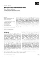
asystole or pulseless electrical activity). The study environ-
ment, the prehospital environment and the characteristics of
cardiac arrest and noncardiac arrest are displayed in
Fig. 1a,b (Utstein style). The causes of asphyxial cardiac
Available online />Figure 1
(a) Cardiac arrests placed into the Utstein template. *It was not
possible to determine the number of resuscitations not attempted
because records for patients who were pronounced dead at the scene
were not available. **Return of spontaneous circulation (ROSC).
#Results before October 2002. (b) Non-cardiac arrests placed into
the Utstein style. EMS, Emergency Medical Service; ICU, intensive
care unit; VF, ventricular fibrillation; VT, ventricular tachycardia.
15. ROSC**
n = 212
12. initial rhythm
asystole
n
=
156
10. initial rhythm
VF
n
=
133
11. initial rhythm
VT
n
=
8
13.initial rh
y
thm
others
n = 38
16. never achieved ROSC
n = 123
7. arrest witnessed
(bystanders)
n
=
127
9. arrest witnessed
(EMS personnel)
n
=
12
8. arrest not witnessed
n = 196
17.efforts ceased expired in field
n = 14
18. admitted to ICU
n = 198
19. expired in hospital
n = 128
20. discharged alive (from ICU)
n = 71
21.# no. expired within one year
of discharge
n = 49
22.# no. alive at one year
n = 22
6. noncardiac aetiology
n = 66
5. cardiac aetiology
n = 335
3. *resuscitation not attempted
not recorded
4.resuscitation attempted
n = 411
1. population served by EMS system
n = 190000
2. confirmed cardiac arrests considered for resuscitation
– not recorded
6. noncardiac aetiology
n= 66
7. arrest witnessed (bystanders)
n=17
9. arrest witnessed
(EMS personnel)
n= 4
12. initial rhythm
asystole
n= 42
10. initial rhythm
VF
n= 11
11. initial rhythm
VT
n= 4
13. initial rhythm
others
n= 9
16.
Never achieved ROSC
n= 37
15. Any ROSC
n= 29
17.effort ceased expired in field
n= 5
18. admitted to ICU
n= 24
19. expired in hospital
n= 11
20. discharged alive
n= 13
21. expired within one year of discharge
n= 6
22. alive at one year
n= 7
8. arrest not witnessed
n= 41
(a)
(b)
R142
arrest were solid foreign body in the airway (seven cases),
aspiration (seven cases), edema or tumor of the upper airway
(five cases), hanging (five cases), acute asthma attack (six
cases), drowning (six cases) and intoxications with respira-
tory arrest (eight cases). Demographic and clinical character-
istics for both groups are presented in Table 2.
The values of the PetCO
2
are presented in Table 3. In the
group of patients who presented with arrest due to asphyxia
there was no significant difference in the initial values of
PetCO
2
, even when we compared those with and without
ROSC (70.1 ±15.3 mmHg versus 62.8± 16.2 mmHg,
P = 0.64). On the contrary, in the group of patients who pre-
sented with VF/VT arrest there was a significant difference in
the initial values of PetCO
2
between patients without and
with ROSC (8.2 ± 4.3mmHg versus 20.3 ± 6.2mmHg,
P = 0.04). In all patients with ROSC the initial PetCO
2
was
higher than 10 mmHg. The values of the PetCO
2
after 1 min
of CPR did not differ significantly among the two groups. In
both groups significantly higher values were achieved in
patients with ROSC than in those without ROSC (asphyxial
arrest group, 35.8 ± 8.6mmHg versus 19.4 ± 8.7mmHg,
P < 0.05; VF/VT arrest group, 30.2± 8.3 mmHg versus
14.2 ± 5.2mmHg, P < 0.05). The values of the final PetCO
2
in
both groups were significantly higher in patients with ROSC
than in the patients without ROSC (asphyxial arrest group,
31.2 ± 8.4mmHg versus 7.2 ± 3.3mmHg, P <0.05; VF/VT
arrest group, 28.1 ± 4.8mmHg versus 6.2 ± 2.8 mmHg,
P < 0.05).
Discussion
In the present study we confirmed that the PetCO
2
was
markedly elevated during the first minute of CPR in asphyxial
cardiac arrest. This study therefore confirmed the results of
the studies that used animal models in which cardiopul-
monary arrest was induced by asphyxia. In the present study
the PetCO
2
values during CPR were initially high, then
decreased to subnormal levels and then increased again to
near-normal levels in patients with ROSC. This pattern of
PetCO
2
changes is different from the pattern observed in
Critical Care December 2003 Vol 7 No 6 Grmec et al.
Table 2
Demographic and clinical characteristics of patients: a group with primary ventricular fibrillation/pulseless ventricular tachycardia
(VF/VT) cardiac arrest and a group with asphyxial arrest
Primary VF/VT cardiac Asphyxial cardiac arrest
arrest (n = 141) (asystole and PEA) (n = 44) P value
Age (years) 65.8 ± 13.8 48.8 ± 20.1 < 0.05
b
Gender (male/female) 82/59 27/17 0.83
c
Response time (min)
a
8.4 ± 5.7 8.9 ± 5.2 0.91
b
Witnessed arrest (yes/no) 68/73 19/25 0.78
c
Resuscitation by medical team (min) 28.3 ± 11.3 24.7 ± 13.4 0.76
b
ROSC (yes/no) 101/40 18/26 < 0.05
c
Discharged alive from ICU (yes/no) 38/103 7/37 < 0.05
c
Average number of PetCO
2
observations 12.3 ± 3.4 (range, 7–22) 13.4 ± 2.8 (range, 9–28) 0.74
b
ICU, intensive care unit; PEA, pulseless electrical activity; PetCO
2
, partial pressure of end-tidal carbon dioxide; ROSC, return of spontaneous
circulation.
a
Time elapsed between the received 112 call to the arrival of Emergency Medical Service professionals at the patient’s side.
b
Student t test.
c
Chi-squared test.
Table 3
The mean values for all patients of the initial, final, average and after 1 min of cardiopulmonary resuscitation (CPR) partial
pressures of end-tidal carbon dioxide (PetCO
2
) for arrest due to asphyxia and for ventricular fibrillation/pulseless ventricular
tachycardia (VF/VT) cardiac arrest
Initial PetCO
2
PetCO
2
after 1 min Average PetCO
2
Final PetCO
2
(mmHg) of CPR (mmHg) (mmHg) (mmHg)
Asphyxial cardiac arrest 66.4 ± 17.3 29.1 ± 4.9 48.2 ± 10.1 27.3 ± 9.2
VT/VF cardiac arrest 16.5 ± 9.2 24.2± 5.1 17.3± 7.1 24.4± 10.3
P value (Student t test) < 0.01 0.73 < 0.05 0.78
1 mmHg = 0.133 kPa.
R143
cardiac arrest caused by VF, since cardiac arrest from VF
results in an abrupt cessation of cardiac output and pul-
monary blood flow. Bhende and colleagues [15], Berg and
colleagues [16] and von Planta and colleagues [20] con-
cluded that, during the period of asphyxia, continued cardiac
output prior to cardiac arrest permits continued delivery of
CO
2
to the lungs, which (in the absence of exhalation) results
in higher alveolar CO
2
. This is reflected as increased PetCO
2
when ventilation is resumed.
Understanding the physiology of CO
2
production, delivery to
the lungs and excretion are important in order to appropriately
interpret PetCO
2
monitoring during CPR. The disposition of
CO
2
can also be represented in a hydraulic model [21]. The
large peripheral tissue compartment drains through a conduit
(cardiac output) into the small central pulmonary compart-
ment. The tissues produce CO
2
, which empties into the
peripheral tissue compartment. Carbon dioxide then flows by
gravity (cardiac output) from the higher level tissue to the
lower level pulmonary compartment. Alveolar ventilation,
which equals expired ventilation minus ventilation of the
anatomical dead space, and the effects of high ratio ventila-
tion/perfusion matching eliminate CO
2
from the lung. In this
model the cardiac output affects the distribution and total
amount of CO
2
in the body and can help to understand the
meaning of the PetCO
2
during CPR.
The present study discovered that we can trace the same
pattern of PetCO
2
changes in the asphyxial arrest as were
described in the animal models in the first minute after arrest
[15,16], even after a longer period of time due to the access
time. The inability to measure the PetCO
2
immediately after
cardiac arrest was the main disability of this study.
We also concluded that the high initial values of the PetCO
2
in asphyxial arrest do not have a prognostic value for the
appearance of ROSC as they do in the VT/VF cardiac arrest.
On the contrary, the values after 1 min of CPR and also the
final values of the PetCO
2
do have the prognostic value for
ROSC. These data, like those from Berg and colleagues [16],
suggest that the PetCO
2
during the initial phase of CPR of
asphyxial arrest (1 min after intubation and cardiac massage)
reflects alveolar CO
2
prior to CPR. In the asphyxial model,
cellular respiration results in continued oxygen consumption
and CO
2
production. The high pressure of CO
2
in the alveo-
lar compartment is reflected in the high PetCO
2
during the
initial phase of CPR.
The fast decline of the high values of the PetCO
2
can there-
fore only be interpreted by ventilation of the alveolar com-
partment, which then rapidly decreases the PetCO
2
.
However, in the next phase and with the beginning of CPR
we can again detect the rise of the PetCO
2
. This rise is
achieved by successful cardiac massage, which washes the
acumulated CO
2
out of the peripheral compartment
[11,21–23].
The acquaintance with this pattern of changes can be helpful
in differentiation of cardiac arrest causes and in identification
of mechanisms that led to cardiac arrest. This is very useful in
the prehospital setting and can lead the course of action as
hypoxia is a potentially reversible cause of cardiac arrest. The
issue is potentially important when deciding upon the most
effective sequence of resuscitation intervention. There is
growing evidence that indicates positive pressure ventilation
may be postponed for several minutes in instances of arryth-
mic arrest whereas it might be life-saving in instances of
asphyxial arrest. The emergency medical doctor can therefore
be orientated with greater reassurance towards the measures
that are useful in asphyxial arrest [24–27]. However, one has
to be aware that the initial values of the PetCO
2
in asphyxial
arrest do not have the prognostic value for the outcome of
CPR that they do have in VF/VT arrest [4–6,8–10].
Conclusions
The initial values of the PetCO
2
in asphyxial cardiac arrest are
significantly higher than in VF/VT cardiac arrest. In asphyxial
arrest there is also no significant difference in inital values of
the PetCO
2
in patients with and without ROSC. In asphyxial
arrest the initial values of the PetCO
2
therefore cannot be
used as a prognostic factor of outcome of CPR, as they can
be used in VF/VT cardiac arrest. This difference, together with
other criteria, can therefore be useful for differentiation
between the causes of cardiac arrest in the prehospital
setting. For standard use of this difference in the PetCO
2
in
the prehospital setting we suggest additional clinical research.
Competing interests
None declared.
References
1. Grmec Š: Comparison of three different methods to confirm
tracheal tube placement in emergency intubation. Intensive
Care Med 2002, 28:701-704.
Available online />Key messages
• PetCO
2
correlates with cardiac output and has a
prognostic value for CPR
• The pattern of PetCO
2
changes in asphyxia is different
from the pattern of PetCO
2
changes in VF/VT cardiac
arrest
• Differences in the initial values of PetCO
2
can be
useful in differentiating between the causes of cardiac
arrest
• Initial values of PetCO
2
cannot be used as a
prognostic factor for CPR in asphyxia arrest
• Values after 1 min of CPR in asphyxia arrest can be
used as a prognostic factor for CPR
R144
2. Falk JL, Rackow EC, Weil MH: End-tidal carbon dioxide con-
centration during cardiopulmonary resuscitation. N Engl J
Med 1988, 318:607-611.
3. Garnett AR, Ornato JP, Gonzales ER, Johnson EB: End-tidal
carbon dioxide monitoring during cardiopulmonary resuscita-
tion. JAMA 1987, 257:512-515.
4. Weil MH, Bisera J, Trevino RP, Rackow EC: Cardiac output and
end-tidal carbon dioxide. Crit Care Med 1985, 13:907-909.
5. Callaham M, Barton C: Prediction of outcome of cardiopul-
monary resuscitation from end-tidal carbon dioxide concen-
tration. Crit Care Med 1990, 18:358-362.
6. Asplin BR, White RD: Prognostic value of end-tidal carbon
dioxide pressures during out-of-hospital cardiac arrest. Ann
Emerg Med 1995, 25:756-761.
7. Grmec Š, Klemen P: Does the end-tidal carbon dioxide (etCO
2
)
concentration have prognostic value during out-of-hospital
cardiac arrest. Eur J Emerg Med 2001, 8:263-269.
8. Ahrens T, Schallom L, Bettorf K, Ellner S, Hurt G, O’Mara V,
Ludwig J, George W, Marino T, Shannon W: End-tidal carbon
dioxide mesurements as a prognostic indicator of outcome in
cardiac arrest. Am J Crit Care 2001, 10:391-398.
9. Sanders AB, Kern KB, Berg RA: Searching for a predictive rule
for terminating cardiopulmonary resuscitation. Acad Emerg
Med 2001, 8:654-657.
10. Levine RL, Wayne MA, Miller CC: End-tidal carbon dioxide and
outcome of out-of-hospital cardiac arrest. N Engl J Med 1997,
337:301-306.
11. Wiklund L, Soderberg D, Henneberg S, Rubertsson S, Stjern-
strom H, Groth T: Kinetics of carbon dioxide during cardiopul-
monary resuscitation. Crit Care Med 1986; 14:1015-1022.
12. Song L, Weil MH, Tang W, Sun S, Pellis T: Cardiopulmonary
resuscitation in the mouse. J Appl Physiol 2002, 93:1222-
1226.
13. Binder JC, Parkin WG: Non-invasive cardiac output determina-
tion: comparison of a new partial-rebreathing technique with
termodilution. Anaesth Intensive Care 2001, 29:19-23.
14. Idris AH, Becker LB, Fuerst RS, Wenzel V, Rush WJ, Melker RJ,
Orban DJ: Effect of ventilation on resuscitation in an animal
model of cardiac arrest. Circulation 1994, 90:3063-3069.
15. Bhende MS, Karasic DG, Karasic RB: End-tidal carbon dioxide
changes during cardiopulmonary resuscitation after experi-
mental asphyxial cardiac arrest. Am J Emerg Med 1996,
14:349-350.
16. Berg RA, Henry C, Otto CW, Sanders AB, Kern KB, Hilwig RW,
Ewy GA: Initial end-tidal CO
2
us markedly elevated during car-
diopulmonary resuscitation after asphyxial cardiac arrest.
Pediatr Emerg Care 1996, 12:245-248.
17. Nollan J, Baskett P, Gabbatt D, Gwinnutt C, de Latorre FJ, Lockey
A, Mitchell S, Soar J: Advanced Life Support Course Manual, 4th
edition. London: Resuscitation Council (UK) & ERC; 2000:19-117.
18. Cummins RO, Hazinski MF, Baskett PJF, Chamberlain D,
Bossaert LL, Callanan V, Carli P, Gay M, Handley AJ, Jacobs A,
Kerber RE, Kloeck WGJ, Mason P, Montgomery WH, Morely PT,
Osmond MH, Robertson C, Shuster M, Steen PA, Tibballs J,
Timerman S, Zideman DA: Guidelines 2000 for Cardiopul-
monary Resuscitation and Emergency Cardiovascular Care —
An International Consensus on Science. Circulation 2000, 102
(Suppl I):1-376.
19. Robertson C, Steen P, Adgey J, Bossaert L, Carli P, Chamberlain
D, Dick W, Ekstrom L, Hapnes SA, Holmberg S, Juchems R, Kette
F, Koster R, de Latorre FJ, Lindner K, Perales N: The 1998 Euro-
pean Resuscitation Council Guidelines for adult advanced life
support. Resuscitation 1998, 37:67-80.
20. von Planta M, Weil MH, Gazmuri RJ, Bisera J, Rackow EC:
Myocardial acidosis associated with CO
2
production during
cardiac arrest and resuscitation. Circulation 1989, 80:684-692.
21. Anderson CT, Breen PH: Carbon dioxide kinetics and capnog-
raphy during critical care. Crit Care 2000, 4:207-215.
22. Kristoffersen MB, Rattenborg CC, Holaday DA: Asphyxial death:
the roles of acute anoxia, hypercarbia and acidosis. Anesthesi-
ology 1967, 28:488-497.
23. DeBehnke DJ, Hilander SJ, Dobler DW, Wickman LL, Swart GL:
The hemodynamic and arterial blood gas response to asphyx-
iation: a canine model of pulseless electrical activity. Resusci-
tation 1995, 30:169-175.
24. Sumann G, Krismer AC, Wenzel V, Adelsmayr E, Schwarz B,
Lindner KH, Mair P: Cardiopulmonary resuscitation near
drowing and hypothermia: restoration of spontaneous circula-
tion after vasopressin. Acta Anaesthesiol Scand 2003, 47:363-
365.
25. Mayr VD, Wenzel V, Voelckel WG, Krismer AC, Mueller T, Lurie
KG, Lindner KH: Developing a vasopressor combination in a
pig model of adult asphyxial cardiac arrest. Circulation 2001,
104:1651-1656.
26. Katz LM, Wang Y, Ebmeyer U, Radovsky A, Safar P: Glucose
plus insulin improves cerebral outcome after asphyxial
cardiac arrest. Neuroreport 1998, 9:3363-3367.
27. Levine RL: End-tidal CO
2
: physiology in pursuit of clinical
applications. Intensive Care Med 2000, 26:1595-1597.
Critical Care December 2003 Vol 7 No 6 Grmec et al.