Báo cáo khoa học: "Bench-to-bedside review: Microvascular and airspace linkage in ventilator-induced lung injury" potx
Bạn đang xem bản rút gọn của tài liệu. Xem và tải ngay bản đầy đủ của tài liệu tại đây (491.51 KB, 10 trang )
435
ALI = acute lung injury; IVP = isolated ventilated and perfused; PEEP = positive end-expiratory pressure; VILI = ventilator-induced lung injury.
Available online />Introduction
Although ventilator-induced lung injury (VILI) is undoubtedly a
complex process that is influenced by many factors, the great
majority of investigative attention has been directed at air-
space mechanics, as exemplified by tidal volume, plateau
pressure, and positive end-expiratory pressure (PEEP).
However, because the fragile alveolus serves as the interface
between gas and blood, and because the intraluminal pres-
sures applied to the airway epithelium also impact on the vas-
cular endothelium, the potential for pressures and flows
within blood vessels to influence the development and/or
evolution of VILI also deserves consideration. This overview
addresses the experimental evidence linking alveolar and vas-
cular events in the generation of barrier breakdown.
Inflation and pulmonary vascular pressure
The vascular pathway from pulmonary artery to left atrium can
be considered as a series of three functional segments: arter-
ial, ‘intermediate’ (which includes alveolar capillaries and con-
tiguous microvessels), and venous [1]. Under normal
conditions the arterial and venous segments, which are
entirely extra-alveolar, contribute most to overall pulmonary
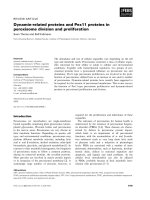
vascular resistance. The compliant intermediate or ‘middle’
segment, however, is influenced primarily by alveolar pres-
sures, and as a consequence it undergoes the greatest
change in overall vascular resistance that occurs during venti-
lation (Fig. 1).
The behaviors of alveolar and extra-alveolar vessels during
lung expansion are fundamentally different. The structural
forces of interdependence cause a fall in interstitial pressure
during inflation, even during positive pressure ventilation [2].
This reduction in interstitial pressure tends to increase the
transmural pressure of the vessels in the immediate environ-
ment, thus dilating them; this in turn increases wall tension, in
particular in the vessels upstream from the alveoli. Something
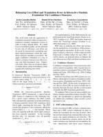
quite different, however, happens at the alveolar level. During
inflation of a normal, fully aerated (‘open’) lung, the majority of
capillaries embedded within the alveolar wall are compressed
by the expansion of adjoining alveoli, even as extra-alveolar
vessels dilate (Fig. 2).
At all lung volumes above functional residual capacity, the
effects of vessel elongation and alveolar capillary compres-
sion outweigh the tendency for extra-alveolar vessels to
dilate, so that pulmonary vascular resistance rises monotoni-
Review
Bench-to-bedside review: Microvascular and airspace linkage in
ventilator-induced lung injury
John J Marini
1
, John R Hotchkiss
2
and Alain F Broccard
3
1
Professor, University of Minnesota, Regions Hospital, St Paul, Minnesota, USA
2
Assistant Professor, University of Minnesota, Regions Hospital, St Paul, Minnesota, USA
3
Associate Professor, University of Minnesota, Regions Hospital, St Paul, Minnesota, USA
Correspondence: John J Marini,
Published online: 17 October 2003 Critical Care 2003, 7:435-444 (DOI 10.1186/cc2392)
This article is online at />© 2003 BioMed Central Ltd (Print ISSN 1364-8535; Online ISSN 1466-609X)
Abstract
Experimental and clinical evidence point strongly toward the potential for microvascular stresses to
influence the severity and expression of ventilator associated lung injury. Intense microvascular
stresses not only influence edema but predispose to structural failure of the gas–blood barrier,
possibly with adverse consequences for the lung and for extrapulmonary organs. Taking measures to
lower vascular stress may offer a logical, but as yet unproven, extension of a lung-protective strategy
for life support in ARDS.
Keywords acute respiratory distress syndrome, capillary stress fracture, mechanical ventilation, vascular injury,
ventilator-induced lung injury
436
Critical Care December 2003 Vol 7 No 6 Marini et al.
cally as a function of lung volume [3]. The so-called ‘corner’
vessels, which are located at the junctions of three or more
alveolar septae, are simultaneously influenced by competing
stresses arising from alveolar and interstitial pressures and do
not behave as the wall-embedded capillaries do. Functionally,
they behave like extra-alveolar vessels. Indeed, they may
serve as a conduit for some blood to flow through the inter-
mediate segment, even when alveolar pressure exceeds pul-
monary arterial pressure [4]. With reference to the vascular
contribution to VILI, it is important to consider that, even for
the normal lung, inflation imposes competing vascular
stresses on different classes of microvessels. As discussed
below, these competing forces are amplified by the hetero-
geneity of acute lung injury (ALI).
Interactions between airway and pulmonary
vascular pressures
The normal lung exhibits up to three perfusion zones, depend-
ing on the relationship between alveolar pressure and pul-
monary arterial and pulmonary venous pressure. According to
the familiar conceptual model popularized by West [5], gas
pressures within aerated alveoli are everywhere equivalent
under static conditions, whereas vascular pressures are influ-
enced by gravity. Zone III conditions, under which both arter-
ial and venous macrovascular pressures exceed alveolar
pressure, allow flow to be governed by vascular pressure gra-
dients and resistances. These conditions are most likely to be
observed in dependent regions during positive pressure ven-
tilation. When alveolar pressure exceeds both arterial and
venous pressures, little blood flow occurs (except through
corner vessels). Zone II exists where alveolar pressure
exceeds pulmonary venous pressure (but not arterial pres-
sure), allowing flow to occur as regulated by the pressure
gradient between arterial and alveolar pressures. These latter
zones tend to develop in less dependent areas, where hydro-
static pressures are lower.
Unfortunately, the application of these potentially useful con-
cepts to the problem of the acutely injured lung is not
straightforward. Indeed, their validity for this circumstance –
in which collapsed, edematous, inflamed, and even fibrotic
lung units may coexist in the same micro-environment – can
rightfully be questioned. In the setting of ALI, both alveolar
and pulmonary arterial pressures are considerably greater
than they are under normal conditions. Moreover, a variety of
perfusion states are likely to exist, even along the same trans-
verse plane. Filling of the small airways, and alveolar and
interstitial spaces with cells and fluid alters the normal rela-
tionships among the pressures and flows of gases and blood.
Independently of any variations in local pathology, increased
lung tissue density and accentuated pleural pressure gradi-
ents tend to collapse dependent lung units, developing shunt
and/or extending zone II conditions to more caudal levels as
the interstitial pressures surrounding the microvasculature
rise. Finally, the hemodynamics of the microvascular environ-
ment are almost certain to vary, depending on ventilation
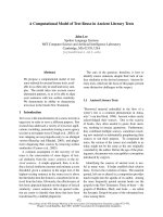
mode. Although the lung is a passive structure, the pressure
drop occurring across the pulmonary circulation is greater for
a positive pressure than it is for a negative pressure breath
(Fig. 3).
Under the high permeability conditions of the first stage of
ALI, even minor increases in pulmonary microvascular pres-
sure will increase edema formation dramatically. Moreover,
unlike in healthy tissue in which the blood–gas barrier is
intact, there is no clear pressure threshold for edema forma-
Figure 1
Relative contributions of pulmonary vascular segments to overall
pulmonary vascular resistance in the normal lung as a function of
transpulmonary pressure (and lung volume). The middle segment that
bridges the alveolus accounts for a progressively greater proportion of
the total as the lung distends.
Transpulmonary pressure
Change in vascular resistance
Arterial segment
Venous segment
Middle segment
Total pulmonary
circulation
Figure 2
Influence of lung expansion on alveolar and extra-alveolar vasculature.
Inflation compresses wall-embedded capillaries but dilates extra-
alveolar microvessels.
437
tion in lung tissue undergoing ALI [6]. The physiologic conse-
quences of pulmonary edema are well understood; alveolar
edema compromises gas exchange, and edematous airways
impede airflow and secretion clearance. From the standpoint
of VILI, however, alveolar flooding may produce competing
effects. A well known if simplistic model of interdependence
proposed by Mead (see below) suggests that collapsed
alveoli are subjected to shearing forces that are proportional
to the disparity in alveolar dimensions between the collapsed
alveolus and its distended neighbors [7]. Therefore, com-
pletely fluid-filled (flooded) alveoli theoretically are subjected
to lower shearing stresses than are atelectatic units, as the
gas–liquid interface is eliminated and alveolar dimensions
increase. On the other hand, elimination of surface tension
would cause capillaries that are fully embedded in the alveo-
lar walls to bulge further into the interior, encouraging their
rupture [8], and the increased weight of the edematous lung
may encourage small airway compression and accentuate the
tendency for tidal opening and closure to occur. Which of
these competing effects predominates cannot be stated with
certainty. Thus, although the influence of preformed edema
on lung mechanics and gas exchange is reasonably well
described, the importance of the microvasculature to the gen-
eration of VILI is less well understood. The remainder of the
present brief review focuses on what is currently known
regarding the interactions between airway pressures and vas-
cular pressures in the generation and maintenance of VILI.
What disrupts the blood–gas barrier during
ventilator-induced lung injury?
Clinicians have long been aware that certain inflammatory
conditions of the lung produce tissue hemorrhage in the
absence of ventilatory stress. These vessel-disrupting inflam-
matory injuries may originate either from the alveolar side (e.g.
pneumonia, abscess) or from the vascular side of the
blood–gas interface. Inflammatory conditions such as
Wegener’s granulomatosis, Goodpasture’s syndrome, and
pulmonary embolism are examples from the latter category.
Each disrupts the delicate barrier between gas and blood,
allowing erythrocytes to breech their vascular confines and
migrate into the interstitium and airspaces.
Although inflammation is of potential importance to the break-
down of the lung’s structural architecture, simply elevating
transmural pulmonary vascular pressure to high levels may
cause vascular rents or tears. Perhaps the clearest example
in this category occurs in severe mitral stenosis, a condition
in which pulmonary venous and capillary pressures can
exceed 35–40 mmHg. Acute edema that forms in this setting
is typically blood tinged, and the presence of hemosiderin-
laden macrophages in expectorated or lavaged samples
strongly suggests that this process originates at the alveolar
level from the pulmonary circulation (rather than the bronchial
circulation). Another circumstance under which elevating
transmural vascular pressures may cause hemoptysis in the
absence of pre-existing inflammation occurs during extreme
exertion, when blood flows through the lung are extremely
high and excursions of alveolar pressure are unusually large.
Postexertional lung hemorrhage is a well described occur-
rence in racehorses [9], and hemoptysis has been reported
after heavy exertion in elite human athletes as well [10].
Finally, forceful inspiratory efforts made during upper airway
obstruction may produce transvascular pressures of sufficient
magnitude to cause hemorrhagic pulmonary edema [11].
In elegant experiments undertaken in the laboratories of West
and colleagues [12–15], electron microscopy was used to
demonstrate the potential for mechanical disruption of the
microvasculature – ‘capillary stress fracture’ – to occur when
microvascular pressures are elevated to very high levels rela-
tive to their usual operating conditions. The pressures neces-
sary to cause capillary stress fracture vary among species,
with disruption being observed in healthy small animal lungs
(e.g. rabbits) at pressures as low as 40 mmHg. Larger
animals, such as dogs, withstand much higher microvascular
pressures without losing the structural integrity of the capil-
lary network [14]. Experimental studies reporting capillary
stress fracture in animals have largely been undertaken in
static preparations in which the airway pressure was held
constant and the intraluminal vascular pressures upstream
and downstream of the alveolus were equivalent. Under such
conditions, structural breakdown is more likely to be seen at
high lung volumes relative to resting conditions [15].
Although the range of microvascular pressure applied in
these studies might appear to preclude their physiologic rele-
vance, much lower vascular pressures might be required if
Available online />Figure 3
Pressure gradient along the normal pulmonary vascular bed as a
function of transpulmonary pressure generated by positive and
negative distension. Note that the pressure drop is greater during
positive pressure breathing, especially at higher lung volumes.
0
5
10
15
20
25
30
35
0 5 10 15 20
Transpulmonary pressure
(mmHg)
Pressure drop across the
pulmonary circulation (mmHg)
Negative pressure inflation
Positive pressure inflation
438
the framework of the lung were degraded by inflammation.
Moreover, there is excellent reason to believe that regional
transmural vascular forces may be dramatically different when
mechanically heterogeneous lungs are ventilated with
adverse ventilatory patterns.
Experimental evidence linking vascular
pressure to ventilator-induced lung injury
Just as with inflammation, mechanical forces that tear the del-
icate alveolar–capillary membrane can originate on either side
of the boundary. That the alveolar epithelium can be disrupted
by sufficient airway pressure is evident when barotrauma
develops. Clinicians recognize this damage radiographically
as air that leaks into the interstitial spaces to cause intrapul-
monary gas cysts, mediastinal emphysema, pneumothorax,
and systemic gas embolism [16]. It is equally clear that the
vasculature can lose its integrity in advance of epithelial frag-
mentation. Postmortem examination of tissues from patients
with acute respiratory distress syndrome often reveals areas
of interstitial and alveolar hemorrhage, findings that have gen-
erally been attributed to the underlying inflammatory process.
However, in both small and large animal models, the applica-
tion of adverse ventilatory patterns to previously healthy lungs
not only causes formation of proteinaceous edema but it also
stimulates neutrophil aggregation and hemorrhage [17,18].
Studies conducted in our laboratory strongly indicate that, in
the supine position, hemorrhagic edema forms preferentially
in dependent areas [18,19]. This proclivity is not subtle, and
has been corroborated by the work of other investigators
using different injury models [20]. It is worth emphasizing that
our experiments demonstrated that purely mechanical forces
originating within the alveolus inflict hemorrhagic injury in the
absence of pre-existing inflammation. It is somewhat counter-
intuitive that tissue disruption should occur in areas where
transmural stretching forces (as defined by plateau pressure
minus pleural pressure) are least. That is to say, ‘alveolar
stretch’ is greatest in the nondependent regions, which are
spared both the hemorrhagic infiltrate and most signs of
inflammation. Why might this occur?
The tendency for hemorrhage to occur preferentially in the
most dependent regions of the lung may have several expla-
nations. One compelling reason to expect microvascular dis-
ruption to occur there is that the mechanical stresses applied
by the tidal inflation cycle are greatly amplified at the interface
of opened and closed lung tissues. More than three decades
ago, Mead and coworkers [7] described a simplified model of
alveolar mechanics in which they proposed that an alveolus
attempting to close in an environment in which it was sur-
rounded by inflated tissue would experience traction forces
that are amplified in nonlinear proportion to the alveolar pres-
sures existing in the open units. According to their reasoning,
the coefficient linking effective pressure (P
eff
) to that actually
applied (P
app
) is the ratio between the alveolar volume that
corresponds to alveolar pressure (V) and the volume of the
collapsed alveolus (V
0
) raised to the power of 2/3:
P
eff
=P
app
× (V/V
0
)
2/3
. That admittedly oversimplified geomet-
ric argument suggested that at 30 cmH
2
O alveolar pressure,
for example, the effective stress applied at the junction of
closed and open tissue might approximate a value 4.5 times
as great as that experienced in the free walls of the open
alveolus.
Whatever its quantitative accuracy, a similar line of reasoning
might apply when tissues are already atelectatic and the lung
is exposed to high ventilating pressures, as in acute respira-
tory distress syndrome. Extrapolating from the Mead equation
presented above, the traction forces applied to junctional
tissues when alveolar pressure is 30 cmH
2
O could approxi-
mate 140 cmH
2
O, or about 100 mmHg. Thus, transvascular
microvascular forces during tidal ventilation could be in the
range that West and colleagues [14] suggested necessary
for a stress fracture to occur in large animals (dogs). Clearly,
such theoretic arguments are widely open to criticism.
However, it does appear reasonable to assume that mechani-
cal shearing forces experienced in ‘junctional’ tissues are
likely to exceed those elsewhere in the lung. Moreover, even
within fully inflated regions, the competing forces of capillary
compression and extraalveolar vessel dilatation/elongation
would be amplified when both lung volumes and vascular
pressures are high. These stresses would tug at the
microvascular conduit that links the alveolar and extra-alveolar
vessels with potentially damaging force. It is not difficult,
therefore, to envision vascular rupture from ventilatory pres-
sure under the pathologic conditions of ALI. Although unstud-
ied, surfactant depletion and inflammatory weakening of the
interstitial structure could amplify the impact of such forces,
whereas other changes of the microenvironment (e.g. flood-
ing by edema) could abrogate the mechanical stresses expe-
rienced in distal lung units.
Another intriguing possibility that may explain disproportion-
ate vascular disruption in dependent lung regions is that dor-
sally situated tissues receive a majority of the lung’s total
blood flow and are subjected to greater hydrostatic pressures
in the supine position. These higher intraluminal vascular
pressures or flows might amplify tensile forces external to the
microvessels or give rise to shearing stresses within the vas-
cular endothelium that initiate inflammation-mediated tissue
breakdown. There are hints in the early experimental literature
for VILI that vascular pressure could play an important if not
pivotal role in VILI development or expression. Dreyfuss and
Saumon [21], for example, found that ventilation with negative
pressure caused damage more severe than that caused by
positive pressure, implicating involvement of increased blood
flow in ventilation-related damage. Those same investigators
provided further support for this hypothesis by showing that
rats given dopamine to increase cardiac output suffered
increased albumin leak when ventilated with high pressure,
and ascribed a major portion of the protective effect of PEEP
in the setting of high pressure ventilation to its reduction of
pulmonary perfusion [22].
Critical Care December 2003 Vol 7 No 6 Marini et al.
439
Our group also explored the vascular contribution to VILI in a
series of experiments using isolated, ventilated, and perfused
(IVP) rodent lungs [23–26]. The IVP system offers numerous
advantages for the investigation of the interactions between
alveolar and vascular pressures. The progress of edema for-
mation can be monitored by continuously weighing the
heart–lung block suspended from a strain gauge. Breakdown
of the alveolar capillary barrier can be inferred from the filtra-
tion constant (K
F
) derived from the weight–time relationship.
Inflow and outflow vascular pressures and perfusion rate can
be precisely measured and/or regulated. Finally, the composi-
tion and physical properties of the perfusate can be adjusted.
In the experiments described below, each heart–lung block
was perfused with Krebs-Henseleit solution, doped with a
small quantity of autologous blood to provide a histologically
visible marker of overt vascular rupture. Sufficient albumin
was added to achieve physiologic tonicity. The cumulative
results of this work demonstrate unequivocally that variations
in vascular pressure and flow have the potential to modulate
the nature and severity of VILI.
Pressure or flow: which is the key variable?
In our first experiment we exposed isolated rabbit lungs to
perfusion levels that were equivalent to or about 50% greater
or less than the normal resting blood flow of that animal
species [23]. All lungs were ventilated identically with airway
pressures that proved damaging in vivo. In this model of VILI
we demonstrated that perfusion amplitude contributed to the
reduced lung compliance resulting from an adverse ventila-
tory pattern and promoted both lung edema and hemorrhage.
We also found a strong correlation between indices of lung
injury and the vascular pressure changes resulting from the
interaction between ventilation and perfusion [23]. Although
data from that experiment strongly suggested the primacy of
perfusion pressure, it was not possible in that initial experi-
ment to determine definitively which of those two variables
was more important in modulating VILI, because vascular
pressure increased in parallel with flow.
In a second IVP lung experiment designed to address that
question, we varied airway pressure profiles to allow arterial
pulmonary pressure to vary while blood flow was held con-
stant [24]. Our results indicated that mean airway pressure
had greater impact than did tidal excursion amplitude in
determining the severity of lung hemorrhage and lung perme-
ability alterations resulting from an adverse pattern of
mechanical ventilation. Histologic injury scores were virtually
identical for large and small tidal volumes when high mean
airway pressures were achieved, whether by lengthening
inspiratory time or by increasing PEEP. A key difference
between high mean airway pressure and low mean airway
pressure preparations was the magnitude of the pulmonary
arterial pressure and the length of time over which it was sus-
tained. The results of those experiments emphasized the
potential for deleterious interactions to occur between lung
volumes and pulmonary hemodynamics. Taken together, our
initial two studies demonstrated that modifications of vascular
pressure within and upstream from the intermediate segment
could influence the severity of VILI inflicted by an unchanging
adverse pattern of ventilation.
How does the number of ventilatory cycles
influence the expression of ventilator-
induced lung injury?
Although ventilation is the product of tidal volume and fre-
quency, surprisingly little attention has been directed at the
role of the latter in the generation of VILI. Therefore, having
concluded that upstream microvascular pressure might be an
important cofactor in the development of VILI, we next
addressed the question of how the number of ventilatory
cycles occurring over a timed interval influences the rate of
edema formation or severity of histologic alterations when
maximum, minimum, and mean airway pressures are held
identical.
Almost 15 years ago, Bshouty and Younes [27] reported that,
for the same minute ventilation target, raising tidal volume at a
constant frequency and raising frequency at a constant tidal
volume produced similar degrees of edema in canine lobes
perfused in situ at elevated hydrostatic pressures. At about
the same time, Kolobow and colleagues [28,29] demon-
strated that sheep ventilated over many hours with high and
moderate airway pressures sustained more lung injury than
did those examined earlier, suggesting the potential for cumu-
lative damage to occur under adverse ventilatory conditions.
We used our IVP model in experiments testing the hypothesis
that cumulative damage occurs as a function of the number of
stress cycles as well as stress magnitude. In these experi-
ments, the pressure controlled mode with a peak pressure of
30 cmH
2
O and a PEEP of 3 cmH
2
O was used in each prepa-
ration [25]. Each experiment was conducted over 30 min. In
one of three experimental groups of isolated and perfused
rabbit lungs, a pulmonary artery peak pressure of 20 mmHg
was matched to a ventilatory frequency of 20 cycles per
minute to serve as our control. In a second set of perfused
lungs, pulmonary artery pressure was allowed to rise to a
maximum of 35 mmHg with each tidal cycle, at a frequency of
20 breaths per minute. In the third group peak pulmonary
artery pressure was again capped at 35 mmHg, but ventilator
frequency was reduced to three cycles per minute with the
same inspiratory time fraction as in the other two groups.
Thus, mean airway pressure was identical for both high-pres-
sure ventilatory patterns. The pH characteristics did not vary
significantly among the groups.
Our main findings were that lungs ventilated at low frequen-
cies and high peak pulmonary artery pressures formed less
edema and exhibited markedly less perivascular hemorrhage
than did those ventilated at higher frequencies but identical
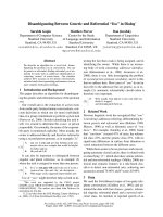
peak pulmonary artery pressures. In addition, lungs ventilated
Available online />440
with high peak pulmonary artery pressures and flows exhib-
ited more extensive histologic alterations and edema forma-
tion than did those subjected to the same ventilatory pattern
but at lower peak vascular pressures and flows [25] (Fig. 4).
Only a very small fraction of this difference was attributable to
differences in mean hydraulic pressure. These data strongly
indicated not only that the characteristics of the tidal cycle
and vascular pressures are of fundamental importance to VILI,
but also that minute ventilation, reflecting the number of
stress cycles of a potentially damaging magnitude per unit
time or their cumulative number, might be as well.
Whereas dependence of edema formation on minute venti-
lation was previously noted in the aforementioned experi-
mental study conducted by Bshouty and Younes in isolated
dog lobes [27], their experiments differed from ours in four
notable ways. First, whereas their study was conducted
with a physiologic ventilatory pattern, we employed ventila-
tory patterns known to be potentially injurious. Second, vas-
cular pressure in the study by Bshouty and Younes was
elevated by raising outflow (left atrial) pressure, thereby
increasing pressure along the entire vascular tree (a simula-
tion of left sided congestive heart failure). We held outflow
pressure constant at a physiologically normal value while
raising pressure selectively in those regions proximal to the
intermediate vascular segment. Third, we used considerably
higher vascular flows on a per-gram-of-lung basis than did
Bshouty and Younes [27]. Finally, we not only measured
edema and but also assessed histologic changes, as
reflected by lung hemorrhage.
Several mechanisms come to mind that may explain the
diminution of lung edema formation and perivascular hemor-
rhage that we observed by decreasing respiratory frequency.
A higher ventilatory frequency could have depleted surfactant
more efficiently, thereby increasing alveolar surface tension,
lowering end-expiratory extravascular pressure, and promot-
ing alveolar flooding. Upstream, the increased transvascular
pressure gradient across extra-alveolar vessels would also
favor fluid transudation, vessel disruption, and perivascular
hemorrhage. Conceivably, the larger number of stress cycles
imposed on the groups receiving 20 breaths per minute
could have induced cumulative damage in a manner similar to
that experienced in a variety of biomaterials that are sub-
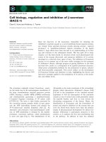
jected to sufficient repeated stress [30]. Overt stress frac-
tures similar to those found by West and colleagues [13–15]
were demonstrated by scanning electron microscopy in our
laboratory to occur in the setting of VILI (Fig. 5), as well as in
a recently reported human patient [31]. A type of ‘materials
failure’ of structural elements seems an attractive explanation,
in that we found that both reducing stress application fre-
quency (respiratory rate) and stress amplitude (pulmonary
artery peak pressure) effectively limited VILI.
If cumulative damage is important, then providing a lower fre-
quency and/or lower pulmonary vascular pressure would both
Critical Care December 2003 Vol 7 No 6 Marini et al.
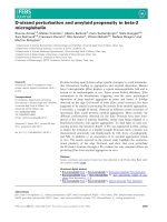
Figure 4
Damaging effect of high vascular pressure and repeated cycling on isolated, ventilated, and perfused rabbit lungs. For identical airway pressure
profiles (plateau, mean, and end-expiratory pressures), higher peak inflow pressure (35 mmHg) was associated with greater damage, as compared
with the control value of 20 mmHg. Reducing cycling frequency from 20 to 3 cycles per minute while holding airway and vascular pressures
constant reduced injury severity. Adapted with permission from Hotchkiss and coworkers [25].
60
50
40
30
20
10
0
Frequency and vascular pressure worsen lung injury
Normal frequency,
and vascular pressure
Normal frequency,
high vascular pressure
Hemorrhage score
Frequency and vascular pressure
F20P20 F3P35 F20P35
441
be expected to reduce the tendency for material stress
failure. Finally, it is interesting to consider that a low fre-
quency may have allowed sufficient time between adjacent
cycles for reparative processes to operate. Surprisingly little
time appears to be needed to reseal small disruptions in
tissue barriers [32,33].
What are the relative roles of vascular and
airspace pressures in ventilator-induced lung
injury?
Because rigorous limitation of pulmonary vascular pressures
significantly attenuated the damage in lungs exposed to a
fixed ventilatory pattern, the work outlined above suggests
that elevations in pulmonary vascular pressure arising from
interactions between lung volume, pulmonary vascular resis-
tance, and pulmonary vascular flow could worsen ventilator-
associated lung injury. Our redirected attention toward the
vascular side of the alveolar capillary barrier stimulated us to
ask whether the mechanism by which pulmonary artery pres-
sure is phasically increased can influence the severity of lung
damage during exposure to high alveolar pressure. In other
words, is periodic inflation a necessary component of the vas-
cular injury that is incurred during VILI?
Knowing that the frequency of ventilation is an important
determinant of VILI, we reasoned that a lung exposed to pul-
satile vascular pressure but not ventilated might experience
significant injury, even without fluctuations in airway pressure.
In an experiment designed to test this, we applied a damag-
ing pattern of airway pressure (plateau 30 cmH
2
O, PEEP
5 cmH
2
O) to one of three sets of lung preparations and
allowed others to remain motionless [26]. In the ventilated
group, peak pulmonary artery pressure was allowed to rise to
35 mmHg. Left atrial pressure was held at 10 mmHg and
mean airway pressure at 17.5 cmH
2
O. This set of ventilated
preparations was compared with two unventilated groups
held without tidal fluctuations in airway pressure (continuous
positive airway pressure 17.5 cmH
2
O) in which in which all
key hemodynamic pressures – peak, mean, and nadir – were
identical to their ventilated counterparts. In the latter two
groups a vascular pump applied pulsatile pulmonary artery
pressure to the motionless lungs at frequencies of 3 or
20 pulses per minute. Each vascular stress cycle, whether
generated by ventilation or by the vascular pump, was char-
acterized by identical peak, mean, and nadir values.
Our main findings were that lungs exposed to cyclic eleva-
tions in pulmonary artery pressure in the absence of ventila-
tion formed less edema and exhibited less perivascular and
alveolar hemorrhage than did ventilated lungs exposed to
similar peak and mean pulmonary artery pressures and mean
airway pressure [26]. Interestingly, under conditions of static
continuous positive airway pressure, the higher pulsing fre-
quency was associated with a greater degree of perivascular
hemorrhage, indicating that the pulsatility of vascular pres-
sure did contribute to VILI. Thus, the effects of respiratory fre-
quency and vascular pressures on VILI are not mediated
primarily by pulsatile vascular pressure per se but rather by a
phenomenon related to cyclic modulation of the vascular
microenvironment induced by ventilation. Because alveolar
and extra-alveolar microvessels are stressed differently by
lung expansion, these experiments focused our attention on
the extra-alveolar microvasculature, suggesting the cyclic
changes in perivascular pressure surrounding extra-alveolar,
Available online />Figure 5
Electron micrographs of rabbit lungs injured solely by high inflation pressures, low positive end-expiratory pressure, and elevated vascular inflow
pressure. (a) Capillary stress fracture with incipient extravasation of an erythrocyte. (b) Higher power view of stress fracture showing exposure of
collagen filaments.
442
juxtacapillary microvessels might be important in the genesis
of VILI.
Should vascular pressures be lowered to
avert ventilator-induced lung injury?
Given that elevation in pulmonary vascular inflow pressure
accentuated VILI, it seems logical that reduction in postalveo-
lar vascular pressure would also be protective. The merit of
reducing left atrial pressure might be expected for at least
two reasons. The edematous lungs tend to collapse under
their own weight and to develop dependent atelectasis,
which could lead to cyclic opening and collapse, intensified
shear stresses, and a tendency toward VILI in dependent
areas. Moreover, exudation of protein-rich fluid has the poten-
tial to inactivate surfactant, further altering membrane perme-
ability by increasing both surface tension and radial traction
on pulmonary microvessels. On the other hand, increased left
atrial pressure might help to limit VILI by flooding the alveoli of
dependent regions, thereby reducing regional mechanical
stresses. Reducing capillary pressure could promote cyclic
vascular recruitment and derecruitment as the lungs transition
from West’s zone III to zone II condition during the course of
the positive pressure inflation/deflation cycle. Hydrodynamic
forces may well be accentuated by the higher velocities and
surface shear stresses that occur along the vascular endothe-
lium under such conditions. Reducing outflow pressure also
increases the gradient of pressure appearing across the
alveoli, and consequently the energy dissipated across the
intermediate segment. For these reasons, the impact of selec-
tively reducing pulmonary venous pressure during ventilation
with high airway pressure cannot easily be predicted.
In a recently published comparison of lungs ventilated with
moderately high peak alveolar pressures with normal and low
left atrial pressures, Broccard and coworkers [34] demon-
strated a striking difference in favor of the normal vascular
pressure subset. This rather surprising result suggests that
cyclic opening and closure of stressed microvessels could be
important in the generation of VILI. Alternatively, decreasing
outflow pressure might amplify microvascular stresses at or
near the alveolar level, presumably acting through interdepen-
dence of the pulmonary vascular network. We speculate that
direct mechanotransduction of inflammatory signals, increased
transalveolar energy dissipation, or materials failure at the
stressed boundary could be important linking mechanisms.
Collectively, the results of the laboratory experiments and
clinical observations discussed above suggest several path-
ways by which microvascular hemodynamics may influence
the development or expression of VILI (Fig. 6).
Potential clinical implications
Manipulation of cardiac output and vascular pressure is of
vital importance in critical care management. The interactions
between vascular pressure and ventilation outlined in this
review suggest strongly that closer attention should be paid
to interventions that impact on vascular pressures, flows, and
resistances when high inflation pressures are in use. Because
microvascular stresses appear to be a potent cofactor in the
development of pulmonary edema as well as lung damage
resulting from an injurious pattern of ventilation, the clinician
managing ALI should reconcile the competing objectives of
ensuring adequate oxygen delivery and minimizing adverse
effects. For example, an increase in cardiac output is gener-
ally held to be a beneficial consequence of management;
however, increases in cardiac output are associated with an
increased prealveolar microvascular pressure and a higher
vascular pressure gradient across the lung. If increased pre-
alveolar microvascular pressure accentuates a tendency
toward VILI, then attempts to raise cardiac output may have
unintended consequences. On the other hand, taking steps
to reduce oxygen consumption demands could benefit the
lung by reducing the pressure gradient developed across the
microvasculature. Similarly, a reduction in left atrial pressure
with maintained cardiac output is generally believed to benefit
lung function, and this perception is almost certainly accurate
with respect to hydrostatic edema formation. However, the
results of the recent work cited above suggest that excessive
reduction in left atrial pressure could amplify the tendency
toward VILI [34]. Because reducing ventilation frequency
decreases the number of stress cycles, our work would
suggest that a reduction in minute ventilation effected either
by a decrease in tidal volume or by a decrease in ventilatory
frequency might have a salutary effect in reducing the ten-
dency toward VILI.
Reduced minute ventilation is generally associated with
increased carbon dioxide retention and hypercapnic acidosis,
which until recently was considered an undesirable but nec-
essary consequence of a ‘lung protective’ ventilation strategy.
However, when the lung’s gas exchanging properties are not
Critical Care December 2003 Vol 7 No 6 Marini et al.
Figure 6
Possible mechanisms by which hemodynamic parameters may incite or
exacerbate ventilator-induced lung injury (VILI). Microvascular strain
may be amplified at the junctions of open and closed lung units. CO,
cardiac output; P
LA
, left atrial pressure; P
PA
, pulmonary artery pressure.
Structural vessel failure
CO, P
PA
, P
LA
or
venous resistance
Lung volume
Microvascular
stretch / strain
Edema
Vascular resistance
(middle segment)
Transmural
pressure
VILI
Pinterstitial
Intramural
pressure
443
dramatically altered and carbon dioxide production remains
unchanged, recently published experiments by Sinclair and
coworkers [35] and by Broccard and colleagues [36]
strongly indicate that the generation of hypercapnic acidosis
may exert a protective effect on the severity of VILI. (This
observation is consistent with elegant work that previously
addressed ischemia/reperfusion injury [37].)
Because interventions such as increasing PEEP or extending
inspiratory time may redirect blood flow and radically alter the
microvascular environment, it is conceivable that both benefit
and harm could potentially result from these maneuvers. The
body of investigative work reviewed here suggests that a
reduction in the demands for cardiac output and ventilation
could dramatically reduce the tendency toward VILI, even
when using patterns that generate similar values for peak and
end-expiratory alveolar pressures. Whether these intriguing
possibilities are relevant to the clinical setting will require
extensive and careful additional study.
Conclusion
Experimental and clinical evidence point strongly toward the
potential for microvascular stresses to influence the severity
and expression of ventilator associated lung injury. Increasing
pressure upstream from the alveolus not only worsens edema
but also predisposes the gas–blood barrier to disrupt in
response to high airspace pressures, especially at rapid venti-
lation frequencies. Paradoxically, reducing venous microvas-
cular pressure (and simultaneously increasing both the
pressure gradient and energy dissipated along the pulmonary
microvasculature) appears to worsen edema and/or accentu-
ate barrier breakdown. Once disruption occurs, bi-directional
interchange between the vascular and gaseous compart-
ments may take place, possibly with adverse consequences
for the lung and for extrapulmonary organs. Although it is haz-
ardous to extrapolate from the available data to the clinical
setting, taking measures to lower vascular stress (e.g. by
reducing the physiologic requirements for ventilation and
cardiac output) is a logical, but as yet unproven, extension of
a lung-protective strategy for life support in ARDS.
Competing interests
None declared.
References
1. Hakim TS, Michel RP, Chang HK: Effect of lung inflation on pul-
monary vascular resistance by arterial and venous occlusion.
J Appl Physiol 1982, 53:1110-1115.
2. Lai-Fook SJ: Perivascular interstitial pressure measured by
micropipettes in isolated dog lung. J Appl Physiol 1982, 52:9-15.
3. Fishman AP: Pulmonary circulation. In Handbook of Physiology.
Section 3: The Respiratory System. Edited by Fishman AP, Fisher
AB, Geiger SR. Bethesda: American Physiology Society;
1987:93-97.
4. Lamm WJ, Kirk KR, Hanson WL, Wagner WW Jr, Albert RK: Flow
through zone 1 lungs utilizes alveolar corner vessels. J Appl
Physiol 1991, 70:1518-1523.
5. West JB, Dollery CT, Naimark A: Distribution of blood flow in
isolated lung; relation to vascular and alveolar pressures. J
Appl Physiol 1964, 19:713-724.
6. Brigham KL, Woolverton WC, Blake LH, Staub NC: Increased
sheep lung vascular permeability caused by pseudomonas
bacteremia. J Clin Invest 1974, 54:792-804.
7. Mead J, Takishima T, Leith D: Stress distribution in lungs: a
model of pulmonary elasticity. J Appl Physiol 1970, 28:218-
233.
8. Namba Y, Kurdak SS, Fu Z, Mathieu-Costello O, West JB: Effect
of reducing alveolar surface tension on stress failure in pul-
monary capillaries. J Appl Physiol 1995, 79:2114-2121.
9. West JB, Mathieu-Costello O, Jones JH, Birks EK, Logemann RB,
Pascoe JR, Tyler WS: Stress failure of pulmonary capilaries in
racehorses with exercise-induced pulmonary hemorrhage. J
Appl Physiol 1993, 75:1097-1109.
10. Hopkins SR, Schoene RB, Martin TR, Henderson WR, Spragg
RG, West JB: Intense exercise impairs the integrity of the pul-
monary blood-gas barrier in elite athletes. Am J Respir Crit
Care Med 1997, 155:1090-1094.
11. Broccard AF, Liaudet L, Aubert JD, Schnyder P, Schaller MD:
Negative pressure post-tracheal extubation alveolar hemor-
rhage. Anesth Analg 2001, 92:273-275.
12. Costello ML, Mathieu-Costello OM, West JB: Stress failure of
alveolar epithelial cells studied by scanning electron
microscopy. Am Rev Respir Dis 1992, 145:1446-1455.
13. West JB, Tsukimoto K, Mathieu-Costello O, Prediletto R: Stress
failure in pulmonary capillaries. J Appl Physiol 1991, 70:1731-
1742.
14. Mathieu-Costello O, Willford DC, Fu Z, Garden RM, West JB:
Pulmonary capillaries are more resistant to stress failure in
dogs than in rabbits. J Appl Physiol 1995, 79:908-917.
15. Fu Z, Costello ML, Tsukimoto K, Prediletto R, Elliott AR, Mathieu-
Costello O, West JB: High lung volume increases stress
failure in pulmonary capillaries. J Appl Physiol 1992, 73:123-
133.
16. Amato MB, Marini JJ: Barotrauma, volutrauma, and the ventila-
tion of acute lung injury. In Physiological Basis of Ventilatory
Support. Edited by Marini JJ, Slutsky AS. New York: Marcel
Dekker; 1998:1187-1245.
17. Dreyfuss D, Saumon G: Ventilator-induced lung injury: lessons
from experimental studies. Am J Respir Crit Care Med 1998,
157:294-323.
18. Broccard A, Shapiro R, Schmitz L, Adams AB, Nahum A, Marini J:
Prone positioning attenuates and redistributes ventilator-
induced lung injury in dogs. Crit Care Med 2000, 28:295-303.
19. Broccard AF, Shapiro RS, Schmitz LL, Ravenscraft SA, Marini JJ:
Influence of prone position on the extent and distribution of
lung injury in a high tidal volume oleic acid model of acute
respiratory distress syndrome. Crit Care Med 1997, 25:16-27.
20. Hirschl RB, Tooley R, Parent A, Johnson K, Bartlett RH: Evalua-
tion of gas exchange, pulmonary compliance, and lung injury
during total and partial liquid ventilation in the acute respira-
tory distress syndrome. Crit Care Med 1996, 24:1001-1008.
21. Dreyfuss D, Saumon G: Role of tidal volume, FRC, and end-
inspiratory volume in the development of pulmonary edema
following mechanical ventilation. Am Rev Respir Dis 1993,
148:1194-1203.
22. Dreyfuss D, Soler P, Basset G, Saumon G: High inflation pres-
sure pulmonary edema. Respective effects of high airway
pressure, high tidal volume, and positive end-expiratory pres-
sure. Am Rev Respir Dis 1988, 137:1159-1164.
23. Broccard AF, Hothchkiss JR, Kuwayama N, Olson DA, Jamal S,
Wangensteen DO, Marini JJ: Consequences of vascular flow on
lung injury induced by mechanical ventilation. Am J Respir Crit
Care Med 1998, 157:1935-1942.
24. Broccard AF, Hotchkiss JR, Suzuki S, Olson D, Marini JJ: Effects
of mean airway pressure and tidal excursion on lung injury
induced by mechanical ventilation in an isolated perfused
rabbit lung model. Crit Care Med 1999, 27:1533-1541.
25. Hotchkiss JR, Blanch LL, Murias G, Adams AB, Olson D, Wan-
gensteen OD, Leo PH, Marini JJ: Effects of decreased respira-
tory frequency on ventilator induced lung injury. Am J Respir
Crit Care Med 2000, 161:463-468.
26. Hotchkiss JR, Blanch LL, Naviera A, Adams AB, Olson D, Marini
JJ: Relative roles of vascular and airspace pressures in venti-
lator induced lung injury. Crit Care Med 2001, 29:1593-1598.
27. Bshouty Z, Younes M: Effect of breathing pattern and level of
ventilation on pulmonary fluid filtration in dog lung. Am Rev
Respir Dis 1992, 145:3672-3676.
Available online />444
28. Kolobow T, Moretti MP, Fumagalli R, Mascheroni D, Prato P, Chen
V, Joris M: Severe impairment in lung function induced by high
peak airway pressuring during mechanical ventilation. An
experimental study. Am Rev Respir Dis 1987, 135:312-315.
29. Tsuno K, Miura K, Takeya M, Kolobow T, Morioka T: Histopatho-
logic pulmonary changes from mechanical ventilation at high
peak airway pressures. Am Rev Respir Dis 1991, 143:1115-
1120.
30. Hashin Z, Rotem A: A cumulative damage theory of fatigue
failure. Mater Sci Eng 1978, 34:147-160.
31. Hotchkiss JR, Simonson DA, Marek DJ, Marini JJ, Dries DJ: Pul-
monary microvascular fracture in a patient with acute respira-
tory distress syndrome. Crit Care Med 2002, 30:2368-2370.
32. Dreyfuss D, Soler P, Saumon G: Spontaneous resolution of pul-
monary edema caused by short periods of cyclic overinflation.
J Appl Physiol 1992, 72:2081-2089.
33. Vlahakis NE, Hubmayr RD: Invited review: plasma membrane
stress failure in alveolar epithelial cells. J Appl Physiol 2000,
89:2490-2496.
34. Broccard A, Vannay C, Feihl F, Schaller MD: Impact of low pul-
monary vascular pressure on ventilator-induced lung injury.
Crit Care Med 2002, 30:2183-2190.
35. Sinclair SE, Kregenow DA, Lamm WJ, Starr IR, Chi EY, Hlastala
MP: Hypercapnic acidosis is protective in an in vivo model of
ventilator-induced lung injury. Am J Respir Crit Care Med
2002, 166:403-408.
36. Broccard AF, Hotchkiss JR, Vannay C, Markert M, Sauty A, Feihl
F, Schaller MD: Protective effects of hypercapnic acidosis on
ventilator-induced lung injury. Am J Respir Crit Care Med
2001, 164:802-806.
37. Laffey JG, Tanaka M, Engelberts D, Luo X, Yuan S, Tanswell AK,
Post M, Lindsay T, Kavanagh BP: Therapeutic hypercapnia
reduces pulmonary and systemic injury following in vivo lung
reperfusion. Am J Respir Crit Care Med 2000, 162:2021-2022.
Critical Care December 2003 Vol 7 No 6 Marini et al.