Báo cáo khoa học: ":Clinically important deep vein thrombosis in the intensive care unit: a survey of intensivists" pot
Bạn đang xem bản rút gọn của tài liệu. Xem và tải ngay bản đầy đủ của tài liệu tại đây (161.63 KB, 8 trang )
Open Access
Available online />R145
June 2004 Vol 8 No 3
Research
Clinically important deep vein thrombosis in the intensive care
unit: a survey of intensivists
Deborah Cook
1
, Maureen Meade
2
, Gordon Guyatt
3
, Lauren Griffith
4
, John Granton
5
,
William Geerts
6
and Mark Crowther
7
, the Canadian Critical Care Trials Group
1
Professor, Departments of Medicine, and Clinical Epidemiology & Biostatistics, McMaster University, Hamilton, Ontario, Canada
2
Associate Professor, Departments of Medicine, and Clinical Epidemiology & Biostatistics, McMaster University, Hamilton, Ontario, Canada
3
Professor, Departments of Medicine, and Clinical Epidemiology & Biostatistics, McMaster University, Hamilton, Ontario, Canada
4
Department of Clinical Epidemiology & Biostatistics, McMaster University, Hamilton, Ontario, Canada
5
Associate Professor, Department of Medicine, University of Toronto, Toronto, Ontario, Canada
6
Professor, Department of Medicine, University of Toronto, Toronto, Ontario, Canada
7
Associate Professor, Department of Clinical Epidemiology & Biostatistics, McMaster University, Hamilton, Ontario, Canada
Corresponding author: Deborah Cook,
Abstract
Introduction Outside the intensive care unit (ICU), clinically important deep vein thrombosis (DVT) is
usually defined as a symptomatic event that leads to objective radiologic confirmation and subsequent
treatment. The objective of the present survey is to identify the patient factors and radiologic features
of lower limb DVT that intensivists consider more or less likely to make a DVT clinically important in ICU
patients.
Methods Our definition of clinically important DVT was a DVT likely to result in short-term or long-term
morbidity or mortality if left untreated, as opposed to a DVT that is unlikely to have important
consequences. We asked respondents to indicate the likelihood that patient factors and
ultrasonographic features make a DVT clinically important using a five-point scale (from 1 = much less
likely to 5 = much more likely).
Results Of the 71 Canadian intensivists who responded, 70 (99%) rated three patient factors as most
likely to make a DVT clinically important: clinical suspicion of pulmonary embolism (mean score 4.6),
acute or chronic cardiopulmonary morbidity that might limit a patient's ability to tolerate pulmonary
embolism (score 4.5), and leg symptoms (score 4.2). Of the ultrasound features, proximal (score 4.7),
large (score 4.2), and totally occlusive (score 3.9) thrombi were considered the three most important.
Conclusion When labeling a DVT as clinically important, intensivists rely on different patient specific
factors and thrombus characteristics than are used to assess the clinical importance of DVT outside
the ICU. The clinical importance of DVT is influenced by unique factors such as cardiopulmonary
reserve among mechanically ventilated patients.
Keywords: deep venous thrombosis, pulmonary embolism, ultrasound
Introduction
Venous thromboembolism (VTE), which includes both deep
vein thrombosis (DVT) and pulmonary embolism (PE), is a
common complication of critical illness [1,2]. Critically ill
patients harbor many coincident risk factors for DVT, such as
the need for surgery, catheters, immobility, and use of seda-
tives and paralytic agents [3-5]. As in the noncritically ill popu-
lation, it is likely that most episodes of DVT are asymptomatic
Received: 26 January 2004
Revisions requested: 11 March 2004
Revisions received: 27 March 2004
Accepted: 05 April 2004
Published: 6 May 2004
Critical Care 2004, 8:R145-R152 (DOI 10.1186/cc2859)
This article is online at: />© 2004 Cook et al.; licensee BioMed Central Ltd. This is an Open
Access article: verbatim copying and redistribution of this article are
permitted in all media for any purpose, provided this notice is preserved
along with the article's original URL.
DVT = deep vein thrombosis; ICU = intensive care unit; PE = pulmonary embolism; SD = standard deviation; VTE = venous thromboembolism.
Critical Care June 2004 Vol 8 No 3 Cook et al.
R146
and confined to the deep veins of the calf. However, with time,
20–30% of untreated calf vein thrombi extend proximally into
the thigh, where they pose a 40–50% risk for PE [6]. Early
studies of the natural history of PE suggest that untreated PE
has a mortality rate of at least 25% [7].
One large study found PE at autopsy in 59 out of 404 hospi-
talized patients (14.6%); among the 20 patients who died from
PE, this diagnosis was not suspected in 14 (70)% [8]. In a 25-
year longitudinal study, 9% of patients had autopsy evidence
of PE; again, the diagnosis of PE was not suspected in 84%
of patients before autopsy [9]. In a landmark study highlighting
this problem in critical care practice, 13 out of 34 (38%) inten-
sive care unit (ICU) patients with known DVT who had no
symptoms of PE were diagnosed with PE by ventilation-per-
fusion scans [10]. It is possible that many mechanically venti-
lated patients with sudden episodes of hypotension,
tachycardia, or hypoxia may have undetected PE [11]. Unsus-
pected PE may also contribute to difficulty in weaning patients
from mechanical ventilation [12]. We posit that in critically ill
patients with impaired cardiopulmonary reserve, a small PE,
which might be of minimal clinical importance in patients who
are less ill, might have severe or fatal consequences [13].
Thus, the clinical consequences of DVT have the potential to
be particularly serious, but may be unrecognized in the ICU. It
is also possible that ICU patients undergoing treatment for
DVT are more likely than other patients to suffer serious com-
plications from anticoagulant therapy.
Among ambulatory patients, most clinicians consider sympto-
matic DVT to be more clinically important than asymptomatic
DVT. Some clinicians may consider clinically important DVT to
be proximal (thigh DVT) rather than distal (calf DVT), based on
the greater propensity of untreated proximal DVT to cause PE.
Thus, clinically important episodes of DVT are usually defined
as symptomatic events that lead to objective radiologic confir-
mation and subsequent treatment [2,14]. However, critically ill
patients cannot reliably communicate their symptoms, and
intensivists rarely use the reference standard of venography to
diagnose DVT [15]. Furthermore, physical signs such as uni-
lateral leg edema, which are important clinical features of DVT
outside the ICU, are uncommon in the ICU because many crit-
ically ill patients are supine and frequently have severe bilateral
edema [16]. These observations suggest that the classic def-
inition of a clinically important DVT is not suitable in the ICU
setting.
There is no objective reference standard for or definition of
clinically important DVT in critically ill patients because, to our
knowledge, no studies have systematically correlated clinical
or radiographic characteristics of acute venous thrombosis
with patient outcomes. Despite the lack of such data, the con-
cept of a clinically important DVT in the critically ill is important
because such an assessment may be used to determine
whether a DVT should be treated; in addition, a definition of
clinically important DVT could be used in future clinical
research. Therefore, as a first step toward defining a 'clinically
important DVT', we surveyed critical care physicians to identify
those patient factors and radiologic features of lower limb DVT
that influence their perceptions about the clinical importance
of DVT. Our primary hypothesis was that intensivists will con-
sider the following patient factors more likely to make a DVT
clinically important: the DVT is symptomatic, is detected by
physical examination, is found in a patient with risk factors,
occurs in a patient who has been receiving DVT prophylaxis or
therapeutic anticoagulation, and occurs in a patient with either
chronic cardiopulmonary comorbidity or acute cardiopulmo-
nary illness that might limit tolerance to a PE. Our secondary
hypothesis was that intensivists will consider the following
compression ultrasound features more likely to make a DVT
clinically important: the DVT is proximal, large, occlusive, and
persists 1 week later.
Methods
Instrument development
We developed a survey for members of the Canadian Critical
Care Trials Group who care for critically ill adults [17]. We
generated items for this instrument based on a review of the
literature, our prior work [15,18,19], and discussion among
the investigators. We reduced items for this instrument by
interviews with five intensivists who did not participate in the
survey.
Instrument formatting
In the survey we provided the rationale for the questionnaire,
defined the concept of clinically important DVT, and requested
basic demographic information about respondents and their
ICU settings. We then surveyed respondents about the extent
to which various factors influence their assessment of the clin-
ical importance of DVT in critically ill patients. A self-adminis-
tered rather than interviewer-administered format was used to
maximize the validity of self-reported information [20].
We used the following conceptual definition of clinically impor-
tant DVT: a DVT that, if untreated, would will lead to increased
short-term morbidity (e.g. cardiopulmonary consequences that
may delay weaning from mechanical ventilation), long-term
morbidity (e.g. patient-centered consequences such as
chronic venous insufficiency), or mortality. We listed potential
characteristics of clinically important DVTs in two domains:
patient factors and ultrasonographic features. For an ICU
patient with a DVT we asked respondents to indicate, using a
Likert scale, whether each of these characteristics was more
or less likely to make a DVT clinically important (1 = much less
likely to make the DVT clinically important, 2 = somewhat less
likely to make the DVT clinically important, 3 = neither more nor
less likely to make the DVT clinically important, 4 = somewhat
more likely to make the DVT clinically important, and 5 = much
more likely to make the DVT clinically important; see Appendix
1).
Available online />R147
Demographic questions included year of graduation from
medical school and clinical discipline (internal medicine or its
subspecialities, surgery, or anesthesia). We also requested
information about the number of ICU beds in which the
respondent works, the type of ICU (medical, surgical or mixed
medical–surgical), and the presence of a thrombosis consult
service. We used closed-ended questions for the ICU demo-
graphic data to maximize the accuracy and completeness of
responses [21].
Instrument administration
We administered the survey instrument by e-mail to all mem-
bers of the Canadian Critical Care Trial Group who care for
critically ill adults. We contacted nonrespondents by facsimile
with a second copy of the questionnaire [22], and then by e-
mail or telephone contact. The survey was conducted in Sep-
tember and October 2003.
Research ethics
Participation was voluntary, individual responses were kept
confidential, and data were anonymized upon receipt. The
study was approved by the Research Ethics Board of St
Joseph's Healthcare in Hamilton, Ontario.
Analysis
Descriptive statistics were used, including proportions with
associated confidence intervals, as well as measures of cen-
tral tendency and dispersion (mean and standard deviation
[SD]). Within each domain (patient factors and ultrasono-
graphic features), we ranked the characteristics according to
which were most likely to make the DVT clinically important.
Results
The response rate was 70 out of 71 (99%). Sixty-six (93%) of
the surveys were returned without a reminder e-mail; five (7%)
required a reminder. Clinical discipline backgrounds were
internal medicine in 52 (75%), anesthesia in eight (12%), sur-
gery in six (9%), and other in three (4%). Respondents had
16.2 (SD 8.2) years of ICU experience, and worked in ICUs
with a mean of 21.5 (SD 8.0) beds. ICUs represented were
from 31 hospitals in 16 cities in Canada; 67 (94%) of
respondents were university affiliated. Twelve out of 31 hospi-
tals (39%) had a thrombosis consultation service.
Patient factors considered to increase the clinical importance
of a DVT, ranked according to the mean score, are shown in
Table 1. A clinical suspicion of PE (mean score 4.6 [SD 0.7]),
acute or chronic cardiopulmonary morbidity that might limit a
patient's ability to tolerate PE (score 4.5 [0.7]), and the pres-
ence of leg symptoms (score 4.2 [0.8]) were most likely to
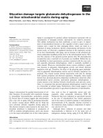
make a DVT clinically important. In Fig. 1 we present the distri-
bution of responses from 1 to 5 for patient factors associated
with clinically important DVT. The absence of risk factors,
symptoms, or physical signs of DVT, DVT associated with cen-
tral venous catheterization, and DVT in the absence of proph-
ylaxis were all considered to be less important than the other
factors in determining the clinical relevance of an ICU-
acquired DVT.
In Table 2 we present the ultrasonographic features consid-
ered likely to make a DVT clinically important, ranked accord-
ing to the mean score. Proximal location of the DVT was rated
as most likely to make a DVT clinically important (score 4.7
[SD 0.5]). Of the other ultrasound features, large (score 4.2
[0.6]) and totally occlusive (score 3.9 [0.8]) thrombi were con-
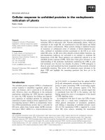
sidered most important. In Fig. 2 we present the distribution of
responses from 1 to 5 for ultrasonographic features associ-
ated with clinically important DVT. A DVT that is small, partially
occlusive, or has flow around it did not make the DVT unlikely
to be considered clinically important.
Discussion
DVT is underdiagnosed and probably undertreated in critically
ill patients. This occurs despite the high likelihood that PE is
clinically more important in the critically ill, who often have
impaired cardiopulmonary reserve [13]. We conducted the
present survey to determine which characteristics of DVT are
considered to be clinically important by intensivists. Determin-
ing which characteristics impart clinical significance is impor-
tant because the thrombi that are considered to be clinically
important are likely to be treated, and these treatments have
adverse effects such as bleeding, inconvenience, and cost
[23].
Outside the ICU setting, many clinicians consider DVT clini-
cally significant primarily when patients have referable symp-
toms and evidence of occlusive thrombosis on
ultrasonography. However, critically ill patients are generally
unable to report symptoms and uncommonly have unilateral
leg edema because they are recumbent. Therefore, 'typical'
clinical symptoms and signs cannot be used to establish the
clinical significance of a DVT in the ICU. Our survey confirmed
that intensivists use some different criteria to assign clinical
significance than are used by nonintensivists, particularly
thrombosis experts [24-26]. For example, asymptomatic DVT
detected on screening ultrasonography in critically ill patients
with severely impaired cardiorespiratory reserve are accorded
substantial clinical importance in the ICU, whereas the signifi-
cance of these thrombi may be debated by clinicians working
outside the ICU setting.
Strengths of the survey include comprehensive lists of both
patient factors and ultrasonographic features that may influ-
ence clinicians' interpretation of the importance of a DVT in the
ICU setting. Ideally, empiric data would inform us about which
DVT characteristics are most likely to be associated with
short-term and long-term morbidity and mortality in critically ill
patients. In the absence of such data, we defined clinically
important DVT as a DVT that is likely, in the opinion of the
intensivists, to result in short-term or long-term morbidity or
Critical Care June 2004 Vol 8 No 3 Cook et al.
R148
mortality if left untreated, as opposed to a DVT that will proba-
bly not have important consequences. We used evidence from
three randomized trials to conduct this survey; these sug-
gested that a self-administered format yields more valid self-
reports than interviewer-administered questionnaires [20],
that closed-ended formats yield more complete and valid
demographic data about the respondents than do open-ended
formats [21], and that appending second questionnaires to
reminders maximizes response rates [22]. Our respondents
represented largely university affiliated ICUs from coast to
coast in Canada, and our response rate was 99%. We believe
that our results are generalizable to similar multidisciplinary
secondary and tertiary care settings in which the majority of
mechanically ventilated, critically ill patients are cared for.
However, in focusing on the short-term consequences of VTE,
there are three major limitations of the survey. The first is that
we did not evaluate thresholds for DVT treatment in the ICU
setting, which is a more complex phenomenon. Treatment
thresholds are also influenced by other factors such as the nat-
ural history of the ICU patient's underlying illness, the type of
therapeutic options available and suitable for each patient,
intensivists' knowledge about the realistic magnitude of thera-
peutic benefits and harms, patient-specific and dynamic bal-
ance between bleeding and thrombosis risks, and alternative
options such as surveillance monitoring. Evaluating treatment
Figure 1
Patient factors considered by respondents to make a deep venous thrombosis (DVT) clinically important in the intensive care unit settingPatient factors considered by respondents to make a deep venous
thrombosis (DVT) clinically important in the intensive care unit setting.
The scale used was as follows: 1 = much less likely to make the DVT
clinically important; 2 = somewhat less likely to make the DVT clinically
important; 3 = neither more nor less likely to make the DVT clinically
important; 4 = somewhat more likely to make the DVT clinically impor-
tant; and 5 = much more likely to make the DVT clinically important.
0
10
20
30
40
50
60
70
80
90
100
Therapeutic
Anticoagulation
No Therapeutic
Anticoagulation
Chronic/Acute
CP Comorbidity
Clinical
Suspicion of PE
Patient factors
0
10
20
30
40
50
60
70
80
90
100
Leg Symptoms Asymptomatic Detected by
PE
Not Detected
by PE
Risk Factor
0
10
20
30
40
50
60
70
80
90
100
No Risk
Factors
Associated
with CVC
Not Associated
with CVC
Receiving
Prophylaxis
No Prophylaxis
Much Less
Somewhat Less
Neither
Somewhat More
Much More
Table 1
Patient factors affecting the clinical importance of deep venous
thrombosis in intensive care unit patients
Patient factor Mean (SD)
rating
Clinical suspicion of pulmonary embolism 4.6 (0.7)
Chronic/acute cardiopulmonary comorbidity 4.5 (0.7)
Leg symptoms 4.2 (0.8)
Occurring while receiving therapeutic anticoagulation 4.0 (1.2)
Detected by physical examination 3.8 (0.7)
Occurring while receiving thromboprophylaxis 3.6 (0.8)
At least one risk factor for venous thromboembolism 3.6 (0.6)
Associated with central venous catheterization 3.3 (0.9)
Not receiving prophylaxis 3.2 (0.7)
Not associated with central venous catheterization 3.1 (0.5)
Not receiving therapeutic anticoagulation 3.1 (0.3)
No risk factors 3.0 (0.4)
Asymptomatic 2.8 (0.5)
Not detected by physical examination 2.8 (0.5)
According to mean scores we ranked the patient factors considered
by intensivists as likely to make a deep venous thrombosis clinically
important. SD, standard deviation
Available online />R149
thresholds is also interesting, although it is a more complex
issue and would require different survey questions (or an alter-
native study design altogether). Second, we did not elicit
views on the risks, costs, and consequences of investigating
and managing treatment complications such as bleeding.
Third, we did not consider the long-term consequences of
DVT such as postphlebitic syndrome, which may be associ-
ated with disabling symptoms and/or ulceration among
survivors.
Experts express varying views on the appropriate end-points
for studies of thromboprophylaxis [27-31]. Over the years, the
majority of thromboprophylaxis trials have used DVT detected
by sensitive screening methods such as venography as the pri-
mary efficacy outcome. Only one of two randomized trials in
the ICU has employed ultrasonographic followed by veno-
graphic screening for DVT [32], and venograms are rarely per-
formed in critical care practice today [15]. The clinical
importance of DVT identified by screening tests outside the
ICU is controversial. Two lines of evidence support the
hypothesis that DVT detected by screening tests is clinically
unimportant. The first line of evidence is the fact that even with
use of appropriate VTE prophylaxis, 15–30% of patients
undergoing major orthopedic surgery will be discharged from
the hospital with venographically detectable calf or more prox-
imal DVT; fewer (about 3% of such patients) ultimately return
with a symptomatic DVT [33].
The second line of evidence is that, even when clinicians per-
form screening ultrasonography at the time of hospital dis-
charge and then treat patients who have DVT with
Figure 2
Ultrasonographic factors considered by respondents to make a deep venous thrombosis (DVT) clinically important in the intensive care unit settingUltrasonographic factors considered by respondents to make a deep
venous thrombosis (DVT) clinically important in the intensive care unit
setting. The scale used was as follows: 1 = much less likely to make the
DVT clinically important; 2 = somewhat less likely to make the DVT clin-
ically important; 3 = neither more nor less likely to make the DVT clini-
cally important; 4 = somewhat more likely to make the DVT clinically
important; and 5 = much more likely to make the DVT clinically
important.
0
10
20
30
40
50
60
70
80
90
100
Totally Occlusive
DVT
Partially
Occlusive DVT
Still Present 1
Week Later
Not Present 1
Week Later
Ultrasonographic Features
0
10
20
30
40
50
60
70
80
90
100
No Flow Flow No Extension to Deep
Syst
em
Much less
Somewhat less
Neither
Some more
Much More
0
10
20
30
40
50
60
70
80
90
100
Proximal DVT Distal DVT Large DVT Small DVT
Table 2
Ultrasonographic features affecting the clinical importance of
deep venous thrombosis in intensive care unit patients
Ultrasonographic feature Mean (SD) Rating
Proximal DVT 4.7 (0.5)
Large DVT 4.2 (0.6)
Totally occlusive DVT 3.9 (0.8)
No flow on color Doppler ultrasound 3.6 (0.7)
DVT still present 1 week after diagnosis 3.6 (0.7)
Partially occlusive DVT 3.3 (0.7)
Flow on color Doppler ultrasound 3.0 (0.6)
DVT not extending into deep system 2.7 (1.0)
Localized DVT 2.7 (0.7)
Small DVT 2.6 (0.6)
DVT not present 1 week after diagnosis 2.3 (0.8)
Distal DVT 2.0 (0.7)
According to mean scores we ranked the ultrasonographic features
considered by intensivists as likely to make a deep venous
thrombosis (DVT) clinically important. SD, standard deviation
Critical Care June 2004 Vol 8 No 3 Cook et al.
R150
antithrombotic therapy, there is no evidence that the subse-
quent risk for thrombosis is decreased. For example, Robinson
and colleagues [34] randomized patients after orthopedic sur-
gery to undergo screening ultrasonography or sham ultra-
sonography. After hospital discharge the risk for patients
returning with clinical symptoms of venous thrombosis was the
same, irrespective of whether patients underwent ultrasonog-
raphy, suggesting that thrombi identified by screening ultra-
sonography in hospitalized patients are not necessarily
destined to produce clinical symptoms. As a result of these
observations, routine screening of such patients for DVT at the
time of hospital discharge has effectively been abandoned [2].
Caution is needed, however, in extrapolating this practice pat-
tern to critically ill patients. For example, patients in the ICU fre-
quently have significantly impaired cardiorespiratory reserve.
Therefore, they may be unable to tolerate a small PE that an
otherwise healthy outpatient might tolerate easily. Better qual-
ity evidence on the clinical impact of DVT diagnosed by
screening ultrasound in the critically ill is needed. Meanwhile,
most such patients with lower limb DVT diagnosed by screen-
ing ultrasonography appear to be treated, based on recent
studies [5]. Interpreting studies about VTE in the ICU requires
consideration of the clinical importance of the DVT end-points.
In future randomized trials testing the efficacy of thrombo-
prophylaxis interventions, we recommend that validated and
feasible screening tests, such as Doppler ultrasonography or
serial Doppler ultrasonography, be used [31]. Large, simple
randomized trials that use clinically important VTE end-points
should follow, consisting of events such as symptomatic or
fatal PE, and ultrasonographically diagnosed proximal DVT
[28-31].
The results of this survey can inform our research program on
VTE in the ICU. We conclude that most forms of VTE are con-
sidered to be clinically serious, perhaps more so than in
patients outside the critical care setting, supporting the need
for ongoing research in this area. From this survey we gener-
ated new information showing how the critical care context is
important to intensivists caring for patients with DVT. Develop-
ment of DVT in the critically ill patient may have unique conse-
quences, particularly in patients requiring mechanical
ventilation, vasopressors, or inotropes, because intensivists
report that cardiopulmonary reserve is crucial in interpreting
the features of a DVT that make it clinically important. Our
results also suggest that treatment thresholds used by inten-
sivists may be lower than the treatment thresholds employed
by other clinicians, although this hypothesis can be formally
tested. On the other hand, the treatment threshold may be
higher for intensivists who take into account other complica-
tions of critical illness. For example, dynamic bleeding and
thrombotic risks in ICU patients must be traded off when con-
sidering whether to treat a DVT, and with what to treat it. Such
factors will influence the associated morbidity and mortality of
VTE in the ICU setting, which need to be more rigorously
evaluated.
Conclusion
We demonstrated that intensivists define clinically important
DVT using criteria that are relevant to critical care patients in
addition to those suitable for general DVT patients. Pending
the availability of data that correlate clinical outcomes with the
criteria we studied in this survey, these concepts could be
used in future studies of the prevention and treatment of DVT
in the critically ill.
Competing interests
MM received grants for investigator initiated studies from
Pharmica, Aventis and Leo Pharmaceuticals (latter for this sur-
vey). JG as a co-investigator, indirectly received funding from
Pharmacia and Leo Pharmaceuticals for the DVT study. GG
received grants for investigator initiated studies from
Pharmica, Aventis and Leo Pharmaceuticals (latter for this sur-
vey). MC received honoraria and research grants from Pfizer,
Pharmica, Aventis, Leo Pharmaceuticals and Novo Nordisk.
LG received a grant from Leo Pharmaceuticals for this survey.
DC has received a grant for this survey from Leo Pharmaceu-
ticals, and additional grants for investigator initiated research
from Pharmacia and Aventis.
Key messages
1. Determining which DVT characteristics impart clinical
significance to intensivists is important because DVT
considered to be clinically important are likely to be
treated, and treatments have adverse effects such as
bleeding, inconvenience and cost.
2. In this survey of intensivists, we defined a clinically
important DVT as one that, if untreated, would will lead
to increased short term morbidity, long term morbidity,
or mortality.
3. The 3 patient factors reported as most likely to make a
DVT clinically important were clinical suspicion of PE,
acute or chronic cardiopulmonary morbidity which
might limit the tolerability of a PE, and leg symptoms.
4. The 3 ultrasound features reported as most likely to
make a DVT clinically important were proximal location,
large size, and total vessel occlusion.
5. The clinical importance of DVT according to intensivists
is influenced by unique ICU factors such as cardiopul-
monary reserve among mechanically ventilated
patients.
Available online />R151
Additional material
Acknowledgements
We appreciate the data management assistance of Nicole Zytaruk. We
are grateful to the Canadian Critical Care Trials Group for their support
of the present study.
This study was funded by an unrestricted grant in aid from Leo Pharma-
ceuticals. Dr D Cook is a Research Chair of the Canadian Institutes for
Health Research. Drs Crowther and Meade are Investigators of the
Canadian Institutes for Health Research.
The authors’ contributions were as follows: conception: D Cook, G Guy-
att, and M Meade; design: D Cook, M Crowther, M Meade, G Guyatt, W
Geerts, and L Griffith; data collection: D Cook; analysis: L Griffith; inter-
pretation: D Cook, M Crowther, M Meade, G Guyatt, J Granton, and W
Geerts; drafting of manuscript: D Cook, M Crowther, and L Griffith; and
critical revision of manuscript: M Meade, G Guyatt, J Granton, and W
Geerts.
References
1. Attia J, Cook DJ, Douketis J, Ginsberg JS, Geerts W: Deep vein
thrombosis and its prevention in critically ill patients. Arch
Intern Med 2001, 161:1268-1279.
2. Geerts WH, Heit JA, Clagett P, Pineo GF, Colwell CW, Anderson
FA, Wheeler HB: Prevention of venous thromboembolism.
Sixth ACCP Antithrombotic Consensus Conference on Anti-
thrombotic Therapy. Chest 2001, 119(1 suppl):132S-175S.
3. Geerts W, Cook DJ, Selby R, Etchells E: Venous thromboembo-
lism and its prevention in critical care. J Crit Care 2002,
17:95-104.
4. Ibrahim EH, Iregui M, Prentice D, Sherman G, Kollef M, Shannon
W: Deep vein thrombosis during prolonged mechanical venti-
lation despite prophylaxis. Crit Care Med 2002, 30:771-774.
5. Cook DJ, Crowther M, Meade M, Rabbat C, Schiff D, Geerts W,
Griffith L, Guyatt GH: Deep venous thrombosis in medical-sur-
gical ICU patients: prevalence, incidence and risk factors. Crit
Care 2003, Suppl 2:S54.
6. Kakkar VV, Howe CT, Flanc C, Clarke MB: Natural history of
postoperative deep vein thrombosis. Lancet 1969, 2:230-232.
7. Barritt DW, Jordan SC: Anticoagulant drugs in the treatment of
pulmonary embolism: A controlled trial. Lancet 1960,
i:1309-1312.
8. Stein PD, Henry JW: Prevalence of acute pulmonary embolism
among patients in a general hospital and at autopsy. Chest
1995, 108:978-981.
9. Karwinski B, Svendsen E: Comparison of clinical and postmor-
tem diagnosis of pulmonary embolism. J Clin Pathol 1989,
42:135-139.
10. Moser KM, Fedullo PF, LittleJohn JK, Crawford R: Frequent
asymptomatic pulmonary embolism in patients with deep
venous thrombosis. JAMA 1994, 271:223-225.
11. McKelvie PA: Autopsy evidence of pulmonary
thromboembolism. Med J Aust 1994, 160:127-128.
12. Hirsch DR, Ingenito EP, Goldhaber SZ: Prevalence of deep
venous thrombosis among patients in medical intensive care.
JAMA 1995, 274:335-337.
13. Douketis JD, Kearon C, Bates S, Duku EK, Ginsberg JS: Risk of
fatal pulmonary embolism in patients with treated venous
thromboembolism. JAMA 1998, 279:458-462.
14. Warwick D, Samama MM: The contrast between venographic
and clinical endpoints in trials of thromboprophylaxis in hip
replacement. J Bone Joint Surg 2000, 82:480-482.
15. Cook D, McMullin J, Hodder R, Heule M, Pinilla J, Dodek P, Stewart
T, for the Canadian ICU Directors Group: Prevention and diagno-
sis of venous thromboembolism in critically ill patients: a
Canadian survey. Crit Care 2001, 5:336-342.
16. Clarke FJ, Griffith L, McDonald E, Hoad N, Rabbat C, Meade M,
Guyatt GH, Devereaux PJ, Geerts W, Wells P, Crowther M, Cook
DJ, for the Canadian Critical Care Trials Group: Deep vein throm-
bosis: Clinically silent in the ICU. Am J Resp Crit Care Med
2004 in press.
17. Cook DJ, Todd TRJ: The Canadian Critical Care Trials Group: a
collaborative educational organization for the advancement of
adult clinical ICU research. Intensive Care World 1997,
14:68-70.
18. Cook D, Attia J, Weaver B, McDonald E, Meade M, Crowther M:
Venous thromboembolic disease: An observational study in
medical-surgical intensive care unit patients. J Crit Care 2000,
15:127-132.
19. Cook DJ, Laporta D, Skrobik Y, Peters S, Sharpe M, Murphy P,
Chin D, Crowther V, for the Canadian ICU Directors Group: Pre-
vention of venous thromboembolism in critically ill surgery
patients. J Crit Care 2001, 16(4):161-166.
20. Cook DJ, Guyatt GH, Juniper E, Griffith LE, McIlroy W, Willan A,
Jaeschke R, Epstein R: Interviewer versus self-administered
questionnaires in developing a disease-specific, health
related quality of life instrument for asthma. J Clin Epidemiol
1993, 46(6):529-534.
21. Griffith LE, Cook DJ, Guyatt GH, Charles C: Comparison of open
versus closed questionnaire formats in obtaining demo-
graphic information from Canadian general internists. J Clin
Epidemiol 1999, 52:997-1005.
22. Asch DA, Jedrziewski MK, Christakis NA: Response rates to mail
surveys published in medical journals. J Clin Epidemiol 1997,
50:1129-1136.
23. Linkins LA, Choi P, Douketis J: Clinical impact of bleeding in
patients taking oral anticoagulant therapy for venous throm-
boembolism: a meta-analysis. Ann Intern Med 2003,
139:893-900.
24. Wells PS, Anderson DR, Bormanis J, Guy F, Mitchell M, Gray L,
Clement C, Robinson KS, Lewandowski B: Application of a diag-
nostic clinical model for the management of hospitalized
patients with suspected deep-vein thrombosis. Thromb
Haemost 1999, 81:493-497.
25. Wells PS, Anderson DR, Bormanis J, Guy F, Mitchell M, Gray L,
Clement C, Robinson KS, Lewandowski B: Value of assessment
of pretest probability of deep-vein thrombosis in clinical
management. Lancet 1997, 350:1795-1798.
26. Wells PS, Hirsh J, Anderson DR, Lensing AW, Foster G, Kearon
C, Weitz J, D'Ovidio R, Cogo A, Prandoni P: Accuracy of clinical
assessment of deep-vein thrombosis. Lancet 1995,
345:1326-1330.
27. Kearon C, Salzman EW, Hirsh J: Epidemiology, pathogenesis,
and natural history of venous thrombosis. In Hemostasis and
Thrombosis: Basic Principles and Clinical Practice 4th edition.
Edited by: Colman RW, Hirsh J, Marder VJ, Clowes AW, George
JN. Philadelphia, PA: JB Lippincott; 2001:1153-1177.
28. Kearon C: Noninvasive diagnosis of deep venous thrombosis
in postoperative patients. Semin Thromb Hemost 2001, 27:3-8.
29. Sonaglia F, Rossi R, Agnelli G: End points in studies on the pre-
vention of deep vein thrombosis. Semin Thromb Hemost 2001,
27:41-46.
30. Leizorovicz A, Kassai B, Becker F, Cucherat M: The assessment
of deep vein thromboses for therapeutic trials. Angiology
2003, 54:19-24.
31. Geerts WH, Pineo GF, Heit JA, Bergqvist D, Lassen MR, Colwell
CW: Prevention of venous thromboembolism. Chest 2004 in
press.
32. Fraisse F, Holzapfel L, Couland JM, Simonneau G, Bedock B, Feis-
sel H, Herbecq P, Pordes R, Poussel JF, Roux L, for the Associa-
tion of Non-University Affiliated Intensive Care Specialist
Physicians in France: Nadroparin in the prevention of deep vein
Appendix 1
Appendix 1
Click here for file
[ />s1.doc]
Critical Care June 2004 Vol 8 No 3 Cook et al.
R152
thrombosis in acute decompensated COPD. Am Rev Resp Crit
Care Med 2000, 161:1109-1114.
33. Eikelboom JW, Quinlan DJ, Douketis JD: Extended-duration
prophylaxis against venous thromboembolism after total hip
or knee replacement: a meta-analysis of the randomised trials.
Lancet 2001, 358:9-15.
34. Robinson SK, Anderson DR, Gross M, Petrie D, Leighton R, Stan-
ish W, Alexander D, Mitchell M, Flemming B, Gent M: Ultrasono-
graphic screening before hospital discharge for deep venous
thrombosis after arthroplasty. Ann Intern Med 1997,
127:439-445.