Báo cáo khoa học: " Bench-to-bedside review: Sepsis is a disease of the microcirculation" pps
Bạn đang xem bản rút gọn của tài liệu. Xem và tải ngay bản đầy đủ của tài liệu tại đây (482.14 KB, 7 trang )
462
DO
2
= oxygen delivery; IVM = intravital microscopy; MODS = multiple organ dysfunction syndrome; NO = nitric oxide; NOS = NO synthase; OPS =
orthogonal polarization spectral; pCO
2
= partial pressure of CO
2
; SIRS = systemic inflammatory response syndrome; SvO
2
= mixed venous oxygen
saturation; VO
2
= oxygen consumption.
Critical Care December 2004 Vol 8 No 6 Spronk et al.
Introduction
The initial treatment of trauma and critically ill patients is
aimed at securing the airway and establishing adequate
breathing, followed by the correction of circulatory
abnormalities (‘ABC’) [1]. These basic principles underline
the fact that optimization of oxygen delivery to the tissues is
one of the cornerstones of critical care medicine, thus
preventing cellular dysfunction and cellular death, and
subsequent organ dysfunction. Disturbance of the delicate
balance between oxygen delivery (DO
2
) and oxygen
consumption (VO
2
) to the tissues can be defined as a state
of shock. Impairment of DO
2
can be caused by severe
anemia, hypoxia, or a low cardiac output. To preserve tissue
DO
2
in several states of shock, especially to the heart and
brain, many compensating physiological reserve mechanisms
come into play. This leads to microvascular derecruitment in
compliant vascular beds such as the skin and the splanchnic
area, redirecting blood flow to more crucial body areas.
During this process, systemic hemodynamics can be
maintained at the expense of impaired microcirculatory
perfusion. Nevertheless, if this microcirculatory state of
hypoperfusion is not reversed in a timely manner, multiple
organ failure can develop, with a high probability of death.
This line of thought can be found in a recent general guideline
for the treatment of patients with septic shock, in which
infusion of volume is judged to be critical to basic care in
these patients [2].
Systemic inflammatory response syndrome (SIRS) is seen
after trauma, major surgery or hemorrhage. A similar
Review
Bench-to-bedside review: Sepsis is a disease of the
microcirculation
Peter E Spronk
1,2
, Durk F Zandstra
3
and Can Ince
2
1
Department of Intensive Care Medicine, Gelre ziekenhuizen, Apeldoorn, The Netherlands
2
Department of Physiology, Academic Medical Center, University of Amsterdam, The Netherlands
3
Department of Intensive Care Medicine, Onze Lieve Vrouwe Gasthuis, Amsterdam, The Netherlands
Corresponding author: Peter E Spronk,
Published online: 16 June 2004 Critical Care 2004, 8:462-468 (DOI 10.1186/cc2894)
This article is online at />© 2004 BioMed Central Ltd
See Commentary, page 419
Abstract
Microcirculatory perfusion is disturbed in sepsis. Recent research has shown that maintaining systemic
blood pressure is associated with inadequate perfusion of the microcirculation in sepsis.
Microcirculatory perfusion is regulated by an intricate interplay of many neuroendocrine and paracrine
pathways, which makes blood flow though this microvascular network a heterogeneous process.
Owing to an increased microcirculatory resistance, a maldistribution of blood flow occurs with a
decreased systemic vascular resistance due to shunting phenomena. Therapy in shock is aimed at the
optimization of cardiac function, arterial hemoglobin saturation and tissue perfusion. This will mean the
correction of hypovolemia and the restoration of an evenly distributed microcirculatory flow and
adequate oxygen transport. A practical clinical score for the definition of shock is proposed and a novel
technique for bedside visualization of the capillary network is discussed, including its possible
implications for the treatment of septic shock patients with vasodilators to open the microcirculation.
Keywords shock, microcirculation, orthogonal polarization spectral imaging
463
Available online />phenomenon is seen in sepsis as a response to infection, and
is still an important cause of death in critically ill patients.
Both can progress to severe shock and multiple organ
dysfunction syndrome (MODS) [3]. This progression is
currently thought to be due to an increased VO
2
, a decreased
peripheral vascular resistence and a maldistribution of tissue
blood flow to preserve central blood volume. As a result,
microcirculatory perfusion is shut down and is the final
common pathway in shock. Especially in septic shock,
alterations in metabolic pathways called ‘cytopathic hypoxia’
can lead to additional tissue damage [4]. This review
discusses briefly the importance of microcirculatory flow in
the pathogenesis of sepsis and the progression to MODS.
Heterogeneous microcirculatory perfusion
The measurement of global hemodynamics reflects only a tiny
part of whole-body circulatory blood flow. The micro-
circulation, with its huge endothelial surface, is in fact the
largest ‘organ’ in the human body. We have come a long way
since the disclosure of human bodily circulation by Harvey [5]
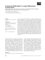
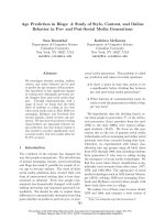
and Malpighi [6]. The number of publications concerning the
microcirculation in humans is steadily increasing (Fig. 1).
However, the microcirculation remains difficult to investigate.
In clinical practice, microcirculatory perfusion is judged on
aspects such as the color, capillary refill and temperature of
the distal parts of the body (i.e. fingers, toes, earlobes and
nose).
Perfusion of the microcirculation is regulated by an intricate
interplay of many neuroendocrine, paracrine, and mechano-
sensory pathways [7]. These mechanisms adapt to the
balance between locoregional tissue oxygen transport and
metabolic needs to ensure that supply matches demand. In
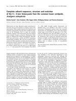
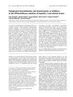
sepsis, this process is severely compromised because of
decreased deformability of red blood cells with inherent
increased viscosity [8], an increased percentage of activated
neutrophils with decreased deformability and increased
aggregability due to the upregulation of adhesion molecules
[9], activation of the clotting cascade with fibrin deposition
and the formation of microthrombi [10], dysfunction of
vascular autoregulatory mechanisms [11], and finally, the
secondary enhanced perfusion of large arteriovenous shunts
[12] (Fig. 2). These processes result in tissue dysoxia, either
from impaired microcirculatory oxygen delivery and/or from
mitochondrial dysfunction [4,13]. Clinically this process is
perceived as an oxygen extraction defect, a prominent feature
of sepsis. A possible mechanism accounting for this
phenomenon could be the shut-down of vulnerable micro-
circulatory units in the organ beds, promoting the shunting of
oxygen transport from the arterial to the venous compartment
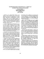
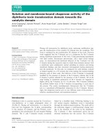
leaving the microcirculation hypoxic [14]. This might be an
explanation for the different findings regarding locoregional
tissue perfusion in shock (Fig. 3). In this so-called shunting
theory of sepsis, correction of this condition should occur by
recruitment of the shunted microcirculatory units. Applying
strategies to ‘open the microcirculation’ by vasodilation would
be expected to promote microcirculatory flow by increasing
the driving pressure at the entrance of the microcirculation
and/or decreasing the capillary afterload [15].
Indeed, in animal studies, these effects occur during
hemorrhage and sepsis caused by microcirculatory shunting
with associated tissue dysoxia [16–18]. Such micro-
circulatory shunting was reversed by vasodilation [14] and by
improvement in regional flow in an animal sepsis model [19].
In addition, oxygen extraction was improved [20] and
microcirculatory shunting was reversed [21] by the use of
nitric donors. To redirect microvascular flow, matters become
more complicated if one realizes that sepsis causes
heterogeneous effects in constriction and dilation in different
organs and at different levels of the microcirculation [22].
Although cardiac output is frequently increased in sepsis,
high lactate levels and increased tonometric partial pressure
of CO
2
(pCO
2
) in tissues indicate at least regional tissue
dysoxia. This has been termed oxygen extraction deficit in
Figure 1
Number of publications on regarding microcirculation in humans
(source: Medline; search term ‘microcirculation’ limited to human data).
Figure 2
A multitude of factors potentially imparing microcirculatory perfusion in
sepsis.
464
Critical Care December 2004 Vol 8 No 6 Spronk et al.
sepsis and has been well documented in different animal
models of shock [23–25]. It is still a matter of debate whether
it can be explained by pathologic flow heterogeneity due to
dysfunctional autoregulatory mechanisms and micro-
circulatory dysfunction causing hypoxic pockets, or by
mitochondrial dysfunction with associated impaired oxidative
phosphorylation [4], or by a combination of both.
How is critical microcirculatory dysfunction
assessed?
Especially in critical illness, function and dysfunction of the
microcirculatory network are of utmost importance in the
cause of disease and the development of organ failure. In
sepsis, all three elements of the microvascular network are
compromised, namely arteriolar hyporesponsiveness to vaso-
contrictors and vasodilators, a reduced number of perfused
capillaries, and venular obstruction by the sequestration of
activated neutrophils [22]. However, an objective and reliable
method of monitoring microcirculatory organ perfusion is still
not available. ‘Downstream’ global derivatives of micro-
circulatory dysfunction such as lactate, tonometry, and mixed
venous oxygen saturation (SvO
2
), in addition to measure-
ments of DO
2
and oxygen uptake VO
2
, are used in daily
intensive care clinical practice. But which parameters should
be used to prevent further deterioration of organ function in a
critically ill patient with septic shock? In this section we
discuss the reasons for, and limitations of, several parameters
that have been used to assess microcirculatory perfusion.
Lactate levels are thought to reflect anaerobic metabolism
associated with tissue dysoxia and might predict a response
to therapy and prognosis [26]. The balance between lactate
production due to global (shock, hypoxia), local (tissue
ischemia), and cellular (mitochondrial dysfunction) factors on
the one hand, and lactate clearance depending on metabolic
liver function on the other hand, make the interpretation of
lactate levels uncertain and difficult [27]. SvO
2
can be
measured with a pulmonary artery catheter and is thought to
reflect the average oxygen saturation of all perfused
microvascular beds. In sepsis, microcirculatory shunting can
cause normal SvO
2
while severe local tissue dysoxia is
present [14]. Delayed therapy aimed at the normalization of
SvO
2
failed to demonstrate a survival benefit [28,29].
Optimization of oxygen delivery might have been instituted
too late in these studies, when irreversible cellular damage
was already present. In addition, the frequent use of
dobutamine to obtain preset goals of oxygen delivery might
have affected the outcome, because dobutamine has been
implicated in the impairment of hepatosplanchnic perfusion in
sepsis [30]. Nevertheless, besides ongoing discussions
about the use of a pulmonary artery catheter in sepsis, the
sole use of SvO
2
seems an inadequate parameter as a
guideline for therapy in the restoration of local tissue
oxygenation in septic shock patients. However, if an
integrative approach is used in the early stage of treating
critically ill patients, states of hypoperfusion are recognized
earlier [31] and, if early treatment is started, can even improve
survival [32]. It is likely that the results of the Rivers study
[32] are due largely to the prevention of irreversible cellular
damage, in contrast to the earlier findings by Hayes and
Gattinoni, who targeted high oxygen delivery levels during
later phases of sepsis [28,29].
An appealing alternative to the evaluation of tissue dysoxia
might be regional intestinal capnography as introduced by
Fiddian-Green and Baker [33]. This method relies on the
principle of CO
2
diffusion from the local anaerobic production
site across tissue and cell membranes. Measurement of the
difference between intestinal pCO
2
and arterial pCO
2
has
been found to be better than that of pH
i
alone, because
arterial pCO
2
fluctuates in ventilated patients [34]. In sepsis,
the interpretation of tonometric results is affected by
microcirculatory shunting. This complicates the clear
establishment of impaired perfusion, because areas with
reduced perfusion and CO
2
offloading are next to hypoxic
regions [35]. Recently, gastric intramucosal pCO
2
values
were found to be well correlated with sublingual pCO
2
values
[36]. The baseline difference between sublingual pCO
2
and
arterial pCO
2
values was a better predictor of survival than
the change in lactate or SvO
2
[37]. Further studies should
demonstrate whether this parameter can be used in clinical
management of patients with septic shock.
All parameters discussed are indirect and downstream from
the pathological process in the microcirculatory network.
Direct assessment of microcirculatory perfusion seems a
superior and more direct approach and has been extensively
studied in vivo by intravital microscopy (IVM) in animals. In
humans, IVM studies are restricted to the eye, the skin and
the nail fold owing to the size of the IVM equipment and the
Figure 3
The shunting theory of sepsis accounts for the condition in which
apparently adequate oxygen delivery is not successful in delivering
oxygen to microcirculatory weak units that are shunted. This leads to
an oxygen extraction deficit of these shunted units with raised levels of
venous partial pressure of CO
2
, lactate and gastric CO
2
, whereas
input oxygen delivery seems adequate. Vasodilation would be
expected to recruit these shunted units by increasing the driving
pressure to the microcirculation and possibly to these shunted units.
465
use of fluorescent dyes for contrast enhancement. IVM
depends on trans- or epi-illumination and thus observations
are limited to superficial layers of thin tissues only. By using
fluorescent dyes a higher contrast is possible as well as
labelling specific cells for visualization and quantification.
Because of the potentially toxic effects of these dyes in
humans, studies are mostly limited to animals [38,39]. We
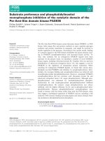
recently introduced [40,41], validated [42], and clinically
applied [43] a new method for observing the microcirculation
in patients, called orthogonal polarization spectral (OPS)
imaging (CYTOSCAN™; Cytometrics Inc., Philadelphia, PA),
which creates high-contrast images without the use of
fluorescent dyes. This technique is based on the reflection of
light from the tissues. Contrast is obtained from the
absorption of linearly polarized light by the haemoglobin in the
blood. As a consequence, red blood cells in the micro-
circulation appear black on the white background of the
surrounding tissue. For OPS imaging a 5 × objective (on-
screen magnification of × 326) is used during measurements.
Data are recorded on a digital video recorder for later analysis
and displayed on a black and white monitor. Because the
OPS machine is a small hand-held device (Fig. 4), it can be
used at the bedside for humans in the visualization of unique
in vivo images of the microcirculation [44]. Although nailfold
microcirculatory blood flow as established by OPS imaging
correlates very well with IVM microvascular flow when
analysed by specific video-analysis-software [42], this
quantitative approach proved not to be usable with sublingual
images owing to movement artefacts induced by tongue
movements or respiration. A semi-quantitative approach was
therefore used successfully to analyse changes in
microcirculatory flow [45,46].
Despite these shortcomings in the assessment of local tissue
oxygenation, several studies have been performed aiming at
recruitment of the tissue microcirculatory flow.
Microcirculatory perfusion as an endpoint
Data from several studies support the idea that the
impairment of microcirculatory perfusion results in organ
failure and increases the risk of death [17,18,22,45,47–50].
In this line of thought, restoring perfusion in disturbed
microcirculatory networks might improve outcome. Indeed,
survival was related to microcirculatory shut-down in rats that
were bled and in which the blood volume was subsequently
resuscitated, although whole-body hemodynamic parameters
were comparable in survivors and non-survivors [51].
Comparable findings have been reported in humans with
septic shock. Bihari found that vasodilation might unmask a
preexisting tissue oxygen debt. After increasing DO
2
with the
vasodilator prostacyclin, all patients survived when the
increase in DO
2
did not coincide with an increase in VO
2
,
whereas all patients died who showed increasing VO
2
[52].
By recruitment of the microcirculation, oxygen might have
become available to previously hypoxic tissues that had shut
down. De Backer and colleagues [45] reported that
sublingual microcirculatory perfusion was compromised to a
greater extent in non-surviving than in surviving septic shock
patients. We observed normal sublingual microcirculatory
perfusion in a septic patient with hepatic failure who received
high doses of norepinephrine (P Spronk, unpublished
observation). Dubois recently reported a comparable
observation in a septic patient treated with vasopressin [53],
whereas others observed sublingual microcirculatory shut-
down with the use of vasopressin (C Boerma, personal
Available online />Figure 4
Orthogonal polarization spectral imaging technique (a) built into a simple hand-held device (b).
466
communication). Larger studies should demonstrate why
these patients behave differently from those in previous
reports. Nevertheless, De Backer and colleagues showed
that microcirculatory perfusion improved over time in
survivors, whereas the disturbance of perfusion in the
microvessels of the non-survivors remained. In addition, they
showed that sublingual microcirculatory perfusion
abnormalities could be corrected by the topical application of
acetylcholine, showing that the local endothelium was still
responsive to nitric oxide (NO), whereas vasoplegia due to
ongoing sepsis might be expected.
NO has been implicated as the major cause for hypotension,
generated from endothelial cells through the expression of
inducible NO synthase (NOS) [54], thus contributing to many
of the manifestations of septic shock such as vasoplegia,
diminished myocardial contractility, hepatic damage, and
vascular and intestinal hyperpermeability. Others, however,
found decreased NO production during sepsis [55], and,
more recently, that NOS activity is diminished in mononuclear
cells from sepsis patients [56]. On the basis of the
hypothesis that NO production is increased in sepsis,
experiments in septic animal models were performed and
indicated that hypotension could be prevented by inhibiting
NOS. This led to clinical studies with several compounds
capable of inhibiting NO synthesis. Early promising data
showed increasing blood pressures and decreasing doses of
vasopressors in septic shock patients treated with NOS
inhibitors [57]. However, a subsequent randomized
controlled multicenter phase III trial was stopped when
interim analysis showed increased mortality in the N
G
-
monomethyl-
L-arginine group compared with placebo [58].
Inhibition of NOS activity seems to result in an improvement
in the general hemodynamic situation, but at the cost of
increased mortality [59]. Apparently, completely inhibiting
vasodilation is not the proper answer to sepsis. A more
specific approach by inhibiting only the inducible form of
NOS might be an attractive alternative. Indeed, after the
application of 1400W (a synthetic blocker of inducible NOS)
in a pig endotoxemia model, microvascular perfusion was
restored by a redistribution within the gut wall and/or an
amelioration of the cellular respiration [60].
NO is an important vasodilator in the microcirculation during
sepsis [61]. Indeed, Ince and colleagues showed recently
that NO donors were highly effective in correcting micro-
circulatory oxygenation after endotoxemia in a pig model of
sepsis, with both mucosal and serosal microvascular PO
2
as
well as intraluminal gastric pCO
2
being restored to baseline
values [21]. In addition, the glucose oxidation rate improves in
septic patients after treatment with prostacyclin [62].
Apparently, the microcirculation in sepsis fails to support
adequate tissue oxygenation. Optimizing DO
2
can result in
lower mortality rates, especially when therapy is started
without delay [63,64]. Others, however, showed comparable
mortality rates [29] or even a higher hospital mortality [65] in
septic shock patients whose treatment sought to increase
DO
2
. In these studies, oxygen supply to the tissues was
increased by manipulating macrohemodynamic endpoints
such as cardiac output, hemoglobin, and central venous
pressure and/or pulmonary artery wedge pressure.
Radermacher and colleagues [66] treated septic shock
patients with prostacyclin when no further increase in DO
2
could be obtained by volume resuscitation and dobutamine
infusion. Gastric pH
i
improved after starting prostacyclin,
suggesting an increase in splanchnic blood flow.
These findings led us to propose that the addition of systemic
NO to adequately volume resuscitated patients with septic
shock results in an improvement of microcirculatory
perfusion. In a small observational study in septic shock
patients, we were indeed able to show an improvement in
sublingual microcirculatory perfusion after the injection of
0.5 mg of nitroglycerin [46]. The observation of capillary
shutdown next to sustained flow in the larger vessels
corroborates the shunting theory of sepsis. Upon the
administration of nitroglycerin, microcirculatory flow increased
not only in large microvessels but also in small microvessels.
The latter finding argues against NO donation’s inducing even
more shunting flow. All patients except one, owing to late
cerebral hemorrhage, were discharged from the hospital alive.
This suggests that one can actively open up the
microcirculatory network and keep it open by volume and
vasodilator therapy. One might argue that oxygen consumption
increases with a concurrent increase in DO
2
under nitrate
administration [67]. However, concentrations of nitrate/nitrite
seem to be increased in septic shock patients anyway [68].
We administered 1 mg/kg dexamethasone intravenously to all
our patients at admission, which might well have attenuated the
production of NO by inhibiting excessive activation of inducible
NOS. With this background, a controlled opening strategy
using NO donors might be a rational approach. Further studies
should demonstrate whether this line of thought regarding
therapy in sepsis can be guided by microcirculatory flow
patterns and might result in a better outcome.
Future aspects
Therapy in shock should be aimed at the optimization of
cardiac function, arterial hemoglobin saturation, and tissue
perfusion. This will mean the correction of hypovolemia and
the restoration of an evenly distributed microcirculatory flow
and inadequate oxygen transport. How can the latter goals in
particular be accomplished? Discussions about the role of
vasodilators, particularly NO, in sepsis with microcirculatory
disturbance will continue. Will the optimization of sublingual
microcirculation become a novel resuscitation endpoint? Do
we need to take mitochondrial function and tissue respiration
into account [69]? Or should we use an integrative approach
incorporating both macrocirculatory and microcirculatory
hemodynamic data, as proposed in Table 1? Several tools
will become available for improving the assessment of
regional oxygen demands in critical illness. This will create
Critical Care December 2004 Vol 8 No 6 Spronk et al.
467
new challenges for the clinician to improve bedside critical
care and optimization of microcirculatory perfusion, thus
preventing the further deterioration of organ function and
keeping the old principle of primum non nocere alive.
Competing interests
The author(s) declare that they have no competing interests.
References
1. Carley S, Driscoll P: Trauma education. Resuscitation 2001, 48:
47-56.
2. Vincent JL: Hemodynamic support in septic shock. Intensive
Care Med 2001, 27 Suppl 1:S80-S92.
3. Bone RC: The pathogenesis of sepsis. Ann Intern Med 1991,
115:457-469.
4. Fink M: Cytopathic hypoxia in sepsis. Acta Anaesthesiol Scand
Suppl 1997, 110:87-95.
5. Harvey W: Exercitatio Anatomica de Motu Cordis et Sanguinis in
Animalibus. 1628.
6. Malpighi M: Opera Omnia. 1687.
7. Lehr HA, Bittinger F, Kirkpatrick CJ: Microcirculatory dysfunction
in sepsis: a pathogenetic basis for therapy? J Pathol 2000,
190:373-386.
8. Astiz ME, DeGent GE, Lin RY, Rackow EC: Microvascular func-
tion and rheologic changes in hyperdynamic sepsis. Crit Care
Med 1995, 23:265-271.
9. Linderkamp O, Ruef P, Brenner B, Gulbins E, Lang F: Passive
deformability of mature, immature, and active neutrophils in
healthy and septicemic neonates. Pediatr Res 1998, 44:946-
950.
10. Diaz NL, Finol HJ, Torres SH, Zambrano CI, Adjounian H: Histo-
chemical and ultrastructural study of skeletal muscle in
patients with sepsis and multiple organ failure syndrome
(MOFS). Histol Histopathol 1998, 13:121-128.
11. Avontuur JA, Bruining HA, Ince C: Nitric oxide causes dysfunc-
tion of coronary autoregulation in endotoxemic rats. Cardio-
vasc Res 1997, 35:368-376.
12. Cronenwett JL, Lindenauer SM: Direct measurement of arteri-
ovenous anastomotic blood flow in the septic canine
hindlimb. Surgery 1979, 85:275-282.
13. Ince C: Microcirculatory weak units – an alternative explana-
tion. Crit Care Med 2000, 28:3127-3129.
14. Ince C, Sinaasappel M: Microcirculatory oxygenation and shunt-
ing in sepsis and shock. Crit Care Med 1999, 27:1369-1377.
15. Buwalda M, Ince C: Opening the microcirculation: can vaso-
dilators be useful in sepsis? Intensive Care Med 2002, 28:
1208-1217.
16. Lam C, Tyml K, Martin C, Sibbald W: Microvascular perfusion is
impaired in a rat model of normotensive sepsis. J Clin Invest
1994, 94:2077-2083.
17. Sinaasappel M, van Iterson M, Ince C: Microvascular oxygen
pressure in the pig intestine during haemorrhagic shock and
resuscitation. J Physiol 1999, 514:245-253.
18. van Iterson M, Sinaasappel M, Burhop K, Trouwborst A, Ince C:
Low-volume resuscitation with a hemoglobin-based oxygen
carrier after hemorrhage improves gut microvascular oxy-
genation in swine. J Lab Clin Med 1998, 132:421-431.
19. Erdmann E: The effect of positive inotropes on the failing
human myocardium. Cardiology 1997, 88 Suppl 2: 7-11.
20. Zhang H, Rogiers P, Smail N, Cabral A, Preiser JC, Peny MO,
Vincent JL: Effects of nitric oxide on blood flow distribution
and O2 extraction capabilities during endotoxic shock. J Appl
Physiol 1997, 83:1164-1173.
21. Siegemund M, van Bommel J, Ince C: Influence of NO donor
SIN-1 on the gut oxygenation in a normodynamic, porcine
model of low-dose endotoxaemia. Intensive Care Med 2000,
26:S362.
22. Lush CW, Kvietys PR: Microvascular dysfunction in sepsis.
Microcirculation 2000, 7:83-101.
23. Cain SM, Curtis SE: Experimental models of pathologic
oxygen supply dependency. Crit Care Med 1991, 19:603-612.
24. Nelson DP, Samsel RW, Wood LDH, Schumacker PT: Experi-
mental models of pathologic oxygen supply dependency. J
Appl Physiol 1988, 64:2410-2419.
25. Vallet B, Lund N, Curtis SE, Kelly D, Cain SM: Gut and muscle
tissue pO
2
in endotoxemic dogs during shock and resuscita-
tion. J Appl Physiol 1994, 76:793-800.
26. Bakker J, Coffernils M, Leon M, Gris P, Vincent JL: Blood lactate
levels are superior to oxygen-derived variables in predicting
outcome in human septic shock. Chest 1991, 99:956-962.
27. De Backer D: Lactic acidosis. Intensive Care Med 2003, 29:699-
702.
28. Hayes MA, Timmins AC, Yau EH, Palazzo M, Hinds CJ, Watson D:
Elevation of systemic oxygen delivery in the treatment of criti-
cally ill patients. N Engl J Med 1994, 330:1717-1722.
29. Gattinoni L, Brazzi L, Pelosi P, Latini R, Tognoni G, Pesenti A,
Fumagalli R: A trial of goal-oriented hemodynamic therapy in
critically ill patients. SvO2 Collaborative Group. N Engl J Med
1995, 333:1025-1032.
30. Creteur J, De Backer D, Vincent JL: A dobutamine test can dis-
close hepatosplanchnic hypoperfusion in septic patients. Am
J Respir Crit Care Med 1999, 160:839-845.
31. Kaplan LJ, McPartland K, Santora TA, Trooskin SZ: Start with a sub-
jective assessment of skin temperature to identify hypoperfusion
in intensive care unit patients. J Trauma 2001, 50:620-627.
32. Rivers E, Nguyen B, Havstad S, Ressler J, Muzzin A, Knoblich B,
Peterson E, Tomlanovich M: Early goal-directed therapy in the
treatment of severe sepsis and septic shock. N Engl J Med
2001, 345:1368-1377.
33. Fiddian-Green RG, Baker S: Predictive value of the stomach
wall pH for complications after cardiac operations: compari-
son with other monitoring. Crit Care Med 1987, 15:153-156.
34. Lowes BD, Tsvetkova T, Eichhorn EJ, Gilbert EM, Bristow MR:
Milrinone versus dobutamine in heart failure subjects treated
chronically with carvedilol. Int J Cardiol 2001, 81:141-149.
35. Vallet B, Ince C: Noninvasive assessment of tissue oxygena-
tion. Semin Respir Crit Care Med 1999, 20:3-10.
36. Marik PE: Sublingual capnography: a clinical validation study.
Chest 2001, 120:923-927.
37. Marik PE, Bankov A: Sublingual capnometry versus traditional
markers of tissue oxygenation in critically ill patients. Crit
Available online />Table 1
Integrative clinical approach to define a state of shock
Item evaluated Points
Hemodynamic variables 2
Heart rate > 100 b.p.m. or
MAP < 50 mmHg and (CVP < 2 or CVP > 15 mmHg) or
CI < 2.2 l min
–1
m
–2
Peripheral circulation 2
Mottled skin or
T
c
–T
p
difference > 5°C or
Pfi < 0.3 or
Impaired peripheral capillary refill
Microvascular variables 1
Increased tonometric CO
2
gap or
Increased sublingual CO
2
gap or
Impaired sublingual microvascular perfusion (OPS imaging)
Systemic markers of tissue oxygenation 1
Lactate > 4 mmol l
–1
or
SvO
2
< 60%
Organ dysfunction
Diuresis < 0.5 ml kg
–1
h
–1 a
1
Decreased mental state
a
1
A state of shock is present if the score exceeds 2 points. CI, cardiac
index; CVP, central venous pressure; MAP, mean arterial pressure;
OPS, orthogonal polarization spectral imaging; Pfi, peripheral perfusion
index; SvO
2
, mixed venous oxygen saturation; T
c
, core temperature;
T
p
, peripheral toe temperature.
a
Due to present disease.
468
Critical Care December 2004 Vol 8 No 6 Spronk et al.
Care Med 2003, 31:818-822.
38. Saetzler RK, Jallo J, Lehr HA, Philips CM, Vasthare U, Arfors KE,
Tuma RF: Intravital fluorescence microscopy: impact of light-
induced phototoxicity on adhesion of fluorescently labeled
leukocytes. J Histochem Cytochem 1997, 45:505-513.
39. Steinbauer M, Harris AG, Abels C, Messmer K: Characterization
and prevention of phototoxic effects in intravital fluorescence
microscopy in the hamster dorsal skinfold model. Langen-
becks Arch Surg 2000, 385:290-298.
40. Groner W, Winkelman JW, Harris AG, Ince C, Bouma GJ,
Messmer K, Nadeau RG: Orthogonal polarization spectral
imaging: a new method for study of the microcirculation. Nat
Med 1999, 5:1209-1212.
41. Mathura KR, Alic L, Ince C: Initial clinical experience with OPS
imaging for observation of the human microcirculation. In
Yearbook of Intensive Care and Emergency Medicine. Edited by
Vincent JL. Berlin: Springer Verlag; 2001:233-245.
42. Mathura KR, Vollebregt KC, Boer K, De Graaff JC, Ubbink DT,
Ince C: Comparison of OPS imaging and conventional capil-
lary microscopy to study the human microcirculation. J Appl
Physiol 2001, 91:74-78.
43. Liu L, Zhao SP: The changes of circulating tumor necrosis
factor levels in patients with congestive heart failure influ-
enced by therapy. Int J Cardiol 1999, 69:77-82.
44. Robin ED: Of men and mitochondria: coping with hypoxic
dysoxia. The 1980 J Burns Amberson Lecture. Am Rev Respir
Dis 1980, 122:517-531.
45. De Backer D, Creteur J, Preiser JC, Dubois MJ, Vincent JL:
Microvascular blood flow is altered in patients with sepsis. Am
J Respir Crit Care Med 2002, 166:98-104.
46. Spronk PE, Ince C, Gardien MJ, Mathura KR, Oudemans-van
Straaten HM, Zandstra DF: Nitroglycerin in septic shock after
intravascular volume resuscitation. Lancet 2002, 360:1395-1396.
47. Baskurt OK, Temiz A, Meiselman HJ: Red blood cell aggregation
in experimental sepsis. J Lab Clin Med 1997, 130:183-190.
48. Siegemund M, Racovitza I, Ince C: The rationale for vasodilator
therapy in sepsis. In Yearbook of Intensive Care and Emergency
Medicine. Edited by Vincent JL. Berlin: Springer Verlag;
2002:221-231.
49. Piagnerelli M, Boudjeltia KZ, Vanhaeverbeek M, Vincent JL: Red
blood cell rheology in sepsis. Intensive Care Med 2003, 29:
1052-1061.
50. Bateman RM, Sharpe MD, Ellis CG: Bench-to-bedside review:
microvascular dysfunction in sepsis – hemodynamics, oxygen
transport, and nitric oxide. Crit Care 2003, 7:359-373.
51. Zhao KS, Junker D, Delano FA, Zweifach BW: Microvascular
adjustments during irreversible hemorrhagic shock in rat
skeletal muscle. Microvasc Res 1985, 30:143-153.
52. Bihari D, Smithies M, Gimson A, Tinker J: The effects of vasodi-
lation with prostacyclin on oxygen delivery and uptake in criti-
cally ill patients. N Engl J Med 1987, 317:397-403.
53. Dubois MJ, De Backer D, Creteur J, Anane S, Vincent JL: Effect
of vasopressin on sublingual microcirculation in a patient with
distributive shock. Intensive Care Med 2003, 29:1020-1023.
54. Vallance P: Exploring vascular nitric oxide in health and
disease. The Goulstonian Lecture 1996. J R Coll Physicians
Lond 1997, 31:321-327.
55. Wang P, Ba ZF, Chaudry IH: Endothelium-dependent relax-
ation is depressed at the macro- and microcirculatory levels
during sepsis. Am J Physiol 1995, 269:R988-R994.
56. Reade MC, Young D, Boyd CAR: Nitric oxide synthases are
decreased in mononuclear cells from sepsis patients. Crit
Care Med 2003, 31:A52.
57. Lopez A, Lorente JA, Steingrub J, Bakker J, McLuckie A, Willatts
S, Brockway M, Anzueto A, Holzapfel L, Breen D, Silverman MS,
Takala J, Donaldson J, Arneson C, Grove G, Grossman S, Grover
R: Multiple-center, randomized, placebo-controlled, double-
blind study of the nitric oxide synthase inhibitor 546C88:
effect on survival in patients with septic shock. Crit Care Med
2004, 32:21-30.
58. Grover R, Lopez A, Lorente JA, Grossman S: Multicenter, ran-
domized, placebo controlled, double-blind study of the nitric
oxide synthase inhibitor 546C88: effect on survival in patients
with septic shock. Crit Care Med 1999, 27 Suppl: A33.
59. Vincent JL, Zhang H, Szabo C, Preiser JC: Effects of nitric oxide
in septic shock. Am J Respir Crit Care Med 2000, 161:1781-
1785.
60. Pittner A, Nalos M, Asfar P, Yang Y, Ince C, Georgieff M, Bruck-
ner UB, Radermacher P, Froba G: Mechanisms of inducible
nitric oxide synthase (iNOS) inhibition-related improvement
of gut mucosal acidosis during hyperdynamic porcine endo-
toxemia. Intensive Care Med 2003, 29:312-316.
61. Li H, Förstermann U: Nitric oxide in the pathogenesis of vascu-
lar disease. J Pathol 2000, 190:244-254.
62. Siostrzonek P, Koreny M, Delle-Karth G, Haumer M, Koller-Stram-
etz J, Heinz G: Milrinone therapy in catecholamine-dependent
critically ill patients with heart failure. Acta Anaesthesiol Scand
2000, 44:403-409.
63. Tuchschmidt J, Fried J, Astiz M, Rackow E: Elevation of cardiac
output and oxygen delivery improves outcome in septic
shock. Chest 1992, 102:216-220.
64. Milani RV, Mehra MR, Endres S, Eigler A, Cooper ES, Lavie CJ Jr,
Ventura HO: The clinical relevance of circulating tumor necro-
sis factor-alpha in acute decompensated chronic heart failure
without cachexia. Chest 1996, 110:992-995.
65. Hayes MA, Yau EH, Timmins AC, Hinds CJ, Watson D: Response
of critically ill patients to treatment aimed at achieving supra-
normal oxygen delivery and consumption. Relationship to
outcome. Chest 1993, 103:886-895.
66. Radermacher P, Buhl R, Santak B, Klein M, Kniemeyer HW,
Becker H, Tarnow J: The effects of prostacyclin on gastric
intramucosal pH in patients with septic shock. Intensive Care
Med 1995, 21:414-421.
67. Cerra FB, Hassett J, Siegel JH: Vasodilator therapy in clinical
sepsis with low output syndrome. J Surg Res 1978, 25:180-
183.
68. Groeneveld AB, Hartemink KJ, de Groot MC, Visser J, Thijs LG:
Circulating endothelin and nitrate–nitrite relate to hemody-
namic and metabolic variables in human septic shock. Shock
1999, 11:160-166.
69. Brealey D, Brand M, Hargreaves I, Heales S, Land J, Smolenski R,
Davies NA, Cooper CE, Singer M: Association between mito-
chondrial dysfunction and severity and outcome of septic
shock. Lancet 2002, 360:219-223.