Báo cáo y học: "Greater meningeal inflammation in lumbar than in ventricular region in human bacterial meningitis" pdf
Bạn đang xem bản rút gọn của tài liệu. Xem và tải ngay bản đầy đủ của tài liệu tại đây (155.58 KB, 4 trang )
Open Access
Available online />R491
December 200 4 Vol 8 No 6
Research
Case report: Greater meningeal inflammation in lumbar than in
ventricular region in human bacterial meningitis
Walid Naija
1
, Joaquim Matéo
2
, Laurent Raskine
3
, Jean-François Timsit
4
, Anne-Claire Lukascewicz
5
,
Bernard George
6
, Didier Payen
7
and Alexandre Mebazaa
8
1
Fellow, Department of Anesthesiology and Critical Care Medicine, Lariboisière University Hospital, Paris, France
2
Attending, Department of Anesthesiology and Critical Care Medicine, Lariboisière University Hospital, Paris, France
3
Attending, Department of Microbiology, Lariboisière University Hospital, Paris, France
4
Professor, Medical ICU, Bichat University, Paris, France
5
Assistant Professor, Department of Anesthesiology and Critical Care Medicine, Lariboisière University Hospital, Paris, France
6
Professor and Chairman, Department of Neurosurgery, Lariboisière University Hospital, Paris, France
7
Professor and Chairman, Department of Anesthesiology and Critical Care Medicine, Lariboisière University Hospital, Paris, France
8
Professor, Department of Anesthesiology and Critical Care Medicine, Lariboisière University Hospital, Paris, France
Corresponding author: Alexandre Mebazaa,
Abstract
Differences in the composition of ventricular and lumbar cerebrospinal fluid (CSF) based on single
pairs of samples have previously been described. We describe a patient that developed post-surgical
recurrent meningitis monitored by daily biochemical and bacteriological CSF analysis, simultaneously
withdrawn from lumbar space and ventricles. A 20-year-old Caucasian man was admitted to the ICU
after a resection of a chordoma that extended from the sphenoidal sinus to the anterior face of C2. CSF
was continuously leaking into the pharyngeal cavity after surgery, and three episodes of recurrent
meningitis, all due to Pseudomonas aeruginosa O12, occurred. Our case showed permanent
ventricular-to-lumbar CSF gradients of leukocytes, protein and glucose that were increased during the
acute phase of meningitis, with the greatest amplitude being observed when bacteria were present in
both ventricular and lumbar CSF. This might suggest a greater extent of meningeal inflammation in the
lumbar than in the ventricular region. Our case also showed that the increase in intravenous antibiotics
(cefepim from 8 to 12 g/day and ciprofloxacine from 1.2 to 2.4 g/day) led to an increase in
concentration in plasma but not in CSF.
Keywords: chordoma, lumbar puncture, meningitis, sepsis
Introduction
Bacterial meningitis and ventriculitis remain the most frequent
complication in neurosurgery. Diagnosis is based almost
exclusively on biochemical and bacteriological analysis of cer-
ebrospinal fluid (CSF) withdrawn either by puncture in the lum-
bar space or through an external drain located either in the
lumbar or ventricular space. It is established that CSF infection
is strongly suspected in the presence of a positive CSF culture
and/or of a CSF : serum glucose ratio of less than 0.6 and/or
of a CSF leukocyte count of more than 11/mm
3
in the lumbar
space [1].
Differences in the composition of ventricular and lumbar CSF,
based on single pairs of CSF samples, were previously
described [2-4]. These studies showed a rostrocaudal gradi-
ent of leukocytes and protein and an inverse gradient of
Received: 1 September 2004
Accepted: 14 September 2004
Published: 27 October 2004
Critical Care 2004, 8:R491-R494 (DOI 10.1186/cc2972)
This article is online at: />© 2004 Naija et al; licensee BioMed Central Ltd.
This is an Open Access article distributed under the terms of the
Creative Commons Attribution License ( />licenses/by/2.0), which permits unrestricted use, distribution, and
reproduction in any medium, provided the original work is properly cited.
CSF = cerebrospinal fluid; D1, day of insertion of EVD; ELD = external lumbar drainage; EVD = external ventricular drainage; ICU = intensive care
unit; i.v. = intravenous.
Critical Care December 2004 Vol 8 No 6 Naija et al.
R492
glucose in the first CSF withdrawn in patients with a confirmed
diagnosis of meningitis. However, the time course of a ven-
tricular-to-lumbar gradient of leukocytes, glucose and protein,
during the occurrence and the relief of meningitis, remains
unknown.
Here we describe a patient who developed, after surgery for a
chordoma of the clivus, three episodes of recurrent meningitis
due to Pseudomonas aeruginosa O12. The last two episodes
were monitored by daily biochemical and bacteriological anal-
ysis of CSF withdrawn in parallel from the lumbar space and
ventricles by external lumbar drainage (ELD) and external ven-
tricular drainage (EVD).
Case report
A 20-year-old Caucasian man with no medical history was
admitted for elective surgery of a chordoma that extended
from the sphenoidal sinus to the anterior face of C2. The first
surgical step consisted of a subtotal removal of the tumour by
a transfrontal approach. An EVD was inserted at day 1 (D1)
because of the appearance of hydrocephalia.
At D10, the second approach consisted in a transoral resec-
tion of the tumour with a reconstruction of the pharyngeal wall
with skin taken from the arm. However, the wall was not totally
occlusive, with a continuous CSF leak into the pharyngeal cav-
ity. Seven days later (D17), the patient developed meningitis
with fever and a white blood cell count of 13,800/mm
3
. CSF
withdrawn through the ventricular drain showed CSF leuko-
cytes at 830/mm
3
, a CSF protein concentration of 0.99 g/l
and a CSF glucose concentration of 3 mmol/l (for a glycaemia
of 6 mmol/l). A Ps. aeruginosa O12 resistant to almost all anti-
biotics except ceftazidime and polymyxin B, similar to that
repeatedly found in the oral cavity, grew in CSF culture. It was
therefore decided to replace the ventricular drain with another
in the controlateral hemisphere for two purposes: first, to with-
draw CSF to reduce CSF leakage by the fistula, and second,
to perform a biochemical and bacteriological analysis. Antibio-
therapy was started with intravenous (i.v.) ceftazidime (6 g/day
for 2 days, followed by 8 g/day for 25 days) combined with
amikacin and polymyxin B both in the ventricles.
A clear improvement in the meningitis allowed us to perform
the third and last approach (at D42): an occipito-cervical fixa-
tion procedure with EVD removed. Three days later (D45), the
patient developed a new episode of hydrocephalia. It was
therefore decided to introduce ELD rather than EVD.
Twelve days later (D57), the patient developed a second epi-
sode of meningitis: fever, lumbar CSF leukocytes at 14,000/
mm
3
, a CSF protein concentration of 1.88 g/l and a glucose
concentration of 0.9 mmol/l (for a glycaemia of 6 mmol/l). A
CSF culture found the same bacteria as in the first episode of
meningitis. This second episode was considered to be related
to the persistent pharyngeal fistula. The ELD was replaced
with a new one and EVD was added because of the suspicion
of an additional obstruction in the 4th ventricle related to post-
surgical oedema. Meningitis was treated with an increasing
dose of i.v. ciprofloxacin (from D61 to D95: 1.2 g continuously
over 24 hours for 4 days, followed by 2.4 g over 24 hours for
a further 31 days) and i.v. cefepim (from D61 to D95: 4 × 2 g/
day for 4 days to 4 × 3 g/day for a further 31 days; see below
for the inhibitory minimal concentration and the plasma and
CSF concentrations of antibiotics) and amikacine and poly-
myxin B colistine both administered directly into the ventricles.
The third episode of meningitis appeared at D66 with identifi-
cation of the same Ps. aeruginosa O12 in CSF culture,
increased CSF protein and decreased CSF glucose levels in
both ELD and EVD. Antibiotics were kept constant and,
despite negative cultures, ELD and EVD were replaced with
new drains. Interestingly, since this last episode of meningitis,
the pharyngeal fistula disappeared, which indicated the end of
pharyngeal contamination of CSF. The patient improved rap-
idly and was discharged home at D108. No further episode of
meningitis during the next 3 years, nor any toxic effect related
to the high doses of antibiotics, was observed. It is noteworthy
that repetitive cerebral computed tomography scans showed
no empyema.
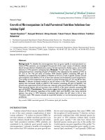
Figure 1 shows the time course of the following parameters:
leukocyte counts, glucose and protein concentrations, meas-
ured in parallel in CSF from EVD and ELD, for 17 days (D57
to D73) corresponding to the second and third episodes of
meningitis. Figure 1 shows strikingly that the leukocytes and
the protein concentration were always higher and the glucose
concentration was always lower in ELD than in EVD. Interest-
ingly, the highest ventriculo-lumbar CSF gradients in leuko-
cytes, protein and glucose concentration were present at the
very acute phase of meningitis, when Ps. aeruginosa O12 was
present in the meningeal cavity.
Our case also showed that the increase in the amount of anti-
biotics given did increase their concentration in plasma but not
in CSF. Indeed, i.v. cefepim was increased from 8 to 12 g/day
and i.v. ciprofloxacin from 1.2 to 2.4 g/day from D64 to D95.
This induced a persistent increase in plasma cefepim concen-
tration from 46 µ g/ml to more than 60 µg/ml and plasma cip-
rofloxacin concentration from 0.2 µg/ml to more than 1.0 µg/
ml. However, only a transient increase in cefepim concentra-
tion (D63, 7 µg/ml; D73, 15 µg/ml; D81 and D95, less than 9
µg/ml) and no increase in ciprofloxacin concentration (0.4–0.5
µg/ml from D63 to 95) were seen in lumbar and ventricular
CSF. It is noteworthy that the inhibitory minimal concentrations
of cefepim and ciprofloxacin for Ps. aeruginosa O12 were 16
and 0.25 mg/ml, respectively.
Discussion
Our case report followed ventriculo-lumbar CSF gradients in
leukocytes, protein and glucose concentration during two
Available online />R493
episodes of post-operative recurrent meningitis due to Ps. aer-
uginosa O12. It showed the presence of a rostrocaudal gradi-
ent of leukocytes and protein and an inverse gradient of
glucose. This confirmed previous work that showed greater
leukocytes and protein concentration in lumbar than in ven-
tricular CSF in patients with a central neural system infection,
mostly after neurosurgery [2,4]. However, patients from those
studies each had only one pair (ventricular and lumbar) of
measurements within a 24-hour interval [2] and glucose con-
centration in CSF was measured in only six patients [4].
We extend previous studies by showing that the greatest
amplitude of ventricular-to-lumbar gradients for all measured
parameters (leukocytes, protein and glucose concentration)
were seen during the very acute phase of meningitis, when
bacteria were present in the meningeal cavity. The mecha-
nisms of such ventricular-to-lumbar gradients are unknown.
Our data strongly suggest a compartmentalization of menin-
geal inflammation in the ventricular and lumbar area. Indeed,
similar bacteria, here Ps. aeroginusa O12, in similar quantities,
seemed to induce a greater alteration of meningeal permeabil-
ity with greater leukocyte and protein concentrations and a
lower glucose concentration in the lumbar than the ventricular
CSF region. Although still debatable, the decrease in glucose
concentration in CSF seems to be less related to a 'leukocyte-
induced glucose consumption' but rather to a meningeal shift
of glucose metabolism to anaerobic glycolysis, as indicated by
the concomitant increase in CSF lactate concentration and/or
a decrease in meningeal glucose transport [5]; the latter is
probably directly related to the degree of meningeal inflamma-
tion. An alternative explanation of the existence of a rostrocau-
dal gradient of leukocytes is that leukocytes from ventricular
CSF might fall by gravity to lumbar CSF. However, as
explained above, a greater concentration of leukocytes cannot
by itself explain a greater protein concentration and a lower
glucose concentration in lumbar CSF. Accordingly, our study
suggests that meningeal inflammation was greater in the lum-
bar than the ventricular region in our patient with CSF infection
due to a pharyngeal fistula.
Recurrent meningitis led us to increase the antibiotic dosage
to achieve a better concentration in CSF [6]. Surprisingly, only
a transient increase in CSF cefepim concentration and no
change in CSF ciprofloxacine concentration were observed
despite a more than 50% increase in plasma concentrations
of both antibiotics. The transient increase in cefepim in CSF
paralleled that of protein in CSF and could be related to the
transient alteration in meningeal permeability.
In summary, this case report shows that the maximal rostro-
caudal gradient of leukocytes, protein and glucose was seen
in the very acute phase of meningitis. This strongly suggests a
greater alteration in the meningeal barrier and very probably a
greater meningeal inflammation in the lumbar than the ven-
tricular regions.
Competing interests
The author(s) declare that they have no competing interests.
Author’s contributions
WN and AM coordinated the data analysis and drafted the
manuscript. JM, LR and J-F T participated in bacteriological
analysis. A-C L and BG participated in analysis of clinical data.
DP helped to draft the manuscript. All authors read and
approved the final manuscript
Figure 1
Time course of the ventricular-to-lumbar gradient of cerebrospinal fluid leukocyte, glucose and protein concentrations in cerebrospinal fluidTime course of the ventricular-to-lumbar gradient of cerebrospinal fluid
leukocyte, glucose and protein concentrations in cerebrospinal fluid.
The arrows represent days of positive cerebrospinal fluid culture.
0
80
160
0
80
160
0
2
4
0
1
2
D55 D60 D65 D70 D75
CSF Glucose conc.
(mmol/l)
CSF Protein conc.
(g/l)
CSF leukocytes
(×100/mm
3
)
External ventricular drain
External lumbar drain
Key messages
• The paper describes a patient that developed, after
surgery for a chordoma of the clivus, three episodes of
recurrent meningitis due to Ps. aeruginosa O12.
• Episodes were monitored by biochemical and
bacteriological daily analysis of CSF withdrawn in
parallel from lumbar space and ventricles by external
lumbar and ventricular damamge.
• We observed a permanent ventricular-to-lumbar CSF
gradients of leukocytes, protein and glucose that
increased during the acute phase of meningitis, with
the greatest amplitude observed when bacteria was
present in both ventricular and lumbar CSF.
• This may suggest a greater extent of meningeal
inflammation in lumbar than in ventricular region
Critical Care December 2004 Vol 8 No 6 Naija et al.
R494
References
1. Lozier A, Sciacca R, Romagnoli M, Connolly EJ: Ventriculostomy-
related infections: a critical review of the literature. Neurosur-
gery 2002, 51:170-181.
2. Gerber J, Tumani H, Kolenda H, Nau R: Lumbar and ventricular
CSF protein, leukocytes, and lactate in suspected bacterial
CNS infections. Neurology 1998, 51:1710-1714.
3. Merritt H, Fremont-Smith F: Acute purulent meningitis. In The
Cerebrospinal Fluid Edited by: Merritt H. Philadelphia: WB
Sanders; 1938:94-103.
4. Sommer J, Gaul C, Heckmann J, Neundorfer B, Erbguth F: Does
lumbar cerebrospinal fluid reflect ventricular cerebrospinal
fluid? A prospective study in patients with external ventricular
drainage. Eur Neurol 2002, 47:224-232.
5. Ernst J, Decazes J, Sande M: Experimental pneumococcal men-
ingitis: role of leukocytes in pathogenesis. Infect Immun 1983,
41:275-279.
6. Wolff M, Boutron L, Singlas E, Clair B, Decazes J, Regnier B: Pen-
etration of ciprofloxacin into cerebrospinal fluid of patients
with bacterial meningitis. Antimicrob Agents Chemother 1987,
31:899-902.