Báo cáo khoa học: "Acetazolamide-mediated decrease in strong ion difference accounts for the correction of metabolic alkalosis in critically ill patients" pps
Bạn đang xem bản rút gọn của tài liệu. Xem và tải ngay bản đầy đủ của tài liệu tại đây (206.96 KB, 6 trang )
Open Access
Available online />Page 1 of 6
(page number not for citation purposes)
Vol 10 No 1
Research
Acetazolamide-mediated decrease in strong ion difference
accounts for the correction of metabolic alkalosis in critically ill
patients
Miriam Moviat
1
, Peter Pickkers
1
, Peter HJ van der Voort
2
and Johannes G van der Hoeven
1
1
Department of Intensive Care Medicine, Radboud University Nijmegen Medical Centre, Nijmegen, The Netherlands
2
Department of Intensive Care Medicine, Medical Centre Leeuwarden
Corresponding author: Peter Pickkers,
Received: 22 Aug 2005 Accepted: 14 Dec 2005 Published: 9 Jan 2006
Critical Care 2006, 10:R14 (doi:10.1186/cc3970)
This article is online at: />© 2006 Moviat et al.; licensee BioMed Central Ltd.
This is an open access article distributed under the terms of the Creative Commons Attribution License ( />),
which permits unrestricted use, distribution, and reproduction in any medium, provided the original work is properly cited.
Abstract
Introduction Metabolic alkalosis is a commonly encountered
acid–base derangement in the intensive care unit. Treatment
with the carbonic anhydrase inhibitor acetazolamide is indicated
in selected cases. According to the quantitative approach
described by Stewart, correction of serum pH due to carbonic
anhydrase inhibition in the proximal tubule cannot be explained
by excretion of bicarbonate. Using the Stewart approach, we
studied the mechanism of action of acetazolamide in critically ill
patients with a metabolic alkalosis.
Methods Fifteen consecutive intensive care unit patients with
metabolic alkalosis (pH ≥ 7.48 and HCO
3
-
≥ 28 mmol/l) were
treated with a single administration of 500 mg acetazolamide
intravenously. Serum levels of strong ions, creatinine, lactate,
weak acids, pH and partial carbon dioxide tension were
measured at 0, 12, 24, 48 and 72 hours. The main strong ions
in urine and pH were measured at 0, 3, 6, 12, 24, 48 and 72
hours. Strong ion difference (SID), strong ion gap, sodium–
chloride effect, and the urinary SID were calculated. Data (mean
± standard error were analyzed by comparing baseline variables
and time dependent changes by one way analysis of variance for
repeated measures.
Results After a single administration of acetazolamide,
correction of serum pH (from 7.49 ± 0.01 to 7.46 ± 0.01; P =
0.001) was maximal at 24 hours and sustained during the period
of observation. The parallel decrease in partial carbon dioxide
tension was not significant (from 5.7 ± 0.2 to 5.3 ± 0.2 kPa; P
= 0.08) and there was no significant change in total
concentration of weak acids. Serum SID decreased significantly
(from 41.5 ± 1.3 to 38.0 ± 1.0 mEq/l; P = 0.03) due to an
increase in serum chloride (from 105 ± 1.2 to 110 ± 1.2 mmol/
l; P < 0.0001). The decrease in serum SID was explained by a
significant increase in the urinary excretion of sodium without
chloride during the first 24 hours (increase in urinary SID: from
48.4 ± 15.1 to 85.3 ± 7.7; P = 0.02).
Conclusion A single dose of acetazolamide effectively corrects
metabolic alkalosis in critically ill patients by decreasing the
serum SID. This effect is completely explained by the increased
renal excretion ratio of sodium to chloride, resulting in an
increase in serum chloride.
Introduction
Metabolic alkalosis is a common acid–base disturbance in the
intensive care unit (ICU) that is associated with increased ICU
mortality and morbidity [1,2], with adverse effects on cardio-
vascular, pulmonary and metabolic function [3,4]. Additionally,
such patients are characterized by compensatory alveolar
hypoventilation, which can result in delayed weaning from
mechanical ventilation. Options for treatment aimed at correct-
ing metabolic alkalosis are fluid and potassium replacement,
and administration of ammonium chloride, hydrochloric acid,
or acetazolamide [5]. These therapeutic interventions poten-
tially increase minute ventilation, allowing patients to be
weaned more rapidly [6].
An advanced understanding of acid–base physiology is cen-
tral to the practice of critical care medicine. Although it is not
difficult to quantify the degree of metabolic alkalosis, it is more
challenging to identify the cause of a metabolic alkalosis and
ICU = intensive care unit; PCO
2
= partial carbon dioxide tension; SID = strong ion difference; SIG = strong ion gap.
Critical Care Vol 10 No 1 Moviat et al.
Page 2 of 6
(page number not for citation purposes)
determine the actions that must be taken to correct it. The
method of quantifying and qualifying an acid–base distur-
bance, as described by Stewart, relies on the accepted phys-
icochemical principles of conservation of mass and
electroneutrality [7,8]. According to Stewart, three variables
independently determine the serum hydrogen concentration.
These variables are the partial carbon dioxide tension (PCO
2
),
the total concentration of nonvolatile weak acids (primarily
serum proteins and phosphate), and the strong ion difference
(SID) [9]. The Stewart approach, in contrast to other
approaches, allows us to quantify an acid–base derangement
as well as determine its cause.
The kidneys are the most important regulators of SID for acid–
base purposes. The concentration of strong ions in plasma
can be altered by adjusting absorption from glomerular filtrate
or secretion into the tubular lumen from plasma. In this respect,
administration of the carbonic anhydrase inhibitor acetazola-
mide during metabolic alkalosis could modulate plasma pH by
influencing the urinary excretion of various strong ions.
Because plasma sodium controls intravascular volume and
osmolality, and because plasma potassium is important for
cardiac and neuromuscular function, plasma chloride appears
to represent the strong ion that the kidney uses to regulate
acid–base status without interfering with other important
homeostatic processes [7]. Furthermore, the basic physico-
chemical principles imply that a change in bicarbonate con-
centration is not a cause but merely a co-phenomenon of an
acid–base disturbance such as metabolic alkalosis.
Acetazolamide decreases proximal tubular bicarbonate reab-
sorption by up to 80% through inhibition of carbonic anhy-
drase in the luminal borders of renal proximal tubule cells, and
it is often effectively used in the treatment of metabolic alkalo-
sis in the ICU. However, the mechanism of action of acetazola-
mide remains unclear. According to the basic
physicochemical principles mentioned above, retention of
bicarbonate cannot causally be related to correction of serum
pH, and acetazolamide-induced effects must be explained by
modulation of the urinary excretion of strong ions.
We hypothesized that acetazolamide, by inhibiting carbonic
anhydrase in the proximal tubules, causes excretion of strong
cations (along with bicarbonate) and retention of chloride, and
in this way decreases the serum SID. Subsequently, the
decrease in SID will correct an alkalosis by causing dissocia-
tion of water and formation of hydrogen ions. The purpose of
the present study was to determine the mechanism of action
of acetazolamide in critically ill patients with a metabolic alka-
losis according to the physicochemical principles described
by Stewart.
Materials and methods
Patients
The local ethics committee granted approval for the study and,
because the indication for acetazolamide was based on clini-
cal grounds, waived the need for informed consent. This pro-
spective study was set in the multidisciplinary ICU of the
Radboud University Nijmegen Medical Centre, Nijmegen, The
Netherlands.
We studied 15 consecutive ICU patients with a metabolic
alkalosis (defined as pH ≥ 7.48) and serum bicarbonate of 28
mmol/l or greater. All patients had an arterial line in situ.
Patients clinically suspected of having volume contraction (for
example, cold extremities, blood pressure increase during pas-
sive leg raising), hypokalaemia (serum potassium ≤ 3.4 mmol/
l), nasogastric tube drainage greater than 50 cc/hour, renal
insufficiency (creatinine clearance <20 ml/min and/or renal
replacement therapy), or intolerance or allergy to acetazola-
mide or sulfonamides were excluded. Also excluded were
patients who were treated with intravenous acetazolamide or
sodium bicarbonate during the previous 72 hours.
Acute Physiology and Chronic Health Evaluation II score was
calculated and recorded for each patient for the first 24 hours
after admission. Data on fluid intake and output, ventilator set-
tings, and relevant medications such as diuretics and steroids
were also recorded. After inclusion, patients received a single
dose acetazolamide (500 mg as an intravenous push).
Experimental design
We measured pH, arterial oxygen tension, arterial PCO
2
,
sodium, potassium, chloride, magnesium, calcium, lactate,
creatinine, urea, phosphate and albumin in a single arterial
Table 1
Patients characteristics
Characteristic Value
Age (years; mean [range]) 67 (35–79)
Sex (male/female; n)10/5
APACHE II score (mean [range]) 21 (12–30)
Mechanical ventilation (%) 87
Diuretics (%) 47
Standardized mortality ratio 0.57
Hospital mortality (%) 20
Diagnosis
Dissecting/ruptured aorta 2
Postoperative bleeding 2
Sepsis 4
Open heart surgery 4
Cardiogenic shock 1
Neurological disease 2
Shown are demographic data of all patients. APACHE, Acute
Physiology and Chronic Health Evaluation.
Available online />Page 3 of 6
(page number not for citation purposes)
blood sample before acetazolamide was administered (t = 0)
and 12, 24, 48 and 72 hours later (t = 12, t = 24, t = 48 and
t = 72).
Urine samples were taken before acetazolamide was adminis-
tered, and 3, 6, 12, 24, 48 and 72 hours later. In these sam-
ples pH was measured immediately. Urine was stored at -
80°C, and sodium, chloride, potassium and creatinine were
measured in a single batch at the end of the study.
Data analysis, calculations and statistics
Bicarbonate was calculated using the Henderson–Hassel-
balch equation (pH = 6.1 + log [HCO
3
-
]/0.0301 arterial
PCO
2
]) and the standard base excess was calculated using
the Siggaard–Andersen formula.
The apparent SID (SID
app
) was calculated using the equation
SID
app
= [Na
+
] + [K
+
] + [Ca
2+
] + [Mg
2+
] - [Cl
-
] - [lactate
-
]. The
effective SID (SID
eff
) was calculated using the equation SID
eff
= 12.2 × PCO
2
/(10
-pH
) + [albumin] × (0.123 × pH - 0.631) +
[PO
4
-
] × (0.309 × pH - 0.469). The strong ion gap (SIG) was
calculated using the equation SIG = SID
app
- SID
eff
[10]. The
sodium–chloride effect was calculated using the formula [Na
+
]
- [Cl
-
] - 38 [11]. Urinary electrolytes to creatinine ratios and
sodium to chloride ratios were calculated, as was the urinary
SID using the following equation: urinary SID = [Na
+
] + [K
+
] -
[Cl
-
].
The effects of acetazolamide were analyzed by comparing
baseline variables and time-dependent changes using one-
way analysis of variance with repeated measures. Power anal-
ysis was based on a presumed standard deviation of 15% for
the measured end-points. A change of 10% was considered
clinically relevant. With α = 0.05, we calculated that a sample
size of 14 would be needed to achieve a power of 80%. There-
fore, 15 patients were included.
Data are expressed as mean ± standard error unless other-
wise specified. P < 0.05 was considered statistically signifi-
cant.
Results
Patients
Patient characteristics are presented in Table 1, and baseline
acid–base and electrolyte data are presented in Table 2. Of
the patients studied, 87% were mechanically ventilated (all in
an assisted mode of ventilation in which spontaneous breath-
ing activity was fully possible). Although 47% of the patients
were treated with diuretics, none exhibited clinical symptoms
of hypovolaemia. Furthermore, low urinary chloride excretion
(< 20 mmol/l), which is indicative of hypovolaemia in patients
who do not use diuretics, was present in only one patient.
Intravenous and enteral intake of sodium chloride, as well as
ventilator settings and diuretic dose, were not changed during
the study period.
Table 2
Acid–base and electrolyte data
Acid–base and electrolyte data Baseline t = 24
PH 7.49 (7.48–7.51) 7.46 (7.44–7.48)
PaCO
2
(kPa) 5.7 (5.1–6.1) 5.3 (4.9–5.9)
Bicarbonate (mmol/l) 31.5 (29.5–33.7) 28.6 (26.3–30.3)
Sodium (mmol/l) 141 (139–145) 142 (139–145)
Potassium (mmol/l) 3.7 (3.7–4) 3.8 (3.4–3.9)
Chloride (mmol/l) 106 (102–107) 108 (107–110)
Creatinine (µmol/l) 64 (49–95) 65 (49–101)
Lactate (mmol/l) 1.4 (1.2–1.8) 1.5 (1.2–1.7)
Albumin (g/l) 16 (14–20) 17 (15–20)
Apparent SID (mEq/l) 41.7 (39.1–44.0) 39.4 (36.4–41.4)
Effective SID (mEq/l) 39.0 (37.3–40.3) 35.6 (32.9–37.7)
SIG (mEq/l) 2.4 (1.5–4.4) 3.1 (2.1–4.8)
Sodium–chloride effect (mEq/l) -2.0 (-3.5 to +0.5) -3.0 (-7.5 to -1.5)
Shown are baseline acid–base and electrolyte data (median [interquartile range]) for 15 patients before administration of 500 mg acetazolamide
(baseline) and after 24 hours (t = 24). The serum apparent SID (SID
app
) was calculated using the following equation: SID
app
= [Na
+
] + [K
+
] +
[Ca
2+
] + [Mg
2+
] - [Cl
-
] - [lactate
-
]. The serum effective SID (SID
eff
) was calculated using the following equation: SID
eff
= 12.2 × PCO
2
/(10
-pH
) +
[albumin] × (0.123 × pH - 0.631) + [PO
4
-
] × (0.309 × pH - 0.469). The SIG was calculated using the following equation: SIG = SID
app
- SID
eff
.
The sodium–chloride effect was calculated using the formula [Na
+
] - [Cl
-
] - 38. PaCO
2
, arterial carbon dioxide tension; SID, strong ion difference;
SIG, strong ion gap.
Critical Care Vol 10 No 1 Moviat et al.
Page 4 of 6
(page number not for citation purposes)
Effects of acetazolamide on Stewart's parameters in
blood
After administration of acetazolamide, correction of serum pH
(7.49 ± 0.01 to 7.46 ± 0.01; P = 0.001) was maximal at 24
hours and was sustained during the period of observation (Fig-
ure 1). The parallel decrease in PCO
2
was not significant (from
5.7 ± 0.2 to 5.3 ± 0.2 kPa; P = 0.08). There was no significant
change in the total concentration of weak acids. When values
of weak acids were expressed as values contributing to the
electrical charge equilibrium in plasma (see formula for SID
eff
),
phosphate decreased from 2.14 ± 0.11 mEq/l to 1.94 ± 0.10
mEq/L (P = 0.02), and albumin remained unchanged (from
4.65 ± 0.30 mEq/l to 4.87 ± 0.35 mEq/L; P = 0.15; Figure 1).
Serum SID decreased significantly during the period of obser-
vation (from 41.5 ± 1.3 mEq/l to 38.0 ± 1.0 mEq/l; P = 0.03)
because of an increase in serum chloride (from 105 ± 1.2
mmol/l to 110 ± 1.2 mmol/l; P < 0.0001, figure 2). There was
a strong relation between the serum SID and the sodium–
chloride effect (R
2
= 0.99; P < 0.001), indicating that the
observed changes in SID are completely accounted for by
changes in serum sodium and/or chloride and not other strong
ions. The decrease in serum SID was caused by a significant
increase in the urinary excretion of sodium without chloride
during the first 24 hours (change in urinary [Na]/[Cl]: from 1.3
± 0.3 to 2.5 ± 0.5; P = 0.02), resulting in an increase in urinary
SID (see Effects of acetazolamide on Stewart's parameters in
urine, below).
In the patients studied here, there was no relevant SIG (mean
baseline value 2.11 ± 0.81 mEq/L), and it exhibited no change
after administration of acetazolamide (3.13 ± 0.48; P = 0.43).
Effects of acetazolamide on Stewart's parameters in
urine
Urinary pH increased significantly from 5.55 ± 0.26 to 6.13 ±
0.37 (P = 0.005) during the first 12 hours after administration
of acetazolamide, and returned to pre-administration value dur-
ing the next 60 hours (Figure 3). Urinary SID exhibited a paral-
lel increase (from 48.4 ± 15.1 to 85.3 ± 7.7; P = 0.02) during
the first 12 hours and a parallel decrease thereafter.
Discussion
Our study is the first to demonstrate that the acetazolamide-
induced correction of metabolic alkalosis in critically ill patients
can completely be accounted for by a significant decrease in
serum SID, using the physicochemical principles described by
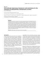
Figure 1
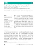
Time course of acetazolamide-induced changes in pH and three inde-pendent variables that determine pHTime course of acetazolamide-induced changes in pH and three inde-
pendent variables that determine pH. Effect of 500 mg acetazolamide
administration (intravenous) in patients with metabolic alkalosis. Data
are expressed as mean ± standard error values for 15 patients. The P
values refer to the time-dependent changes analyzed using one-way
analysis of variance. pCO
2
, partial carbon dioxide tension; SIDa, appar-
ent strong ion difference.
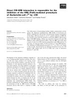
Figure 2
Time course of acetazolamide-induced changes in serum potassium, sodium and chlorideTime course of acetazolamide-induced changes in serum potassium,
sodium and chloride. Effect of 500 mg acetazolamide administration
(intravenous) in patients with metabolic alkalosis. Serum chloride exhib-
ited a significant increase, whereas there were no significant changes
in serum potassium and sodium concentration. Data are expressed as
mean ± standard error values for 15 patients. The P values refer to the
time-dependent changes analyzed using one-way analysis of variance.
Available online />Page 5 of 6
(page number not for citation purposes)
Stewart. Although analysis using the Henderson–Hasselbalch
equation is useful for describing and classifying acid–base
disorders, the physicochemical approach described by Stew-
art is better suited to quantifying these disorders and for gen-
erating hypotheses regarding mechanisms.
Use of the Stewart model has improved our understanding of
the pathophysiology that leads to changes in acid–base bal-
ance. SID, total concentration of nonvolatile weak acids, and
PCO
2
are biological variables that are regulated mainly by
renal tubular transport, metabolism and ventilation. The relative
complexity of the Stewart approach comes from the fact that
several variables are needed. However, when these variables
are absent or assumed to be normal, the approach becomes
essentially indistinguishable from the more traditional descrip-
tive methods. For example, our study does not dispute the con-
tention that acetazolamide, through inhibition of carbonic
anhydrase in the proximal tubule, increases urinary bicarbo-
nate excretion. However, according to the Stewart approach it
is not the loss of bicarbonate that determines the fall in pH,
because bicarbonate is not an independent parameter.
According to Stewart, it is the change in SID (due to a rise in
chloride) that explains the decrease in pH. In our patients,
acetazolamide-induced loss of bicarbonate facilitated the
renal reabsorption of chloride, while sodium could still be
excreted. In other words, acetazolamide-induced bicarbonate
excretion permits urinary excretion of sodium without loss of
any strong anions, resulting in a lower SID and thereby a
decrease in pH.
Apart from the acetazolamide-induced change in SID, our
study demonstrates that inhibition of carbonic anhydrase does
not significantly alter the other independent determinants of
serum pH. In contrast, the nonsignificant decrease in PCO
2
and small decrease in weak acid phosphate cause the oppo-
site effect on serum pH. The small decrease in PCO
2
in our
patients can be explained by an increase in minute ventilation
in response to correction of serum pH by acetazolamide. This
increase in minute ventilation, as a result of an increased res-
piratory drive, was possible in an assisted mode of mechanical
ventilation. Finally, the observed small increase in serum albu-
min does not have a significant lowering effect on serum pH
and could probably be explained by the hemo-concentrating
effect of diuretics during the study period.
The acetazolamide-induced decrease in SID is entirely caused
by a change in serum concentration of chloride, as shown by
the strong relation between the SID and the sodium–chloride
effect. These changes in sodium and chloride are explained by
an increase in urinary sodium excretion (along with a weak
anion) while chloride excretion is maintained, as shown by the
increased urinary sodium–chloride ratios. The intravenous and
enteral salt intake of patients was unchanged during the
observation period. Thus, the renal effect of acetazolamide
results in a relative increase in serum chloride. Because
sodium and chloride are the most abundant and therefore the
most important strong ions, an increase in chloride relative to
sodium will have a significant lowering effect on serum SID.
Stewart proposed that H
+
and therefore pH cannot change
unless one or more of the three independent variables (PCO
2
,
weak acids, and SID) change. Our study demonstrates that
the acetazolamide-induced effects on pH are solely mediated
by a decrease in serum SID through renal excretion of sodium
without chloride. Although the Stewart approach has proved
to be valuable in critically ill acidotic patients [12-14], this
paper represents the first report using the Stewart approach
during metabolic alkalosis.
Our study confirms previous reports in patients with metabolic
alkalosis that, despite corrected fluid and electrolyte abnor-
malities, a single dose of acetazolamide is an effective and
safe form of therapy, with a quick onset and long duration of
action [5,15]. Our findings suggest that the duration of the
pharmacologic effect of a single administration of 500 mg
acetazolamide exceeds its serum half-life (6–8 hours). This
long effect is reflected by the 24-hour duration of altered uri-
nary sodium and chloride excretion. Furthermore, after normal-
ization of serum pH at 24 hours, this correction was sustained
although urinary electrolyte excretion and pH returned to pre-
administration values. Apparently, once the serum SID is cor-
rected by acetazolamide because of the increased sodium
excretion without a strong anion, this new equilibrium is main-
tained. The Stewart approach does not help us to explain the
long-lasting effects of acetazolamide, and it is unclear how the
new equilibrium is maintained after correction of the SID and
Figure 3
Effect acetazolamide on urinary pH and sodium–chloride ratioEffect acetazolamide on urinary pH and sodium–chloride ratio. Effect of
500 mg acetazolamide administration (intravenous) in patients with
metabolic alkalosis. Data are expressed as mean ± standard error val-
ues for 15 patients. The P values refer to the time-dependent changes
analyzed using one-way analysis of variance.
Critical Care Vol 10 No 1 Moviat et al.
Page 6 of 6
(page number not for citation purposes)
what the regulating mechanism is that induces the permanent
hyperchloraemia. The pharmacokinetics of acetazolamide in
tissue (not plasma) may explain this observation. Another
explanation could be that the alkalizing factors that were orig-
inally present in our patients are corrected during the course
of the observation period. Although clinical suspicion of a
hypovolaemic state was an exclusion criterion in our study, one
of the alkalizing factors could very well be some degree of vol-
ume contraction induced by the administration of diuretics.
Whatever the cause, it is highly unlikely that the presence of
some degree of hypovolaemia in our patients would influence
our conclusions regarding the effects – as determined using
the Stewart approach – of acetazolamide on metabolic alkalo-
sis.
The SIG – indicative of the presence of unmeasured anions,
which are often present in metabolic acidosis, particularly in
patients with renal failure [14] – was not found to be elevated
in our study, as was expected. Furthermore, administration of
acetazolamide had no influence on the SIG.
Conclusion
Our study is the first to report the mechanism by which aceta-
zolamide-induced correction of metabolic alkalosis in critically
ill patients is mediated. Applying the quantitative biophysical
principles of acid–base analysis described by Stewart, the
acetazolamide-induced effects on serum pH are completely
accounted for by an increased renal excretion of sodium with-
out chloride, resulting in an increase in serum chloride and a
decrease in serum SID.
Competing interests
The authors declare that they have no competing interests.
Authors' contributions
MM collected all of the data and drafted the manuscript. PP
conceived the study and cowrote the manuscript. PvdV and
JGvdH participated in the design of the study and corrected
the manuscript. All authors read and approved the final manu-
script.
Acknowledgements
We thank our research nurses and the nurses of our ICUs for their help
with the collection of the blood and urine samples.
PP is a recipient of a Clinical Fellowship grant of the Netherlands Organ-
isation for Scientific Research (ZonMw).
References
1. Anderson LE, Henrich WL: Alkalemia-associated morbidity and
mortality in medical and surgical patients. South Med J 1987,
80:729-733.
2. Hodgkin JE, Soeprono FF, Chan DM: Incidence of metabolic
alkalemia in hospitalized patients. Crit Care Med 1980,
8:725-728.
3. Krintel JJ, Haxholdt OS, Berthelsen P, Brockner J: Carbon dioxide
elimination after acetazolamide in patients with chronic
obstructive pulmonary disease and metabolic alkalosis. Acta
Anaesthesiol Scand 1983, 27:252-254.
4. Berthelsen P: Cardiovascular performance and oxyhemoglobin
dissociation after acetazolamide in metabolic alkalosis. Inten-
sive Care Med 1982, 8:269-274.
5. Mazur JE, Devlin JW, Peters MJ, Jankowski MA, Iannuzzi MC,
Zarowitz BJ: Single versus multiple doses of acetazolamide for
metabolic alkalosis in critically ill medical patients: a rand-
omized, double-blind trial. Crit Care Med 1999, 27:1257-1261.
6. Berthelsen P, Gothgen I, Husum B, Jacobsen E: Oxygen uptake
and carbon dioxide elimination after acetazolamide in the crit-
ically ill. Intensive Care Med 1985, 11:26-29.
7. Kellum JA: Determinants of blood pH in health and disease.
Crit Care 2000, 4:6-14.
8. Stewart PA: Modern quantitative acid-base chemistry. Can J
Physiol Pharmacol 1983, 61:1444-1461.
9. Figge J, Rossing TH, Fencl V: The role of serum proteins in acid-
base equilibria. J Lab Clin Med 1991, 117:453-467.
10. Kellum JA, Kramer DJ, Pinsky MR: Strong ion gap: a methodol-
ogy for exploring unexplained anions. J Crit Care 1995,
10:51-55.
11. Story DA., Morimatsu H, Bellomo R: Strong ions, weak acids and
base excess: a simplified Fencl–Stewart approach to clinical
acid–base disorders. Br J Anaesth 2004, 92:54-60.
12. Moviat M, van Haren F, van der Hoeven HH: Conventional or
physicochemical approach in intensive care unit patients with
metabolic acidosis. Crit Care 2003, 7:R41-R45.
13. Dondorp AM, Chau TT, Phu NH, Mai NT, Loc PP, Chuong LV, Sinh
DX, Taylor A, Hien TT, White NJ, Day NP: Unidentified acids of
strong prognostic significance in severe malaria. Crit Care
Med 2004, 32:1683-1688.
14. Rocktaeschel J: Acid–base status of critically ill patients with
acute renal failure: analysis based on Stewart-Figge method-
ology. Crit Care 2003, 7:R60.
15. Marik PE, Kussman BD, Lipman J, Kraus P: Acetazolamide in the
treatment of metabolic alkalosis in critically ill patients. Heart
Lung 1991, 20:455-459.
Key messages
• The Stewart approach offers an advanced understand-
ing of acid–base physiology that is central to the prac-
tice of critical care medicine.
• Although the Stewart approach has proved to be valua-
ble in critically ill acidotic patients, no reports exist in
which the approach is used in ICU patients with meta-
bolic alkalosis.
• In ICU patients with metabolic alkalosis, the carbonic
anhydrase inhibitor acetazolamide corrects pH by
decreasing the SID, with no effect on the other inde-
pendent determinants of pH.
• The decrease in serum SID is completely explained by
an increase in plasma chloride, caused by an increase
in the urinary excretion of sodium without chloride.
• The Stewart approach explains that acetazolamide-
induced loss of bicarbonate is not the cause of the
decrease in serum pH, but only facilitates the renal rea-
bsorption of chloride while sodium can still be excreted.