Báo cáo y học: "Inhibition of HIV-1 integrase nuclear import and replication by a peptide bearing integrase putative nuclear localization signal" pot
Bạn đang xem bản rút gọn của tài liệu. Xem và tải ngay bản đầy đủ của tài liệu tại đây (2.95 MB, 16 trang )
BioMed Central
Page 1 of 16
(page number not for citation purposes)
Retrovirology
Open Access
Research
Inhibition of HIV-1 integrase nuclear import and replication by a
peptide bearing integrase putative nuclear localization signal
Aviad Levin
†1
, Ayelet Armon-Omer
†1,4
, Joseph Rosenbluh
1
, Naomi Melamed-
Book
1
, Adolf Graessmann
2
, Elisabeth Waigmann
3
and Abraham Loyter*
1
Address:
1
Department of Biological Chemistry, The Alexander Silberman Institute of Life Sciences, The Hebrew University of Jerusalem, Jerusalem
91904, Israel,
2
Institut fur Molekularbiologie und Biochemie, Free University of Berlin, Germany,
3
Max F. Perutz Laboratories, University
Departments at the Vienna Biocenter, Institute of Medical Biochemistry, Medical University of Vienna, Austria and
4
Ziv Medical Center, Zefat
13100, Israel
Email: Aviad Levin - ; Ayelet Armon-Omer - ; Joseph Rosenbluh - ;
Naomi Melamed-Book - ; Adolf Graessmann - ;
Elisabeth Waigmann - ; Abraham Loyter* -
* Corresponding author †Equal contributors
Abstract
Background: The integrase (IN) of human immunodeficiency virus type 1 (HIV-1) has been
implicated in different steps during viral replication, including nuclear import of the viral pre-
integration complex. The exact mechanisms underlying the nuclear import of IN and especially the
question of whether it bears a functional nuclear localization signal (NLS) remain controversial.
Results: Here, we studied the nuclear import pathway of IN by using multiple in vivo and in vitro
systems. Nuclear import was not observed in an importin α temperature-sensitive yeast mutant,
indicating an importin α-mediated process. Direct interaction between the full-length IN and
importin α was demonstrated in vivo using bimolecular fluorescence complementation assay (BiFC).
Nuclear import studies in yeast cells, with permeabilized mammalian cells, or microinjected
cultured mammalian cells strongly suggest that the IN bears a NLS domain located between
residues 161 and 173. A peptide bearing this sequence -NLS-IN peptide- inhibited nuclear
accumulation of IN in transfected cell-cycle arrested cells. Integration of viral cDNA as well as HIV-
1 replication in viral cell-cycle arrested infected cells were blocked by the NLS-IN peptide.
Conclusion: Our present findings support the view that nuclear import of IN occurs via the
importin α pathway and is promoted by a specific NLS domain. This import could be blocked by
NLS-IN peptide, resulting in inhibition of viral infection, confirming the view that nuclear import of
the viral pre-integration complex is mediated by viral IN.
Background
Active nuclear import begins in the cytoplasm with recog-
nition of the transported cargo molecules by nuclear
transport receptors designated as importins [1]. Proteins
targeted to the nucleus contain a specific amino acid
sequence, termed nuclear localization signal (NLS),
which is recognized by either a member of the importin α
family, or directly by importin β. The resultant complex
then interacts with the nuclear pore complexes (NPCs),
through which it is subsequently transported into the
Published: 5 December 2009
Retrovirology 2009, 6:112 doi:10.1186/1742-4690-6-112
Received: 30 September 2009
Accepted: 5 December 2009
This article is available from: />© 2009 Levin et al; licensee BioMed Central Ltd.
This is an Open Access article distributed under the terms of the Creative Commons Attribution License ( />),
which permits unrestricted use, distribution, and reproduction in any medium, provided the original work is properly cited.
Retrovirology 2009, 6:112 />Page 2 of 16
(page number not for citation purposes)
nucleus [2]. This nuclear translocation machinery is
highly conserved among lower and higher eukaryotes [3].
Human immunodeficiency virus type 1 (HIV-1) belongs
to the lentivirus family, which in contrast to other retrovi-
ruses can infect terminally differentiated cells [4,5]. The
capability of HIV-1 to infect cell-cycle arrested cells has
been ascribed to the ability of its pre-integration complex
(PIC) [6,7] to translocate across the nuclear envelope via
the NPC [1]. The karyophilic properties of the viral PIC
have been attributed mainly to three viral proteins: matrix
(MA), Vpr, and integrase (IN) [8-10]. The cellular Lens
Epithelium-Derived Growth Factor p75 (LEDGF/p75)
protein as well as the DNA flap structure of the viral cDNA
have also been implicated in promoting the translocation
of the PIC into nuclei of virally infected cells [11-13].
Yamashipa et al. have proposed that the HIV capsid pro-
tein plays a crucial role in controlling the nuclear import
of the HIV genome [14]. However, despite these extensive
studies and numerous reports, the nuclear import mecha-
nism of the PIC and the involvement of viral or cellular
factors driving such a process remain unclear and contro-
versial [15].
The HIV-1 IN protein consists of 288 amino acids and
three functional domains: the N-terminal domain (resi-
dues 1-50), which bears a zinc-binding motif [16,17]; the
central core domain (residues 51-212), which includes
the catalytic DDE motif [18-20]; and the C-terminal
domain (residues 213-288), which has been shown to
non-specifically bind the DNA [19-21]. To achieve inte-
gration of the viral DNA into the host chromosome, the
IN must be translocated into the nuclei of infected cells
[15].
Various studies have showed that IN is a karyophilic pro-
tein. Transfection of cultured mammalian cells with
expression vectors bearing IN results in nuclear accumula-
tion of the encoded protein [22]. Import of fluorescently
labeled IN into the nuclei of digitonin-permeabilized
mammalian cells was shown to be ATP- and temperature-
dependent; and this import could be blocked by the addi-
tion of unlabeled IN, clearly indicating an active, receptor-
mediated process [23,24]. Based on the ability of recom-
binant IN protein to bind in vitro to importin α and the
ability of a peptide bearing the prototypic simian virus 40
T-antigen NLS (SV40-NLS) to block such binding, as well
as nuclear import, nuclear transport of IN has been sug-
gested to occur via the importin α pathway [8,23]. More-
over, interaction of IN with the importin α family has
recently been reported [25].
The possibility of the IN protein being carried into the
cell's nuclei by other cellular components has also been
suggested [13,26,27]. The LEDGF/p75 was initially impli-
cated in mediating the nuclear import of IN [13]. How-
ever, studies on the specific contributions of LEDGF/p75
demonstrated that it facilitates the interaction between IN
and nuclear chromatin, but is not directly involved in the
import process [28]. An interaction with importin 7, via a
sequence located at the C terminus of IN [26], has been
proposed. However conflicting results have been obtained
regarding the necessity of this receptor [29,30]. Further-
more anti-importin 7 antibodies did not block nuclear
import of IN [25]. More recently, the involvement of the
transportin-SR2 (TNPO3) in the nuclear import of IN has
been suggested [27]. This conclusion is based mainly on
experiments showing that the knockdown of transportin-
SR2 (TNPO3) resulted in the reduction of nuclear cDNA
[27].
In the present work, we further confirm and emphasize
the role that importin α plays in promoting nuclear
import of the viral IN and thus in virus infection. Multiple
approaches and various experimental systems such as
transfected mammalian and yeast cells as well as virally
infected cells have been used to answer the question of
whether nuclear import of IN may be mediated by its own
NLS via interaction with importin α. Our results clearly
demonstrate that IN accumulates within wild-type yeast
cell nuclei, but fails to do so in importin α-defective yeast
mutants (srp1-31) [31]. A full-length IN, as well as a pep-
tide bearing the IN amino acid sequence 161-173 (NLS-
IN), interacted in vivo with mammalian importin α, as
demonstrated by a bimolecular fluorescence complemen-
tation (BiFC) assay [32] in yeast. The involvement of
amino acids 161-173 in mediating nuclear import of IN
was also demonstrated by microinjection and transfection
experiments in cultured mammalian cells. Furthermore,
the putative NLS-IN peptide inhibited nuclear accumula-
tion of IN as well as of cDNA in IN-transfected and virally
infected cells. This appears to be due to the ability of the
NLS-IN peptide to compete for the interaction between
the viral IN and the cellular importin α. This peptide has
also been found to significantly inhibit HIV-1 replication
in TZM-bl cells and inhibit the integration of viral cDNA
in infected cells. Thus, the present results support our [23]
and others' previous results [33] claiming that the IN pro-
tein contains a specific functional NLS sequence, which is
located between amino acids 161 and 173 and which con-
fers to this protein the karyophilic property required to
ensure productive viral infection.
Results
The NLS-IN peptide is functional in transfected and
microinjected mammalian cells as well as in yeast cells
The results in Fig. 1 clearly show that in stably transfected
aphidicolin treated cell-cycle arrested HeLa/P4 cells, HIV-
1 IN accumulates within the nuclei, confirming previously
published results [22]. We next evaluated the ability of the
Retrovirology 2009, 6:112 />Page 3 of 16
(page number not for citation purposes)
NLS-IN peptide [23] to block the nuclear import of IN.
However, the NLS-IN peptide was found to be cell-imper-
meable (not shown). Addition of the cell-permeable pen-
etrating peptide (Pen-peptide) [34] sequence to the NLS-
IN converted the latter to a cell-permeable peptide (not
shown) which was designated NLS-IN-Pen. No toxic effect
was exerted by this peptide during the time of the experi-
ment, as estimated by MTT assay (data not shown), thus
allowing for studies on its effect in cultured cells. As can
be seen (Fig. 1), following the incubation of the trans-
fected cells with the NLS-IN-Pen peptide, very little - if any
- IN was intranuclear; most of it was located within the
cytoplasm, clearly demonstrating the inhibition of
nuclear import. Incubation with the Pen-peptide alone
did not have any effect on nuclear import of IN (Fig. 1),
strongly indicating a specific effect of the NLS-IN. The
same results were obtained when the transfected cells
were incubated with a cell-permeable SV40-NLS-Pen pep-
tide (Fig. 1), indicating an importin α-dependent nuclear
import pathway [35].
Due to the high ambiguity surrounding the nuclear
import pathway of IN and its NLS domain, we studied its
translocation into nuclei in a non-mammalian cell envi-
ronment as well, namely in yeast cells. W303 cells were
transformed with expression vectors encoding the full-
length and truncated IN fused to the green fluorescence
protein (GFP) (expressed proteins are schematized in Fig.
2A). As can be seen (Fig. 2B) in cells expressing GFP-IN,
the fluorescence is packed into small intranuclear dots, as
confirmed by DAPI staining of the cell's DNA, while both
the cytosol and cell vacuoles appear relatively dark. The
same was observed with the truncated GFP-180-IN, which
includes the NLS-IN (Fig. 2B). Next, the ability of the NLS-
IN sequence to promote nuclear import was studied. To
create a molecule of high molecular weight, thereby
avoiding passive nuclear import [2], the NLS-IN coding
sequence was fused to the coding region of a double-GFP
(GFP)
2
(Fig. 2B). Similar to GFP-IN and GFP-180-IN, the
expressed GFP
2
-NLS-IN fusion protein also accumulated
within the yeast cell nuclei (Fig. 2B). In contrast, no
nuclear import was observed in yeast cells transformed
with an expression vector encoding the truncated GFP-
152-IN, which lacks the putative NLS-IN (Fig. 2B). IN-
mediated nuclear import can be inferred from the results
showing that in yeast cells transformed with vectors
expressing GFP molecules alone, the fluorescence distrib-
uted within the intracellular space (Fig. 2B). Yeast nuclei
in all described experiments were identified by DAPI
staining: GFP fluorescence appeared mostly in the nuclei
(Fig. 2B).
Following the results in yeast cells, the karyophilic prop-
erties of the recombinant full-length IN and those of the
truncated IN proteins were compared in microinjected
cultured COS-7 cells. Microinjection of FITC-BSA-IN into
COS-7 cells resulted in its translocation into the mamma-
lian cells' nuclei (Fig. 3A). The same results were obtained
following microinjection of FITC-BSA-180-IN or FITC-
BSA-NLS-IN (Fig. 3B and 3C, respectively). On the other
hand, very little, if any, nuclear import was observed when
FITC-BSA-152-IN conjugates (truncated IN lacking the
putative NLS-IN sequence) were microinjected into the
COS-7 cells (see empty nuclei, arrows in Fig. 3D). Moreo-
ver, no nuclear import was observed when only FITC-BSA
molecules were microinjected (Fig. 3E). It should be men-
tioned that the various recombinant IN proteins were
attached to BSA molecules in order to increase their solu-
bility as well as their molecular size, thus avoiding passive
diffusion via the NPC.
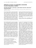
Immunostaining experiments for intracellular localization of IN in transfected cellsFigure 1
Immunostaining experiments for intracellular locali-
zation of IN in transfected cells. HeLaP4/IN-expressing
cells were generated by stable transfection into HeLaP4 cells
using pcDNA3.1 plasmid bearing the full wt IN gene. Cells
were fixed and immunostained using 1:100 rabbit a-IN and
second antibody, Cy3-conjugated anti-rabbit antibody as
described in Methods. Staining of IN (red) and DAPI (blue)
was observed under confocal microscope. Bar 10 μm.
Retrovirology 2009, 6:112 />Page 4 of 16
(page number not for citation purposes)
Essentially similar results were obtained when the degree
of nuclear import was quantitatively estimated using an
ELISA-based system and biotinylated BSA (Bb) conjugates
(Fig. 3F). A relatively high degree of nuclear import was
observed with both Bb-NLS-IN and Bb-IN, whereas
almost no import was observed with Bb-152-IN (Fig. 3E),
again emphasizing the role of NLS-IN in mediating
nuclear import of IN.
Sub-cellular localization of the full-length and truncated IN fused to GFP in transformed yeast cellsFigure 2
Sub-cellular localization of the full-length and trun-
cated IN fused to GFP in transformed yeast cells. (A)
Schematic presentation of the various expressed GFP-IN
fusion proteins used in this experiment. (B) W303 yeast cells
were transformed, using lithium acetate method, with
expression vectors coding for the following: GFP-IN, GFP-
180-IN, GFP
2
-NLS-IN, GFP-152-IN and GFP. Left panel, GFP
fluorescence (green); middle panel, DAPI staining (blue);
merged fluorescence is shown in the right panel. Bottom, a
line profile through the overlay image showing that maximum
GFP fluorescence and DAPI staining are co-localized (in the
nucleus). Yeast cells were grown to exponential phase in
selective minimal medium. After induction with galactose,
cells were harvested and GFP fluorescence was observed
under confocal microscope; all other conditions were as
described in Methods. Bar 7 μm.
Nuclear import mediated by recombinant HIV-1 IN protein: studies with microinjected and permeabilized mammalian cellsFigure 3
Nuclear import mediated by recombinant HIV-1 IN
protein: studies with microinjected and permeabi-
lized mammalian cells. Solutions containing the following
conjugates: (A) FITC-BSA-IN, (B) FITC-BSA-180-IN, (C)
FITC-BSA-NLS-IN, (D) FITC-BSA-152-IN and (E) FITC-
BSA*, were microinjected into the cytoplasm of cultured
COS-7 cells. All other experimental conditions were as
described in Methods. (F) Nuclear import was quantitatively
estimated by an ELISA-based assay system. Digitonin-perme-
abilized Colo-205 cells were incubated for 1 h with Bb-IN,
Bb-NLS-IN or Bb-152-IN (4 μg) in a final volume of 40 μL of
transport buffer containing ATP regeneration system. The
nuclear import experiments were repeated at least three
times; data given in the figure represent results obtained
from a single experiment. Error bars represent standard
deviation which is about +/-5%. Bar 10 μm.
Retrovirology 2009, 6:112 />Page 5 of 16
(page number not for citation purposes)
NLS-IN mediates binding to importin in yeast cells and in
vitro
Our results showing inhibition of IN nuclear import by
the SV40-NLS peptide indicated the involvement of
importin α in the translocation process. To verify this, we
used the yeast srp1-31 temperature-sensitive mutant [31]
in which importin α is inactivated following growth at the
non-permissive temperature of 37°C. GFP-IN appeared as
small fluorescent dots when expressed in the wild-type
W303 or in the srp1-31 mutant yeast cells grown at 25°C
(Fig. 4). These results clearly demonstrate accumulation
within the cells nuclei, a localization which was verified
by DAPI staining. Neither the cytosol nor the cell vacuoles
were strongly fluorescent. The same fluorescently stained
dots were observed after 4 h growth of the W303 cells at
37°C (Fig. 4), again indicating accumulation of GFP-IN
within the nuclei under these conditions. On the other
hand, the srp1-31 mutant yeast cells lost their nuclear
import ability at the non-permissive temperature (37°C):
most of the GFP-IN was distributed within the yeast cell's
cytoplasm (Fig. 4). However, nuclear localization was
restored in these mutant cells following re-incubation at
the permissive temperature of 25°C (not shown). To con-
firm that at the non-permissive temperature only impor-
tin α-dependent nuclear import is blocked, we repeated
previous experiments in which Pik1 protein [36] had been
shown to be imported into nuclei of srp1-31 cells at 37°C
(not shown and see [36]), and in which it was established
that nuclear import of this protein is importin α-inde-
pendent [36]. Thus, the blockage is specific to the impor-
tin α pathway in srp1-31 cells at the non-permissive
temperature.
A specific IN-importin α interaction in vivo can be inferred
also from the results obtained using the BiFC assay system
in yeast cells ([32] and Fig. 5). As a control system, to con-
firm that restoration of fluorescence following the use of
labeled IN is due to specific protein-protein interactions,
the BiFC assay system was first employed to study the
dimerization of the IN molecules themselves [37].
Indeed, intracellular fluorescence was seen in yeast cells
which expressed both the GN-IN and GC-IN constructs
(Fig. 5). No such fluorescence appeared in yeast cells
expressing the combination of GN-IN and GC-linker (Fig.
5), or the combination of GN-linker and GC-IN (not
shown), strengthening the view that the appearance of flu-
orescent dots resulted from specific IN-IN interaction.
Next, we examined the interaction between the transcrip-
tional co-activator LEDGF/p75 and IN (Fig. 5). LEDGF/
p75 is the dominant cellular binding partner of HIV-1 IN
in human cells [38]. Yeast cells were thus transformed
with the combination of mammalian importin α (impα)
and vectors expressing the various IN polypeptides. Fluo-
rescence, indicating a direct interaction between GN-IN
and GC-Impα, appeared in the yeast cell nuclei (Fig. 5).
Nuclear fluorescence was also detected following transfor-
mation with either GN-180-IN or GN-NLS-IN and GC-
Impα (Fig. 5). On the other hand, no fluorescence was
detected in yeast cells transformed with the combination
of GN-152-IN and GC-Impα (Fig. 5). Moreover, almost
no complementation occurred in yeast cells transformed
with the combination of GN-IN and GC-Impβ (importin
β) (Fig. 5), again indicating that the appearance of fluores-
cence resulted from specific interaction of the IN with
importin α. The same could be inferred from the negative
results obtained following transformation of yeast cells
with GN-Rev and GC-Impα: these yeast cells remained
completely dark, with no fluorescent signal (Fig. 5). It has
been well established that nuclear import of HIV-1 Rev is
mediated by importin β and not α [39].
Nuclear import of HIV-1 IN is importin α-dependentFigure 4
Nuclear import of HIV-1 IN is importin α-dependent.
W303 and in srp1-31 yeast cells were transformed with plas-
mid bearing the full length of the IN fused to GFP
(pYES
2
yEGFP-IN for the construction of the plasmid see
Methods). Following transformation using the lithium acetate
method, the fusion protein GFP-IN was expressed in the
yeast cells, as described in Methods. GFP fluorescence
(green) and DAPI (blue) were observed under confocal
microscope following growth of W303 yeast cells at 25°C or
at 37°C, or of srp1-31 yeast cells at 25°C or at the non-per-
missive temperature, 37°C. Bar 5 μm.
Retrovirology 2009, 6:112 />Page 6 of 16
(page number not for citation purposes)
Similar results were obtained when interactions were
tested by the ELISA-based system with various IN conju-
gates and the receptor importin α. Our quantitative esti-
mation revealed lower binding abilities by importin α
with the Bb-152-IN conjugates as compared to the bind-
ing ability of Bb-IN, of Bb-NLS-IN and Bb-SV40-NLS con-
jugates (Fig. 6). These results again indicate that amino
acids 161-173 are required for interaction with the impor-
tin α receptor.
IN interaction as observed by the BiFC assay systemFigure 5
IN interaction as observed by the BiFC assay system. EGY48 yeast cells were transformed using the lithium acetate
method with plasmids encoding the following combinations: GN-IN and GC-IN, GN-IN and GC-LEDGF, GN-IN and GC-
linker (control), GN-IN and GC-Impα (importin α), GN-180-IN and GC-Impα, GN-NLS-IN and GC-Impα, GN-152-IN and
GC-Impα, GN-IN and GC-Impβ (importin β), GN-Rev (HIV-1) and GC-Impα. Restoration of GFP fluorescence was observed
by confocal microscopy. All other experimental conditions were as described in Methods. Bar 10 μm.
Retrovirology 2009, 6:112 />Page 7 of 16
(page number not for citation purposes)
NLS-IN inhibits IN and cDNA nuclear import as well as
HIV-1 replication in cultured cells
In the light of the results showing the requirement of NLS-
IN for nuclear import of IN in mammalian as well as in
yeast cells, it became of relevance to study its effect in
virally infected cells.
Co-immunoprecipitation (co-IP) experiments using a
lysate obtained from HIV-1-infected cells revealed an
interaction between the virus IN protein and the cellular
importin α (Fig. 7A). Interestingly, when the virus-
infected cells were treated with either the NLS-IN-Pen or
the SV40-NLS-Pen peptides, no such interaction between
the two proteins could be detected (Fig. 7A). Specificity of
the peptide effect can be inferred from the results showing
that neither the SV40-mut-NLS-Pen (or a scrambled NLS-
IN [23], not shown) nor the Pen peptide itself promoted
dissociation of the IN-importin α complex (Fig. 7A).
Inhibition of nuclear import of the viral IN in HIV-1
infected cells as well is evident from the immunofluores-
cence microscopic study shown in Fig. 7B. From the
immunostaining results, it appears that in infected cells,
the IN is localized both within the cytosol and within the
nuclei (Fig. 7B (no peptide)). However, no intranuclear
fluorescence was observed in cells treated with the NLS-
IN-Pen or the SV40-NLS-Pen peptides, indicating the inhi-
bition of nuclear import (Fig. 7B). In contrast, some intra-
nuclear fluorescently labeled IN could be observed when
the infected cells were incubated in the presence of the
SV40-mut-NLS-Pen or the Pen peptide itself (Fig. 7B).
This is also evident from the fact that more cytosolic IN
was present in such peptide-treated cells than in those
incubated in the absence of any peptide or with the non-
active peptides (Fig. 7B).
Similar to their effect on IN nuclear import, both NLS-IN-
Pen and SV40-NLS-Pen blocked nuclear import of the
viral cDNA (Fig. 8A), as is particularly evident from the
absence of 2LTR circles (Fig. 8B) in cells infected with
wild-type HIV-1.
Inhibition of IN nuclear import is expected to result in the
inhibition of virus replication, especially in cell-cycle
arrested cells. Using TZM-bl cells [40] treated with aphidi-
colin to obtain cell-cycle arrested cells as an experimental
system, a reduction in HIV-1 infection as is reflected by
the inhibition of reporter gene expression was observed
in the presence of NLS-IN-Pen or SV40-NLS-Pen (Fig. 9A).
As expected, the inhibition was less pronounced when
dividing cells were treated with the NLS-bearing peptides
(Fig. 9B). The specific effect of NLS-IN and the require-
ment for cell permeability can be inferred from the results
showing that no inhibition of HIV-1 infection was pro-
moted by the Pen peptide alone or by the impermeable
NLS-IN peptides. The results in Fig. 9C and 9D clearly
demonstrate that the NLS-IN-Pen peptide due to its inhib-
itory effect on IN nuclear import inhibited the process of
viral cDNA integration, reaching a higher degree of inhi-
bition in non-dividing (cell-cycle arrested) than in divid-
ing cells. Detailed kinetics studies (Fig. 9E) further
support the view that the step which is blocked by the two
NLS-Pen peptides (IN-NLS and SV40-NLS) is the nuclear
import process. Evidently, nuclear import of the IN-DNA
complex is required for the integration process to proceed.
In addition, the time-dependent inhibitory pattern of the
NLS-IN-Pen (Fig. 9E) is almost exactly half the way
between that observed following the addition of AZT and
that of the LEDGF 402-411 peptide, which has been dem-
onstrated to directly block HIV-1 IN and thus the integra-
tion process [41]. Inhibition was not observed following
the addition of the non-permeable NLS-IN peptide, a pep-
tide bearing a SV40-mut-NLS-Pen or the Pen peptide,
again indicating the specific effect of the NLS sequence
(Fig. 9).
Discussion
The question of how retroviruses and particularly HIV-1
cross the nuclear envelope in cell-cycle arrested cells is of
specific scientific interest. After long and extensive
research, it appears that no clear mechanism has yet
emerged and the possibility that several pathways simul-
taneously mediate nuclear import of the viral PIC cannot
be excluded. In the present work-following our previous
experiments using in vitro systems [23]-we focused on the
nuclear import of IN protein using yeast and mammalian
Binding of IN to importin α as estimated by an ELISA-based systemFigure 6
Binding of IN to importin α as estimated by an
ELISA-based system. Importin α-coated ELISA plates
were incubated with increasing amounts of the following
biotinylated conjugates: SV40 (black circles), IN (white dia-
mond), NLS-IN (black squares) and 152-IN (black triangles).
The degree of binding was estimated as described in Meth-
ods. Error bars represent standard deviation which is about
+/-5%.
Retrovirology 2009, 6:112 />Page 8 of 16
(page number not for citation purposes)
NLS-IN-Pen inhibits IN nuclear import by the dissociation of IN-importin α interaction in HIV-infected cellsFigure 7
NLS-IN-Pen inhibits IN nuclear import by the dissociation of IN-importin α interaction in HIV-infected cells.
(A) H9 lymphocytes were infected by wild-type HIV-1, and after infection half of the cells' lysate volume was subjected to SDS-
PAGE, then immunoblotted with either by anti-IN, anti-importin α (anti-Impα) antibody or an anti-actin antibody. The comple-
mentary HRP-conjugated antibodies were used as the second antibody. The remaining lysate or isolated fractions were co-IP
with either the anti-Impα or anti-IN antibodies and were immunoblotted with these antibodies, and the complementary HRP-
conjugated antibodies as second antibodies. When peptides were used, cells were incubated with 150 μM of the indicated pep-
tide. All others experimental details were as described in Methods. (B) HeLaP4 cells were infected and immunostained as
described in Methods. IN (red); DAPI (blue); the area marked in the merge picture was magnified for a better view of IN local-
ization within the infected cell. Bar 10 μm.
Retrovirology 2009, 6:112 />Page 9 of 16
(page number not for citation purposes)
cells, as well as on the contribution of its putative NLS-IN
[23] on the HIV-1 replication process.
An interaction between HIV-1 IN and importin α was first
demonstrated by Gallay et al. [8]. Similarly an interaction
between the IN and members of the importin family and
that its nuclear transport appears to be dependent on the
importin α/β heterodimer have also demonstrated by
Hearps and Jans [25]. A functional NLS sequence was
identified between amino acids 161 and 173 of the IN
protein whose mutation disrupted IN translocation into
nuclei [12,42]. However, later studies indicated that this
sequence may be required for promoting viral DNA inte-
gration and not necessarily nuclear import [43]. However,
our previous work clearly demonstrated that a peptide
bearing IN 161-173 residues can mediate import of a con-
jugated protein into the nuclei of permeabilized cells,
confirming the view that it can function as a NLS [23].
In the present work, we have further studied the involve-
ment of IN amino acids 161-173 in promoting its nuclear
import. Nuclear accumulation was observed in yeast
transformed with an expression vector bearing only the
NLS-IN sequence, which in order to avoid diffusion into
the nuclei, was fused to two molecules of GFP, resulting in
a molecule of about 58 kDa. It is assumed that molecules
of up to about 30 to 40 kDa can passively diffuse via the
NPCs into cell nuclei [2]. Therefore, the nuclear import
observed here with the various GFP-IN conjugates, the
molecular weights of which varied between 48 and 60
kDa, should be ascribed to a receptor-mediated, active
process. Nuclear import was practically not observed
when yeast cells were transformed with the 152-IN trun-
cated protein, which lacks the putative NLS sequence.
Results obtained in permeabilized or microinjected cells,
as well as in IN-transfected intact mammalian cells, fur-
ther supported these results. The failure of 152-IN to pen-
etrate the cell nuclei suggests that if an additional NLS
sequence, besides the one located between 161 and 173,
were present, it must be located closer to the C terminus
of the IN protein, a possibility that has been suggested
previously [25,44]. However, the fact that the truncated
180-IN protein was translocated into the cell nuclei indi-
cates that the identified NLS-IN is sufficient to provide the
IN with karyophilic properties. Our results demonstrating
inhibition of IN nuclear import in transfected cells by a
peptide carrying the NLS-IN sequence further emphasize
the view that this sequence gives IN its karyophilic prop-
erties.
SRP1 is the only known importin α protein in budding
yeast, and previous studies have demonstrated that it is
essential for proper maintenance of nucleocytoplasmic
trafficking [45]. Indeed, a temperature-sensitive mutant in
SRP1 has been isolated (srp1-31) and shown to be defec-
tive in nuclear import at the non-permissive temperature
[31]. Thus, yeast, including SRP1-mutated strains, has
been instrumental in studying various aspects of the
nuclear import machinery [36,46] and in characterizing
karyophilic properties. The availability of the tempera-
ture-sensitive srp1-31 mutant offers an advantage for stud-
ying nuclear transport of the IN protein in yeast cells. The
involvement of importin α in the IN nuclear import path-
way can be inferred from our experiments showing
nuclear import at the permissive, but not at the non-per-
missive temperature where importin α is inactivated [31].
A direct and specific interaction between IN or the NLS-IN
sequence and the mammalian importin α in vivo, within
the intracellular environment, was demonstrated using
the BiFC assay. The view that restoration of GFP fluores-
cence using the BiFC assay in yeast cells results from a spe-
cific protein-protein interaction has already been
established [47,48]. Indeed, our results clearly demon-
strated the well-known IN-IN and IN-LEDGF/p75 interac-
tions in yeast cells. Next we showed restoration of
fluorescence in yeast cells expressing the combination of
importin α and the full-length IN or the truncated 180-IN.
The combination of importin α and the truncated 152-IN
protein did not result in the appearance of fluorescence,
strongly indicate that the NLS-IN sequence is located
between amino acids 152 and 180, and is necessary to
mediate the interaction with the nuclear receptor. How-
NLS-IN-Pen inhibits nuclear import of viral DNAFigure 8
NLS-IN-Pen inhibits nuclear import of viral DNA. H9
lymphocytes were infected by wild-type HIV-1 at a MOI of 1;
and (A) following infection, the nuclei fraction was isolated
from half of the cells, and the amount of viral DNA was esti-
mated using real time PCR method. (B) The amount of 2LTR
circles was estimated using real time PCR method. All other
experimental details are as described in Methods. Error bars
represent standard deviation which is about +/-5%
Retrovirology 2009, 6:112 />Page 10 of 16
(page number not for citation purposes)
NLS-IN-Pen peptide inhibits HIV-1Figure 9
NLS-IN-Pen peptide inhibits HIV-1. (A) Cell-cycle arrested TZM-b1 cells (non-dividing cells) were incubated with the des-
ignated peptides at the indicated concentrations and after HIV-1 infection were tested for β-galactosidase activity. (B) Experi-
mental conditions were as in (A), but with dividing TZM-b1 cells. The number of integration events per cell was determined in
cell-cycle arrested (non-dividing) cells (C) or dividing cells (D) following incubation with the designated peptides at different
concentrations. Cells were infected with HIV-1 at a MOI of 1 as described in Methods. (E) Inhibition of HIV-1 replication by
NLS-IN-Pen as well as SV40-NLS-Pen is dependent on its time of addition. Sup-T1 cells were infected with HIV-1 at a MOI of
2, and the indicated elements were added at different time points after infection (0, 2, 4, , 24 h). Viral p24 production was
determined 48 h PI. Error bars represent standard deviation which is about +/-5%. All other experimental conditions are as
described in Methods.
Retrovirology 2009, 6:112 />Page 11 of 16
(page number not for citation purposes)
ever, our results do not exclude the possibility that addi-
tional NLS sequences are located between amino acid 180
and the C terminus, as suggested previously [25,44].
Attempts to study the interaction of fragments bearing
these regions failed due to non-specific restoration of flu-
orescence (not shown). A similar pattern also character-
ized the in vitro interaction of the various INs. Our co-IP
experiments confirmed the interaction between IN and
importin α in virally infected cells. Furthermore, the view
that such an interaction is mediated by the putative NLS-
IN was supported by the results showing disruption of this
interaction by the cell permeable NLS-IN or SV40-NLS
peptides.
The NLS-IN-Pen peptide almost totally blocked viral
infection of TZM-bl cells, caused high inhibition of the
cDNA integration process, and affected p24 production
when added up to 18 h post-infection (PI). Inhibitory
effects were much higher in cell-cycle arrested than in
dividing cells, clearly supporting the notion that the NLS-
IN-Pen bears a sequence which is involved in mediating
nuclear import of the IN. Inhibition of IN as well as of
viral cDNA nuclear import by the NLS-IN-Pen peptide was
demonstrated directly in the present work using immun-
ofluorescence staining of IN and quantitative estimation
of nuclear viral DNA.
The amino acid residues of NLS-IN described in the
present work have been implicated in additional viral-
related functions, such as specific binding of IN to the
LEDGF/p75 protein and to the viral LTR region
[38,49,50]. Due to its multifunctional activity, this region
may be useful as a target for the development of inhibi-
tors. As mentioned above our present as well as previous
results describing the involvement of importin α in medi-
ating nuclear import of IN do not exclude the possibility
of an additional nuclear import pathway for IN in which
TNPO3 is involved.
Methods
Mammalian, bacterial and yeast cells
Monolayer adherent HeLaP4 and HeLa TZM-bl cells
(obtained through the NIH AIDS Research and Reference
Reagent Program) expressing the β-galactosidase gene
under regulation of the transactivation response element
[51] were grown in Dulbecco's modified Eagle's medium.
COS-7 and Colo-205 mammalian cells and the T-lym-
phocyte cell lines H9 and Sup-T1 were grown in RPMI
1640. All media were supplemented with 10% (v/v) fetal
calf serum, 0.3 g/l L-glutamine, 100 units/ml penicillin,
and 100 units/ml streptomycin (Biological Industries,
Beit Haemek, Israel). Cells were incubated at 37°C in a
5% CO
2
atmosphere and re-cultured every 4 days.
HeLaP4/IN-expressing cells were generated by stable
transfection into HeLaP4 cells [52] of pcDNA3.1 plasmid
bearing the full wt IN gene. Selection was carried out for
four weeks with 400 μg/ml Hygromycin B. Escherichia coli
strain DH5α served as the host for general plasmid con-
struction and maintenance, and E. coli strain BL21 (DE3)
was used for protein overexpression. The yeast strains
were congenic to Saccharomyces cerevisiae W303. The
W303 (MAT a, leu2-3, 112 trp1-1 ura3-1 ade2-1 his3-
11,15) and srp1-31 (MAT a, leu2-3, 112 trp1-1 ura3-1 ade2-
1 his3-11,15, srp1-31) strains were kind gifts from G. Fink
(Whitehead Institute, Massachusetts Institute of Technol-
ogy, USA). The EGY48 strain (MAT a, his3-11,15, trp1-1,
ura3-52, leu2::LexA6op-LEU2) was a kind gift from Y.
Gafni (The Volcani Center, Israel). Yeast strains were
grown in yeast peptone dextrose-rich medium (2% pep-
tone, 2% glucose, 1% yeast extract, w/v). Transformation
was performed by the lithium acetate method [53,54] and
then the yeast cells were grown in standard yeast nitrogen
(YNB) minimal media, prepared by adding the appropri-
ate amino acids to 0.67% (w/v) YNB without amino acids
(Difco) and supplemented with 2% glucose or 2% galac-
tose (w/v) as carbon sources.
Viruses
Wild-type HIV-1 was generated by transfection of
HEK293T cells with pSVC21 plasmid containing the full-
length HIV-1 HXB2 viral DNA. Wild-type viruses were
harvested from HEK293T cells 48 and 72 h post-transfec-
tion. The viruses were stored at -75°C.
Virus stock titration
Quantitative titration of HIV-1 was carried out using the
MAGI assay, as described by Kimpton and Emerman [40].
Briefly, TZM-b1 cells were grown in 96-well plates at 1 ×
10
4
cells per well. the cells were infected with 50 μl of seri-
ally diluted virus as described [40]. Two days post-infec-
tion (PI), cultured cells were fixed and β-galactosidase was
estimated exactly as described previously [40]. Blue cells
were counted under a light microscope at 200× magnifica-
tion.
Synthesis of peptides
Peptides were synthesized on Rink amide resin using a
model 433A Applied Biosystems peptide synthesizer with
FastMoc chemistry, exactly as described previously [55].
The Pen peptide had the following sequence: RQIKIW-
FQNRRMKWKK (Ant 43-58) [34].
Plasmid construction
All of the plasmids used in this study were constructed
using PCR cloning techniques with the high-fidelity
enzyme Platinum Pfx DNA polymerase (Invitrogen).
Clones were subjected to automated DNA sequencing.
1. Site-directed mutagenesis to create a stop codon at position 152
or 180
The plasmid pT7-7-IN [56] was used as the template for
IN mutagenesis. A QuikChange site-directed mutagenesis
Retrovirology 2009, 6:112 />Page 12 of 16
(page number not for citation purposes)
kit (Stratagene) was adapted to create an in-frame stop
codon at the desired position within the IN sequence,
according to the manufacturer's protocol. The mutagenic
primers were designed to contain a stop codon after resi-
dues 152/180 of the IN giving pT7-7-152-IN/180-IN,
respectively, as well as a new DraI site, to facilitate screen-
ing. The primers used were:
152-IN: 5'-CCCGCAGTCTCAGGGTGTTGTTTAAACTAT-
GAACAAAGAGCTC-3'
180-IN: 5'-CCGCGGTTCAGATGGCTGTTTAAACCACAA
CAAGAAACG-3'
2. Construction of plasmids for GFP in yeast cells
The yeast expression cloning plasmid pYES
2
yEGFP with
the GFP codon optimized (a kind gift from T. Gilon, Alex-
ander Silberman Institute, Israel) was linearized with the
BsrGI restriction enzyme at the stop codon site of the
yEGFP sequence [57], and then dephosphorylated and
purified. The DNA products of IN, 152-IN, and 180-IN
were obtained by PCR amplification from the pT7-7-152-
IN/180-IN, respectively. The primers used were:
BsrGI, SalI-IN: 5'-CCGGCGTGTACAAAAGTCGACTAAT-
GCACCACCATCACCAT-3'
IN-BamHI, BsrGI: 5'-GCCGGATGTACAGGATCCCCG-
GGCGCG-3'
These DNA products were then cloned into the BsrGI sites
of the linearized pYES
2
yEGFP, resulting in the formation
of pYES
2
yEGFP-IN (GFP-IN), pYES
2
yEGFP-152-IN (GFP-
152-IN) and pYES
2
yEGFP-180-IN (GFP-180-IN). A GFP
2
-
NLS-IN (amino acids 161 to 173 of IN) expression vector
was prepared by PCR amplification from pT7-7-180-IN.
The primers used were: 5'BsrGI: 5'-CCGCCATGTACAAA-
GAGCTCAAAAAAATCATCGGTCAG-3' 3 BsrGI: 5'-GCG-
GTATGTACACCAGCAGAGTAACCACCGATAC-3' The
resultant DNA products were cloned into pBS-yEGFP,
resulting in pBS-yEGFP-NLS-IN, which was digested by
SalI and BamHI, and the resultant product was subcloned
into pYES
2
yEGFP to give pYES
2
yEGFP
(2)
-NLS-IN (GFP
2
-
NLS-IN).
3. Construction of expression vectors for the BiFC assay
The yeast multicopy shuttle vectors pRS423 (with HIS3 as
the selective marker) and pRS426 (with URA3 as the selec-
tive marker), both with the ADH1 promoter, were used as
the cloning plasmids (a kind gift from D. Engelberg, Alex-
ander Silberman Institute, Israel). The DNA coding region
of the two GFP fragments [58], namely the N terminus
(GN) and C terminus (GC), were cloned into pRS423 and
pRS426 [59] to give pRS423-GN and pRS426-GC, respec-
tively. A linker consisting of (GGS)
5
was used to separate
the inserted genes and the GFP fragments. The coding
sequences of IN and HIV-1 Rev [60] were amplified by
PCR, digested by XmaI and NotI and inserted in-frame
into the corresponding sites of pRS423-GN at the C-termi-
nal fragments of the GN, resulting in pRS423-GN-IN (GN-
IN) and pRS423-GN-Rev (GN-Rev), respectively. The PCR
products of 152-IN and 180-IN were ligated into the SalI
and SacII sites of pRS423-GN to give pRS423-GN-152/
180 (GN-152-IN/GN-180-IN). The plasmid pRS423-GN-
NLS-IN (GN-NLS-IN) was obtained by PCR amplification
of the NLS-IN sequence, digested by SacI and EcoRI and
ligated at the corresponding sites of pRS423-GN. The PCR
products of IN, importin α (hSRP1-α), importin β and
LEDGF were cloned into the corresponding XmaI and
NotI sites of pRS426-GC, to construct the following plas-
mids, respectively: pRS426-GC-IN (GC-IN), pRS426-GC-
mammalian importin α (GC-Impα), pRS426-GC-impor-
tin β (GC-Impβ) and pRS426-GC-LEDGF (GC-LEDGF,
PCR amplification from pET28-LEDGF a kind gift from
C.A. Casiano, Loma Linda University, USA). The
sequences of all primers used in this work can be obtained
directly from the authors.
Nuclear import of IN molecules in transformed yeast cells
Expression vectors bearing the GFP, GFP-IN, GFP-180-IN,
GFP-152-IN, GFP
2
-NLS-IN and GFP-Pik1 coding regions
under the galactose promoter (a generous gift from Dr.
Thorner, UC Berkeley [36]) were introduced into the yeast
strain W303. Expression was induced with 2% galactose
for 4 h at 25°C. Following removal of the medium, the
appearance of intracellular fluorescence was examined by
confocal microscopy using an MRC 1024 confocal imag-
ing system (Bio-Rad). Similarly, W303 and the srp1-31
yeast mutant were transformed by pYES2yEGFP-IN (GFP-
IN). The strain srp1-31 contains a temperature-sensitive
mutation in the SRP1 protein (importin α) [31,61]. The
transformed yeast cells were grown for 24 h at 25°C to
reach the logarithmic phase, then divided into two cul-
tures: one remained at 25°C (permissive temperature)
and the other was transferred to 37°C (non-permissive
temperature at which more than 95% of the SRP1 is inac-
tivated [61]). After 4 h of growth, yeast cells were washed;
GFP-IN expression was induced by the addition of galac-
tose and the yeast cells were incubated for an additional 4
h at either 25 or 37°C. At the end of the growth period,
yeast cells were harvested and then observed by confocal
microscope. Yeast nuclei were identified by DNA staining
in fixed yeast cells (4% v/v paraformaldehyde and 3.4%
w/v sucrose) />Protocols/MICROSCOPY/gfpfix.html with 4',6-diamid-
ino-2-phenylindole (DAPI). To confirm expression of all
proteins, western blot with anti-GFP antibody was per-
formed. Each experiment was repeated at least three times.
Retrovirology 2009, 6:112 />Page 13 of 16
(page number not for citation purposes)
Analysis of protein-protein interaction by the BiFC assay
In this approach, a molecule of GFP is separated into two
portions: the N-terminal part (GN) ending at amino acid
residue 154 and the C-terminal part (GC) beginning with
the methionine residue preceding residue 155 of GFP
[62]. Neither of these two halves of GFP fluoresce when
expressed alone; however, the fluorescence is restored
when GN and GC are brought together as fusions with
interacting proteins [32]. The two halves of the GFP were
cloned separately with each of the indicated proteins as
described above and the different plasmids were trans-
formed into the yeast strain EGY48. After 48 h at 30°C,
the plates were transferred to 23°C for 3 days and then
yeast cells were visualized by confocal microscopy. Nuclei
were stained with DAPI as described above. Each experi-
ment was repeated at least three times.
Recombinant proteins
Expression and purification of the recombinant mamma-
lian importin α (hSRP1-α) and HIV-1 IN proteins were
performed essentially as described previously [23]. The
vectors, as well as the methods used to obtain purified
recombinant truncated IN bearing amino acids 1-180
(180-IN) or amino acids 1-152 (152-IN) were the same as
described for the full-length IN [23].
Nuclear import transport substrates
Proteins and peptides used in this work as transport sub-
strates were covalently attached to either fluorescein iso-
thiocyanate-labeled bovine serum albumin (FITC-BSA) or
biotinylated BSA (Bb) molecules (Sigma). Sulfosuccinim-
idyl-4-(N-maleimidomethyl) cyclohexane-1-carboxylate
(sulfo-SMCC) was used as the cross-linker to give FITC-
BSA-protein/peptide and Bb-protein/peptide conjugates,
as described previously [23].
Binding of transport substrates to importin
Binding to importin α was estimated using an ELISA-
based system essentially as described in [23,63]. Briefly,
Maxisorp plates (Nunc) were coated overnight at 4°C
with a solution containing recombinant importin α (5 μg)
in carbonate buffer (NaHCO
3
/Na
2
CO
3
buffer at pH 9.6).
All subsequent steps were as described previously [23].
Microinjection of fluorescently labeled transport
substrates
FITC-BSA-IN, FITC-BSA-180-IN, FITC-BSA-152-IN, FITC-
BSA-NLS-IN, as well as unlabeled FITC-BSA, were micro-
injected into the cytoplasm of cultured COS-7 cells as
described previously [64]. The CompInject AIS2 auto-
mated microinjection system (Cell Biology Trading, Ham-
burg, Germany) was used following the method
developed by Graessmann and Graessman [65]. Cultured
mammalian cells were incubated at 37°C for 2 h after
injection and observed by fluorescence microscopy.
Nuclear import in permeabilized mammalian cells:
quantitative estimation
Import of Bb-IN proteins or of Bb-NLS-IN conjugates into
nuclei of digitonin-permeabilized Colo-205 cells was
determined quantitatively by an ELISA-based system,
exactly as described previously [23].
Localization studies of transfected IN in cultured cells by
immunostaining
HeLaP4/IN-expressing cells were grown on chamber
slides (Nunc). After reaching 70-80% confluence, cells
were arrested in the cell cycle by treatment with 5 μg/ml
of aphidicolin, and then incubated with 150 μM of the
indicated peptide for 6 h. Cells were fixed and immunos-
tained essentially as described previously [66] with several
modifications. Briefly, after fixation cells were blocked
with 5% (w/v) BSA (IgG free) (Jackson) in PBS for 60 min.
For detection of HIV-1 IN, cells were incubated with 1:100
rabbit α-IN (NIH AIDS Research & Reference Reagent Pro-
gram, cat. no. 758) at room temperature for 60 min each.
Cells were washed five times with PBS + 0.05% (v/v)
Tween 20. Then the cells were incubated with a second
antibody, Cy3-conjugated anti-rabbit antibody (Jackson)
(1:200) at room temperature for 60 min followed by five
washes with PBS + 0.05% Tween 20. For detection of
DNA, cells were stained with DAPI according to the man-
ufacturer's protocol. The slides were prepared with
mounting media (Bio-Rad) and immunofluorescent cells
were detected with a confocal microscope.
Localization studies of IN in HIV-1-infected cultured cells
by immunostaining
HeLaP4 cells were grown on chamber slides. Cells were
arrested in the cell cycle by treatment with 5 μg/ml aphidi-
colin, and then incubated with 150 μM of the indicated
peptide for 2 h. After incubation with the peptides, cells
were infected with wild-type HIV-1 at a multiplicity of
infection (MOI) of 25. Cells were fixed and stained as
described above with the following modifications: fixa-
tion was performed at 10 h PI, the first antibody was used
at a dilution of 1:50, the second antibody at a dilution of
1:100.
Study of in vivo protein-protein interactions using co-IP
Cells were infected with a MOI of 15 of the indicated
viruses, harvested at 10 h post-infection (PI), washed
three times with PBS and lysed by the addition of PBS con-
taining 1% (v/v) Triton X-100. Half of the lysate volume
was subjected to SDS-PAGE, then immunoblotted with
either antiserum raised against IN amino acids 276-288
(anti-IN) (NIH AIDS Research & Reference Reagent Pro-
gram cat. no. 758), anti-importin α (anti-Impα) antibody
(Santa Cruz), or an anti-actin (anti-Actin) antibody (Santa
Cruz) The complementary HRP-conjugated antibodies
(Jackson) were used as the second antibody.
Retrovirology 2009, 6:112 />Page 14 of 16
(page number not for citation purposes)
The remaining lysate or isolated fractions were incubated
for 1 h at 4°C with either the anti-Impα or anti-IN anti-
bodies. Following 3 h incubation with protein G-agarose
beads (Santa Cruz) at 4°C, the samples were washed three
times with PBS containing 1% (v/v) Nonidet P-40. SDS
buffer was added to the samples and after boiling and run-
ning on an SDS polyacrylamide gel, the membranes were
immunoblotted with either anti-Impα or anti-IN antibod-
ies, and the complementary HRP-conjugated antibodies
(Jackson) as second antibodies.
When peptides were used, cells were incubated with 150
μM of the indicated peptide for 2 h prior to infection.
HIV-1 titration by multinuclear activation of a
galactosidase indicator (MAGI) assay
Quantitative titration of HIV-1 was carried out using the
MAGI assay, as described previously [40]. Briefly, TZM-b1
cells were grown in 96-well plates at 10
4
cells/well and
incubated for 12 h at 37°C. Peptides were then added and
after an additional 2 h of incubation, the cells were
infected with 50 μl of serially diluted HIV-1. To obtain
cell-cycle arrested cells, 5 μg/ml of aphidicolin was added
2 h before the experiment. Cultured cells were fixed 2 days
PI and β-galactosidase was estimated [47]. Blue cells were
counted under a light microscope at 200× magnification.
Quantitative analysis of integration
Real-time PCR experiments were performed to estimate
integration as described previously [67].
Time-of-addition assay
Sup-T1 cells were infected with wild-type HIV-1 at a MOI
of 2, and the test compounds were added at different time
points after infection (0, 2, 4, , 24 h). Viral p24 produc-
tion was determined at 48 h PI [67]. Dextran sulfate was
tested at 20 μM, AZT at 2 μM, NLS-IN-Pen, NLS-IN and
Pen at 62.5 μM, LEDGF 402-411 at 12.5 μM [41].
Isolation of cytoplasm and nuclei from infected cells
The various fractions were obtained from virus-infected
cells essentially as described previously [68,69] with sev-
eral modifications. Briefly, cells were harvested and
washed twice in buffer A (20 mM Hepes pH 7.3, 150 mM
KCl, 5 mM MgCl
2
, 1 mM DTT and 0.1 mM PMSF). Cells
were then suspended in 200 μl of buffer A with 0.025%
(w/v) digitonin and incubated at room temperature for 10
min. Cells were centrifuged for 3 min at 1000 g at room
temperature. The supernatant was then centrifuged at
8000 g and separated into supernatant (cytoplasm) and
pellet (nuclei) and stored at -70°C.
Quantitation of total and nuclear viral DNA
Total viral DNA was estimated using SYBR green real-time
quantitative PCR at 10 h PI from the total or nuclear-iso-
lated fractions of the infected cells. DNA was isolated by
phenol chloroform method. Briefly, DNA samples (1 μg
of DNA) were added to 95 μl containing 1× Hot-Rescue
Real Time PCR Kit-SG (Diatheva s.r.l, Fano, Italy), and
100 nM of each PBS (primer-binding site) primer: F5 (5'
primer, 5'-TAGCAGTGGCGCCCGA-3') and R5 (3'
primer, 5-TCTCTCTCCTTCTAGCCTCCGC-3'). All ampli-
fication reactions were carried out using an ABI Prism
7700 Sequence Detection System (Applied Biosystems):
One cycle at 95°C for 10 min, followed by 45 cycles of 15
s at 95°C and 35 s at 68°C. In each PCR run, three repli-
cates were performed. All other details are exactly as
described in Casabianca et al. [70].
Quantitation of 2LTR circles
Quantification of 2LTR circles was estimated exactly as
described in Butler et al. [71].
Competing interests
The authors declare that they have no competing interests.
Authors' contributions
ALevin designed and performed experiments (immunos-
taining, co-IP and HIV-1 tests), analyzed data and contrib-
uted to writing the paper; AAO designed and performed
experiments, analyzed data and contributed to writing the
paper; JR contributed to the study design of the BiFC
assay; NMB provided technical support and contributed
to the confocal images; AG performed the microinjection
experiments; EW evaluated the manuscript; ALoyter
designed the study, contributed to writing the paper and
coordinated the study. All authors have read and
approved the manuscript.
Acknowledgements
The authors are grateful to J. Thorner (Berkeley) for providing us with the
GFP-Pik1 plasmids. This work was supported by grants from the Israel Sci-
ence Foundation (ISF; grant no. 888/05) and Jubiläumsfonds of the Austrian
National Bank (Project No. 11462 to A. Loyter and EW).
References
1. Stewart M: Molecular mechanism of the nuclear protein
import cycle. Nat Rev Mol Cell Biol 2007, 8:195-208.
2. Gorlich D, Kutay U: Transport between the cell nucleus and
the cytoplasm. Annu Rev Cell Dev Biol 1999, 15:607-660.
3. Feldherr C, Akin D, Littlewood T, Stewart M: The molecular
mechanism of translocation through the nuclear pore com-
plex is highly conserved. J Cell Sci 2002, 115:2997-3005.
4. Lewis P, Hensel M, Emerman M: Human immunodeficiency virus
infection of cells arrested in the cell cycle. EMBO J 1992,
11:3053-3058.
5. Katz RA, Greger JG, Boimel P, Skalka AM: Human immunodefi-
ciency virus type 1 DNA nuclear import and integration are
mitosis independent in cycling cells. J Virol 2003,
77:13412-13417.
6. Miller MD, Farnet CM, Bushman FD: Human immunodeficiency
virus type 1 preintegration complexes: studies of organiza-
tion and composition. J Virol 1997, 71:5382-5390.
7. Suzuki Y, Craigie R: The road to chromatin - nuclear entry of
retroviruses. Nat Rev Microbiol 2007, 5:187-196.
8. Gallay P, Hope T, Chin D, Trono D: HIV-1 infection of nondivid-
ing cells through the recognition of integrase by the impor-
Retrovirology 2009, 6:112 />Page 15 of 16
(page number not for citation purposes)
tin/karyopherin pathway. Proc Natl Acad Sci USA 1997,
94:9825-9830.
9. Haffar OK, Popov S, Dubrovsky L, Agostini I, Tang H, Pushkarsky T,
Nadler SG, Bukrinsky M: Two nuclear localization signals in the
HIV-1 matrix protein regulate nuclear import of the HIV-1
pre-integration complex. J Mol Biol 2000, 299:359-368.
10. Jenkins Y, McEntee M, Weis K, Greene WC: Characterization of
HIV-1 vpr nuclear import: analysis of signals and pathways. J
Cell Biol 1998, 143:875-885.
11. Zennou V, Petit C, Guetard D, Nerhbass U, Montagnier L, Charneau
P: HIV-1 genome nuclear import is mediated by a central
DNA flap. Cell 2000, 101:173-185.
12. Llano M, Vanegas M, Fregoso O, Saenz D, Chung S, Peretz M, Poe-
schla EM: LEDGF/p75 determines cellular trafficking of
diverse lentiviral but not murine oncoretroviral integrase
proteins and is a component of functional lentiviral preinte-
gration complexes. J Virol 2004, 78:9524-9537.
13. Maertens G, Cherepanov P, Pluymers W, Busschots K, De Clercq E,
Debyser Z, Engelborghs Y: LEDGF/p75 is essential for nuclear
and chromosomal targeting of HIV-1 integrase in human
cells. J Biol Chem 2003, 278:33528-33539.
14. Yamashita M, Perez O, Hope TJ, Emerman M: Evidence for direct
involvement of the capsid protein in HIV infection of nondi-
viding cells. PLoS Pathog 2007, 3:1502-1510.
15. Sherman MP, Greene WC: Slipping through the door: HIV entry
into the nucleus. Microbes Infect 2002, 4:67-73.
16. Woodward CL, Wang Y, Dixon WJ, Htun H, Chow SA: Subcellular
localization of feline immunodeficiency virus integrase and
mapping of its karyophilic determinant. J Virol 2003,
77:4516-4527.
17. Zheng R, Jenkins TM, Craigie R: Zinc folds the N-terminal
domain of HIV-1 integrase, promotes multimerization, and
enhances catalytic activity. Proc Natl Acad Sci USA 1996,
93:
13659-13664.
18. Kulkosky J, Jones KS, Katz RA, Mack JP, Skalka AM: Residues critical
for retroviral integrative recombination in a region that is
highly conserved among retroviral/retrotransposon inte-
grases and bacterial insertion sequence transposases. Mol Cell
Biol 1992, 12:2331-2338.
19. Chiu TK, Davies DR: Structure and function of HIV-1 integrase.
Curr Top Med Chem 2004, 4:965-977.
20. Esposito D, Craigie R: HIV integrase structure and function. Adv
Virus Res 1999, 52:319-333.
21. Chen Z, Yan Y, Munshi S, Li Y, Zugay-Murphy J, Xu B, Witmer M,
Felock P, Wolfe A, Sardana V, Emini EA, Hazuda D, Kuo LC: X-ray
structure of simian immunodeficiency virus integrase con-
taining the core and C-terminal domain (residues 50-293)
an initial glance of the viral DNA binding platform. J Mol Biol
2000, 296:521-533.
22. Pluymers W, Cherepanov P, Schols D, De Clercq E, Debyser Z:
Nuclear localization of human immunodeficiency virus type
1 integrase expressed as a fusion protein with green fluores-
cent protein. Virology 1999, 258:327-332.
23. Armon-Omer A, Graessmann A, Loyter A: A synthetic peptide
bearing the HIV-1 integrase 161-173 amino acid residues
mediates active nuclear import and binding to importin
alpha: characterization of a functional nuclear localization
signal. J Mol Biol 2004, 336:1117-1128.
24. Depienne C, Mousnier A, Leh H, Le Rouzic E, Dormont D, Benichou
S, Dargemont C: Characterization of the nuclear import path-
way for HIV-1 integrase. J Biol Chem 2001, 276:18102-18107.
25. Hearps AC, Jans DA: HIV-1 integrase is capable of targeting
DNA to the nucleus via an importin alpha/beta-dependent
mechanism. Biochem J 2006, 398:475-484.
26. Ao Z, Huang G, Yao H, Xu Z, Labine M, Cochrane AW, Yao X: Inter-
action of human immunodeficiency virus type 1 integrase
with cellular nuclear import receptor importin 7 and its
impact on viral replication. J Biol Chem 2007, 282:13456-13467.
27. Christ F, Thys W, De Rijck J, Gijsbers R, Albanese A, Arosio D, Emil-
iani S, Rain JC, Benarous R, Cereseto A, Debyser Z: Transportin-
SR2 imports HIV into the nucleus. Curr Biol 2008,
18:1192-1202.
28. Emiliani S, Mousnier A, Busschots K, Maroun M, Van Maele B, Tempe
D, Vandekerckhove L, Moisant F, Ben-Slama L, Witvrouw M, Christ
F, Rain JC, Dargemont C, Debyser Z, Benarous R: Integrase
mutants defective for interaction with LEDGF/p75 are
impaired in chromosome tethering and HIV-1 replication. J
Biol Chem 2005, 280:25517-25523.
29. Zielske SP, Stevenson M: Importin 7 may be dispensable for
human immunodeficiency virus type 1 and simian immuno-
deficiency virus infection of primary macrophages. J Virol
2005, 79:11541-11546.
30. Zaitseva L, Cherepanov P, Leyens L, Wilson SJ, Rasaiyaah J, Fassati A:
HIV-1 exploits importin 7 to maximize nuclear import of its
DNA genome. Retrovirology 2009, 6:11.
31. Yano R, Oakes M, Yamaghishi M, Dodd JA, Nomura M: Cloning and
characterization of SRP1, a suppressor of temperature-sen-
sitive RNA polymerase I mutations, in Saccharomyces cere-
visiae. Mol Cell Biol 1992, 12:5640-5651.
32. Kerppola TK: Visualization of molecular interactions by fluo-
rescence complementation. Nat Rev Mol Cell Biol 2006,
7:449-456.
33. Bouyac-Bertoia M, Dvorin JD, Fouchier RA, Jenkins Y, Meyer BE, Wu
LI, Emerman M, Malim MH: HIV-1 infection requires a functional
integrase NLS. Mol Cell 2001, 7:1025-1035.
34. Pietersz GA, Li W, Apostolopoulos V: A 16-mer peptide
(RQIKIWFQNRRMKWKK) from antennapedia preferen-
tially targets the Class I pathway. Vaccine 2001, 19:1397-1405.
35. Conti E, Kuriyan J: Crystallographic analysis of the specific yet
versatile recognition of distinct nuclear localization signals
by karyopherin alpha. Structure Fold Des 2000, 8:329-338.
36. Strahl T, Hama H, DeWald DB, Thorner J: Yeast phosphatidyli-
nositol 4-kinase, Pik1, has essential roles at the Golgi and in
the nucleus. J Cell Biol 2005, 171:967-979.
37. Kalpana GV, Goff SP: Genetic analysis of homomeric interac-
tions of human immunodeficiency virus type 1 integrase
using the yeast two-hybrid system.
Proc Natl Acad Sci USA 1993,
90:10593-10597.
38. Cherepanov P, Ambrosio AL, Rahman S, Ellenberger T, Engelman A:
Structural basis for the recognition between HIV-1 integrase
and transcriptional coactivator p75. Proc Natl Acad Sci USA 2005,
102:17308-17313.
39. Truant R, Cullen BR: The arginine-rich domains present in
human immunodeficiency virus type 1 Tat and Rev function
as direct importin beta-dependent nuclear localization sig-
nals. Mol Cell Biol 1999, 19:1210-1217.
40. Kimpton J, Emerman M: Detection of replication-competent
and pseudotyped human immunodeficiency virus with a sen-
sitive cell line on the basis of activation of an integrated beta-
galactosidase gene. J Virol 1992, 66:2232-2239.
41. Hayouka Z, Rosenbluh J, Levin A, Loya S, Lebendiker M, Veprintsev
D, Kotler M, Hizi A, Loyter A, Friedler A: Inhibiting HIV-1 inte-
grase by shifting its oligomerization equilibrium. Proc Natl
Acad Sci USA 2007, 104:8316-8321.
42. Dvorin JD, Bell P, Maul GG, Yamashita M, Emerman M, Malim MH:
Reassessment of the roles of integrase and the central DNA
flap in human immunodeficiency virus type 1 nuclear import.
J Virol 2002, 76:12087-12096.
43. Limon A, Devroe E, Lu R, Ghory HZ, Silver PA, Engelman A: Nuclear
localization of human immunodeficiency virus type 1 pre-
integration complexes (PICs): V165A and R166A are pleio-
tropic integrase mutants primarily defective for integration,
not PIC nuclear import. J Virol 2002, 76:10598-10607.
44. Ao Z, Fowke KR, Cohen EA, Yao X: Contribution of the C-ter-
minal tri-lysine regions of human immunodeficiency virus
type 1 integrase for efficient reverse transcription and viral
DNA nuclear import. Retrovirology 2005, 2:62.
45. Silver P, Sadler I, Osborne MA: Yeast proteins that recognize
nuclear localization sequences. J Cell Biol 1989, 109:983-989.
46. Wendler P, Lehmann A, Janek K, Baumgart S, Enenkel C: The bipar-
tite nuclear localization sequence of Rpn2 is required for
nuclear import of proteasomal base complexes via karyo-
pherin alphabeta and proteasome functions. J Biol Chem 2004,
279:37751-37762.
47. Rosenbluh J, Hayouka Z, Loya S, Levin A, Armon-Omer A, Britan E,
Hizi A, Kotler M, Friedler A, Loyter A: Interaction between HIV-
1 Rev and Integrase Proteins: A BASIS FOR THE DEVELOP-
MENT OF ANTI-HIV PEPTIDES. J Biol Chem 2007,
282:15743-15753.
48. Cole KC, McLaughlin HW, Johnson DI: Use of bimolecular fluo-
rescence complementation to study in vivo interactions
Publish with BioMed Central and every
scientist can read your work free of charge
"BioMed Central will be the most significant development for
disseminating the results of biomedical research in our lifetime."
Sir Paul Nurse, Cancer Research UK
Your research papers will be:
available free of charge to the entire biomedical community
peer reviewed and published immediately upon acceptance
cited in PubMed and archived on PubMed Central
yours — you keep the copyright
Submit your manuscript here:
/>BioMedcentral
Retrovirology 2009, 6:112 />Page 16 of 16
(page number not for citation purposes)
between Cdc42p and Rdi1p of Saccharomyces cerevisiae.
Eukaryot Cell 2007, 6:378-387.
49. Chen A, Weber IT, Harrison RW, Leis J: Identification of amino
acids in HIV-1 and avian sarcoma virus integrase subsites
required for specific recognition of the long terminal repeat
Ends. J Biol Chem 2006, 281:4173-4182.
50. Xu Z, Zheng Y, Ao Z, Clement M, Mouland AJ, Kalpana GV, Bel-
humeur P, Cohen EA, Yao X: Contribution of the C-terminal
region within the catalytic core domain of HIV-1 integrase to
yeast lethality, chromatin binding and viral replication. Ret-
rovirology 2008, 5:102.
51. Derdeyn CA, Decker JM, Sfakianos JN, Wu X, O'Brien WA, Ratner
L, Kappes JC, Shaw GM, Hunter E: Sensitivity of human immun-
odeficiency virus type 1 to the fusion inhibitor T-20 is modu-
lated by coreceptor specificity defined by the V3 loop of
gp120. J Virol 2000, 74:8358-8367.
52. Cullen BR: Use of eukaryotic expression technology in the
functional analysis of cloned genes. Methods Enzymol 1987,
152:684-704.
53. Schiestl RH, Gietz RD: High efficiency transformation of intact
yeast cells using single stranded nucleic acids as a carrier.
Curr Genet 1989, 16:339-346.
54. Gietz D, St Jean A, Woods RA, Schiestl RH: Improved method for
high efficiency transformation of intact yeast cells. Nucleic
Acids Res 1992, 20:1425.
55. Armon-Omer A, Levin A, Hayouka Z, Butz K, Hoppe-Seyler F, Loya
S, Hizi A, Friedler A, Loyter A: Correlation between shiftide
activity and HIV-1 integrase inhibition by a peptide selected
from a combinatorial library. J Mol Biol 2008, 376:971-982.
56. Chen JC, Krucinski J, Miercke LJ, Finer-Moore JS, Tang AH, Leavitt
AD, Stroud RM: Crystal structure of the HIV-1 integrase cata-
lytic core and C-terminal domains: a model for viral DNA
binding. Proc Natl Acad Sci USA 2000, 97:8233-8238.
57. Cormack BP, Bertram G, Egerton M, Gow NA, Falkow S, Brown AJ:
Yeast-enhanced green fluorescent protein (yEGFP) a
reporter of gene expression in Candida albicans. Microbiology
1997, 143(Pt 2):303-311.
58. Magliery TJ, Wilson CG, Pan W, Mishler D, Ghosh I, Hamilton AD,
Regan L: Detecting protein-protein interactions with a green
fluorescent protein fragment reassembly trap: scope and
mechanism. J Am Chem Soc 2005, 127:146-157.
59. Christianson TW, Sikorski RS, Dante M, Shero JH, Hieter P: Multi-
functional yeast high-copy-number shuttle vectors. Gene
1992, 110:119-122.
60. Fineberg K, Fineberg T, Graessmann A, Luedtke NW, Tor Y, Lixin R,
Jans DA, Loyter A: Inhibition of nuclear import mediated by
the Rev-arginine rich motif by RNA molecules. Biochemistry
2003, 42:2625-2633.
61. Loeb JD, Schlenstedt G, Pellman D, Kornitzer D, Silver PA, Fink GR:
The yeast nuclear import receptor is required for mitosis.
Proc Natl Acad Sci USA 1995, 92:7647-7651.
62. Hu CD, Chinenov Y, Kerppola TK: Visualization of interactions
among bZIP and Rel family proteins in living cells using
bimolecular fluorescence complementation. Mol Cell 2002,
9:789-798.
63. Rosenbluh J, Kapelnikov A, Shalev DE, Rusnati M, Bugatti A, Loyter A:
Positively charged peptides can interact with each other, as
revealed by solid phase binding assays. Anal Biochem 2006,
352:157-168.
64. Hariton-Gazal E, Feder R, Mor A, Graessmann A, Brack-Werner R,
Jans D, Gilon C, Loyter A: Targeting of nonkaryophilic cell-per-
meable peptides into the nuclei of intact cells by covalently
attached nuclear localization signals. Biochemistry 2002,
41:9208-9214.
65. Graessmann M, Graessmann A: Microinjection of tissue culture
cells. Methods Enzymol 1983, 101:482-492.
66. Levin A, Kutznetova L, Kahana R, Rubinstein-Guini M, Stram Y:
Highly effective inhibition of Akabane virus replication by
siRNA genes. Virus Res 2006, 120:121-127.
67. Levin A, Hayouka Z, Helfer M, Brack-Werner R, Friedler A, Loyter A:
Peptides derived from HIV-1 integrase that bind Rev stimu-
late viral genome integration. PLoS ONE 2009, 4:e4155.
68. Zhang J, Scadden DT, Crumpacker CS:
Primitive hematopoietic
cells resist HIV-1 infection via p21. J Clin Invest 2007,
117:473-481.
69. Levin A, Rosenbluh J, Hayouka Z, Friedler A, Loyter A: Integration
of HIV-1 DNA is regulated by interplay between viral Rev
and cellular LEDGF/p75 proteins. Mol Med 2009 in press.
70. Casabianca A, Gori C, Orlandi C, Forbici F, Federico Perno C, Mag-
nani M: Fast and sensitive quantitative detection of HIV DNA
in whole blood leucocytes by SYBR green I real-time PCR
assay. Mol Cell Probes 2007, 21:368-378.
71. Butler SL, Hansen MS, Bushman FD: A quantitative assay for HIV
DNA integration in vivo. Nat Med 2001, 7:631-634.