Báo cáo y học: " The mutation T477A in HIV-1 reverse transcriptase (RT) restores normal proteolytic processing of RT in virus with Gag-Pol mutated in the p51-RNH cleavage site" ppt
Bạn đang xem bản rút gọn của tài liệu. Xem và tải ngay bản đầy đủ của tài liệu tại đây (884.88 KB, 9 trang )
RESEARC H Open Access
The mutation T477A in HIV-1 reverse
transcriptase (RT) restores normal proteolytic
processing of RT in virus with Gag-Pol mutated in
the p51-RNH cleavage site
Michael E Abram
1
, Stefan G Sarafianos
2
, Michael A Parniak
3*
Abstract
Background: The p51 subunit of the HIV-1 reverse transcriptase (RT) p66/p51 heterodimer arises from proteolytic
cleavage of the RT p66 subunit C-terminal ribonuclea se H (RNH) domain during virus maturation. Our previous
work showed that mutations in the RT p51↓RNH cleavage site resulted in virus with defects in proteolytic
processing of RT and significantly attenuated infectivity. In some cases, virus fitness was restored after repeated
passage of mutant viruses, due to reversion of the mutated sequences to wild-type. However, in one case, the
recovered virus retained the mutated p51↓RNH cleavage site but also developed an additional mutation, T477A,
distal to the cleavage site. In this study we have characterized in detail the impact of the T477A mutation on
intravirion processing of RT.
Results: While the T477A mutation ar ose during serial passage only with the F440V mutant background,
introduction of this substitution into a variety of RT p51↓RNH cleavage site lethal mutant backgrounds was able to
restore substantial infectivity and normal RT processing to these mutants. T477A had no phenotypic effect on wild-
type HIV-1. We also evaluated the impact of T477A on the kinetics of intravirion Gag-Pol polyprotein processing of
p51↓RNH cleavage site mutants using the protease inhibitor ritonavir. Early processing intermediates accumulated
in p51↓RNH cleavage site mutant viruses, whereas introduction of T477A promoted the completion of processing
and formation of the fully processed RT p66/p51 heterodimer.
Conclusions: This work highlights the extraordinary plasticity of HIV-1 in adapting to seemingly lethal mutations
that prevent RT heterodimer formation during virion polyprotein maturation. The ability of T477A to restore RT
heterodimer formation and thus intravirion stability of the enzyme may arise from increased conformation flexibility
in the RT p51↓RNH cleavage site region, due to loss of a hydrogen bond associated with the normal threonine
residue, thereby enabling proteolytic cleavage near the normal RT p51↓RNH cleavage site.
Background
Human immunodeficiency virus type 1 (HIV-1) reverse
transcriptase (RT) is a multifunctional viral enzyme that
catalyzes all chemical steps in the conversion of HIV-1
genomic RNA into double stranded viral DNA. While
the RT gene encodes a polypeptide of 66 kDa (translated
as a part of a much larger 160 kDa Gag-Pol polypro-
tein), RT in infectious virions is a heterodimer of 66
kDa (p66) and 51 kDa (p51) subunits [1]. The latter
subunit, p51, is derived from the larger p66 subunit (or
a larger RT precursor) by HIV-1 protease (PR)-catalyzed
cleavage of the p51↓RNH junction during viral matura-
tion. This event results in the removal of a 15 kDa C-
terminal ribonuclease H (RNH) domain [2-5]. The ter-
tiary folding of each subunit in RT differs, resulting in
an asymmetric heterodimer [6,7]. RT cata lytic activities
are located in the p66 subunit, whereas the p51 subunit
of the RT h eterodimer is believed to play primarily a
structural role [8-10]. In addition to its catalytic func-
tion, the RNH domain of the p66 subunit has been
* Correspondence:
3
University of Pittsburgh School of Medicine, Department of Microbiology
and Molecular Genetics, Pittsburgh, PA, 15219, USA
Abram et al. Retrovirology 2010, 7:6
/>© 2010 Abram et al; licensee BioMed Central Ltd. This is an Open Access artic le distribute d under the terms o f the Creative Commons
Attribution License ( which permits unrestricted use, distribution, and reproductio n in
any medium, provided the original work is properly ci ted.
suggested to play a structural role in the maintenance of
RT stability [11-16].
Since HIV-1 virions contain essentially equivalent
amounts of p66 and p51 RT subunits [17,18], proteolytic
cleavage of the p51↓RNH junction may possibly be an
important factor in the production of replication-com-
petent virions. Furthermore, both recombinant RT p66/
p66 homodimers and RT p66/p51 heterodimers show
similar catalytic properties (DNA polymerase a nd RNH
activities) in vitro [19-21], which begs the question, why
is additional proteo lytic cleavage of the p51↓RNH junc-
tion needed in vivo during virus maturation? We
recently showed that mutagenesis of the R T p51↓RNH
protease recognition sequence (AETF
440↓
Y
441
VDG)
resulted in aberrant proteolytic processing producing
HIV-1 virions with greatly decreased levels of RT that in
many cases was primarily RT p51, leading to substan-
tially reduced replication capacity [22]. We hypothesized
that the p51↓RNH cleavage event was essential to confer
proteolytic stability to RT. Repeated passage of some of
these p51↓RNH cleavage site mutant viruses eventually
led to the appearance of relatively normal replication
kinetics. These recovered viruses possessed normally
processed heterodimeric p66/p51 RT. In some cases, the
recovery was due to reversion of the mutant sequence
to the normal wild-type p51↓RNH protease recognition
sequence. However, in one case, the recovered virus
maintained the mutated protease recognition sequence
(F440V), but n ow possessed a single additional amino
acid substitution, T477A, distal to the normal the
p51↓RNH cleavage site between F440 and Y441 [22].
In the present work we examined in detail the e ffect
of the conservative T477A substitution on alleviating
the detrimental phenotypic effect of the F440V mutation
in the p51↓ RNH protease recognition se quence. Inter-
estingly, the T477A substitution also alleviated the phe-
notypic impact of many other mutations in the
p51↓RNH cleavage region, despite the fact that this
compensatory substitution did not normally arise in
revertants of these mutant viruses. Furthermore, the
T477A compensatory substitution was also effective at
restoring infectivity to some p51↓ RNH mutants that
never recovered infectivity during repeated passage. In
all cases, the addition of the T477A substitution resulted
in virions containing seemingly wild-type levels of het-
erodimeric p66/p51 RT despite the continued presence
of mutat ions in the p51↓RNH protease recognition
sequence. We propose that when the p51↓RNH junction
is mutated, the T477A compensatory substitution may
enab le HIV-1 PR-mediated proteolytic processing of RT
p66 at another site close to the normal proteolytic clea-
vage point, thereby enabling formation of an RT hetero-
dimer refractory to additional proteolytic degradation.
Results
Effect of the RT T477A substitution on infectivity and
virion RT content of p51↓RNH cleavage site mutants
In order to validate the role of T477A in the reversion
phenotype of F440V, we introduced this amino acid
substitution into HIV-1 clones containing a variety of
other p51↓RNH cleavage site mutations that we pre-
viously showed to be detrimental to proper RT proces-
sing,aswellasintowild-typeHIV-1.Introductionof
the T477A mutation into a wild-type HIV-1 background
had no effect on virus replication (Fig. 1; Table 1), or on
virion Pol protein content (Fig. 2). However, introduc-
tion of this substitution into a molecular clone of the
F440V p51↓RNH cleavage site mutant HIV-1 resulted in
a significant increase in infectivity of t his mutant virus
(Fig. 1, Table 1) as well as a considerable acceleration in
viral spread (data not shown), thereby validating the
compensatory nature of the T477A substitution in the
context of the F440V p51↓RNH cleavage site mutation.
Interestingly, the T477A substitution also significantly
improved the infectivity (Fig. 1, Table 1) and replication
kineticsof3outof6otherp51↓RN H cleavage site
mutants (data not shown) that originally showed severe
attenuations in infectivity, despite the fact that this com-
pensatory substitution a ros e only du ring passage of the
F440V mutant.
The p51↓RNH cleavage site mutants that showed
improved infectivity due to the presence of the T477A
substitution also showed significantly increased virion
levels of RT (Fig. 2B) and integrase (IN) (Fig. 2C) and
normalized the ratio of the p66 RT and p51 RT subunit
content in mo st mutant viruses, suggesting near normal
proteolytic processing and stability of RT p66 in these
mutant viruses (Fig. 2B). The impact of the T477A sub-
stitution was not due to increased virion incorporation
of Pr160
gag-pol
, as normal levels of PR were present in
all mutant viruses (Fig. 2A), and the presence of T477A
did not alter virion incorporation of P r160
gag-pol
posses-
sing inactive PR (data not shown).
Effect of the RT T477A substitution on intravirion
processing of the Pr160
Gag-Pol
polyprotein
To better evaluate the effect of p51↓RNH cleavage site
mutations in the absence and in the presence of the
T477A substitution on the formation of mature RT p66/
p51 heterodimers, we compared the intravirion accumu-
lation of Pr160
Gag-Pol
polyprotein proteolytic processing
intermediates by preparing virions in the presence of
increasing concentrations of the HIV-1 PR inhibitor
ritonavir (RTV). Wild-type virions with or without the
T477A substitution showed similar RTV dose-depen-
dent diminutions in Pr55
Gag
and Pr160
Gag-Pol
proteolytic
processing as assessed by decreasing levels of RT p66/
p51 and Gag p24, and increasing levels of higher
Abram et al. Retrovirology 2010, 7:6
/>Page 2 of 9
molecular weight polyprot eins reactive with either RT-
specific or Gag-p24-specific antibodies (Fig. 3A and 3B).
The apparent masses of these larger processing inter-
mediates are consistent with previous findings and pre-
dictions based on protease cleavage sites in the
polyproteins [23-26].
Consistent with our p revious findings [22], mutations
in the p51↓RNH cleavage site resulted in significantly
diminished leve ls of RT p66/p51 in virions produced in
the absence of RTV. In fact, with most mutants,
p51↓RNH cleavage site mutant virions contained vir-
tually no RT immunoreactive proteins (Fig. 3A, left
panel). RT antibody reacti ve proteins increased substan -
tially in virions produced in the presence of RTV con-
centrations above 0.1 μM. However, in no case did RTV
treatment lead to the appearance of RT p66/p51 in the
mutant virions. Instead, the RT antibody immunoreac-
tive proteins corresponded to Gag-Pol polyprotein pro-
cessing intermediates between 160 and 100 kDa.
Addition of the T477A su bstitution to the p51↓RNH
cleavage site mutants resulted in virions with elevated
levels of p66 RT at RTV concentrations less than 0.1
μM and p66/p51 RT in the absence of RTV, suggesting
relatively normal processing and proteolytic stability of
RT (Fig. 3A, right panel). These virions produced in the
presence of RTV concentrations above 0.1 μMshowed
higher molecular weight Gag-Pol polyprotein intermedi-
ate profiles similar to those seen in virions lacking the
T477A substitution, due to RTV inhibition of normal
viral polyprotein processing.
Discussion
The proteolytic processing of Gag and Gag-Pol polypro-
teins into their respective structural proteins and func-
tional enzymes is an essential stage in HIV replicat ion.
This processing does not occur randomly, but rather
appears to comprise some degree of ordered cleavage
events to provide functional and infectious virus
[23,24,27]. However, the kinetics of and factors defining
these cleavage events in vivo remain poorly defined.
One of the most intriguing polypr otein proteolytic clea-
vage events is the RT p51↓RNH cleavage needed to
form the obligate p66/p51 RT heterodimer. While all
three pol-derived enzymes (PR, RT, IN) are active only
as oligomers, only RT is a heterodimer. We previously
showed that mutations introduced into the RT
p51↓RNH protease recognition and cleavage site, which
Table 1 Replication capacity of HIV-1 with mutations in the p51↓RNH cleavage site ± T477A
RT p51↓RNH mutation Virus titer (% wild-type control)
a
- T477A + T477A
WT 100 100 ± 30 (N.S.)
b
F440V < 1 ± 1 10 ± 3 (p < 0.01)
T439S/V442G —
c
—
Y441I/V442K ——
F440A <1 ± 1 10 ± 5 (p < 0.01)
F440A/Y441A <1 ± 1 1 ± 1 (N.S.)
F440W/Y441W <1 ± 1 32 ± 14 (p < 0.01)
E438N <1 ± 1 5 ± 4 (p < 0.5)
a
Infectious virus titer (TCID
50
/ml) was determined in MT-2 cells and was normalized by dividing by the amount of viral p24 (ng/ml), as described in Materials and
Methods. Data are presented as mean % wild-type virus titer ± standard deviation from four individual experiments.
b
P values were calculated using a one-tailed Student’s t-test assuming equal variance. N.S., not significant.
c
No virus titer noted
Figure 1 Infectivity of WT and p51↓RNH ± T477A mutant virus
in single cycle replication assays. HIV-1 derived from transfection
of 293T cells was added to P4R5 indicator cells (5 × 10
3
), and
infectivity was assessed 48 h post infection as indicated in Materials
and Methods. Black and white bars indicate p51↓RNH - T477A or
p51↓RNH + T477A mutant viruses respectively, derived from
transfected 293T cells, normalized to 25 ng viral p24 at time of
infection. Infectivity was determined after 48 h of culture by
fluorescent measurement of b-galactosidase gene expression, as
described in Materials and Methods. Data are means ± S.D. from 16
independent experiments. Asterisks (*) indicate statistical
significance (p < 0.001) between -T477A and +T477A mutant
viruses, calculated using a one-tailed Student’s t-test assuming equal
variance.
Abram et al. Retrovirology 2010, 7:6
/>Page 3 of 9
Figure 2 Effect of p51↓RNH ± T477A mutations on viral particle protein composition. Western blots of wild-type (WT) and p51↓RNH ±
T477A mutant viruses (1 μg viral p24) generated by transfection of 293T cells and probed with (A) anti-PR, (B) anti-RT, (C) anti-IN, and (D) anti-
p24 antibodies. The positions of molecular size markers are shown to the left of each panel. Arrows to the right of each panel indicate the
positions and molecular masses of immunoreactive viral proteins. The relative mean proportion of p66 RT to p51 RT (p66:p51) and the total viral
content of RT, IN and CA were determined from multiple experiments (n = 3) by densitometric scanning analysis of ECL-exposed blots under
subsaturating conditions. Statistical significance of the T477A compensatory effect was determined for each individual mutant virus relative to its
non-substituted counterpart using a one-tailed Student’s t-test assuming equal variance. Asterisks indicate the degree of statistical significance in
relation to the size of the type I error: (*)p < 0.10, *p < 0.05, **p < 0.01. In Figure 2A and 2B, WT and WT+T477A samples (two leftmost lanes) are
from a different gel than the rest of the samples, as the number of wells in the electrophoresis apparatus was unable to accommodate all
samples simultaneously. However, all electrophoresed samples had the same amount of p24 (see Methods) and were processed simultaneously
(using two identical electrophoresis apparatus). Both resultant gels were imaged simultaneously by chemiluminescence as described in Materials
and Methods.
Abram et al. Retrovirology 2010, 7:6
/>Page 4 of 9
we predicted would result in accumulation of unpro-
cessed RT p66, instead resulted in severe attenuations of
HIV-1 infectivity due t o inappropriate intravirion degra-
dation of RT by the viral protease [22]. Based on these
findings, we suggested that the proteolytic cleavage at
the RT p51↓RNH junction to form the RT p66/p51 het-
erodimer was essential to stabilize RT and to prevent
extensive intravirion HIV PR-mediated degradation of
RT.
HIV has an extraordinary adaptive capacity, and we
asked whether virions mutated to prevent RT p51↓RNH
cleavage could surmount this barrier to normal phenoty-
pic maturation. We tested this by carrying out long term
passage of RT p51↓RNH cleavage site mutants in per-
missive cells. Three different phenotypes were found,
depending on the initial cleave site mutations intro-
duced [22]. Some mutants never recovered replication
capacity. Some mutants recovered replication capacity
due to reversion of the p51↓RNH cleavage site muta-
tions to wild-type sequences. In some cases however,
the p51↓RNH cleavage site mutations remained, but
additional amino acid changes in RT were found. The
predominant consensus change was the conservative
second-site substitution T477A, initially identified in the
background of an F 440V p51↓RNH cleavage site
mutant.
Addition of T477A into the F440V mutant virus
resulted in more normal virion RT p66/p51 content and
an increase in virion infectivity (Fig. 1). Importantly,
addition of this mutation into several other RT
Figure 3 Effect of p51↓RNH ± T477A mutations on ordered intravirion processing of Gag and Gag-Pol polyproteins. V irus-containing
culture supernatants derived from the transfection of COS-7 cells in the presence of various concentrations of ritonavir were subjected to SDS-
10% PAGE resolution and Western blotting analysis. (A) Pr160
Gag-Pol
and (B) Pr55
gag
polyprotein processing intermediates were visualized with
anti-RT and anti-p24 monoclonal antibodies respectively, followed by ECL exposure. Analyses of p51↓RNH mutant viruses containing the wild-
type 477T or the mutant 477A are in the left and right panels, respectively. The positions of molecular size markers are shown to the left of each
panel. Lines to the right of each panel indicate the positions and estimated molecular masses of predicted polyprotein processing intermediates
[24,27].
Abram et al. Retrovirology 2010, 7:6
/>Page 5 of 9
p51↓RNH cleavage site mutants also resulted in restora-
tion of virion RT p66/p51 content and infectivity to
near wild-type levels. As an example, introduction of
T477A in the background of the “ lethal” F440W/
Y441WmutationresultedinanincreaseofHIV-1
infectivity from near zero to about 70% of wild-type
levels (Fig. 1).
The compensatory nature of the T477A second-site
mutation highlights the importance of maintaining a
proteolytically stable form of RT during virus matura-
tion. The Thr to Ala change at residue 477 is poly-
morphic, present in about 4% of HIV-1 sequences [28],
suggesting that it may confer some advantage or at least
is benign under normal replication conditions. This is
consistent with the lack of phenotypic effect follo wing
introduction of T477A into a wild-type RT background
(Fig. 1 and 2). Our studies suggest that virtually any
mutation in the PR-recognition sequence defining the
p51↓R NH cleavage site (residues 437-44 4) leads to non-
infectious virus, and work by others had indirectly sug-
gested that residue 438 was important for correct RT
heterodimer processing [29]. So how can a conservative
change such as T477A in the RNH domain of RT possi-
bly alleviate this detrimental phenotype? Proteolytic pro-
cess ing occurs with intermedi ate forms of RT preced ing
the final p66/51 heterodimer, and RT structures are
available only for the latte r. Nonetheless, examination of
the RT p66/51 structure enables us to propose the fol-
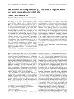
lowing model. The p51↓RNH cleavage site, defined by
residues F440 and Y441, is part of a b 1sheetthatis
nicely packed against the a A helix that carries T477.
This places the p 51↓RNH cleavage site proximal in
space to the T477 residue whose side chain OH engages
in a hydrogen bond with the main chain of residue
A445 (Fig. 4). Despite the extensive hydrophobic inter-
actions between this helix and the b-sheet throughout
their length, the hydrogen bond between residues A445
and T477 is the only electrostatic interaction between
these secondary structure elements. We surmise that a
T477A substitution would eliminate this hydrogen bond,
resulting in inc reased regional flexibility s uch that pro-
teolytic cleavage occurs at an a lternate site despite the
continued presence of the p51↓RNH mutations. Such
alternate cleavage sites have been previously suggested
[5,30], but have yet to be ide ntified directly from virus-
derived RT. A comparison of several HIV-1 RT crystal
structures provides indirect evide nce for the potential
flexibility in this region. Specifically, the superposition of
the RNH domains of several RT structu res shows nota-
ble variability in the main chain conformation; and in
some cases (e.g., PDB 1FK9[31]), parts of the b-sheet
(residues 444 - 454) are missing, possibly due to poor
electron density resulting from multiple conformations
in this region.
The intravirion RT processing defects imparted by th e
p51↓RNH cleavage site m utations are similar to those
noted with mutations such as W401A [32], a mutation
which impacts RT dimerization [33]. It is not inconcei-
vable that the RT p66 monomer would be more proteo-
lytically labile than the dimer, thus mutations that
prevent RT dimerization would lead to virions with
reduced RT content such as seen with the p51↓RNH
cleavage site mutants. Despite the similarity in the phe-
notyp e imparted by the W401A and the p51↓RNH clea-
vage site mutations, we do not think that the latter act
by reducing RT dime r formation. The p 51↓RNH clea-
vage site is not involved in significant subunit interfa ce
interactions, and as well is quite removed from the Trp
motif that plays a major role in RT dimer stabilization.
Conclusion
In summary, we have demonstrated that both virion
infectivity and proteolytic stability of RT with p51↓RNH
cleavage site mutations can be restored to various
extents by the second-site compensatory mutation
Figure 4 Positions of the p51↓RNH cleavage site and the
residue T477 in HIV-1 RT. Ribbon diagram of amino acid residues
425-560 depicting the RNH domain of HIV-1 RT, adapted from PDB
1LDO[42]. The H-bond between the hydroxyl of T477 and the main
chain of A445 is indicated by a dashed line. Details are provided in
the text. The molecular graphics image was produced using the
UCSF Chimera package from the resource for Biocomputing,
Visualization, and Informatics at the University of California, San
Francisco (supported by NIH P41 RR-01081) [43].
Abram et al. Retrovirology 2010, 7:6
/>Page 6 of 9
T477A. Studies are currently in progress to characterize
the C-terminal amino acid sequence of the RT p51 sub-
unit from HIV-1 with p51↓RNH cleavag e site mutatio ns
and the T477A substitution in RT in order to determine
whether this compensatory mutation enables proteolytic
cleavage at a positio n other than the normal F440↓Y441
location.
Materials and methods
Reagents
The following reagents were obta ined through the AIDS
Research and Reference Reagent Program, Division of
AIDS, NIAD, NIH: anti-HIV-1
SF2
p24/25 IgG mAb
(76C) from Dr. Kathelyn Ste imer, Chiron Corporation,
and anti-HIV-1
HXB2
IN (2C11 and 8G4) IgG mAb from
Dr. Dag Helland. Rabbit anti-HIV-1 PR polyclonal
serum directed against PR residues 86-108 [34,35] was a
generous gift from Dr. Stuart Le Grice, NCI-Fred erick
(Frederick, MD). Anti-HIV-1
IIIB
RT IgG mAbs specifi-
cally reacting with HIV-1 RT were previously generated
in our laboratory [36]. Goat anti-mouse-HRP and don-
key anti-rabbit secondarymAbwereproductsofGE
HealthCare (formerly Amersham Pharmacia Biotech,
Piscataway, NJ). The SuperPico ECL Substrate System
for detection of peroxidase-labeled antibody was
obtained from PIERCE (Rockford, IL). 4-methylumbelli-
feryl-b-D-galactopyranoside (4-MUG), a fluorescent sub-
strate for b-galactosidase, was obtained from Sigma-
Aldrich (St. Louis, M O). HIV-1 p24 antigen ELISA kits
were obtained from SAIC-Frederick (Frederick, MD).
Sequencing, PCR amplification and mu tation-containing
oligonucleotide primers were purchased from Invitrogen
(Carlsbad, CA).
Cell lines
The human T-lymphocytoid MT-2 and MT-4 cell lines
were maintained in RPMI 1640 supplemente d with 10%
fetal bovine serum (FBS). Human 293T and monkey
COS-7 fibroblast cel l lines were maintained in Dulbec-
co’ s modified Eagle medium (DMEM) supplemented
with 10% FBS. The P4R5 HIV infection indicator cells
were obtained fr om Dr. John Mellors, Univers ity of
Pittsburgh, and maintained in DMEM/10% FBS supple-
mented with puromycin (0.5 μg/mL). P4R5 cells express
CD4, CXCR4 and CCR5 as well as a b-galactosidase
reporter gene under the control of an HIV LTR promo-
ter [37].
Preparation, cloning and sequencing of p51↓RNH mutant
revertants
As described in our previous report [22], MT-2 cells
were inoculated with p51↓RNHcleavagesitemutant
viruses and then maintained in culture for up to 30 d or
until cytopathic effects were noted. Virus-containing
cell-free culture supernatants were then used to infect
fresh MT-2 cells. Cells we re isolated 5 d post-infection
and then chromosomal DNA was extracted using the
QIAamp DNA Mini Kit protocol (Qiagen Inc., Valencia,
CA). The HIV-1 RT-encoding region was amplified by
PCR and cloned into pCR-T7/CT TOPO (Invitrogen,
Carlsbad, CA) for sequencing analysis.
Mutagenesis of HIV-1 molecular clones and production of
recombinant virus
Pla smid pSVC21 -BH10 encodes an infectious molecular
clone of HIV-1 IIIB (HxB2) and carries an SV40 origin
of replication for expression in 293T and COS-7 cells
[38]. In our previous study [22], we used pSVC21-BH10
to prepare ten different variants mutated in the RT
p51↓RNH cleavage site (amino acid residues 437-443),
namely A437I, V442S, F440W, F440V, T439S/V442G,
Y441I/V442K, F440A, F440A/Y441A, F440W/Y441W,
and E438N. We introduced the mutation T477A into
each of these p51↓RNH cleavage site mutant s as well as
into the wild-type clone using the Quick Change™ Site-
Directed Mutagenesis kit protocol (Stratagene, La Jolla,
CA). In order t o assess the incorporation of the
Pr160
Gag-Pol
polyprotein precursor into recombinant vir-
ions, we also prepared a second set of HIV-1 clones
containing the D25A inactivating mutation in the PR
coding region to prevent proteolytic processing o f
Pr160
Gag-Pol
. The presence of all mutations were verified
by sequencing. Recombinant virus was prepared by
transfection of 293T cells using calcium phosphate co-
precipitation. Virus-c ontaining culture supernatants
were harvested 60 h post-transfection and clarified by
centrifugation (3,000×g, 1 h at 4°C). The level of recom-
binant virus production was quantified by measurement
of HIV-1 p24 antigen. Virus preparations were then ali-
quoted and stored at -80°C until use.
HIV-1 infectivity assays
Infectivity of virus particles produced by transfection of
293T cells was determined by addition of defined quanti-
ties of HIV-1 p24 antigen to target infectable cells. Single-
cycle viral infectivity was assessed in 96-well microplate
assays using P4R5 cells (5 × 10
3
cells/well). Cells were
inoculated with 25 ng HIV-1 p24/well and the extent of
infection was evaluated 48 h post-infection using a fluores-
cence-based b-galactosidase detection assay. Briefly,
infected cells were washed, then incubated with 100 μL
lysis buffer (60 mM Na
2
HPO
4
,40mMNaH
2
PO
4
(pH 7.2),
1 mM MgSO
4
, 100 mM b-mercaptoethanol, 2% [v/v] Tri-
ton X-100) for 1 h at 37°C. b-galactosidase activity was
assessed by addition of 50 μL 4-MUG to a final concentra-
tion of 0.5 mM, incubation for 1 h at 37°C, and then
quenched with 150 μL0.2MNa
2
CO
3
,pH11.2.Fluores-
cence intensity was assessed with a SPECTRAmax
GEMINI XS dual-scanning microplate spectrofluorometer
(Molecular Devices, Sunnyvale, CA) using an excitation
wavelength of 355 nm and an emission wavelength of 480
nm, with cutoff filter set to 475 nm.
Abram et al. Retrovirology 2010, 7:6
/>Page 7 of 9
Multiple-round viral replication (virus spread) was
assessed using MT-2 cells cultured in 96-well micro-
plates (6.5 × 10
4
cells/well). Cells were inoculated with
25 ng HIV-1 p24/well. HIV-1 induced cytopathic effects
were evaluated daily by microscopic observation of HIV-
1 induced syncytium formation (data not shown), as
previously described [39,40]. In a separate, but comple-
menting experiment (Table 1), each virus was titered on
MT-2 cells to evaluate the median tissue culture infec-
tive dose (TCID
50
/ng p24) after seven d post-infection,
as described [41].
Analysis of virion proteins
HIV-1 virions were isolated by centrifugation of aliquots
of cell-free culture supernatants (corresponding to 1 μg
viral p24) at 175,000×g for 1.5 h at 4°C through a 20%
(w/v) sucrose cushion. Pelleted virions were lysed in 16
μL of 20 mM Tris-Cl (pH 8.0) containing 120 mM
NaC l, 2 mM EDTA, 0.5% deoxycholate, 0.5% NP-40 (v/
v), as well as the protease inhibitors phenylmethyl sulfo-
nyl fluoride (2 μg/mL), aprotinin (10 μg/mL) and pep-
statin A (10 μg/mL). Virion protein composition was
assessed by Western blotting after resolution of the pro -
teins by 10% SDS-PAGE. Specific viral proteins were
detected by incubating the blots with anti-HIV-1 RT
mAbs (6 μg/mL), anti-HIV-1 IN (mixed 2C11 and 8G4,
1:40 dilution), anti-HIV-1 PR monospecific antiserum
(1:40 dilution) or anti-HIV-1 p24 mAb (3 μg/ml) fol-
lowed by incubation with the appropriate HRP-conju-
gated secondary antibody (1:1000 dilution). Non-specific
binding was minimized by blocking the blots with 7%
(w/v) skim milk/0.05% (v/v) Tween 20 in phosphate buf-
fered saline. Normal goat or donkey serum was added to
the blocking solution at a 1:100 (v/v) dilution where
appropriate. Immunoreactive protein bands were visua-
lized and quantified by enhanced chemiluminescence
(ECL) using a BioRad VersaDoc Imaging System.
Analysis of intravirion proteolytic processing of Pr55
Gag
and Pr160
Gag-Pol
polyproteins
The accumulation of polyprotein intermediates formed
during HIV-1 PR-mediated processi ng of Pr160
Gag-Pol
in
nascent HIV-1 virions was a ssessed by immunoprobing
of Western blots of recombinant virions produced in
the presence of varying concentrations of the PR inhibi-
tor ritonavir (RTV). Briefly, COS-7 cells (1.6 × 10
5
cells/
well) were transfected with 3 μg of proviral plasmid
DNA (pSVC21-BH10) using LipofectAMINE Plus (Invi-
trogen, Carlsbad, CA) for 3 h. The transfection medium
was then replace d with cell culture medium containing
varying concentrations of RTV. Cell culture superna-
tants were harvested 48 h post-transfection and HIV-1
virions were isolated b y centrifugation at 175,000×g for
1.5 h at 4°C through a 20% (w/v) sucrose cushion. Puri-
fiedvirionswerequantifiedbyanalysisofHIV-1p24
antigen, and virion protein composition was assessed
after lysis and SDS-PAGE resolution as described above.
Abbreviations
HIV-1: human immunodeficiency virus type 1; PR: protease; RT: reverse
transcriptase; RNH: ribonuclease H; IN: integrase; WT: wild-type; ritonavir: RTV.
Acknowledgements
Research in the Parniak laboratory is supported in part by NIH grants
AI073975, AI077424 and AI079801. Research in the Sarafianos laboratory is
supported in part by NIH grants AI074389 and AI076119.
Author details
1
HIV Drug Resistance Program, National Cancer Institute, Frederick, MD,
21702, USA.
2
University of Missouri-Columbia, Department of Molecular
Microbiology and Immunology, Columbia, MO, 65211, USA.
3
University of
Pittsburgh School of Medicine, Department of Microbiology and Molecular
Genetics, Pittsburgh, PA, 15219, USA.
Authors’ contributions
MEA designed the study and carried out most of the experimental
procedures and data analysis, and drafted the manuscript. SGS contributed
to analysis of the structural basis for the observed phenotype. MAP made
substantial contributions to the conception and design of the study, data
interpretation, and in preparation of the final manuscript.
Competing interests
The authors declare that they have no competing interests.
Received: 5 October 2009
Accepted: 1 February 2010 Published: 1 February 2010
References
1. Tisdale M, Ertl P, Larder BA, Purifoy DJ, Darby G, Powell KL: Characterization
of human immunodeficiency virus type 1 reverse transcriptase by using
monoclonal antibodies: role of the C terminus in antibody reactivity and
enzyme function. J Virol 1988, 62(10):3662-7.
2. Chattopadhyay D, Evans DB, Deibel MR Jr, et al: Purification and
characterization of heterodimeric human immunodeficiency virus type 1
(HIV-1) reverse transcriptase produced by in vitro processing of p66
with recombinant HIV-1 protease. J Biol Chem 1992, 267(20):14227-32.
3. Fan N, Rank KB, Leone JW, et al: The differential processing of
homodimers of reverse transcriptases from human immunodeficiency
viruses type 1 and 2 is a consequence of the distinct specificities of the
viral proteases. J Biol Chem 1995, 270(22):13573-9.
4. Hostomska Z, Matthews DA, Davies JF, Nodes BR, Hostomsky Z: Proteolytic
release and crystallization of the RNase H domain of human
immunodeficiency virus type 1 reverse transcriptase. J Biol Chem 1991,
266(22):14697-702.
5. Tomasselli AG, Sarcich JL, Barrett LJ, Reardon IM, Howe WJ, Evans DB,
Sharma SK, Heinrikson RL: Human immunodeficiency virus type-1 reverse
transcriptase and ribonuclease H as substrates of the viral protease.
Protein Sci 1993, 2(12):2167-76.
6. Kohlstaedt LA, Wang J, Friedman JM, Rice PA, Steitz TA: Crystal structure at
3.5 A resolution of HIV-1 reverse transcriptase complexed with an
inhibitor. Science 1992, 256(5065):1783-90.
7. Wang J, Smerdon SJ, Jager J, Kohlstaedt LA, Rice PA, Friedman JM,
Steitz TA: Structural basis of asymmetry in the human immunodeficiency
virus type 1 reverse transcriptase heterodimer. Proc Natl Acad Sci USA
1994, 91(15):7242-6.
8. Jacobo-Molina A, Arnold E: HIV reverse transcriptase structure-function
relationships. Biochemistry 1991, 30(26):6351-6.
9. Prasad VR, Goff SP: Structure-function studies of HIV reverse transcriptase.
Ann N Y Acad Sci 1990, 616:11-21.
10. Goff SP, Prasad VR: Linker insertion mutagenesis as probe of structure-
function relationships. Methods Enzymol 1991, 208:586-603.
11. Mizrahi V, Brooksbank RL, Nkabinde NC: Mutagenesis of the conserved
aspartic acid 443, glutamic acid 478, asparagine 494, and aspartic acid
498 residues in the ribonuclease H domain of p66/p51 human
Abram et al. Retrovirology 2010, 7:6
/>Page 8 of 9
immunodeficiency virus type I reverse transcriptase. Expression and
biochemical analysis. J Biol Chem 1994, 269(30):19245-9.
12. Prasad VR, Goff SP: Linker insertion mutagenesis of the human
immunodeficiency virus reverse transcriptase expressed in bacteria:
definition of the minimal polymerase domain. Proc Natl Acad Sci USA
1989, 86(9):3104-8.
13. Hizi A, Hughes SH, Shaharabany M: Mutational analysis of the
ribonuclease H activity of human immunodeficiency virus 1 reverse
transcriptase. Virology 1990, 175(2):575-80.
14. Mizrahi V, Lazarus GM, Miles LM, Meyers CA, Debouck C: Recombinant HIV-
1 reverse transcriptase: purification, primary structure, and polymerase/
ribonuclease H activities. Arch Biochem Biophys 1989, 273(2):347-58.
15. Tisdale M, Schulze T, Larder BA, Moelling K: Mutations within the RNase H
domain of human immunodeficiency virus type 1 reverse transcriptase
abolish virus infectivity. J Gen Virol 1991, 72(1):59-66.
16. Hizi A, Barber A, Hughes SH: Effects of small insertions on the RNA-
dependent DNA polymerase activity of HIV-1 reverse transcriptase.
Virology 1989, 170(1):326-9.
17. Starnes MC, Gao WY, Ting RY, Cheng YC: Enzyme activity gel analysis of
human immunodeficiency virus reverse transcriptase. J Biol Chem 1988,
263(11):5132-4.
18. Wu J, Amandoron E, Li X, Wainberg MA, Parniak MA: Monoclonal
antibody-mediated inhibition of HIV-1 reverse transcriptase polymerase
activity. Interaction with a possible deoxynucleoside triphosphate
binding domain. J Biol Chem 1993, 268(14):9980-5.
19. Fletcher RS, Holleschak G, Nagy E, Arion D, Borkow G, Gu Z, Wainberg MA,
Parniak MA: Single-step purification of recombinant wild-type and
mutant HIV-1 reverse transcriptase. Protein Expr Purif 1996, 7(1):27-32.
20. Bathurst IC, Moen LK, Lujan MA, Gibson HL, Feucht PH, Pichuantes S,
Craik CS, Santi DV, Barr PJ: Characterization of the human
immunodeficiency virus type-1 reverse transcriptase enzyme produced
in yeast. Biochem Biophys Res Commun 1990, 171(2):589-95.
21. Hansen J, Schulze T, Mellert W, Moelling K: Identification and
characterization of HIV-specific RNase H by monoclonal antibody. EMBO
J 1988, 7(1):239-43.
22. Abram ME, Parniak MA: Virion instability of human immunodeficiency
virus type I reverse transcriptase (RT) mutated in the protease cleavage
site between RT p51 and the RT RNase H domain. J Virol 2005,
79(18):11952-61.
23. Pettit SC, Everitt LE, Choudhury S, Dunn BM, Kaplan AH: Initial Cleavage of
the Human Immunodeficiency Virus Type 1 GagPol Precursor by Its
Activated Protease Occurs by an Intramolecular Mechanism. J Virol 2004,
78(16):8477-85.
24. Pettit SC, Sheng N, Tritch R, Erickson-Viitanen S, Swanstrom R: The
regulation of sequential processing of HIV-1 Gag by the viral protease.
Adv Exp Med Biol 1998,
436:15-25.
25. Speck RR, Flexner C, Tian CJ, Yu XF: Comparison of human
immunodeficiency virus type 1 Pr55(Gag) and Pr160(Gag-pol) processing
intermediates that accumulate in primary and transformed cells treated
with peptidic and nonpeptidic protease inhibitors. Antimicrob Agents
Chemother 2000, 44(5):1397-403.
26. Lindhofer H, von der HK, Nitschko H: In vivo processing of Pr160gag-pol
from human immunodeficiency virus type 1 (HIV) in acutely infected,
cultured human T-lymphocytes. Virology 1995, 214(2):624-7.
27. Wiegers K, Rutter G, Kottler H, Tessmer U, Hohenberg H, Krausslich HG:
Sequential steps in human immunodeficiency virus particle maturation
revealed by alterations of individual Gag polyprotein cleavage sites. J
Virol 1998, 72(4):2846-54.
28. Rhee SY, Gonzales MJ, Kantor R, Betts BJ, Ravela J, Shafer RW: Human
immunodeficiency virus reverse transcriptase and protease sequence
database. Nucleic Acids Res 2003, 31(1):298-303.
29. Navarro JM, Damier L, Boretto J, Priet S, Canard B, Quérat G, Sire J:
Glutamic Residue 438 within the Protease-Sensitive Subdomain of HIV-1
Reverse Transcriptase Is Critical for Heterodimer Processing in Viral
Particles. Virology 2001, 290(2):300-8.
30. Graves MC, Meidel MC, Pan YC, Manneberg M, Lahm HW, Gruninger-
Leitch F: Identification of a human immunodeficiency virus-1 protease
cleavage site within the 66,000 Dalton subunit of reverse transcriptase.
Biochem Biophys Res Commun 1990, 168(1):30-6.
31. Ren J, Milton J, Weaver KL, Short SA, Stuart DI, Stammers DK: Structural
basis for the resilience of efavirenz (DMP-266) to drug resistance
mutations in HIV-1 reverse transcriptase. Structure 2000, 8(10):1089-94.
32. Chiang C-C, Wang S-M, Tseng Y-T, Huang K-J, Wang C-T: Mutations at
human immunodeficiency virus type 1 reverse transcriptase tryptophan
repeat motif attenuate the inhibitory effect of efavirenz on virus
production. Virology 2009, 383(2):261-270.
33. Tachedjian G, Orlova M, Sarafianos SG, Arnold E, Goff SP: Nonnucleoside
reverse transcriptase inhibitors are chemical enhancers of dimerization
of the HIV type 1 reverse transcriptase. Proc Natl Acad Sci USA 2001,
98(13):7188-7193.
34. Le Grice SF, Mills J, Mous J: Active site mutagenesis of the AIDS virus
protease and its alleviation by trans complementation. EMBO J 1988,
7(8):2547-53.
35. Mous J, Heimer EP, Le Grice SF: Processing protease and reverse
transcriptase from human immunodeficiency virus type I polyprotein in
Escherichia coli. J Virol 1988, 62(4):1433-6.
36. Li X, Amandoron E, Wainberg MA, Parniak MA: Generation and
characterization of murine monoclonal antibodies reactive against N-
terminal and other regions of HIV-1 reverse transcriptase. J Med Virol
1993, 39(3):251-9.
37. Munk C, Brandt SM, Lucero G, Landau NR: A dominant block to HIV-1
replication at reverse transcription in simian cells. Proc Natl Acad Sci USA
2002, 99(21):13843-8.
38. Fisher AG, Collalti E, Ratner L, Gallo RC, Wong-Staal F: A molecular clone of
HTLV-III with biological activity.
Nature 1985, 316(6025):262-5.
39. Borkow G, Fletcher RS, Barnard J, Arion D, Motakis D, Dmitrienko GI,
Parniak MA: Inhibition of the ribonuclease H and DNA polymerase
activities of HIV-1 reverse transcriptase by N-(4-tert-butylbenzoyl)-2-
hydroxy-1- naphthaldehyde hydrazone. Biochemistry 1997, 36(11):3179-85.
40. Motakis D, Parniak MA: A tight-binding mode of inhibition is essential for
anti-human immunodeficiency virus type 1 virucidal activity of
nonnucleoside reverse transcriptase inhibitors. Antimicrob Agents
Chemother 2002, 46(6):1851-6.
41. Johnson VA, Byington RE: Techniques in HIV research New York, N.Y.:
Stockton PressAldovini A, Walker BD 1990, 71-6.
42. Hsiou Y, Ding J, Das K, Clark AD Jr, Hughes SH, Arnold E: Structure of
unliganded HIV-1 reverse transcriptase at 2.7 A resolution: implications
of conformational changes for polymerization and inhibition
mechanisms. Structure 1996, 4(7):853-60.
43. Pettersen EF, Goddard TD, Huang CC, Couch GS, Greenblatt DM, Meng EC,
Ferrin TE: UCSF Chimera–a visualization system for exploratory research
and analysis. J Comput Chem 2004, 25(13):1605-12.
doi:10.1186/1742-4690-7-6
Cite this article as: Abram et al.: The mutation T477A in HIV-1 reverse
transcriptase (RT) restores normal proteolytic processing of RT in virus
with Gag-Pol mutated in the p51-RNH cleavage site. Retrovirology 2010
7:6.
Submit your next manuscript to BioMed Central
and take full advantage of:
• Convenient online submission
• Thorough peer review
• No space constraints or color figure charges
• Immediate publication on acceptance
• Inclusion in PubMed, CAS, Scopus and Google Scholar
• Research which is freely available for redistribution
Submit your manuscript at
www.biomedcentral.com/submit
Abram et al. Retrovirology 2010, 7:6
/>Page 9 of 9