Báo cáo Y học: The presence of a helix breaker in the hydrophobic core of signal sequences of secretory proteins prevents recognition by the signal-recognition particle in Escherichia coli doc
Bạn đang xem bản rút gọn của tài liệu. Xem và tải ngay bản đầy đủ của tài liệu tại đây (261.2 KB, 8 trang )
The presence of a helix breaker in the hydrophobic core
of signal sequences of secretory proteins prevents recognition
by the signal-recognition particle in
Escherichia coli
Hendrik Adams
1
, Pier A. Scotti
2
*, Hans de Cock
1
, Joen Luirink
2
and Jan Tommassen
1
1
Department of Molecular Microbiology and Institute of Biomembranes, Utrecht University, The Netherlands;
2
Department of
Microbiology, Institute of Molecular Biological Sciences, Biocentrum Amsterdam, The Netherlands
Signal sequences often contain a-helix-destabilizing amino
acids within the hydrophobic core. In the precursor of the
Escherichia coli outer-membrane protein PhoE, the glycine
residue at position )10 (Gly
)10
) is thought to be responsible
for the break in the a-helix. Previously, we showed that
substitution of Gly
)10
by a-helix-promoting residues (Ala,
Cys or Leu) reduced the proton-motive force dependency of
the translocation of the precursor, but the actual role of the
helix breaker remained obscure. Here, we considered
the possibility that extension of the a-helical structure in
the signal sequence resulting from the Gly
)10
substitutions
affects the targeting pathway of the precursor. Indeed, the
mutations resulted in reduced dependency on SecB for tar-
geting in vivo. In vitro cross-linking experiments revealed that
the G-10L and G-10C mutant PhoE precursors had a dra-
matically increased affinity for P48, one of the constituents of
the signal-recognition particle (SRP). Furthermore, in vitro
cross-linking experiments revealed that the G-10L mutant
protein is routed to the SecYEG translocon via the SRP
pathway, the targeting pathway that is exploited by integral
inner-membrane proteins. Together, these data indicate
that the helix breaker in cleavable signal sequences prevents
recognition by SRP and is thereby, together with the
hydrophobicity of the signal sequence, a determinant of the
targeting pathway.
Keywords: outer-membrane protein; Sec translocon; SecB;
signal-recognition particle; translocation.
Most cell envelope proteins of Escherichia coli are translo-
cated across or inserted into the cytoplasmic membrane via
the membrane-embedded Sec translocon. Targeting of
precursor proteins to the translocon can be mediated by
components of the Sec pathway or by the signal-recognition
particle (SRP) pathway [1,2]. The Sec pathway utilizes a
cytosolic chaperone, SecB, which interacts with the mature
portion of presecretory proteins [3,4]. The SecB-preprotein
complexisthentargetedtoSecA,whichinturninteracts
with components of the Sec translocon [5,6]. At the onset of
translocation, SecB is released [7] and the preprotein is
translocated by an insertion–deinsertion cycle of SecA into
the SecYEG translocon [8]. Energy for the translocation
process is provided by ATP hydrolysis by SecA [8,9] and by
the proton-motive force (pmf) [9]. At the periplasmic side of
the membrane, leader peptidase removes the signal sequence
from the precursor, and the mature protein is released into
the periplasm [10]. The bacterial SRP-targeting route is
homologous with, but less complex than, the eukaryotic
SRP system [11,12]. The E. coli SRP consists of a single
protein, P48, and a 4.5S RNA, and binds cotranslationally
to hydrophobic sequences [13,14]. The ribosome-nascent
chain (RNC) complex subsequently binds to FtsY and is
targeted to the Sec translocon in the inner membrane
[15,16]. Whereas the SecB route is predominantly used by a
subset of periplasmic and most, if not all, outer-membrane
proteins, inner-membrane proteins are particularly depend-
ent on a functional SRP pathway [17].
We are using outer-membrane protein PhoE as a model
to study protein export. PhoE is targeted via its signal
sequence in a SecB-dependent way to the Sec translocon [3].
Whereas the signal sequence is necessary and, in most cases,
sufficient for translocation across the cytoplasmic mem-
brane, its exact role in the export mechanism is far from
understood. Despite the common function of signal
sequences, i.e. to direct the translocation of the attached
polypeptide chain, there is little sequence homology among
them [18]. Nevertheless, a common structural organization
can be recognized (Fig. 1). Signal sequences are character-
ized by a positively charged N-terminal region (N domain),
followed by a 10–15 residues long hydrophobic core (H
domain) and a polar C-terminus (C domain) containing the
signal-peptidase cleavage site [19]. The importance of a-helix
formation in the signal sequence is well documented [20–24].
However, NMR studies on the conformation of signal
sequences in a membrane mimetic environment showed
that the stable a-helix is disrupted towards the C-terminus
of the hydrophobic core [25–27]. Furthermore, a statistical
Correspondence to J. Tommassen, Department of Molecular
Microbiology, Utrecht University, Padualaan 8, 3584 CH Utrecht,
The Netherlands. Fax: + 31 30 2513655, Tel.: + 31 30 2532999,
E-mail:
Abbreviations: SRP, signal-recognition particle; pmf, proton-motive
force; RNC, ribosome-nascent chain; BS
3
, bis(sulfosuccinimidyl)-
suberate; DSS, disuccinimidyl-suberate; IMV, inverted inner-
membrane vesicles; TF, trigger factor.
*Present address:IECB-E
´
cole polytechnique ENSCPB, Talence cedex,
France.
(Received 29 May 2002, revised 10 September 2002,
accepted 16 September 2002)
Eur. J. Biochem. 269, 5564–5571 (2002) Ó FEBS 2002 doi:10.1046/j.1432-1033.2002.03262.x
analysis of signal sequences revealed the common
occurrence of a-helix-destabilizing amino acids in the
hydrophobic core [28]. In a previous study, the role of the
a-helix-breaking glycine residue at position )10 (Gly
)10
)of
the signal sequence of PhoE was examined [29]. It was
shown that substitution of this residue by a-helix-promoting
residues (Ala, Cys or Leu) reduced the pmf dependency of
the translocation across the inner membrane, but the actual
role of the helix breaker remained obscure. It should be
noted that such substitutions extend the a-helix not just by a
single residue, but, probably, over the entire H domain
(Fig. 1). Whereas the a-helix in the wild-type signal
sequence is too short to span the inner membrane, the
resulting mutant signal sequences would more closely
resemble the membrane-spanning domains of inner-mem-
brane proteins and might therefore be turned into substrates
for the SRP. In this paper, we considered the possibility that
the extended a-helix resulting from the Gly
)10
substitutions
affects the targeting pathway of the precursor.
EXPERIMENTAL PROCEDURES
Reagents and biochemicals
Restriction enzymes were purchased from either Boehringer
Mannheim or Pharmacia. MEGAshortscript T7 transcrip-
tion kit was from Ambion, and [
35
S]methionine and Tran
35
S-label were from Amersham International. Bis(sulfo-
succinimidyl)-suberate (BS
3
) and disuccinimidyl-suberate
(DSS) were from Pierce, and oligonucleotides were pur-
chased from Isogen Bioscience (Maarsen, the Netherlands).
Bacterial strains
The E. coli K-12strainsusedinthisstudyarelistedin
Table 1. Strains CE1514 and CE1515 were obtained by P1
transduction using strain CE1224 as the recipient and
strains IQ85 and strain MM152, respectively, as donor
strains. To obtain strain CE1513, strain MM88 was used as
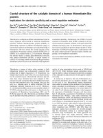
Fig. 1. Physical characteristics of the PhoE signal sequence. The signal sequence consists of the positively charged N domain, the hydrophobic H
domain and the C-terminal C domain. The a-helix in the H domain is predicted to extend up to the Gly at position )10 in the signal sequence.
Introduction of an a-helix-stabilizing residue (Ala, Cys or Leu) at position )10 results in extension of the a-helical core region as indicated. The
leader peptidase cleavage site is depicted with an arrow.
Table 1. Bacterial strains and plasmids used in this study. Ts, temperature sensitive. Cam
r
and Amp
r
, resistance to chloramphenicol and ampicillin,
respectively.
Designation Relevant characteristics Description/reference
Strains
CE1224 F
–
, thr leu D(proA-proB-phoE-gpt) his thi argE lacY galK xyl rpsL supE ompR [49]
MC4100 F
–
, DlacU169 araD139 rpsL thi relA [50]
MM88 F
–
, DlacU169 araD139 thiA rpsL relA leu::Tn10 secAtsA51 B. Oudega (pers. comm.)
NT1060 F
–
, DlacU169 araD139 rpsL thi relA ptsF25 deoC1 lamBD60 T.J. Silhavy (pers. comm.)
MM152 MC4100 secB::Tn5 [51]
IQ85 Tn10 thiA Dlac araD rpsL rpsE relA secYts24 [51]
CE1513 CE1224 secAts51 leu::Tn10 This study
CE1514 CE1224 Tn10 secYts24 This study
CE1515 CE1224 secB::Tn5 This study
FF283 F
–
, lacDx74 araD139 (araABOIC-leu) D7679 galU galK rpsL ffs::kan/F¢ lac-pro,
lacI
q
Ptac::ffs
[52]
Plasmids
pJP29 Cam
r
, wild-type phoE [30]
pNN5 pJP29 derivative encoding (G-10A)prePhoE [29]
pNN7 pJP29 derivative encoding (G-10C)prePhoE [29]
pNN8 pJP29 derivative encoding (G-10L)prePhoE [29]
pC4Meth101FtsQ-WT Amp
r
, encodes truncated 101FtsQ [13]
pC4Meth94PhoE Amp
r
, encodes truncated 94PhoE [13]
pC4Meth(G-10C)94PhoE pC4Meth94PhoE derivative encoding (G-10C) mutant 94PhoE This study
pC4Meth(G-10L)94PhoE pC4Meth94PhoE derivative encoding (G-10L) mutant 94PhoE This study
Ó FEBS 2002 Re-routing a secretory protein via the SRP pathway (Eur. J. Biochem. 269) 5565
the donor and CE1224 as the recipient in a P1 transduction
experiment.
Plasmid construction
Plasmid pJP29 and derivatives carrying mutations in the
PhoE signal-sequence-encoding region and other plasmids
are listed in Table 1. Plasmid pC4Meth94PhoE was used to
generate truncated phoE mRNA, encoding a 94-residue
PhoE polypeptide exposing the signal sequence just outside
the ribosome [13]. Plasmid pC4Meth101FtsQ-WT was used
to generate truncated FtsQ mRNA, encoding a 101-residue
FtsQ polypeptide exposing the signal-anchor domain just
outside the ribosome. To compensate for the loss of
methionines from the deleted domains of the proteins,
both constructs contain a C-terminal tetra-methionine tag
sequence for labeling. To introduce the Cys and Leu
mutations for the Gly
)10
residue into pC4Meth94PhoE, the
EcoRI/BamHI fragment of the plasmid was replaced by
PCR fragments created using the PhoE forward primer
(5¢-GCCGGAATTCTAATATGAAAAAGAGCACTCT
GGC-3¢) and the 94PhoE reverse primer (5¢-CGCGGA
TCCTTTTTGCTGTCAGTATCAC-3¢), pNN7 and pNN8
as the templates, respectively, and Pfu polymerase. The
resulting plasmids were designated pC4Meth(G-10C)94
PhoE and pC4Meth(G-10L)94PhoE, respectively.
In vivo
pulse–chase experiments
Cells of strain CE1224 or its derivatives each containing a
plasmid expressing (mutant) phoE from its own promoter,
were grown under phosphate limitation at 30 °Cas
described previously [30]. Cells of the 4.5S RNA conditional
strain FF283 were grown to D
660
¼ 1.0 in Hepes-buffered
synthetic medium, supplemented with 660 l
M
K
2
HPO
4
.
For the depletion of 4.5S RNA, isopropyl b-
D
-thiogalacto-
pyranoside was omitted from the growth medium. To
induce the expression of (mutant) phoE from its own
promoter, cells were collected by centrifugation and washed
with Hepes-buffered synthetic medium with no phosphate
added. The cell pellets were resuspended in the latter
medium at the original absorbance, followed by incubation
at 37 °C for 30 min. For pulse-labeling, cells were incubated
for 45 s with Tran
35
S-label followed by a chase period with
an excess of nonradioactive methionine/cysteine. After
precipitation with 5% (w/v) trichloroacetic acid, radio-
labeled proteins were separated either directly or after
immunoprecipitation with a polyclonal PhoE-specific anti-
serum [31] by SDS/PAGE [32] and visualized by autoradio-
graphy.
In vitro
transcription, translation, targeting
and cross-linking analysis
To generate truncated mRNA, plasmids (Table 1) encoding
truncated nascent chains were linearized and transcribed as
described previously [13]. The resulting mRNAs were
translated in vitro in a lysate of strain MC4100 as described
[13,33]. The mixture containing RNCs was chilled on ice
and treated with 1 m
M
BS
3
at 25 °C for 10 min before
addition of 0.1 vol. quench buffer (1
M
glycine/0.1
M
NaHCO
3
, pH 8.5). After incubation for 20 min at 0 °C,
cross-linked products were immunoprecipitated [34], and
the precipitates were analyzed by SDS/PAGE (12% gels).
Radiolabeled proteins were visualized with a Phosphor-
Imager 473 (Molecular Dynamics) and quantified using the
Imagequant software (Molecular Dynamics). To test the
targeting of wild-type prePhoE RNCs, truncated mRNAs
were translated in the presence of inverted inner-membrane
vesicles (IMVs) [33] from strain MC4100. After cross-
linking with 1 m
M
DSSfor10minat25°C, the cross-link
reaction was stopped with quench buffer. Peripheral and
soluble cross-linked complexes were separated from
integral-membrane cross-linked complexes by Na
2
CO
3
extraction as described [35]. Samples were analyzed either
directly or after immunoprecipitation on 12% polyacryla-
mide gels and visualized as described above.
To probe the molecular environment of membrane-
associated RNCs, SRP was reconstituted in vitro from
purified 4.5S RNA and purified hexa-His-tagged P48 as
described [35]. To allow SRP–RNC complex formation
(G-10L)94PhoE and 101FtsQ were synthesized in vitro and
incubated at 25 °C with 350 n
M
reconstituted SRP, and
SRP–RNC complexes were purified from the translation
mixture by centrifugation through a high-salt sucrose
cushion [36]. The SRP–RNC complexes were incubated
with IMVs from strain NT1060 under conditions as
described previously [35]. After cross-linking with 2 m
M
DSS at 25 °C for 10 min, 0.1 vol. quench buffer was added
and incubation was continued on ice for 15 min. Subse-
quently, peripheral and soluble cross-linked complexes were
separated from integral-membrane cross-linked complexes
by Na
2
CO
3
extraction as described [35]. Samples were
analyzed either directly or after immunoprecipitation on
12% or 15% gels and visualized as described above.
RESULTS
SecB dependency of the targeting of mutant prePhoE
By the substitution of an a-helix-promoting residue (Leu,
Ala or Cys) for the helix-breaking Gly
)10
of the signal
sequence of PhoE, the a-helix is expected to be extended
considerably (Fig. 1). As these mutant signal sequences
resemble more closely the membrane-spanning domains of
integral-membrane proteins, the mutations might affect the
targeting route of the precursors to the SecYEG translocon.
This possibility was first tested in vivo in pulse–chase
experiments. The processing kinetics of the wild-type and
mutant PhoE proteins were compared in a secB null mutant
strain. Previously, it was demonstrated that introduction of
an a-helix-stabilizing residue (Ala, Cys or Leu) instead of
the Gly
)10
did not result in dramatic differences in the
processing kinetics of prePhoE in wild-type cells [29]. As the
export of wild-type PhoE is SecB dependent [3], its
precursor strongly accumulated in a secB mutant (Fig. 2A).
Interestingly, the mutant precursors showed considerably
improved processing kinetics compared with wild-type
prePhoE in the secB mutant (Fig. 2A). After a 5-min chase
period, hardly any mutant prePhoE was detected anymore,
whereas the vast majority of the wild-type precursor was still
not processed. Together with the previously reported
reduced pmf dependency for translocation of the mutant
precursors [29], our results suggest that the SecB depend-
ency of prePhoE targeting correlates with its DlH
+
dependency for in vitro translocation.
5566 H. Adams et al.(Eur. J. Biochem. 269) Ó FEBS 2002
Of all the precursors tested, the mutant precursor with the
strongest a-helix-promoting residue (Leu) at position )10
appeared to be most efficiently processed in the secB mutant
strain. This mutant precursor was used to verify if
translocation is still dependent on the membrane-embedded
SecYEG complex and on SecA. For this purpose, pulse–
chase experiments were performed in secA51 and secY24
mutant strains at their nonpermissive temperature. In both
strains, processing of the (G-10L)prePhoE protein, like that
of the wild-type precursor, was strongly impaired in
comparison with the processing in the wild-type strain
(Fig. 2B). Apparently, substitution of the glycine residue at
position )10 by an a-helix-promoting residue does not alter
the dependency of the precursor on SecA and SecY,
whereas its SecB dependency is reduced.
Affinity of mutant prePhoE nascent chains for P48
As the SecB dependency of the translocation of the mutant
prePhoE proteins was clearly decreased, we next considered
the possibility that they had become substrates for the
SRP pathway. To determine whether components of the
SRP pathway are indeed involved in the targeting of
(G-10L)prePhoE to the translocon, in vitro cross-linking
studies were performed. Previously, Valent et al.[13]
analyzed the interaction of nascent prePhoE protein with
soluble proteins in an E. coli lysate. Nascent PhoE 94-mer
extended with a tetra-methionine tag-sequence (94PhoE)
was synthesized in an E. coli lysate and treated with the
water-soluble cross-linker BS
3
. Whereas, in these experi-
ments, nascent chains of integral inner-membrane proteins
could be cross-linked to the P48 component of SRP, this
was not the case for nascent 94PhoE [13]. To investigate
whether substitution of the Gly
)10
residue by an a-helix-
stabilizing residue resulted in a higher affinity for P48,
(G-10L)94PhoE and 94PhoE were synthesized and tested
for cross-linking to P48 present in the E. coli lysate.
Whereas hardly any cross-linked 94PhoE could be immu-
noprecipitated with anti-P48 antibodies, strong cross-link-
ing of (G-10L)94PhoE to P48 was observed (Fig. 3A). To
determine whether the improved cross-linking of (G-10L)
94PhoE to P48 was due solely to the increased hydropho-
bicity of this mutant signal sequence, similar cross-linking
experiments were also performed for the (G-10C)94PhoE
mutant PhoE protein. Even though cysteine has an even
lower hydrophobicity than glycine on the consensus
hydrophobicity scale of Eisenberg et al. [37], the (G-10C)
94PhoE protein was also cross-linked to P48 (Fig. 3),
although not as efficiently as (G-10L)94PhoE. In all cases,
antiserum against trigger factor (TF) efficiently precipitated
cross-linked complexes (Fig. 3A,B), confirming the earlier
observation that TF, a cytosolic chaperone, binds to E. coli
nascent polypeptides [13]. Quantification of the data
indicated that the cross-linking efficiency of the mutant
nascent chains was somewhat reduced (Fig. 3B). In conclu-
sion, our results show an increased affinity of the G-10C and
G-10L prePhoE for the P48 component of SRP.
G-10L nascent PhoE interacts with Sec translocon
components
As (G-10L)94PhoE nascent chains apparently have a high
affinity for P48 in vitro, we subsequently examined whether
these nascent chains are targeted to SecY via SRP by
performing cross-linking experiments in vitro in the presence
of IMVs. To obtain a high cross-linking efficiency, recon-
stituted E. coli SRP was added after translation of nascent
(G-10L)94PhoE polypeptides to saturate the RNCs with
SRP. The SRP–RNC complexes were purified over a high-
salt sucrose cushion and incubated with IMVs to allow
targeting. After cross-linking with the membrane-permeable
cross-linking reagent DSS, peripheral and soluble cross-
linked complexes were separated from integral-membrane
cross-linked complexes by Na
2
CO
3
extraction and analyzed
by SDS/PAGE (Fig. 4). In the Na
2
CO
3
pellet, at least two
major (G-10L)94PhoE cross-linked complexes could be
detected, one at 110 kDa and one at 46 kDa (Fig. 4A,
lane 3). The 110-kDa complex could be immunoprecipitated
with antiserum directed against SecA, indicating that it is a
complex of the radiolabeled (G-10L)94PhoE and SecA
(Fig. 4B, lane 1). In addition, cross-linking adducts of
220 kDa and 40 kDa were also immunoprecipitated
from the Na
2
CO
3
pellet with anti-SecA serum. We assume
that the 220-kDa product corresponds to cross-linked
complexes between (G-10L)94PhoE and the dimeric form
Fig. 2. In vivo processing kinetics of prePhoE and mutant prePhoE
proteins in sec mutants. (A) Cells of secB mutant strain CE1515
carrying plasmid pJP29 encoding wild-type PhoE (WT) or derivatives
were grown under phosphate limitation to express PhoE. The cells
were pulse-labeled, followed by a chase for the indicated periods.
PhoE proteins were immunoprecipitated, separated by SDS/PAGE
followed by autoradiography. G-10A (G-10A)prePhoE; G-10C
(G-10C)prePhoE; G-10L (G-10L)prePhoE. (B) SecAts51 and sec-
Yts24 derivatives of CE1224 or their isogenic wild-type parental
strain (wt) carrying plasmids pJP29 or pNN8, encoding prePhoE or
(G-10L)prePhoE, respectively, were grown under phosphate limitation
for 3 h at the permissive temperature (30 °C), subsequently for 2 h at
the restrictive temperature (42 °C), and pulse-labeled at 42 °Cfor45s
with Tran
35
S-label and chased with an excess of unlabeled methionine/
cysteine. Aliquots were removed at the indicated periods and analyzed
as described for panel (A). The precursor and mature forms of the
PhoE proteins are indicated by p and m, respectively.
Ó FEBS 2002 Re-routing a secretory protein via the SRP pathway (Eur. J. Biochem. 269) 5567
of SecA. The 40-kDa product in the Na
2
CO
3
pellet
probably contains proteolytic fragments of the SecA dimer
and monomer cross-linking products, which is in agreement
with earlier reports [38]. The fuzzy 46-kDa product
(Fig. 4A, lane 3) was immunoprecipitated with anti-SecY
serum (Fig. 4B, lane 2), showing that the (G-10L)94PhoE
nascent chains are targeted to the SecYEG translocon.
In the Na
2
CO
3
supernatant, at least three major cross-
linking adducts, of apparent molecular mass 110, 65
and 55 kDa, could be detected (Fig. 4A, lane 5). In
addition, several cross-linking adducts of low molecular
mass (< 30 kDa) were detected. Immunoprecipitation
revealed that the high-molecular-mass adducts represent
cross-linking to SecA (data not shown), TF and P48
(Fig. 4B, lane 5 and 6), respectively. The identity of the low-
molecular-mass adducts is unknown. As the signal sequence
of 94PhoE has no affinity for P48 (Fig. 3), and the SecB-
binding sites in the mature domain are not exposed from the
ribosome in RNCs of 94PhoE, these RNCs cannot be
targeted to the translocon. Consistently, no cross-linking
adducts similar to those obtained with (G-10L)94PhoE were
obtained, when 94PhoE and (G-10L)94PhoE nascent
chains were incubated with IMVs after cross-linking with
DSS (Fig. 4C, lanes 1–4). To investigate whether the cross-
linking adducts of (G-10L)94PhoE that were obtained are
similar to the cross-linking adducts with a known substrate
of the SRP pathway, FtsQ was used as a model. This class II
membrane protein, with a short N-terminal cytoplasmic tail
[39], was synthesized as a slightly longer nascent chain (101
residues) than (G-10L)94PhoE to expose properly its signal-
anchor domain. Indeed, 101FtsQ interacted properly with
SecY and SecA (Fig. 4A, lane 8; Fig. 4B, lane 3 and 4).
Furthermore, the same cross-linking efficiency was obtained
for P48 (Fig. 4B, lane 8) as was observed for the (G-10L)
prePhoE (Fig. 4B, lane 6), but TF was hardly cross-linked if
at all (Fig. 4B, compare lane 5 and 7). In conclusion, these
results show that (G-10L)94PhoE nascent chains are
correctly targeted to the SecY protein in the translocon
via the SRP pathway.
SRP dependency of (G-10L)prePhoE
in vivo
As the experiments described above show that (G-10L)
prePhoE is targeted in vitro to the Sec translocon via the
SRP pathway, it was of interest to determine whether it is
dependent on this pathway in vivo. To test this possibility,
wild-type and the (G-10L)prePhoE were expressed in
FF283 cells which were depleted of 4.5S RNA. After
radioactive labeling of the cells, the PhoE forms were
immunoprecipitated and analyzed by SDS/PAGE (Fig. 5).
Depletion of 4.5S RNA did not result in the accumulation
of precursors of either wild-type prePhoE or (G-10L)pre-
PhoE. Apparently (G-10L)prePhoE translocation is not
dependent on the SRP pathway in vivo.
DISCUSSION
NMR studies of the signal peptides of LamB [25], OmpA
[26] and PhoE [27] showed that the a-helical conformation is
disrupted toward the C-terminus of the hydrophobic core
near a helix-breaking residue, such as Gly
)10
in the case of
prePhoE. Furthermore, a statistical analysis of signal
sequences revealed the common occurrence of helix-break-
ing residues within the hydrophobic core [28], suggesting
that the disruption of the a-helix is a common feature of
signal sequences. In a previous study, it was shown that the
DlH
+
dependency of prePhoE translocation was dramati-
cally reduced when a helix-promoting residue, such as
leucine or cysteine, was substituted for the helix-breaking
Gly
)10
of the signal sequence [29]. Such a substitution is
expected to result in considerable elongation of the a-helix
in the signal sequence. Consistent with a considerable
conformational change, these substitutions resulted in a
higher electrophoretic mobility of the mutant precursors
compared with that of wild-type prePhoE [29] (see also
Fig. 2A), suggesting a more compact conformation of the
Fig. 3. Cross-linking of soluble E. coli proteins to PhoE nascent chains
and mutant derivatives. (A) [
35
S]methionine-labeled nascent 94PhoE or
mutant derivatives were synthesized in an E. coli lysate and treated
with the homo-bifunctional chemical cross-linker BS
3
. After quench-
ing, both P48- and TF-cross-linked complexes were immunoprecipi-
tated with antisera directed against P48 and TF, analyzed on SDS/
PAGE and visualized with a PhosphorImager. (B) Quantification of
data presented in panel (A), after correction for translation efficiency.
The highest amounts of immunoprecipitated cross-linked nascent
chains were obtained for (G-10L)prePhoE in the case of P48 cross-
linked complexes and for WT prePhoE in the case of the TF cross-
linked complexes. These amounts were set to 100%, and the relative
cross-linking efficiencies of the other prePhoE forms to TF and P48 are
shown.
5568 H. Adams et al.(Eur. J. Biochem. 269) Ó FEBS 2002
signal sequence. In addition, CD measurements on synthetic
signal peptides showed a considerable increase in the
a-helical content by the G-10L substitution [40]. Because
of the extension of the a-helix, the mutant signal sequences
more closely resemble the signal-anchor sequences of
integral-membrane proteins than does the wild-type signal
sequence. Therefore, we considered the possibility that the
Gly
)10
mutations affected the targeting pathway. The
results from the in vivo pulse–chase experiments showed
that targeting of the mutant PhoE precursors is less
dependent on SecB, indicating that they are targeted to
the Sec translocon via another targeting pathway. In vitro
cross-linking with the water-soluble cross-linker BS
3
revealed that the G-10C and G-10L 94PhoE nascent chains
had an increased affinity for the P48 component of SRP
compared with wild-type 94PhoE nascent chains. Further-
more, cross-link experiments with nascent chains in the
presence of IMVs showed SRP-mediated targeting of
(G-10L)94PhoE to the Sec translocon. However, in vivo
pulse–chase experiments revealed normal translocation
kinetics of (G-10L)prePhoE in a 4.5S RNA-depletion
strain. This result is understandable, as the SecB-binding
sites, which are located in the mature domain of the PhoE
precursor [3], are not affected in the G-10L mutant
precursor. Thus, in the absence of SRP, SecB can target
the mutant prePhoE to the SecYEG translocon. Consis-
tently, the processing of the mutant precursors was not
completely SecB independent in a strain expressing SRP
(Fig. 2A). It has been reported previously that the SRP-
targeting pathway is easily overloaded by overexpression of
SRP substrates [17]. Therefore, at the high expression levels
used in these experiments, a proportion of the mutant
prePhoE molecules may still rely on the SecB pathway,
because of overloading of the SRP pathway. The re-routing
of (G-10L)prePhoE to the Sec translocon via the SRP
instead of the SecB pathway could be explained by the
increased hydrophobicity of the hydrophobic core of the
mutant signal sequence, because hydrophobicity was previ-
ously reported to be an important variable in the interaction
with SRP [14,41,42]. However, the hydrophobicity of
cysteine is even slightly lower than that of glycine [37].
Therefore, the cross-linking of (G-10C)prePhoE to P48
indicates that another variable, in addition to hydropho-
bicity, contributes to the interaction of signal sequences with
Fig. 4. Targeting of SRP–RNCs to the Sec
translocon. [
35
S]Methionine-labeled
(G-10L)94PhoE or 101FtsQ was incubated
with 350 n
M
reconstituted SRP. SRP–RNCs
were purified and targeted to IMVs as des-
cribed in Experimental procedures. The cross-
linker DSS was used to analyze SRP–RNC
interactions. After quenching, peripherally
bound and soluble proteins were separated
from the inner membranes by carbonate
extraction. Samples were either (A) directly or
(B) after immunoprecipitation (IP) with the
indicated antisera, subjected to SDS/PAGE,
and cross-linked complexes were visualized
with a PhosphorImager. The positions of
molecular mass marker proteins (MW) are
indicated on the right. Relevant cross-linked
complexes are indicated with arrowheads. (C)
RNCs of wild-type and (G-10L)prePhoE were
synthesized in the presence of IMVs and sub-
sequently incubated with DSS. After quench-
ing, cross-linked products were examined as
described above.
Fig. 5. SRP dependency of (G-10L)prePhoE translocation in vivo.
Wild-type prePhoE and (G-10L)prePhoE were expressed in cells of
strain FF283 either depleted or not depleted of 4.5S RNA. The cells
were pulse-labeled, followed by a chase for the indicated periods. PhoE
proteins were immunoprecipitated, separated by SDS/PAGE and
detected by autoradiography.
Ó FEBS 2002 Re-routing a secretory protein via the SRP pathway (Eur. J. Biochem. 269) 5569
P48. We propose that this additional variable is a-helix
propensity. Apparently, the a-helix propensity of cysteine
compensates for its low hydrophobicity, resulting in a better
interaction of the (G-10C)94PhoE protein with P48.
The mechanism by which secretory proteins are routed
into the SRP-targeting or the SecB-targeting pathways in
E. coli is not fully understood. Although E. coli SRP has
been shown to interact with cleavable signal sequences
in vitro [41,43–46], it is generally assumed that it binds
efficiently, under physiological conditions, only to signal-
anchor sequences, which contain a longer stretch of
consecutive hydrophobic amino acids. Recent studies have
indicated that the hydrophobicity of the targeting signal is
the parameter discriminating between SRP-dependent and
SRP-independent pathways [14]. On the other hand, in vitro
cross-linking studies have revealed that the binding of TF to
a sequence within the first 125 amino-acid residues of pro-
OmpA (but beyond the signal peptide) excluded the
association of the precursor to SRP [47]. This observation
led to the proposal that secretory precursors are targeted to
the SecB pathway when they emerge from the ribosome by
means of their preferential recognition by TF. However, we
found that a single amino-acid substitution (G-10L or
G-10C) in the signal sequence of PhoE results in a high
affinity for P48, even though TF is still bound to the G-10L
PhoE precursor. Therefore, TF binding apparently does not
prevent the binding to P48, although we cannot exclude the
possibility that different (G-10L)prePhoE or (G-10C)pre-
PhoE molecules bind to either TF or P48, but not to both at
the same time. In the case of the 101FtsQ substrate, TF was
not cross-linked efficiently whereas P48 was, in accordance
with previous observations [35]. In general, our results are in
agreement with the reported binding of TF to secretory
precursors [47], but the basis for routing of secretory
proteins to the SecB pathway appears not to be the exclusion
of SRP by TF. More likely, the helix breaker present in the
wild-type prePhoE signal sequence prevents interaction with
SRP, whereas the hydrophobic core of the mutant signal
sequences adopts a longer a-helical structure, which is
recognized by SRP as a substrate. It is interesting to note
that the natural signal sequences of at least some secreted
proteins of Gram-positive bacteria, which do not possess a
SecB pathway and might therefore be entirely dependent on
the SRP pathway for protein secretion, also contain an
extended a-helix and have functional characteristics similar
to those of the G-10L mutant PhoE [48]. In conclusion, our
results indicate that the helix breaker in cleavable signal
sequences prevents recognition by SRP, and it appears that
besides hydrophobicity the a-helix propensity of the hydro-
phobic core of the signal sequence helps to determine the
targeting pathway.
ACKNOWLEDGEMENTS
We would like to thank Elaine Eppens and Margot Koster for helpful
discussions and interest in the work, and Nico Nouwen for construction
of strain CE1513. Our thanks also go to William Wickner and Arnold
Driessen for providing antibodies against SecY and SecA, respectively.
Further, we thank Bauke Oudega for providing strain MM88, and
Tom Silhavy for his gift of strain NT1060. Finally, we thank Malene
Urbanus for her efforts with the cross-linking experiments. This work
was supported by EU grant HPRN-CT-2000-00075 from the European
Community.
REFERENCES
1. Fekkes, P. & Driessen, A.J. (1999) Protein targeting to the
bacterial cytoplasmic membrane. Microbiol. Mol. Biol. Rev. 63,
161–173.
2. Mu
¨
ller, M., Koch, H.G., Beck, K. & Scha
¨
fer, U. (2001) Protein
traffic in bacteria: multiple routes from the ribosome to and across
the membrane. Prog. Nucleic Acids Res. Mol. Biol. 66, 107–157.
3. de Cock, H., Overeem, W. & Tommassen, J. (1992) Biogenesis of
outer-membrane protein PhoE of Escherichia coli. Evidence for
multiple SecB-binding sites in the mature portion of the PhoE
protein. J. Mol. Biol. 224, 369–379.
4. Knoblauch, N.T., Rudiger, S., Schonfeld, H.J., Driessen, A.J.,
Schneider-Mergener, J. & Bukau, B. (1999) Substrate specificity of
the SecB chaperone. J. Biol. Chem. 274, 34219–34225.
5. Manting, E.H., van der Does, C. & Driessen, A.J. (1997) In vivo
cross-linking of the SecA and SecY subunits of the Escherichia coli
preprotein translocase. J. Bacteriol. 179, 5699–5704.
6. van der Does, C., Manting, E.H., Kaufmann, A., Lutz, M. &
Driessen, A.J. (1998) Interaction between SecA and SecYEG in
micellar solution and formation of the membrane-inserted state.
Biochemistry 37, 201–210.
7. Fekkes, P., van der Does, C. & Driessen, A.J. (1997) The mole-
cular chaperone SecB is released from the carboxy-terminus of
SecA during initiation of precursor protein translocation. EMBO
J. 16, 6105–6113.
8. Economou, A., Pogliano, J.A., Beckwith, J., Oliver, D.B. &
Wickner, W. (1995) SecA membrane cycling at SecYEG is driven
by distinct ATP binding and hydrolysis events and is regulated by
SecD and SecF. Cell 83, 1171–1181.
9. Duong, F. & Wickner, W. (1997) The SecDFyajC domain of
preprotein translocase controls preprotein movement by regulat-
ing SecA membrane cycling. EMBO J. 16, 4871–4879.
10. Dalbey, R.E. & Wickner, W. (1985) Leader peptidase catalyzes
the release of exported proteins from the outer surface of the
Escherichia coli plasma membrane. J. Biol. Chem. 260, 15925–
15931.
11. Rapoport, T.A., Jungnickel, B. & Kutay, U. (1996) Protein
transport across the eukaryotic endoplasmic reticulum and bac-
terial inner membranes. Annu. Rev. Biochem. 65, 271–303.
12. Johnson, A.E. & van Waes, M.A. (1999) The translocon: a
dynamic gateway at the ER membrane. Annu. Rev. Cell. Dev. Biol.
15, 799–842.
13. Valent, Q.A., de Gier, J.W., von Heijne, G., Kendall, D.A., ten
Hagen-Jongman, C.M., Oudega, B. & Luirink, J. (1997) Nascent
membrane and presecretory proteins synthesized in Escherichia
coli associate with signal-recognition particle and trigger factor.
Mol. Microbiol. 25, 53–64.
14. Lee, H.C. & Bernstein, H.D. (2001) The targeting pathway of
Escherichia coli presecretory and integral membrane proteins is
specified by the hydrophobicity of the targeting signal. Proc. Natl.
Acad. Sci. USA 98, 3471–3476.
15. Luirink, J., ten Hagen-Jongman, C.M., van der Weijden, C.C.,
Oudega, B., High, S., Dobberstein, B. & Kusters, R. (1994) An
alternative protein targeting pathway in Escherichia coli:studies
on the role of FtsY. EMBO J. 13, 2289–2296.
16. de Leeuw, E., Poland, D., Mol, O., Sinning, I., ten Hagen-
Jongman, C.M., Oudega, B. & Luirink, J. (1997) Membrane as-
sociation of FtsY, the E.coliSRP receptor. FEBS Lett. 416,
225–229.
17. Ulbrandt, N.D., Newitt, J.A. & Bernstein, H.D. (1997) The E.coli
signal-recognition particle is required for the insertion of a subset
of inner-membrane proteins. Cell 88, 187–196.
18. Watson, M.E. (1984) Compilation of published signal sequences.
Nucleic Acids Res. 12, 5145–5164.
19. von Heijne, G. (1985) Signal sequences. The limits of variation.
J. Mol. Biol. 184, 99–105.
5570 H. Adams et al.(Eur. J. Biochem. 269) Ó FEBS 2002
20. Emr, S.D. & Silhavy, T.J. (1983) Importance of secondary struc-
ture in the signal sequence for protein secretion. Proc. Natl. Acad.
Sci. USA 80, 4599–4603.
21. Briggs, M.S. & Gierasch, L.M. (1984) Exploring the conforma-
tional roles of signal sequences: synthesis and conformational
analysis of k receptor protein wild-type and mutant signal pep-
tides. Biochemistry 23, 3111–3114.
22. Batenburg, A.M., Demel, R.A., Verkleij, A.J. & de Kruijff, B.
(1988) Penetration of the signal sequence of Escherichia coli PhoE
protein into phospholipid model membranes leads to lipid-specific
changes in signal peptide structure and alterations of lipid orga-
nization. Biochemistry 27, 5678–5685.
23. McKnight, C.J., Briggs, M.S. & Gierasch, L.M. (1989) Functional
and nonfunctional LamB signal sequences can be distinguished by
their biophysical properties. J. Biol. Chem. 264, 17293–17297.
24. Keller, R.C., Killian, J.A. & de Kruijff, B. (1992) Anionic phos-
pholipids are essential for a-helix formation of the signal peptide
of prePhoE upon interaction with phospholipid vesicles.
Biochemistry 31, 1672–1677.
25. Wang, Z., Jones, J.D., Rizo, J. & Gierasch, L.M. (1993) Mem-
brane-bound conformation of a signal peptide: a transferred
nuclear Overhauser effect analysis. Biochemistry 32, 13991–13999.
26. Rizo, J., Blanco, F.J., Kobe, B., Bruch, M.D. & Gierasch, L.M.
(1993) Conformational behavior of Escherichia coli OmpA signal
peptides in membrane mimetic environments. Biochemistry 32,
4881–4894.
27. Chupin, V., Killian, J.A., Breg, J., de Jongh, H.H., Boelens, R.,
Kaptein, R. & de Kruijff, B. (1995) PhoE signal peptide inserts
into micelles as a dynamic helix-break-helix structure, which is
modulated by the environment. A two-dimensional
1
HNMR
study. Biochemistry 34, 11617–11624.
28. Shinde, U.P., Guru Row, T.N. & Mawal, Y.R. (1989) Export of
proteins across membranes: the helix reversion hypothesis. Biosci.
Rep. 9, 737–745.
29. Nouwen, N., de Kruijff, B. & Tommassen, J. (1996) DlH
+
dependency of in vitro protein translocation into Escherichia
coli inner-membrane vesicles varies with the signal sequence core
region composition. Mol. Microbiol. 19, 1205–1214.
30. Bosch, D., Leunissen, J., Verbakel, J., de Jong, M., van Erp, H. &
Tommassen, J. (1986) Periplasmic accumulation of truncated
forms of outer-membrane PhoE protein of Escherichia coli K-12.
J. Mol. Biol. 189, 449–455.
31. Bosch, D., de Boer, P., Bitter, W. & Tommassen, J. (1989) The role
of the positively charged N-terminus of the signal sequence of
E.coliouter-membrane protein PhoE in export. Biochim. Biophys.
Acta 979, 69–76.
32. Lugtenberg, B., Meijers, J., Peters, R., van der Hoek, P. & van
Alphen, L. (1975) Electrophoretic resolution of the Ômajor outer-
membrane proteinÕ of Escherichia coli K-12 into four bands. FEBS
Lett. 58, 254–258.
33. de Vrije, T., Tommassen, J. & de Kruijff, B. (1987) Optimal
posttranslational translocation of the precursor of PhoE protein
across Escherichia coli membrane vesicles requires both ATP and
the proton-motive force. Biochim. Biophys. Acta 900, 63–72.
34. Ro
¨
misch, K., Webb, J., Lingelbach, K., Gausepohl, H. &
Dobberstein, B. (1990) The 54-kD protein of signal-recognition
particle contains a methionine-rich RNA binding domain. J. Cell
Biol. 111, 1793–1802.
35. Valent, Q.A., Scotti, P.A., High, S., de Gier, J.W., von Heijne, G.,
Lentzen, G., Wintermeyer, W., Oudega, B. & Luirink, J. (1998)
The Escherichia coli SRP and SecB-targeting pathways converge
at the translocon. EMBO J. 17, 2504–2512.
36. High, S., Flint, N. & Dobberstein, B. (1991) Requirements for the
membrane insertion of signal-anchor type proteins. J. Cell Biol.
113, 25–34.
37. Eisenberg, D., Schwarz, E., Komaromy, M. & Wall, R. (1984)
Analysis of membrane and surface protein sequences with the
hydrophobic moment plot. J. Mol. Biol. 179, 125–142.
38.Scotti,P.A.,Valent,Q.A.,Manting,E.H.,Urbanus,M.L.,
Driessen, A.J., Oudega, B. & Luirink, J. (1999) SecA is not
required for signal-recognition particle-mediated targeting and
initial membrane insertion of a nascent inner-membrane protein.
J. Biol. Chem. 274, 29883–29888.
39. Carson, M.J., Barondess, J. & Beckwith, J. (1991) The FtsQ
protein of Escherichia coli: membrane topology, abundance, and
cell division phenotypes due to overproduction and insertion
mutations. J. Bacteriol. 173, 2187–2195.
40. van Dalen, A., Killian, A. & de Kruijff, B. (1999) DW stimulates
membrane translocation of the C-terminal part of a signal
sequence. J. Biol. Chem. 274, 19913–19918.
41. Kim, J., Rusch, S., Luirink, J. & Kendall, D.A. (2001) Is Ffh
required for export of secretory proteins? FEBS Lett. 505, 245–
248.
42. Neumann-Haefelin, C., Scha
¨
fer, U., Mu
¨
ller, M. & Koch, H.G.
(2000) SRP-dependent co-translational targeting and SecA-de-
pendent translocation analyzed as individual steps in the export of
a bacterial protein. EMBO J. 19, 6419–6426.
43. Powers, T. & Walter, P. (1997) Co-translational protein targeting
catalyzed by the Escherichia coli signal-recognition particle and its
receptor. EMBO J. 16, 4880–4886.
44. Valent, Q.A., Kendall, D.A., High, S., Kusters, R., Oudega, B. &
Luirink, J. (1995) Early events in preprotein recognition in E.coli:
interaction of SRP and trigger factor with nascent polypeptides.
EMBO J. 14, 5494–5505.
45. Bernstein, H.D., Zopf, D., Freymann, D.M. & Walter, P. (1993)
Functional substitution of the signal-recognition particle 54-kDa
subunit by its Escherichia coli homologue. Proc. Natl. Acad. Sci.
USA 90, 5229–5233.
46. Luirink, J., High, S., Wood, H., Giner, A., Tollervey, D. &
Dobberstein, B. (1992) Signal-sequence recognition by an
Escherichia coli ribonucleoprotein complex. Nature (London) 359,
741–743.
47. Beck, K., Wu, L.F., Brunner, J. & Mu
¨
ller, M. (2000) Dis-
crimination between SRP- and SecA/SecB-dependent substrates
involves selective recognition of nascent chains by SRP and trigger
factor. EMBO J. 19, 134–143.
48. Adams, H., Scotti, P.A., Luirink, J. & Tommassen, J. (2002) De-
fective translocation of a signal sequence mutant in a prlA4 sup-
pressor strain of Escherichia coli. Eur. J. Biochem. 269, 5572–5580.
49. Tommassen, J., van Tol, H. & Lugtenberg, B. (1983) The ultimate
localization of an outer-membrane protein of Escherichia coli
K-12 is not determined by the signal sequence. EMBO J. 2, 1275–
1279.
50. Casadaban, M.J. (1976) Transposition and fusion of the lac genes
to selected promoters in Escherichia coli using bacteriophage k and
l. J. Mol. Biol. 104, 541–555.
51. Shiba, K., Ito, K., Yura, T. & Cerretti, D.P. (1984) A defined
mutation in the protein export gene within the spc ribosomal
protein operon of Escherichia coli: isolation and characterization
of a new temperature-sensitive secY mutant. EMBO J. 3, 631–635.
52. Ribes, V., Ro
¨
misch, K., Giner, A., Dobberstein, B. & Tollervey,
D. (1990) E.coli4.5S RNA is part of a ribonucleoprotein particle
that has properties related to signal-recognition particle. Cell 63,
591–600.
Ó FEBS 2002 Re-routing a secretory protein via the SRP pathway (Eur. J. Biochem. 269) 5571