Báo cáo y học: "GPG-NH2 acts via the metabolite aHGA to target HIV-1 Env to the ER-associated protein degradation pathway" pptx
Bạn đang xem bản rút gọn của tài liệu. Xem và tải ngay bản đầy đủ của tài liệu tại đây (981.2 KB, 9 trang )
RESEA R C H Open Access
GPG-NH
2
acts via the metabolite aHGA to target
HIV-1 Env to the ER-associated protein
degradation pathway
Alenka Jejcic
1
, Stefan Höglund
2
, Anders Vahlne
1*
Abstract
Background: The synthetic peptide glycyl-prolyl-glycine amide (GPG-NH
2
) was previously shown to abolish the
ability of HIV-1 particles to fuse with the target cells, by reducing the content of the viral envelope glycoprotein
(Env) in progeny HIV-1 particles. The loss of Env was found to result from GPG-NH
2
targeting the Env precursor
protein gp160 to the ER-associated protein degradation (ERAD) pathway during its maturation. However, the anti-
viral effect of GPG-NH
2
has been shown to be mediated by its metabolite a-hydroxy-glycineamide (aHGA), which
is prod uced in the presence of fetal bovine serum, but not human serum. In accordance, we wanted to investigate
whether the targeting of gp160 to the ERAD pathway by GPG-NH
2
was attributed to its metabolite aHGA.
Results: In the presence of fetal bovine serum, GPG-NH
2
, its intermediary metab olite glycine amide (G-NH
2
), and
final metabolite aHGA all induced the degradation of gp160 through the ERAD pathway. However, when fetal
bovine serum was replaced with human serum only aHGA showed an effect on gp160, and this activity was
further shown to be completely independent of serum. This indicated that GPG-NH
2
acts as a pro-drug, which was
supported by the observation that it had to be added earlier to the cell cultures than aHGA to induce the
degradation of gp160. Furthermore, the substantial reduction of En v incorporation into HIV-1 particles that occurs
during GPG-NH
2
treatment was also achieved by treating HIV-1 infected cells with aHGA.
Conclusions: The previously observed specificity of GPG-NH
2
towards gp160 in HIV-1 infe cted cells, resulting in the
production of Env (gp120/gp41) deficient fusion incompetent HIV-1 particles, was most probably due to the action
of the GPG-NH
2
metabolite aHGA.
Background
The HIV-1 envelope glycoprotein (Env) is co-transla-
tionally translocated into the endoplasmic reticulum
(ER) as the precursor protein gp160. It is a is a type 1
membrane protein that in the ER obtains ~30 N-linked
glycans and forms 10 disulphide bonds during a slow
and extensive folding process [1]. The mature gp160 tri-
merizes prior to its export to the Golgi, where it is
being processed into the trans-membrane unit, gp41,
and the highly glycosylated surface unit, gp120, which
remain non-covalently associated to each other [2,3].
These trimeric gp120/gp41 complexes are then trans-
ported to the cell surface for incorporation into the
assembling particles.
The HIV-1 infection is initiated by its Env, where
gp120 directs binding to the target cell, and gp41 med-
iates the fusion of the viral membrane with the host cell
plasma membrane, which results in the delivery of the
viral content into the cell [4]. Prevention of viral spread-
ing by targeting viral entry can be achieved by inhibiting
the function of gp120/gp41 [5,6]. However, it might also
be accomplished late in the viral replication cycle by
negatively affecting the maturation of gp160. This has
been attempted by targeting the glycosylation of gp160
through the use of various glycosylation inhibitors, but
these compounds are very non-specific and have thus
far failed as therapeutic agents [7-9]. We have recently
shown that the maturation of gp160 within the ER can
be targete d rather specifical ly. Treatment of HIV-1
infected cells with the synthetic peptide glycyl-prolyl-
glycine amide (GPG-NH
2
) targets gp160 to the
* Correspondence:
1
Department of Laboratory Medicine, Division of Clinical Microbiology,
Karolinska Institutet, SE-141 86 Stockholm, Sweden
Jejcic et al. Retrovirology 2010, 7:20
/>© 2010 Jejcic et al; licensee BioMed Central Ltd. This is an Open Access article dis tributed under the terms of the Creative Commons
Attribution License ( which permits unrestricted us e, distribution, an d reproduction in
any medium, provided the original work is properly cited.
ER-associated protein degradation (ERAD) pathway. To
be initiated, this process requires the ER quality control
machinery to recognize gp160 as terminally misfolded
and results in its retro-translocation to the cytoplasm.
In the cytoplasm the N-linked glycans are removed
from the peptide chain by the N-glycanase, which gra-
dually decreases the gp160 molecular mass prior to its
degradation by the proteasome (Fig. 1) [10]. Thus, HIV-
1 particles produced in the presence of GPG-NH
2
have
a significantly reduced content of gp120/gp41 on their
surface [10].
During the course of studying its anti-viral mechanism
it was discovered that GPG-NH
2
is metaboli zed via gly-
cine amide (G-NH
2
)intoa-hydroxy-glycine amide
(aHGA) in cell culture media containing fetal bovine
serum (FBS) (Fig. 2A) [11,12]. Both metabolites have
been found to retain the ability to inhibit HIV-1 propa-
gation in the presence of FBS and in serum from several
other species [11]. However, in HS only aHGA still pos-
sesses its anti-viral activity against HIV-1, which indi-
cates that the unidentified enzyme responsible for the
transition of G-NH
2
into aHGA is not present i n HS
[11]. This strongly suggests that the anti-viral activity
previously ascribed to GPG-NH
2
is actually an attribute
of its final metabolite aHGA. In th is study we therefore
further examined if the potent ability of GPG-NH
2
to
target gp160 for ERAD is also dependent on it metabo-
lizing into a HGA.
Results
GPG-NH
2
, G-NH
2
and aHGA treatment all decrease the
molecular mass, steady-state levels and processing
of gp160
To evaluate whether the targeting of gp160 to the ERAD
pathway is due to the action of GPG-NH
2
, its intermedi-
ate metabolite G-NH
2
, or its final metabolit e a HGA
(the structures are depicted in Fig. 2A) the respective
drugs were added to HeLa-tat III cells at indicated con-
centrations 2 h after transfection with the gp160 expres-
sing plasmid pNL1.5EU. Twenty hours post transfection,
the cells were lysed and analyzed by immunoblotting
against gp41. The mobility and steady-state levels of
gp160 were affected at 5 0 μMand100μMGPG-NH
2
(Fig. 2B, lanes 2-4). In comparison to GPG-NH
2
,both
G-NH
2
and aHGA showed a more potent activity as
neither gp160 nor its processing to gp41 were detectable
at 50 μM (Fig. 2B, compare lanes 6 and 9 to 3, Fig. 2C).
aHGA does not require FBS to affect gp160
To examine if the previously shown anti-viral activity of
aHGA in HS correlates with its ability to target gp160
for ERAD, HeLa-tat III cells were transfected to express
gp160 and cultured in RPMI containing HS and various
concentrations of the respective drugs. As expected,
GPG-NH
2
and G-NH
2
showed no eff ect, while aHGA
retained its ability to target gp160 (Fig. 3, upper panel).
To further test if HS is a requirement for the activity of
aHGA on gp160, the transfected HeLa-t at III cells were
cultured in Advanced RPMI without serum and treated
withtherespectivedrugs.Undertheseserum-freecon-
ditions aHGA was still able to target gp160 (Fig. 3,
lower panel). Surprisingly, G-NH
2
still had some activity
towards gp160 in the absence of serum (Fig. 3, lower
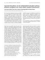
Figure 1 A proposed model for how GPG-NH
2
or its
metabolites target gp160 for ERAD. Initially, gp160 is co-
translationally translocated into the ER, where its growing peptide
backbone becomes glycosylated and starts to fold. (1) In the
presence of GPG-NH
2
or its metabolites gp160 folds incorrectly
which targets it to ERAD. (2) Subsequently, gp160 is retro-
translocated to the cytoplasm, (3) where it becomes deglycosylated
by the cytosolic N-glycanase prior to (4) degradation of its peptide
backbone by the proteasome.
Jejcic et al. Retrovirology 2010, 7:20
/>Page 2 of 9
Figure 2 GPG-NH
2
and its metabolites G-NH
2
and aHGA decrease gp160 mobility and steady-state levels. (A) Scheme of GPG-NH
2
being
metabolized in cell culture medium supplemented with 10% FBS. GPG-NH
2
is processed by CD26 (peptidyl peptidase V) to G-NH
2
and
subsequently modified into aHGA by an unidentified enzyme. (B) HeLa-tat III cells were transfected to express gp160. Two hours post
transfection the cells were treated with the indicated concentrations of GPG-NH
2
, G-NH
2
or aHGA and harvested 20 h post transfection. The cell
lysates were separated by SDS-PAGE and immunoblotted with mAb towards gp41. (C) Densitometric measurement of gp160 and degradation
products (left panel) and gp41 (right panel) given as percentage of total gp160 or gp41 respectively in untreated cells in (B), lane 1. The results
represent the average of two experiments.
Jejcic et al. Retrovirology 2010, 7:20
/>Page 3 of 9
pane l). Together these results support that the target ing
of gp160 to for ERAD is dependent on t he GPG-NH
2
metabolite aHGA.
aHGA targets gp160 for degradation more rapidly
than GPG-NH
2
To investigate the temporal processing of GPG-NH
2
to
the active metabol ite aHGA, the required time of cellu-
lar exposure to the respective drug for a detectable
effect on gp160 was examined. HeLa-tat III cells were
transfec ted to express gp160 and treated with 20 μMor
100 μMGPG-NH
2
or aHGA at various time points
prior to or post transfect ion and the cells were har-
vested 24 h post transfection. The strongest effect of
GPG-NH
2
on gp160, at both concentrations, was
obtained when treatment was initiated 18 h prior to
transfection (Fig. 4A, upper and lower panels, lane 2,
and Fig. 4B). Treatment with GPG-NH
2
starting at 4
and 8 h post transfection still significantly affected
gp160 at 100 μM, but addition at 20 h and 23 h post
transfection, i.e. 4 h an d 1 h prior to harvesting, did not
affect gp160 (Fig. 4A, lower panel, and Fig. 4B). Interest-
ingly, the addition of 20 μM and 100 μM aHGA 18 h
prior to transfection had a slightly milder effect on
gp160 as compared to GPG-NH
2
(Fig. 4C, compare lane
2 to 4A, lane 2). Thus, aHGA treatment did not benefit
from early addition to the cell cultures as did GPG-
NH
2
. Instead, the strongest decrease in the gp160
steady-state levels and molecular mass occurred when
aHGA was added 4 or 8 h post transfection (Fig. 4C,
upper and lower panels, lanes 3 and 4, Fig. 4D). Addi-
tion of aHGA, 20 h post transfection, i.e. 4 hours prior
to harvest of the cells, still had an effect on gp160, while
addition at 1 h prior to h arvest did not (Fig. 4C upper
Figure 3 aHGA acts on gp160 independently of supplemented serum in cell culture medium. HeLa-tat III cells were cultured in cell
culture medium supplemented with 10% FBS and transfected to express gp160 for 20 h. Two hours upon transfection the cell culture
supernatants were carefully removed, the cells rinsed twice in PBS and provided with culture medium containing either 10% HS (upper panel) or
no serum (lower panel) and indicated concentrations of GPG-NH
2
, G-NH
2
or aHGA. The cell lysates were immunoblotted with mAb towards
gp41.
Jejcic et al. Retrovirology 2010, 7:20
/>Page 4 of 9
Figure 4 aHGA t argets gp160 for degradation more rapidly than GPG-NH
2
. (A) HeLa-tat III cells were transfected to express gp160 and
treated with 20 μM (upper panel) or 100 μM GPG-NH
2
(lower panel) for the indicated times pre- or post-transfection. The cells were harvested
24 h post transfection and immunoblotted with mAb towards gp41. (B) Densitometric measurements of gp160 and degradation products in
samples treated with 20 μM (left panel) or 100 μM GPG-NH
2
(right panel) as described in (A) and given as percentage of total gp160 in
untreated cells in (A), lane 1. (C) As in (A), except the cells were treated with aHGA at 20 μM (upper panel) or 100 μM (lower panel). (D)
Densitometric measurements as described in (B) of samples treated with aHGA at 20 μM (left panel) or 100 μM (right panel) described in (C). (E)
Glycoprotein blot of HeLa-tat III cell lysates collected from cells treated with the indicated concentrations of aHGA for 24 h and stained for total
protein and subsequently probed with the lectin Concanavalin A. The asterisks highlight the decreased molecular mass species.
Jejcic et al. Retrovirology 2010, 7:20
/>Page 5 of 9
and lower panels, lanes 5 and 6, Fig. 4D). Thus, the
activity of aHGA towards gp160 requires a much
shorter exposure time than that of G PG-NH
2
,support-
ing that GPG-NH
2
must first be metabolized into
aHGA to become active towards gp160.
We have previously shown that GPG-NH
2
does not
generally effect cellular glycoproteins, but acts rather
selectively on gp160 [10]. Here, we examined the glyco-
protein expression profile in the HeLa-tat III cells upon
treatment with aHGA added to the cultures at seeding
and collected 24 h and 48 h later. The total protein con-
tent increased two fold and three fold, respectively, dur-
ing incubation time (data not shown). As for GPG-NH
2
,
aHGA showed no general effect on glycoproteins at
24 h or 48 h as only a single unidentified high-molecular-
mass-protein (~150 kDa) slightly increased its mobility at
50 μMand100μM aHGA (Fig. 4E; only 24 h blot is
shown).
aHGA decreases the content of Env in HIV-1 particles
The production of viral particles from the chronically
infected ACH-2 cells, monito red by measuri ng the extra
cellular capsid protein p24, was not affected in the pre-
sence of 10-100 μM aHGA (Fig. 5A). In addition,
aHGAdidnotaffecttheviral particle content of the
precursor protein p55Gag or its processing to p24 ( Fig.
5B). However, treatment with aHGA resulted in a sig-
nificant dose-dependent decrease in the gp120/gp41
content in the v iral particles as the ratio of gp 41 to p24
decreased by 85% at 20 μM aHGA to undetecta ble
levels of gp41 at 50 μM aHG A (Fig. 5C). Also HIV-1
particles generated from ACH-2 cells in the absence or
presence of 50 μM aHGA w ere examined f or their
gp120/gp41 content by immunogold labeling and trans-
mission electron microscopy (TEM) (Fig. 5 D). This
further showed that aHGA decreased the inco rporation
of gp120/gp41 as the ratio of immuno gold labeled gp41
to the number of viral particles decreased from 0.46
(total particle number: 984) in the untreated sample to
0.07 (total particle number: 1841).
Discussion
In this study we examined whether either of the two
GPG-NH
2
-metabolites retained the a bility to target
gp160 for destruction in the same manner as GPG-NH
2
.
Here we show that when replacing FBS with HS or in
complete absence of serum the effect of GPG-NH
2
on
gp160 was completely abolished, w hich strongly indi-
cates that GPG-NH
2
is not the molecule responsible for
targeting gp160 for ERAD. aHGA, on the other hand
was active against gp160 both in the presence of HS and
under se rum free condi tions. The intermediate metabo-
lite G-NH
2
was not able to target gp160 for destructio n
in HS but showed some activity in absence of serum.
This means that either some o f the enzymatic activity
converting G-NH
2
to aHGA remained after washing of
the cells and HS prevented its conversion to aHGA or
G-NH
2
was able to affect gp160 by itself but was inhib-
ited by HS. GPG-NH
2
had to be added much earlier
than aHGA to the cell cultures in order to be effective
against gp160. The comparably slow on set of GP G-NH
2
also supports that GPG-NH
2
needs conversion to
aHGA to target gp160 for ERAD. In addit ion, viral par-
ticles produced in the presence of a HGA showed a dra-
matic loss in their gp120/gp41 content with respe ct to
the capsid protein p24. Therefore, the effect on gp160
resulting in reduced gp120/gp41 content in progeny
viral particles rendering them fusion incompetent that
was previously ascribed to GPG-NH
2
is most likely due
to its metabolite aHGA. Although, deletion of the 19
N-terminal amino acids (aa) of the 30 aa long gp160 sig-
nal sequence has been shown to render g p160 resistant
to aHGA treatment, the exact site of aHGA interaction
remains to be identified [10].
We have previously shown that aHGA also causes a
diversity of abnormal capsid formations in progeny viral
particles [11]. These two effects may be complete ly
independent of each other as aHGA is believed to bind
to the hinge region of p24 thereby preventing it from
forming proper capsids [11]. However, the gp41 defi-
ciency in the particles could also contribute to the dis-
torted capsid formation. The exceptionally long
cytosolic tail of gp41, which stretches 150 aa into the
particles, interacts with p55Gag and cellular proteins
and may therefore play a role in the formation of proper
internal viral structures [13-16]. Although important, it
is difficult to evaluate which of the two effects is mostly
responsible for the overall antiviral effect and whether
they are related or are two separate phenomena. In an
effort to solve this, we are now trying to induce the
aHGA resistant gp160 signal sequence mutations into
infectious clones of HIV-1 to see if the resulting clones
are infectious and if so whether aHGA retains its anti-
viral activity to such mutated virus.
Conclusions
In this study, we have reported that it is not GPG-NH
2
but its small metabolite (90 Da) aHGA that t argets
gp160 for destruction via the ERAD pathway, which
results in production of gp120/gp41 deficient HIV-1
progeny particles.
Methods
Reagents and Antibodies
GPG-NH
2
and G-NH
2
were purchas ed from Bachem
Feinchemikalien and aHGA from Chemilia AB. The
monoclonal antibody to gp41 (Chessie 8) [17] was
obtained through the NIH AIDS Research and
Jejcic et al. Retrovirology 2010, 7:20
/>Page 6 of 9
ReferenceReagentProgram,andtheantibodytop24
(EF7) has previously been described [18].
Cell Lines and Plasmids
The cell lines HeLa-tat III and ACH-2 [19,20] and the
infectious HIV-1 expressing plasmid pNL4-3 [21] were
obtained through NIH AIDS Research and Reference
Reagent Program. The expression plasmids for gp160
from the HIV-1 strain NL43 (pNL1.5EU) [22] and for
Rev (pBRev) were kindly pr ovided by Dr. S. Schwartz
(Uppsala U niversity, Sweden). PCR
R
3.1/CAT expresses
chloroamphenichol acetyltransferase and was purchased
from Invitrogen.
Transfection and drug treatments
HeLa-tat III cells (~3 × 10
5
cells/dish) were treated with
the indicated concentrations of GPG-NH
2
,G-NH
2
and
aHGA prior to or post transfection with the gp160, and
the transfection efficiency control CAT expressing plas-
mids using FuGENE 6 (Roche). The cells were rinsed
Figure 5 aHGA treatment reduces HIV-1 particle content of Env. (A) Chronically infected ACH-2 cells were induced with PMA for HIV-1
production and treated with the indicated concentrations of aHGA for 72 h. The viral production was determined by measuring extracellular
p24 concentrations by ELISA. (B) Virus particles were produced as described in (A) and precipitated with polyethylene glycol followed by
immunoblotting towards p24. (C) Immunoblot showing the amount of gp41 present in polyethylene glycol-precipitated HIV-1 particles,
produced by ACH-2 as described in (A) for 48 h. The HIV-1 particle content was standardized to the extracellular p24 concentrations measured
by ELISA and the gp41/p24 ratio was calculated by densitometry. (D) EM images of immuno-gold labeled gp41 in viral particles surrounding
untreated or treated ACH-2 cells with 50 μM aHGA and induced with PMA for 72 h prior to fixation. Arrows indicate labeling of gp41 and the
bars represent 100 nm.
Jejcic et al. Retrovirology 2010, 7:20
/>Page 7 of 9
twice in PBS and lysed 20-24 h post transfection in
RIPA buffer containing 50 mM Tris-HCl pH 7.4, 1%
Triton-X-100, 1% deoxycholate, 150 mM NaCl, 1 mM
EDTA, 0.1% SDS and supplemented with Complete pro-
tease inhibitor cocktail (Roche).
PNGase F digestion
Cell lysates in RIPA buffer were supplemented with 1%
b-mercaptoethenol and denaturated for 10 min at 95°C.
Addition of 1% NP-40 a nd 16 U PNGase F (New Eng-
land Biolabs) was followed by incubation at 37°C for
1h.
Western Blot and ELISA
Cells and precipitated virus were lysed in RIPA buffer,
standardized to CAT or p24 levels respectively, dena-
tured and resolved by SDS-PAGE, transferred to nitro-
cellulose membranes and immunoblotted. The
membranes were exposed to film for the appropriate
time and band intensities were quantified using Gene-
Toolsanalysissoftware(SynGene). For probing against
cellular glycoproteins peroxidase conjugated Concanava-
lin A (Sigma) was used according to manufacturer’s pro-
tocol. In brief, the membranes were incubated in PBS
containing 2% Tween, rinsed in PBS and probed over
night in solution containing 2 μg/ml Concanavalin A,
0,05%Tween, 1 mM of CaCl
2
,MnCl
2
and MgCl
2
. For
detecti on of total pr otein the membranes were stained
with 0.1% Naphthol Blue Black (Sigma) dissolved in 25%
isopropanol and 10% acetic acid. P24 levels in cell cul-
ture supernatants were quantified using p24-ELISA [23]
and CAT concentrations in cell lysates were quantified
using the CAT ELISA kit (Roche).
Virus expression, precipitation of HIV-1 particles and
immune EM
ACH-2 cells (8 × 10
5
cells/ml) were cultured with
100 nM 12-phorbol-13-myristate acetate (PMA) and
with or without aHGA. Three days later the cell culture
supern atants were collected, cleared by centrifugation at
300 × g for 10 min, passed through 0.45 μmfiltersand
the particles were precipitated at 4°C for 48 h in 1:6 (v/v)
with 40% poly ethylene glycol 6000 containing 0.667 M
NaCl. The precipitated particles were allowed to sedi-
ment at 16,000 × g for 20 m inutes at 4°C and the virus
pellets were then dissolved in RIPA buffer. Sample pre-
paration of hydrated ACH-2 cells for immunocytochem-
ical analysis was performed as previously described using
10 nm colloidal gold labeling of anti-gp41 monoclonal
antibody [17,24]. Areas surrounding the infected cells
were used for calculating the number of Au-labeled
particles.
Acknowledgements
We thank Dr Robert Daniels for critical reading of the manuscript. We also
thank the original donors and the NIH AIDS Research and Reference
Reagent Program, Division of AIDS, NIAID for the cell lines HeLa-tat III from
Dr William Haseltine and Dr. Ernest Terwilliger and ACH-2 from Dr Thomas
Folks. We are grateful for the anti-gp41 antibody (Chessie 8) from Dr.
George Lewis and the plasmid pNL4-3 from Dr Malcolm Martin. This work
was supported by grants from the Swedish Medical Foundation (grant no.
K2000-06X-09501-10B), Swedish International developm ent Cooperation
Agency, SIDA (grant no. HIV-2006-050) and by Tripep AB.
Author details
1
Department of Laboratory Medicine, Division of Clinical Microbiology,
Karolinska Institutet, SE-141 86 Stockholm, Sweden.
2
Department of
Biochemistry, Uppsala Universitet, SE-751 23 Uppsala, Sweden.
Authors’ contributions
AJ and AV designed the study. AJ conducted the experiments and analyzed
the results. SH performed the immune TEM work and analyzed the
corresponding results. AJ and AV wrote the article. All authors commented
on and approved the final manuscript.
Competing interests
AV is a founder and shareholder of Tripep AB and a member of its board of
directors.
Received: 13 December 2009 Accepted: 15 March 2010
Published: 15 March 2010
References
1. Land A, Zonneveld D, Braakman I: Folding of HIV-1 envelope glycoprotein
involves extensive isomerization of disulfide bonds and conformation-
dependent leader peptide cleavage. Faseb J 2003, 17:1058-1067.
2. Lu M, Blacklow SC, Kim PS: A trimeric structural domain of the HIV-1
transmembrane glycoprotein. Nat Struct Biol 1995, 2:1075-1082.
3. McCune JM, Rabin LB, Feinberg MB, Lieberman M, Kosek JC, Reyes GR,
Weissman IL: Endoproteolytic cleavage of gp160 is required for the
activation of human immunodeficiency virus. Cell 1988, 53:55-67.
4. Gomez C, Hope TJ: The ins and outs of HIV replication. Cell Microbiol
2005, 7:621-626.
5. Kilgore NR, Salzwedel K, Reddick M, Allaway GP, Wild CT: Direct evidence
that C-peptide inhibitors of human immunodeficiency virus type 1 entry
bind to the gp41 N-helical domain in receptor-activated viral envelope.
J Virol 2003, 77:7669-7672.
6. Ray N: Maraviroc in the treatment of HIV infection. Drug Des Devel Ther
2009, 2:151-161.
7. Dwek RA, Butters TD, Platt FM, Zitzmann N: Targeting glycosylation as a
therapeutic approach. Nat Rev Drug Discov 2002, 1:65-75.
8. Jacob GS: Glycosylation inhibitors in biology and medicine. Curr Opin
Struct Biol 1995, 5:605-611.
9. Tierney M, Pottage J, Kessler H, Fischl M, Richman D, Merigan T,
Powderly W, Smith S, Karim A, Sherman J, et al: The tolerability and
pharmacokinetics of N-butyl-deoxynojirimycin in patients with advanced
HIV disease (ACTG 100). The AIDS Clinical Trials Group (ACTG) of the
National Institute of Allergy and Infectious Diseases. J Acquir Immune
Defic Syndr Hum Retrovirol 1995, 10:549-553.
10. Jejcic A, Daniels R, Goobar-Larsson L, Hebert DN, Vahlne A: Small molecule
targets Env for ER-associated protein degradation and inhibits HIV-1
propagation. J Virol 2009, 83(19):10075-84.
11. Abdurahman S, Vegvari A, Levi M, Hoglund S, Hogberg M, Tong W,
Romero I, Balzarini J, Vahlne A: Isolation and characterization of a small
antiretroviral molecule affecting HIV-1 capsid morphology. Retrovirology
2009, 6:34.
12. Balzarini J, Andersson E, Schols D, Proost P, Van Damme J, Svennerholm B,
Horal P, Vahlne A: Obligatory involvement of CD26/dipeptidyl peptidase
IV in the activation of the antiretroviral tripeptide
glycylprolylglycinamide (GPG-NH(2)). Int J Biochem Cell Biol 2004,
36:1848-1859.
Jejcic et al. Retrovirology 2010, 7:20
/>Page 8 of 9
13. Blot G, Janvier K, Le Panse S, Benarous R, Berlioz-Torrent C: Targeting of
the human immunodeficiency virus type 1 envelope to the trans-Golgi
network through binding to TIP47 is required for env incorporation into
virions and infectivity. J Virol 2003, 77:6931-6945.
14. Dorfman T, Mammano F, Haseltine WA, Gottlinger HG: Role of the matrix
protein in the virion association of the human immunodeficiency virus
type 1 envelope glycoprotein. J Virol 1994, 68:1689-1696.
15. Kim JT, Kim EM, Lee KH, Choi JE, Jhun BH, Kim JW: Leucine zipper domain
of HIV-1 gp41 interacted specifically with alpha-catenin. Biochem Biophys
Res Commun 2002, 291:1239-1244.
16. Murakami T, Freed EO: Genetic evidence for an interaction between
human immunodeficiency virus type 1 matrix and alpha-helix 2 of the
gp41 cytoplasmic tail. J Virol 2000, 74:3548-3554.
17. Abacioglu YH, Fouts TR, Laman JD, Claassen E, Pincus SH, Moore JP,
Roby CA, Kamin-Lewis R, Lewis GK: Epitope mapping and topology of
baculovirus-expressed HIV-1 gp160 determined with a panel of murine
monoclonal antibodies. AIDS Res Hum Retroviruses 1994, 10:371-381.
18. Devito C, Levi M, Broliden K, Hinkula J: Mapping of B-cell epitopes in
rabbits immunised with various gag antigens for the production of HIV-
1 gag capture ELISA reagents. J Immunol Methods 2000, 238:69-80.
19. Clouse KA, Powell D, Washington I, Poli G, Strebel K, Farrar W, Barstad P,
Kovacs J, Fauci AS, Folks TM: Monokine regulation of human
immunodeficiency virus-1 expression in a chronically infected human T
cell clone. J Immunol 1989, 142:431-438.
20. Terwilliger E, Proulx J, Sodroski J, Haseltine WA: Cell lines that express
stably env gene products from three strains of HIV-1. J Acquir Immune
Defic Syndr 1988, 1:317-323.
21. Adachi A, Gendelman HE, Koenig S, Folks T, Willey R, Rabson A, Martin MA:
Production of acquired immunodeficiency syndrome-associated
retrovirus in human and nonhuman cells transfected with an infectious
molecular clone. J Virol 1986, 59:284-291.
22. Schwartz S, Felber BK, Fenyo EM, Pavlakis GN: Env and Vpu proteins of
human immunodeficiency virus type 1 are produced from multiple
bicistronic mRNAs. J Virol 1990, 64:5448-5456.
23. Horal P, Hall WW, Svennerholm B, Lycke J, Jeansson S, Rymo L, Kaplan MH,
Vahlne A: Identification of type-specific linear epitopes in the
glycoproteins gp46 and gp21 of human T-cell leukemia viruses type I
and type II using synthetic peptides. Proc Natl Acad Sci USA 1991,
88:5754-5758.
24. Hoglund S, Su J, Reneby SS, Vegvari A, Hjerten S, Sintorn IM, Foster H,
Wu YP, Nystrom I, Vahlne A: Tripeptide interference with human
immunodeficiency virus type 1 morphogenesis. Antimicrob Agents
Chemother 2002, 46:3597-3605.
doi:10.1186/1742-4690-7-20
Cite this article as: Jejcic et al.: GPG-NH
2
acts via the metabolite aHGA
to target HIV-1 Env to the ER-associated protein degradation pathway.
Retrovirology 2010 7:20.
Submit your next manuscript to BioMed Central
and take full advantage of:
• Convenient online submission
• Thorough peer review
• No space constraints or color figure charges
• Immediate publication on acceptance
• Inclusion in PubMed, CAS, Scopus and Google Scholar
• Research which is freely available for redistribution
Submit your manuscript at
www.biomedcentral.com/submit
Jejcic et al. Retrovirology 2010, 7:20
/>Page 9 of 9