Báo cáo y học: " Inhibition of HIV-1 replication by small interfering RNAs directed against Glioma Pathogenesis Related Protein (GliPR) expression" potx
Bạn đang xem bản rút gọn của tài liệu. Xem và tải ngay bản đầy đủ của tài liệu tại đây (1.1 MB, 10 trang )
RESEARC H Open Access
Inhibition of HIV-1 replication by small interfering
RNAs directed against Glioma Pathogenesis
Related Protein (GliPR) expression
Gianni Capalbo
1
, Thea Müller-Kuller
1
, Ursula Dietrich
2
, Dieter Hoelzer
1
, Oliver G Ottmann
1
, Urban J Scheuring
1*
Abstract
Background: Previously, we showed that glioma pathogenesis related protein (GliPR) is induced in CEM T cells
upon HIV-1 infection in vitro. To examine whether GliPR plays a role as HIV dependency factor (HDF), we tested the
effect of GliPR suppression by siRNA on HIV-1 replication.
Results: Induction of GliPR expression by HIV-1 was confi rmed in P4-CCR5 cells. When GliPR was suppressed by
siRNA, HIV-1 replication was significantly reduced as measured by HIV-1 transcript levels, HIV-1 p24 protein levels,
and HIV-1 LTR-driven reporter gene expression, suggesting that GliPR is a cellular co-factor of HIV-1. Microarray
analysis of uninfected HeLa cells following knockdown of GliPR revealed, among a multitude of gene expression
alterations, a down-regulation of syndecan-1, syndecan-2, protein kinase C alpha (PRKCA), the catalytic subunit b of
cAMP-dependent protein kinase (PRKACB), nuclear receptor co-activator 3 (NCOA3), and cell surface protein CD59
(protectin), all genes having relevance for HIV-1 pathology.
Conclusions: The up-regulation of GliPR by HIV-1 and the early significant inhibition of HIV-1 replication mediated
by knockdown of GliPR reveal GliPR as an important HIV-1 dependency factor (HDF), which may be exploited for
HIV-1 inhibition.
Background
The replication of HIV-1 depends on specific host fac-
tors [1-4]. A recent report identified 273 cellular HIV-1
dependency factors (HDF), that are important for HIV-1
replic ation [5]. Furthermore, HIV-1 modifies the mRNA
expression of a relatively large number of host cell
genes, as shown by several reports [6-10]. Differential
display experiments suggested that the expression of
~700 host genes (approximately 3% of all cellular genes)
is modified by HIV-1 infection in vitro [9]. A microarray
analysis using a limited subset of 1500 cDNAs identified
20 differentially expressed mRNAs from several cellular
pathways [7]. Specific HIV-1 proteins including Tat,
Nef, gp1 20 and Vpr were examined to dissect their role
in modifying the transcription of cellular genes [11-14].
While some of the differentially expressed cellular genes
may play a role in host defense mechanisms, others may
facilitate HIV-1 replication, infectivity, species propaga-
tion and survival. A sub group of differential cellular
gene expressions may even support both host defense
and viral replication, since HIV-1 replication is linked to
immune activation of CD4+ T cells. Due to evolutionary
selection, HIV-1 is expected to induce specific host fac-
tors, favorable for viral replication or propagation, and
to suppress unfavorable cellular gene products [15-17].
Therefore, the examination of host cell genes, that are
up-regulated upon HIV-1 infectio n, is expected to iden-
tify potential targets for inhibition of HIV-1 replication.
Previously, we found an ear ly up-regulation of GliPR
expression by more than 5-fold in CEM T ce lls infecte d
with HIV-1 by a differential display screen [9]. There-
fore, we were interested in delineating the role of GliPR
for HIV-1 replication.
GliPR was identified originally in human glioblastomas
[18] and was also described as related to testes-specific,
vespid, and pathogenesis protein 1 (RTVP-1) [19].
Increased expressi on of GliPR was associated with mye-
lomonocytic differentiation in macrophages [20].
* Correspondence:
1
Department of Hematology/Oncology and Infectious Diseases, J. W.
Goethe-University Hospital, Theodor Stern Kai 7, 60590 Frankfurt/Main,
Germany
Capalbo et al. Retrovirology 2010, 7:26
/>© 2010 Capalbo et a l; licensee BioMed Central Ltd. This is an Open Access article distributed under the terms of the Creative Commons
Attribution License ( which permits unrestricted use, distribution, and reproduction in
any medium, provided the original work is p roperly cited.
Whereas GliPR has been reported to act as a tumor
suppressor gene inducing apoptosis in prostate cancer
[21-24], it appears to be an oncogene in glioblastomas
[25] and Wilms tumors [26]. RTVP-1 protein was
reported to contain a N-terminal signal peptide
sequence and a transmembrane domain [27]. Further-
more, homology studies revealed a putative activ e enzy-
matic center in GliPR [27] . GliPR is homolo gous to
group 1 plant pathogenesis-related proteins (PR-1) that
are implicated in plant defense responses to viral, bac-
terial, and fungal infection [28,29]. Since GliPR shows
structural similarities with its homologous plant PR-1
proteins, mammalian testis proteins (TPX1) and the
insect venom Ag-5 protein, which are secretory proteins
[29,30], it has been suspected that GliPR is also secreted.
GliPR’ s homology with plant PR-1 proteins that have
been attributed with a defense function may raise the
question whether GliPR has an evolutionarily conserved
role in innate immune response and human host
defense of viral infection including HIV-1. Alternatively
or additionally, HIV-1 may induce and exploit GliPR for
viral replication.
The effect of GliPR knockdown on HIV-1 replication
was studied, in order to test the hypotheses of GliPR being
a host defense protein against or a co-factor of HIV-1.
Furthermore, in order to identify downstream targets of
GliPR, the effect of GliPR suppression on cellular gene
expression was also investigated using cDNA microarrays.
Results
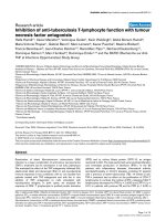
GliPR is induced upon HIV-1 infection in P4-CCR5 cells
Since HIV-1 infection induced GliPR expression in HIV-
1 infected human T cell line cells, as described
previously [9], we tested whether this modification
could be reproduced in P4-CCR5 HeLa cells infected
with HIV-1
LAI
. P4-CCR5 HeLa cells were employed for
thepresentstudybecausethey are more amenable to
efficient transfection of synthetic siRNA compared to
lymphocytic cell lines. Quantitative PCR demonstrated
an up-regulation of GliPR transcripts by approximately
2-fold at day 4 after infection compared to uninfected
cells (Fig. 1a). In order t o display HIV-1 infection
kinetics, real-time quantitative PCR was also utilized to
determine levels of intracellular HIV-1 viral mRNA nor-
malized by cell number (house keeping gene GAPDH)
at different time points following infection (Fig. 1b). The
data show that HIV-1 replication is still in the early
logarithmic phase at day 4 in this cell culture system
and that GliPR expression is induced in this early phase.
Suppression of GliPR mediated by short interfering RNA
P4-CCR5 cells were transfected with siRNAs specific for
GliPR or a non-silencing siRNA, which was 5-prime
labeled with rhodamine. Flow cytometry analysis of cells
transfected with non-silencing siRNA 24 h po st trans-
fection revealed transfection efficiencies on average of
90% in all samples. Forty-eight hours after transfection,
the relative levels of GliPR mRNA transcripts were
decreased by at least 90%, as shown by quan titative real-
time PCR (Fig. 2a). Even four and six days after trans-
fection a markedly reduced G liPR expression by at lea st
80% compared with non-transfec ted cells (mock) or
cells transfected with non-silencing siRNA was observed
(Fig. 2a).
Viability and proliferation rate of P4-CCR5 cells
transfected with siRNAs against GliPR or with the
Figure 1 Up-regulation of GliPR expression by HIV-1 infection. (A) HIV-1
LAI
-infected P4-CCR5 cells and controls were subjected to
quantitative PCR of GliPR expression at day 4 after HIV-1 infection. (B) In order to display HIV-1 infection kinetics, real-time quantitative PCR was
also utilized to determine levels of intracellular HIV-1 viral mRNA normalized by cell number (house keeping gene GAPDH) in triplicate at day 0,
2, 4 and 6 post infection. Bars represent the standard deviation of the mean of determinations.
Capalbo et al. Retrovirology 2010, 7:26
/>Page 2 of 10
non-silencing siRNA remained unchanged as deter-
mined by WST-1 cell proliferation assay (Fig. 2b).
In order to establish a test system in a T cell line as
well, a predominant type of host cell for HIV-1, Jurkat
cells were transfected with 2 different siRNAs targeting
GliPR, control non-silencing siRNA, or mock transfec-
tion without any siRNA. G liPR mRNA expression was
reduced by around 64% to 69% at 48 hours after trans-
fection with specific siRNAs compared to controls
(Fig. 2c). The less pronounced reduction of GliPR
expression compared to P4-CCR5 HeLa cells may be
attributed to the lower transfection efficiency generally
observed in T cell lines. In this experiment, approxi-
mately 70% of Jurkat cells were transfected, while 90%
of P4-CCR5 HeLa cells were transfected.
In general, GliPR-directed siRNAs reduced the expres-
sion of GliPR effectively in P4-CCR5 and Jurkat cells
without affecting cell viability.
Down-regulation of GliPR by siRNA inhibits HIV-1
replication in P4-CCR5 and Jurkat cells
In order to examine the effect of GliPR knockdown on
HIV-1 replication, P4-CCR5 cells were transfected with
GliPR-specific siRNAs and subsequently infected with
HIV-1
LAI
. As a negative control, the non-silencing
siRNA (si-n ons-Rho) was utilized while a siRNA target-
ing HIV-1 p24 was used as a positive control, since it
was able to inhibit viral r eplication very effectively, as
previously demonstrated [31]. HIV-1 infection was per-
formed 24 h post siRNA transfection with a MOI of
Figure 2 Efficacy of siRNA-mediated suppression of GliPR. (A) Quantitative PCR analysis of GliPR expression in P4-CCR5 cells which were
transfected with 2 different siRNAs against GliPR or the control non-silencing siRNA labeled with rhodamine. Results are presented as mean
values of triplicate samples ± standard deviation (SD). (B) Cell viability was determined with the WST-1 assay 24 h and 48 h after siRNA
transfection. Results are expressed as absorbance (OD
450
). Bars represent the standard deviation of the mean of determinations. (C) Quantitative
PCR analysis of GliPR expression in Jurkat cells 2 days after transfection with 2 different siRNAs against GliPR or the control non-silencing siRNA
labeled with rhodamine.
Capalbo et al. Retrovirology 2010, 7:26
/>Page 3 of 10
0.01 or 0.05. Sequential cell-associated HIV-1 viral
mRNA levels were determined by real-time quantitative
PCR during 6 days after infection. As expected, the
positive control siRNA (si-p24) exhibited a marked inhi-
bition in viral mRNA transcription. Similarly, the
siRNA-mediated reduction of GliPR expression was fol-
lowed by significantly reduced viral mRNA transcript
levels compared to HIV-1 infected controls, which were
mock-transfected (mock) or transfected with the non-
silencing siRNA (si-nons-Rho) at both MOI of 0.01 and
0.05 (Fig. 3a and 3b).
The effect of GliPR suppression on HIV-1 replication
was confirmed by p24 ELISA, showing a significantly
reduced p24 expression at day 4 post infection in cul-
tures with GliPR knock-down compared to controls
with non-silencing siRNA (Fig. 3c).
In order to test this phenomenon in T cells, Jurkat
cells transfected with siRNAs specific to GliPR or con-
trol siRNA were infected with HIV-1 at a MOI of 0.01.
GliPR-speci fic siRNAs resulted in a significant reduction
of HIV-1 replication, similar to the positive control with
siRNA against p24 (Fig 3 d). Thus the T cell line results
are in line with the data in P4-CCR5 cells.
Furthermore, the effect on HIV-1 replication was
examined by the integrated HIV-1-LTR-driven reporter
vector expressing b-galactosidase in P4-CCR5 cells.
Figure 3 Effects of siRNA transfections on HIV-1 replication. P4-CCR5 cells were transfected with siRNAs directed against GliPR, viral p24 or
an unspecific sequence (non-silencing control) and subsequently infected with HIV-1
LAI
with a multiplicity of infection of 0.01 (A) and 0.05 (B),
respectively. HIV-1 RNA copy numbers were normalized per cell count by house keeping gene GAPDH. (C) P4-CCR5 cells were transfected with
siGliPR-2, si-p24 or non-silencing control siRNA and subsequently infected with HIV-1
LAI
with a multiplicity of infection of 0.01. Concentrations of
viral p24 at day 0, 2 and 4 represent mean values of triplicate samples. (D) Jurkat cells were transfected with siRNAs directed against GliPR, viral
p24 or an unspecific sequence (non-silencing control) and subsequently infected with HIV-1
LAI
with a multiplicity of infection 0.01. HIV-1 RNA
copy numbers were normalized per cell count by house keeping gene GAPDH.
Capalbo et al. Retrovirology 2010, 7 :26
/>Page 4 of 10
HIV-1 Tat-mediated transactivation of t he LTR leads to
expression of measurable b-galact osidase activity, allow-
ing measurments of inhibitory effects on HIV-1 replica-
tion as reductions in b-galactosidase activity. The
expression of b-galactosidase was markedly decreased by
siGliPR on day four after infection, comparable to the
degree of the positive control with p24 siRNA (Fig. 4a).
The inhibition of LTR-driven transcription was
confirmed by microscopy of these cell cultures after
X-Gal staining on day six after infection (Fig. 4b).
These results demonstrated that siRNA-mediated sup-
pression of GliPR inhibited HIV-1 replication implicat-
ing that GliPR promotes HIV-1 replicatio n. It was not
possible to employ the opposite approach by assessing
the effect of GliPR’s over-expression on H IV-1 replica-
tion, since forced expression of GliPR caused rapid
Figure 4 Expression of b-galactosidase driven by HIV-1-LTR in HIV-infected cells after siRNA transfection. P4-CCR5 cells containing a HIV-
1-LTR driven b-galactosidase reporter vector were transfected with siGliPR-2, non-silencing control siRNA or no siRNA (mock) and subsequently
infected with HIV-1
LAI
with a multiplicity of infection of 0.01. (A) b-galactosidase units at day 4 normalized by relative WST-1 values represent
mean values from triplicate samples. (B) Photomicrograph of b-gal stained P4-CCR5 cells infected with HIV-1
LAI
(MOI 0.01) after transfection with
mock, GliPR-siRNA, HIV-1 p24-siRNA or non-silencing control siRNA at day 6 post infection.
Capalbo et al. Retrovirology 2010, 7 :26
/>Page 5 of 10
induction of apoptosis in HeLa cells and Jurkat cells
(unpublished data).
Differentially expressed genes after GliPR suppression
with relevance for HIV-1 replication
In order to examine the effect of GliPR knock-down on
cellular gene expression, microarray analyses were per-
formed to identify cellular target genes of GliPR
involved in HIV-1 pathology. The list of genes differen-
tially expressed in uninf ected HeLa cells following sup-
pression of GliPR was screened for those genes which
had been reported in the context of HIV-1 infection
previously. S ix genes with potential role in HIV-1
pathology were identified within the list of genes (n =
262) that were differentially regula ted after GliPR sup-
pression (Table 1).
Discussion
The present investigation confirmed t he up-regulation
of GliPR induced by HIV-1 infection that we had found
in a lymphocytic cell line [9] in P4-CCR5 HeLa cells.
The suppression of GliPR expression by siRNA was
associated with a significant inhibition of HIV-1 replica-
tion compared to controls as determined by quantitativ e
PCR for HIV-1 transcripts, p24 ELISA, and HIV-1 LTR
driven b-galactosidase expression. The inhibition of
HIV-1 transcript ion following knockdown of Gli PR was
confirmed in Jurkat T cells. Furthermore, the knock-
down of GliPR in uninfected HeLa cells revealed 6 dif-
ferentially expressed genes, which had been reported to
be associated with HIV-1 pathology.
The first hypothesis that GliPR induction is a cellular
defense reaction hindering HIV-1 replication has to be
questioned. If GliPR had exerted an anti-viral effect
against HIV-1, the suppression of GliPR would have
been expected to show an enhancing effect on HIV-1
replication, which was not found to be the case.
The second hypothesis that GliPR acts as a HIV-1 co-
factor has to be favored, since down-regulation of GliPR
expression caused a significant inhibition of HIV-1
replication, implying that GliPR promotes HIV-1 repli-
cation. This result could not be corroborated by the
inverse technique of over-expressing GliPR because
over-expression of GliPR caused apoptosis as reported
for prostate cancer cell lines [22] and confirmed by us
for HeLa and Jurkat cells (unpublished data). Apoptosis
would have disguised a putative promoting effect on
HIV-1 replication, since apoptosis per se would have
affected the kinetics of HIV-1 replication.
The initiation of apoptosis in an infected cell may be
considered to be an ancient defense mechanism aimed
at the abortion of infection for the organism. Appar-
ently, HIV-1 escapes this putative mec hanism of defense
as long as its replication has not been completed by
blocking apoptosis initially via up-regulat ion of bc l-2 via
Tat [32-34] or by other mechanisms, e.g. reduction of
Bax or inhibition of ISG15 [35,36].
Moreover, HIV-1 appears to exploit a specific function
of GliPR or GliPR-induced gene products for its replica-
tion. Therefore, GliPR may be considered as an HDF
[1-4]. A recent report identified 237 HDF in a broad
siRNA screen, in addition to 36 already described HDF
[5]. GliPR was not found by this HDF screen, although
our results suggest that it meets the criteria for an HDF.
Apparently the detection of HDF depends on the speci-
fic experimental conditions and the selecti on criteria set
for sensitivity and specificity of a screening approach.
It has been shown that tumor suppressor protein p53
is a direct transcriptional activator of GliPR. Gl iPR may
also be induced independently of p53 [22]. It has been
reported that HIV-1 induces or activates p53 [37,38],
suggesting a p53-mediated pathway for up-regulat ion of
GliPR by HIV-1.
It is also of interest to understand the mechanism by
which GliPR promotes HIV-1 r eplication. The modeling
of GliPR’ s protein structure revealed an active site [27].
Protein Tex31, which i s a member of the PR-1 protein
family and a component of the c one snail venom, is a
substrate specific protea se [39]. Golgi associated PR-1
related protein-1 (GAPR-1) also called GliPR2 due to its
Table 1 Differentially expressed genes in siGliPR transfected HeLa cells relevant to HIV-1 pathology.
Probe Set ID Gene Title Gene Symbol fold change
201286_at syndecan 1 SDC1 - 3.37
202741_at protein kinase, cAMP-dependent, catalytic b PRKACB - 3.52
207700_at nuclear receptor coactivator 3 NCOA3 - 6.53
212154_at
212157_at
212158_at
syndecan 2 (heparan sulfate proteoglycan) SDC2 - 3.05
- 20.23
- 16.54
212463_at CD59 antigen p18-20 CD59 -3.11
213093_at
215195_at
protein kinase C, alpha PRKCA - 3.55
- 4.09
Cellular genes that were found >3-fold induced (+) or suppressed (-) by siGliPR transfection of uninfected HeLa cells are listed with the factor of induction or
suppression. Only genes that appeared to be relevant for HIV-1 infection according to a PubMed literature screen are listed.
Capalbo et al. Retrovirology 2010, 7 :26
/>Page 6 of 10
close homology with GliPR, is a putative serine protease,
too [40]. It may be hypothesized that a potential GliPR
protease activity may be involved in the processing of a
specific HIV-1 protein as it has been demonstrated for
other cellular proteases such as furin and PC7 in proces-
sing of gp160 [41]. The interaction of GliPR with HIV-1
polypeptides is possible since GliPR is localized in the
endoplasmic reticulum (unpublished data).
Our microarray screen found 6 gene products down-
regulated by GliPR suppression that have been reported
in the context of HIV:
Syndecan-1 and syndecan-2 are heparansulfate proteo-
glycans and membrane proteins involved in cellular pro-
liferation, migration and matrix interactions. Syndecan-1
functions as receptor f or internalizing extracellular Tat
protein [42] and syndecan-2 as regulator of T c ell acti-
vation [43].
The catalytic subunit b of cAMP-de pendent protein
kinase (PRKACB) phosphorylates HIV-1 precursor pro-
tein and matrix protein p17 thereby initiating their
translocation [44]. PRKACB is incorporated in HIV-1
virio ns and regulates viral infectivity [45]. PRKCA phos-
phorylates Tat and transactivates HIV-1-LTR [46].
The nuclear recept or coacti vator 3 (NCOA 3) interacts
with Tat enhancing Tat’s transactivating effect [47].
The cell surface protein CD59 (protectin) prevents the
formation of membrane attack complexes (MAC). CD59
co-localizes with HIV-1 matrix protein p17 in v irions
[48].
Conclusions
GliPR is i nduced in early HIV-1 infection in vitro.
According to the profound inhibitory effect of GliPR
knock-down on HIV-1 replication, GliPR is an impor-
tant HDF. Future research needs to address whether
GliPR directly functions as a co-factor of HIV-1 proces-
sing or whether it exerts its effect via other cellular tar-
get genes as identified by our microarray screen. It is
also important to define the role of GliPR in cellular
defense against other viral infections in general.
Methods
Cell culture and HIV-1 infection
For siRNA knock-down experiments, HeLa cells and P4-
CCR5 [49] cells (HeLa CD4
+
CCR5
+
long terminal
repeat-LacZ) were cultured in Dulbecco’ smodified
Eagle’ s medium (Invitrogen, Karlsruhe, Germany).
C8166 and Jurkat cells were cultured in RPMI 1640
medium (Invitrogen). All media were supplem ented
with 10% fetal calf serum (Gibco-BRL, Karlsruhe, Ger-
many), 1% glutamine (Gibco-BRL) and 1% antibiotic
solution (penicillin and streptomycin; Gibco-BRL). P4-
CCR5 cells were cultured in the presence of 100 μg/ml
G418 (PAA Laboratories, Coelbe, Germany) and 1 μg/
ml Puromycin (PAA Laboratories). Transfection and
infection of the cells were carried out in t he absence of
any antibiotics. T he HIV-1 strain LAI was taken from
the supernatant fluid of freshly infected H9 cells. Viral
titer (TCID
50
units/ml) was determined by titration on
C8166 cells as described [50].
24 h after siRNA transfection, P4-CCR5 or Jurkat cells
were infected with HIV-1
LAI
in triplicate at a multipli-
city of infection (MOI) of 0.01 or 0.05. Infection of P4-
CCR5 cells was carried out in the presence of 50 μg/ml
DEAE-Dextran (Sigma, Taufkirchen, Germany). After
incubation for 4 h the cells were washed with PBS and
re-fed with fresh medium. Cells and supernatant sam-
ples were collected for quantitative PCR analysis, b-
galactosidase enzyme assay and HIV-1 p24 antigen
ELISA at indicated time points.
siRNA transfection
siRNAs with the following sequences were used: a
positive control siRNA against HIV-1 p24 [31], (si-p24:
5’-GAU UGU ACU GAG AGA CAG Gdtdt-3’); a nega-
tive control non-silencing siRNA with no known
homology to mammalian genes (si-nons-Rho: 5’-UUC
UCC GAA CGU GU C ACG Udtdt-3’; 5-prime labeled
with rhodamine); and two different siRNAs specific to
GliPR (si-GliPR-1: 5’-GGU GAA ACC AAC AGC CAG
Udtdt-3’ ; si-GliPR-2: 5’ -GGA CUA UGA CUU CAA
GAC Udtdt-3’ ). All siRNAs were synthesized by
Ambion (Darmstadt, Germany) and were purchased a s
annealed RNA-duplexes. 24 h before transfection,
HeLa or P4-CCR5 cells were plated in 24-well plates
(Corning, Kaiserslautern, Germany) at 5 × 10
4
cells per
well in Dulbecco’s minimal essential medium contain-
ing 10% FBS with no antibiotics. Transfections were
performed with Lipofectamine 2000 transfection
reagent (Invitrogen) with siRNA at a final concentra-
tion of 20 nM according to the manufacturer’s recom-
mendations. After incubating for 6 h, the lipid/siRNA
complexes were removed and replaced with fresh med-
ium. For further analysis, cells were removed from the
culture dish by trypsinization with 0.25% trypsin/0.02%
EDTA in PBS (Cambrex, Verviers, Belgium) at differ-
ent time points after transfection. Transfection of J ur-
kat cells was performed with HiPerFect Transfection
Reagent (Qiagen , Hilden, Germany) with siRNA at a
final concentration of 100 nM according to the manu-
facturer’ s recommendations. Transfection efficiency
was analyzed by flow cytometry 24 h after transfection.
Data were acquired and analyzed on FACScan with
Cell Quest software (Becton Dickinson, Heidelberg,
Germany). Effects on cellular viability after siRNA
treatment were measured using the cell proliferation
reagent WST-1 according to the manufacturer’ s
instructions (Roche, Penzberg, Germany).
Capalbo et al. Retrovirology 2010, 7 :26
/>Page 7 of 10
Real time PCR quantification of viral and cellular RNA
RNA was extracted using the RNeasy mini kit (Invitro-
gen) including treatment with RNase-free DNase I (Qia-
gen). Syn thesis of cDNA was carried out using random
hexamer primers and Superscript-II RNaseH-reverse
transcriptase according to the manufacturer’s specifica-
tions (Invitrogen).
Real-time PCR was per formed in duplicate reactions
employing ABI PRISM 7700 (Applied B iosystems,
Darmstadt, Germany) with standard conditions (50°C
for 2 min, 95°C for 10 min and 40 cycles at 95°C for 15
s and 60°C for 1 min). The 25 μlPCRincluded2,5μl
cDNA, 1× TaqMan® Universal PCR Mast er Mix
(Applied Biosystems), 0.2 μMTaqMan®probe,0.2μM
forward primer and 0.2 μM re verse primer. Primers and
probes were designed using Primer Express v.1.0 so ft-
ware (Applied Biosystems) and were synthesized by
Thermohybaid (Ulm, Germany). In order to quant ify
GliPR, HIV-pol and GAPDH cDNA, the following pri-
mers and probes were used: GliPR (sense: 5’-TGC CAG
ACA AAG CAT GCG T-3’ ;antisense:5’ -GCT GTG
TGT GAA TAA TTG GAG ACA A-3’; probe: 5’-FAM-
TCA CAC TTG CTA CAA TAG CCT GGA TGG TTT
C-3’-TAMRA), HIV-pol(sense:5’-AAT TTC A CC AGT
ACTACGGTTAAGGC-3’;antisense:5’-CTT TAA
TTC TTT ATT CAT AGA TTC TAC TAC TCC TTG-
3’ ;probe:5’ -FAM-TGT TGG T GG GCG GGA ATC
AAG C-3’ -TAMRA) and GAPDH (sense: 5’ -GAA GGT
GAA GGT CGG AGT C-3’ ;antisense:5’ -G AA GAT
GGT GAT GGG ATT TC-3’; probe: 5’ -FAM-CAA GCT
TCC CGT TCT CAG CC-3’-TAMRA). The probes were
labeled with FAM at the 5’ end and TAMRA at the 3’
end. Copy numbers of the respective transcripts were
calculated by plasmid standard curves, normalized by
GAPDH housekeepi ng gene transcripts. Standard curves
were obtained after amplification of log step dilutions
between 10 to 10
6
copy numbers of purified plasmids
carrying the amplicons of GliPR, HIV-1-pol (modified
plasmidpLAI.2obtainedfromtheNIHAIDSResearch
& Reference Reagent Progr am) or human GAPDH [51],
respectively. The plasmid standard for the quantification
of GliPR was prepared by inserting a PCR-generated
fragment (sense: 5’-GGA TCC ATG CGT GTC ACA
CTT GCT ACA ATA GC-3’ and antisense: 5’ -GTC
GAC TTA GTC CAA AAG AAC TAA ATT AGG
GTA CTT GAG C-3’) into pCR2.1 (Invitrogen), which
was amplified using HeLa cDNA as template.
b-Gal staining of cells
At indicated time points after HIV-1
LAI
infection, P4-
CCR5 cells were washed twice with PBS and fixed for 5
min in fixative (0.25% glutaraldehyde in PBS) at room
temperature. After two washes with PBS, cells were
covered with staining solution (PBS containing 4 mM
potassium ferrocyanide, 4 mM potassium ferricyanide, 2
mM MgCl
2
, and 0.4 mg/ml of X-Gal [5-bromo-4-
chloro-3-indolyl-b-D-galactopyranoside]) and incubated
at 37°C. Subsequently plates were washed twice with
PBS and numbers of b-Gal-positive (blue) cells were
examined microscopically.
b-galactosidase enzyme assay
Cell lysates were prepared b y using Reporter Lysis Buf-
fer (Promega, Mannheim, Germany). To perform 96-
well plate b-galactosidase assays 50 μl of cell lysates and
50 μlof2×b-galactosidase assay buffer (Promega) were
mixed and incubated at 37°C for 30 min. To stop the
reacti on, 150 μl of 1 M sodium carbonate was added to
the mixture and mixed well by vortex ing briefly. Absor-
bance of the reaction mixture was read immediately at
420 n m. A standard curve was created, using sta ndards
between 0 and 6.0 × 10
-3
units of Galactosidase (Pro-
mega). Reporter assay results were normalized according
to relative cell numbers, whic h were estimated by using
the cell pro liferation reagent WST- 1 according to the
manufacturer’s instructions (Roche).
HIV-1 p24 antigen ELISA
HIV-1 p24 ELISA was performed using a commercially
available kit (Beckmann Coulter, Krefeld, Germany)
according to the manufacturer’s instr uctions. For mea-
suring p24 in the supernatant s, 100-fold dilutions of the
supernatants were used. All ELISA measurements were
done in triplicate.
Gene expression analysis by microarrays
The microarray analysis of the effect of GliPR suppres-
sion by siRNA was performed using the HG-U133 Plus
2.0 microarray of Affymetrix (Santa Clara, CA USA) per
manufacturer ’s instructions (GeneChip® Expression Ana-
lysis Manual). This chip contains 47,000 transcripts
which represent 39,000 annotated genes. The data ana-
lysis was carried out according to established standards
for Affymetric microarrays using GeneChip Operating
Software (GCOS; Affymetrix) and GeneSpring (Agilent
Technologies).
Acknowledgements
We are very grateful to Sandra Markovic for excellent technical assistance.
We are very grateful to the H. W. & J. Hector Stiftung (foundation),
Mannheim, Germany that provided a grant to support this research.
Author details
1
Department of Hematology/Oncology and Infectious Diseases, J. W.
Goethe-University Hospital, Theodor Stern Kai 7, 60590 Frankfurt/Main,
Germany.
2
Georg-Speyer-Haus, Institute for Biomedical Research, Paul-Ehrlich-
Str. 42-44, 60596 Frankfurt/Main, Germany.
Capalbo et al. Retrovirology 2010, 7 :26
/>Page 8 of 10
Authors’ contributions
GC performed the experiments and contributed to draft the manuscript.
TMK contributed to the experiments and to the analysis of data. DH, UD
and OGO participated in the design of the study and helped to draft the
manuscript. UJS directed the work and completed the manuscript. All
authors read and approved the final manuscript.
Competing interests
The authors declare that they have no competing interests.
Received: 22 August 2008 Accepted: 31 March 2010
Published: 31 March 2010
References
1. Agbottah ET, Traviss C, McArdle J, Karki S, St Laurent GC, Kumar A: Nuclear
Factor 90(NF90) targeted to TAR RNA inhibits transcriptional activation
of HIV-1. Retrovirology 2007, 4:41.
2. Lama J, Planelles V: Host factors influencing susceptibility to HIV infection
and AIDS progression. Retrovirology 2007, 4:52.
3. Vardabasso C, Manganaro L, Lusic M, Marcello A, Giacca M: The histone
chaperone protein Nucleosome Assembly Protein-1 (hNAP-1) binds HIV-
1 Tat and promotes viral transcription. Retrovirology 2008, 5:8.
4. Ye Y, De Leon J, Yokoyama N, Naidu Y, Camerini D: DBR1 siRNA inhibition
of HIV-1 replication. Retrovirology 2005, 2:63.
5. Brass AL, Dykxhoorn DM, Benita Y, Yan N, Engelman A, Xavier RJ,
Lieberman J, Elledge SJ: Identification of host proteins required for HIV
infection through a functional genomic screen. Science 2008, 319:921-926.
6. Bosinger SE, Hosiawa KA, Cameron MJ, Persad D, Ran L, Xu L, Boulassel MR,
Parenteau M, Fournier J, Rud EW, Kelvin DJ: Gene expression profiling of
host response in models of acute HIV infection. J Immunol 2004,
173:6858-6863.
7. Geiss GK, Bumgarner RE, An MC, Agy MB, van ‘t Wout AB, Hammersmark E,
Carter VS, Upchurch D, Mullins JI, Katze MG: Large-scale monitoring of
host cell gene expression during HIV-1 infection using cDNA
microarrays. Virology 2000, 266:8-16.
8. Krishnan V, Zeichner SL: Host cell gene expression during human
immunodeficiency virus type 1 latency and reactivation and effects of
targeting genes that are differentially expressed in viral latency. J Virol
2004, 78:9458-9473.
9. Scheuring UJ, Corbeil J, Mosier DE, Theofilopoulos AN: Early modification
of host cell gene expression induced by HIV-1. Aids 1998, 12:563-570.
10. Yin J, Chen MF, Finkel TH: Differential gene expression during HIV-1
infection analyzed by suppression subtractive hybridization. Aids 2004,
18:587-596.
11. Cicala C, Arthos J, Selig SM, Dennis G Jr, Hosack DA, Van Ryk D,
Spangler ML, Steenbeke TD, Khazanie P, Gupta N, Yang J, Daucher M,
Lempicki RA, Fauci AS: HIV envelope induces a cascade of cell signals in
non-proliferating target cells that favor virus replication. Proc Natl Acad
Sci USA 2002, 99:9380-9385.
12. de la Fuente C, Santiago F, Deng L, Eadie C, Zilberman I, Kehn K,
Maddukuri A, Baylor S, Wu K, Lee CG, Pumfery A, Kashanchi F: Gene
expression profile of HIV-1 Tat expressing cells: a close interplay
between proliferative and differentiation signals. BMC Biochem 2002, 3:14.
13. Janket ML, Manickam P, Majumder B, Thotala D, Wagner M, Schafer EA,
Collman RG, Srinivasan A, Ayyavoo V: Differential regulation of host
cellular genes by HIV-1 viral protein R (Vpr): cDNA microarray analysis
using isogenic virus. Biochem Biophys Res Commun 2004, 314:1126-1132.
14. Joseph AM, Kumar M, Mitra D: Nef: “necessary and enforcing factor” in
HIV infection. Curr HIV Res 2005, 3:87-94.
15. Goff SP: Genetic control of retrovirus susceptibility in mammalian cells.
Annu Rev Genet 2004, 38 :61-85.
16. Komano J, Futahashi Y, Urano E, Miyauchi K, Murakami T, Matsuda Z,
Yamamoto N: The interaction of HIV-1 with the host factors. Jpn J Infect
Dis 2005, 58:125-130.
17. Wahl SM, Greenwell-Wild T, Vazquez N: HIV accomplices and adversaries
in macrophage infection. J Leukoc Biol 2006, 80:973-983.
18. Murphy EV, Zhang Y, Zhu W, Biggs J: The human glioma pathogenesis-
related protein is structurally related to plant pathogenesis-related
proteins and its gene is expressed specifically in brain tumors. Gene
1995, 159:131-135.
19. Rich T, Chen P, Furman F, Huynh N, Israel MA: RTVP-1, a novel human
gene with sequence similarity to genes of diverse species, is expressed
in tumor cell lines of glial but not neuronal origin. Gene 1996,
180:125-130.
20. Gingras MC, Margolin JF: Differential expression of multiple unexpected
genes during U937 cell and macrophage differentiation detected by
suppressive subtractive hybridization. Exp Hematol 2000, 28:65-76.
21. Naruishi K, Timme TL, Kusaka N, Fujita T, Yang G, Goltsov A, Satoh T, Ji X,
Tian W, Abdelfattah E, Men T, Watanabe M, Tabata K, Thompson TC:
Adenoviral vector-mediated RTVP-1 gene-modified tumor cell-based
vaccine suppresses the development of experimental prostate cancer.
Cancer Gene Ther 2006, 13:658-663.
22. Ren C, Li L, Goltsov AA, Timme TL, Tahir SA, Wang J, Garza L, Chinault AC,
Thompson TC: mRTVP-1, a novel p53 target gene with proapoptotic
activities. Mol Cell Biol 2002, 22:3345-3357.
23. Ren C, Li L, Yang G, Timme TL, Goltsov A, Ren C, Ji X, Addai J, Luo H,
Ittmann MM, Thompson TC: RTVP-1, a tumor suppressor inactivated by
methylation in prostate cancer. Cancer Res 2004, 64:969-976.
24. Satoh T, Timme TL, Saika T, Ebara S, Yang G, Wang J, Ren C, Kusaka N,
Mouraviev V, Thompson TC: Adenoviral vector-mediated mRTVP-1 gene
therapy for prostate cancer. Hum Gene Ther 2003, 14:91-101.
25. Rosenzweig T, Ziv-Av A, Xiang C, Lu W, Cazacu S, Taler D, Miller CG,
Reich R, Shoshan Y, Anikster Y, Kazimirsky G, Sarid R, Brodie C: Related to
testes-specific, vespid, and pathogenesis protein-1 (RTVP-1) is
overexpressed in gliomas and regulates the growth, survival, and
invasion of glioma cells. Cancer Res 2006, 66:4139-4148.
26. Chilukamarri L, Hancock AL, Malik S, Zabkiewicz J, Baker JA, Greenhough A,
Dallosso AR, Huang TH, Royer-Pokora B, Brown KW, Malik K:
Hypomethylation and aberrant expression of the glioma pathogenesis-
related 1 gene in Wilms tumors. Neoplasia 2007, 9:970-978.
27. Szyperski T, Fernandez C, Mumenthaler C, Wuthrich K: Structure
comparison of human glioma pathogenesis-related protein GliPR and
the plant pathogenesis-related protein P14a indicates a functional link
between the human immune system and a plant defense system. Proc
Natl Acad Sci USA 1998, 95:2262-2266.
28. Klessig DF, Durner J, Noad R, Navarre DA, Wendehenne D, Kumar D,
Zhou JM, Shah J, Zhang S, Kachroo P, Trifa Y, Pontier D, Lam E, Silva H:
Nitric oxide and salicylic acid signaling in plant defense. Proc Natl Acad
Sci USA 2000, 97:8849-8855.
29. van Loon LC, Rep M, Pieterse CM: Significance of inducible defense-
related proteins in infected plants. Annu Rev Phytopathol 2006, 44:135-162.
30. Foster JA, Gerton GL: Autoantigen 1 of the guinea pig sperm acrosome is
the homologue of mouse Tpx-1 and human TPX1 and is a member of
the cysteine-rich secretory protein (CRISP) family. Mol Reprod Dev 1996,
44:221-229.
31. Novina CD, Murray MF, Dykxhoorn DM, Beresford PJ, Riess J, Lee SK,
Collman RG, Lieberman J, Shankar P, Sharp PA: siRNA-directed inhibition
of HIV-1 infection. Nat Med 2002, 8:681-686.
32. Scheuring UJ, Sabzevari H, Corbeil J, Theofilopoulos AN: Differential
expression profiles of apoptosis-affecting genes in HIV-infected cell lines
and patient T cells. Aids 1999, 13:167-175.
33. Selliah N, Shackelford J, Wang JF, Traynor F, Yin J, Finkel TH: Tcell
signaling and apoptosis in HIV disease. Immunol Res 2003, 27:247-260.
34. Zheng L, Yang Y, Guocai L, Pauza CD, Salvato MS: HIV Tat protein
increases Bcl-2 expression in monocytes which inhibits monocyte
apoptosis induced by tumor necrosis factor-alpha-related apoptosis-
induced ligand. Intervirology 2007, 50:224-228.
35. Fernandez Larrosa PN, Croci DO, Riva DA, Bibini M, Luzzi R, Saracco M,
Mersich SE, Rabinovich GA, Martínez Peralta L: Apoptosis resistance in HIV-
1 persistently-infected cells is independent of active viral replication and
involves modulation of the apoptotic mitochondrial pathway.
Retrovirology 2008, 5:19.
36. Zhao C, Denison C, Huibregtse JM, Gygi S, Krug RM: Human ISG15
conjugation targets both IFN-induced and constitutively expressed
proteins functioning in diverse cellular pathways. Proc Natl Acad Sci USA
2005, 102:10200-10205.
37. Moon HS, Yang JS: Role of HIV Vpr as a regulator of apoptosis and an
effector on bystander cells. Mol Cells 2006, 21:7-20.
38. Perfettini JL, Castedo M, Roumier T, Andreau K, Nardacci R, Piacentini M,
Kroemer G: Mechanisms of apoptosis induction by the HIV-1 envelope.
Cell Death Differ 2005, 12(Suppl 1):916-923.
Capalbo et al. Retrovirology 2010, 7 :26
/>Page 9 of 10
39. Milne TJ, Abbenante G, Tyndall JD, Halliday J, Lewis RJ: Isolation and
characterization of a cone snail protease with homology to CRISP
proteins of the pathogenesis-related protein superfamily. J Biol Chem
2003, 278:31105-31110.
40. Serrano RL, Kuhn A, Hendricks A, Helms JB, Sinning I, Groves MR: Structural
analysis of the human Golgi-associated plant pathogenesis related
protein GAPR-1 implicates dimerization as a regulatory mechanism. J
Mol Biol 2004, 339:173-183.
41. Decroly E, Benjannet S, Savaria D, Seidah NG: Comparative functional role
of PC7 and furin in the processing of the HIV envelope glycoprotein
gp160. FEBS Lett 1997, 405:68-72.
42. Tyagi M, Rusnati M, Presta M, Giacca M: Internalization of HIV-1 tat
requires cell surface heparan sulfate proteoglycans. J Biol Chem 2001,
276:3254-3261.
43. Teixe T, Nieto-Blanco P, Vilella R, Engel P, Reina M, Espel E: Syndecan-2 and
-4 expressed on activated primary human CD4(+) lymphocytes can
regulate T cell activation. Mol Immunol 2008, 45:2905-19.
44. Yu G, Shen FS, Sturch S, Aquino A, Glazer RI, Felsted RL: Regulation of HIV-
1 gag protein subcellular targeting by protein kinase C. J Biol Chem 1995,
270:4792-4796.
45. Cartier C, Hemonnot B, Gay B, Bardy M, Sanchiz C, Devaux C, Briant L:
Active cAMP-dependent protein kinase incorporated within highly
purified HIV-1 particles is required for viral infectivity and interacts with
viral capsid protein. J Biol Chem 2003, 278:35211-35219.
46. Jakobovits A, Rosenthal A, Capon DJ: Trans-activation of HIV-1 LTR-
directed gene expression by tat requires protein kinase C. Embo J 1990,
9:1165-1170.
47. Kino T, Slobodskaya O, Pavlakis GN, Chrousos GP: Nuclear receptor
coactivator p160 proteins enhance the HIV-1 long terminal repeat
promoter by bridging promoter-bound factors and the Tat-P-TEFb
complex. J Biol Chem 2002, 277:2396-2405.
48. Nguyen DH, Hildreth JE: Evidence for budding of human
immunodeficiency virus type 1 selectively from glycolipid-enriched
membrane lipid rafts. J Virol 2000, 74:3264-3272.
49. Charneau P, Alizon M, Clavel F: A second origin of DNA plus-strand
synthesis is required for optimal human immunodeficiency virus
replication. J Virol 1992, 66:2814-2820.
50. Osborne R, Mason H, Browning M, Mitchell R, Jarrett W: A sensitive assay
for detection and measurement of neutralising antibody to human
immunodeficiency virus. J Virol Methods 1992, 39:15-26.
51. Scheuring UJ, Pfeifer H, Wassmann B, Bruck P, Atta J, Petershofen EK,
Gehrke B, Gschaidmeier H, Hoelzer D, Ottmann OG: Early minimal residual
disease (MRD) analysis during treatment of Philadelphia chromosome/
Bcr-Abl-positive acute lymphoblastic leukemia with the Abl-tyrosine
kinase inhibitor imatinib (STI571). Blood 2003, 101:85-90.
doi:10.1186/1742-4690-7-26
Cite this article as: Capalbo et al.: Inhibition of HIV-1 replication by
small interfering RNAs directed against Glioma Pathogenesis Related
Protein (GliPR) expression. Retrovirology 2010 7:26.
Submit your next manuscript to BioMed Central
and take full advantage of:
• Convenient online submission
• Thorough peer review
• No space constraints or color figure charges
• Immediate publication on acceptance
• Inclusion in PubMed, CAS, Scopus and Google Scholar
• Research which is freely available for redistribution
Submit your manuscript at
www.biomedcentral.com/submit
Capalbo et al. Retrovirology 2010, 7 :26
/>Page 10 of 10