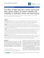
Báo cáo y học: "Oxygenation effect of interventional lung assist in a lavage model of acute lung injury: a prospective experimental study" pdf
Bạn đang xem bản rút gọn của tài liệu. Xem và tải ngay bản đầy đủ của tài liệu tại đây (341.35 KB, 8 trang )
Open Access
Available online />Page 1 of 8
(page number not for citation purposes)
Vol 10 No 2
Research
Oxygenation effect of interventional lung assist in a lavage model
of acute lung injury: a prospective experimental study
Günther Zick, Inéz Frerichs, Dirk Schädler, Gunnar Schmitz, Sven Pulletz, Erol Cavus,
Felix Wachtler, Jens Scholz and Norbert Weiler
Department of Anesthesiology and Intensive Care Medicine, University Hospital Schleswig-Holstein, Campus Kiel, Germany
Corresponding author: Günther Zick,
Received: 20 Jan 2006 Revisions requested: 21 Feb 2006 Revisions received: 27 Feb 2006 Accepted: 13 Mar 2006 Published: 7 Apr 2006
Critical Care 2006, 10:R56 (doi:10.1186/cc4889)
This article is online at: />© 2006 Zick et al.; licensee BioMed Central Ltd.
This is an open access article distributed under the terms of the Creative Commons Attribution License ( />),
which permits unrestricted use, distribution, and reproduction in any medium, provided the original work is properly cited.
Abstract
Introduction The aim of the study was to test the hypothesis
that a pumpless arteriovenous extracorporeal membrane
oxygenator (interventional lung assist (ILA)) does not
significantly improve oxygenation in a lavage model of acute lung
injury.
Methods The study was designed as a prospective
experimental study. The experiments were performed on seven
pigs (48–60 kg body weight). The pigs were anesthetized and
mechanically ventilated. Both femoral arteries and one femoral
vein were cannulated and connected with ILA. Acute lung injury
was induced by repeated bronchoalveolar lavage until the
arterial partial pressure of O
2
was lower than 100 Torr for at
least 30 minutes during ventilation with 100% O
2
.
Results ILA was applied with different blood flow rates through
either one or both femoral arteries. Measurements were
repeated at different degrees of pulmonary gas exchange
impairment with the pulmonary venous admixture ranging from
35.0% to 70.6%. The mean (± standard deviation) blood flow
through ILA was 15.5 (± 3.9)% and 21.7 (± 4.9)% of cardiac
output with one and both arteries open, respectively. ILA
significantly increased the arterial partial pressure of O
2
from 64
(± 13) Torr to 71 (± 14) Torr and 74 (± 17) Torr with blood flow
through one and both femoral arteries, respectively. O
2
delivery
through ILA increased with extracorporeal shunt flow (36 (± 14)
ml O
2
/min versus 47 (± 17) ml O
2
/min) and reduced
arterialization of the inlet blood. Pulmonary artery pressures
were significantly reduced when ILA was in operation.
Conclusion Oxygenation is increased by ILA in severe lung
injury. This effect is significant but small. The results indicate that
the ILA use may not be justified if the improvement of
oxygenation is the primary therapy goal.
Introduction
The mortality of patients with acute respiratory distress syn-
drome (ARDS) has remained high at about 30–50% despite
all efforts in research and treatment [1]. Different strategies of
mechanical ventilation focusing on the avoidance of ventilator-
induced lung injury [2,3] and on the recruitment of diseased
lung areas [4,5] are considered in the management of respira-
tory failure. Additional approaches applied are prone position-
ing [6], high-frequency oscillatory ventilation [7,8] and
extracorporeal membrane oxygenation [9-11].
The venovenous and venoarterial application of extracorporeal
membrane oxygenation is often associated with coagulation
and bleeding complications, with activation of the inflamma-
tory cascade, with damage of red blood cells and with techni-
cal problems [12-14]. These adverse effects mainly result from
the use of long tubings, heat exchangers and external pumps.
To reduce the incidence of such complications, pumpless
arteriovenous systems for extracorporeal gas exchange have
recently been developed.
These pumpless arteriovenous systems have low priming vol-
umes, short tubings and small foreign surface areas, and they
therefore exhibit less adverse effects than classical extracor-
poreal membrane oxygenation. The pumpless lung assist
devices are only effective in hemodynamically stable patients,
however, because the natural arteriovenous blood pressure
gradient determines the flow through the oxygenator. An ani-
mal experimental study has even shown that continuous hemo-
ARDS = acute respiratory distress syndrome; CO
2
= carbon dioxide; FiO
2
= inspired fraction of oxygen; ILA = interventional lung assist; O
2
= oxygen;
PaCO
2
= arterial partial pressure of carbon dioxide; PaO
2
= arterial partial pressure of oxygen.
Critical Care Vol 10 No 2 Zick et al.
Page 2 of 8
(page number not for citation purposes)
dynamic support was necessary with pumpless extracorporeal
lung assist [15].
Although the pumpless extracorporeal lung assist has been
shown to improve the CO
2
removal [16-19], its oxygenation
effect is difficult to assess. This is mainly because the blood
entering the oxygenator is already of arterial origin and the
amount of oxygen that can be added by the oxygenator is lim-
ited. The lower the oxygen saturation of the inlet blood, how-
ever, the greater the effect expected. Another limitation of the
extracorporeal lung assist is the fact that only a small fraction
of the cardiac output passes through the oxygenator and only
this blood is supplied with oxygen. On the return of blood into
the systemic circulation, the oxygen saturation decreases con-
siderably due to the mixture with venous blood. A significant
oxygenation effect of the extracorporeal lung assist device can
only be expected when sufficient flow through the oxygenator
is secured.
Since the oxygenation effect of the pumpless extracorporeal
lung assist has not extensively been studied until now and only
the effective CO
2
removal has been well described, partly
under the conditions of normal lung function [20], the primary
aim of our study was to test the hypothesis that a pumpless
arteriovenous extracorporeal membrane oxygenator does not
significantly improve oxygenation in a lavage model of severe
acute lung injury. We expect that our study may provide new
information on the possible use of the extracorporeal lung
assist to improve oxygen supply in ARDS patients.
Materials and methods
The study was approved by the university committee for animal
care and adhered to the guidelines on animal experimentation.
The experiments were performed on seven domestic pigs
(Deutsches Landschwein, Institute of Animal Breeding and
Husbandry, Christian-Albrechts-University, Kiel, Germany)
with a body weight of 48–60 kg. The animals were sedated
with azaperon (8 mg/kg) in combination with atropine (0.1 mg/
kg). Anesthesia was induced with ketamine (5 mg/kg) and,
after cannulation of an ear vein, sufentanil (0.2 µg/kg) and pro-
pofol (1 mg/kg) were added. The pigs were intubated and ven-
tilated with a Siemens servo 900 C ventilator (Siemens-Elema,
Solna, Sweden) with an inspired oxygen fraction (FiO
2
) of 1.0,
a tidal volume of 9 ml/kg body weight at a positive end-expira-
tory pressure of 5 cmH
2
O and a respiratory rate of 20 breaths/
minute. During preparation and instrumentation the ventilator
settings were set to attain normal levels of arterial partial pres-
sure of oxygen (PaO
2
) and of arterial partial pressure of carbon
dioxide (PaCO
2
). Anesthesia was maintained with propofol
(6–8 mg/kg per hour) and sufentanil (10 µg/kg per hour).
A catheter was introduced into the carotid artery, allowing
continuous analysis of PaO
2
and PaCO
2
(Paratrend 7+; Dia-
metrics Medical Inc, High Wycombe, UK) and arterial pressure
measurement. This access was also used for arterial blood
sampling. The samples were processed by a blood gas ana-
lyzer (ABL System 615; Radiometer Medical Inc., Copenha-
gen, Denmark), which was also applied for the measurement
of hemoglobin concentration. A pulmonary artery catheter was
inserted through the internal jugular vein to provide central
venous, pulmonary artery and capillary wedge pressures, as
well as continuous cardiac output (Baxter Healthcare, Irvine,
CA, USA). Mixed venous blood samples were drawn through
this line. The heart rate, the partial pressure of CO
2
in respired
gas, the airway pressures, and the pulmonary artery, arterial
and central venous pressures were monitored using the S/5
anesthesia monitoring system (Datex Ohmeda, Helsinki, Fin-
land).
The interventional lung assist (ILA) (Novalung, Hechingen,
Germany) was installed using the femoral blood vessels. One
17-Fr cannula was inserted into the femoral vein and two 13-
Fr cannulae were inserted into both femoral arteries either via
surgical preparation or via direct cannulation using Seldinger's
technique with ultrasound guidance. Once the instrumentation
was completed, 5,000 units heparin were administered. The
ILA device was then filled with saline, connected with the can-
nulae and the extracorporeal circuit was established. The tub-
ing for the O
2
delivery into the ILA system was attached and
the flow measurement through the arteriovenous shunt was
initiated.
Induction of acute lung injury
Acute lung injury was induced by bronchoalveolar lavage with
1.5 l warm saline, a modification of the method described by
Lachmann and colleagues [21]. The lavage was repeated until
the PaO
2
was well below 100 Torr and remained stable for a
period of 30 minutes at an FiO
2
of 1.0. To maintain hemody-
namic stability after the induction of acute lung injury, nore-
pinephrine was continuously administered at 0.02–0.3 µg/kg
per minute with an increasing dosage up to 0.1–1.8 µg/kg per
minute by the end of the experiment. Basic volume therapy
was initiated after induction of anesthesia using lactated
Ringer solution. After induction of lung injury, when the blood
pressure and heart rate indicated volume depletion, 6%
hydroxyethyl starch solution was added.
Protocol
The baseline data were collected after the completion of
instrumentation before ILA was put into operation and lung
injury was induced. The ventilator settings, the arterial, central
venous, pulmonary artery and capillary wedge pressures, the
cardiac output, the arterial and venous O
2
pressures, the CO
2
pressure and the respective hemoglobin concentrations and
hemoglobin O
2
saturations were determined.
The same data were collected after ILA was started before the
initiation of lung lavage. Additionally, the O
2
pressure, CO
2
pressure, hemoglobin concentration and hemoglobin O
2
satu-
ration were determined in blood samples drawn from the outlet
Available online />Page 3 of 8
(page number not for citation purposes)
of ILA. Afterwards, the measurements were performed during
the following three combinations of blood and gas flows
through ILA: blood flow through one arterial cannula with no
gas flow, blood flow through one arterial cannula with a gas
flow of 2 lO
2
/minute, and blood flow through both arterial can-
nulae with a gas flow of 2lO
2
/minute.
Identical series of three measurements were repeatedly per-
formed after the induction of severe lung injury. A total of three
to four measuring series were acquired in each animal.
Between the individual series, the extent of intrapulmonary
arteriovenous shunting was varied by application of different
positive end-expiratory pressures in the range 0–8 cmH
2
O
and/or additional lavage. Data acquisition was started when
the online PaO
2
was stable. Care was taken to keep the con-
ditions within each measuring series stable: no changes in
ventilator settings or cardiocirculatory support were allowed
until the data acquisition was completed.
After the completion of measurements, additional parameters
such as the O
2
content in arterial, mixed venous and ILA outlet
blood, the O
2
consumption and the O
2
delivery through ILA
were determined from the data acquired using basic physio-
logical calculations. The intrapulmonary venous admixture (for
instance, intrapulmonary right-to-left shunt) was calculated by
the Fick equation.
Statistical analysis
The results are presented as mean ± standard deviation val-
ues. Statistical analysis was performed using GraphPad Prism
version 4.0 (GraphPad Software, San Diego, CA, USA). One-
way analysis of variance followed by the Bonferroni multiple
comparison test was applied to test the significance of differ-
ences between the measurements. The paired Student's t test
was used to check the effect of the extracorporeal shunt flow
on O
2
delivery through ILA. Statistical significance was
accepted at P < 0.05. The reported P values are two-tailed.
Results
The results presented were obtained in seven animals during
the following study periods: baseline conditions without ILA,
ILA in operation before lung lavage, and ILA in operation after
lung lavage. A total of 25 series of measurements were per-
formed during the final period (for instance, after the induction
of lung injury).
In the present study, the effectiveness of ILA was followed
under conditions of severe impairment of pulmonary gas
exchange in a possibly large range of pulmonary arteriovenous
shunting. During baseline conditions, in anesthetized and arti-
ficially ventilated animals, the pulmonary venous admixture was
12.5 (± 4.9)% (Figure 1, left). After ILA was put into operation
the pulmonary venous admixture remained in the same range
(Figure 1, left). The induction of acute lung injury by repeated
bronchoalveolar lavage significantly raised the venous admix-
ture to 50.5 (± 9.3)% (P < 0.001). During the subsequent
measuring period, the pulmonary venous admixtures were in
the range 35.0–70.6% (Figure 1, right).
Arterial systolic and diastolic blood pressures did not signifi-
cantly differ among the measurements performed during base-
line conditions, before and after lung lavage. Pulmonary
capillary wedge pressures also remained unaffected: 8 (± 2)
mmHg during baseline and before lavage, and 9 (± 3) mmHg
after lavage. Cardiac output increased slightly but insignifi-
cantly after ILA was put into operation, from 6.8 (± 1.3) l/
minute to 7.2 (± 1.8) l/minute, 7.7 (± 1.5) l/minute and 7.9 (±
1.5) l/minute under the three measuring conditions studied.
After the induction of lung injury, cardiac outputs of 8.4 (± 2.6)
l/minute, 8.7 (± 2.1) l/minute and 8.6 (± 2.3) l/minute were
Figure 1
Venous admixture calculated in animals with normal and lavaged lungsVenous admixture calculated in animals with normal and lavaged lungs. B, baseline; N-, normal lung, one arterial cannula open, no gas flow; N+, nor-
mal lung, one arterial cannula open, gas flow of 2 lO
2
/minute; N++, normal lung, two arterial cannulae open, gas flow of 2 lO
2
/minute; L-, lavaged
lung, one arterial cannula open, no gas flow; L+, lavaged lung, one arterial cannula open, gas flow of 2 lO
2
/minute; L++, lavaged lung, two arterial
cannulae open, gas flow of 2 lO
2
/minute.
Critical Care Vol 10 No 2 Zick et al.
Page 4 of 8
(page number not for citation purposes)
determined. These values did not significantly differ from those
obtained before lavage.
The blood flow through the oxygenator was virtually independ-
ent of the lung condition and of the gas flow through the oxy-
genator (Figure 2). With one arterial cannula open and without
gas flow, the blood flow through the oxygenator was 1.29 (±
0.37) l/minute before lung injury (Figure 2, left) and 1.32 (±
0.15) l/minute after bronchoalveolar lavage (Figure 2, right).
After the addition of a sweep gas flow of 2 l O
2
/minute, the
blood flow through the oxygenator remained unchanged at
1.29 (± 0.38) l/minute and 1.30 (± 0.14) l/minute, respec-
tively. With two cannulae open and a sweep gas flow of 2 l O
2
/
minute, the oxygenator flow increased to 1.85 (± 0.52) l/
minute before lavage and to 1.86 (± 0.21) l/minute after the
induction of lung injury (P < 0.001).
Before lung lavage and with one cannula open, the relative
blood flow through the oxygenator corresponded to 18.5 (±
5.1)% and 17.0(± 4.5)% of the cardiac output during the
measurements without and with gas flow through the oxygen-
ator, respectively. The proportion of the oxygenator flow
increased to 23.4 (± 5.2)% (P < 0.001) when both arterial
cannulae were open. After the induction of lung injury, the cor-
responding relative blood flows through the ILA device were
16.6 (± 4.6)%, 15.5 (± 3.9)% and 21.7(± 4.9)% (P < 0.001)
of the cardiac output, respectively.
Our measurements revealed a significant removal of CO
2
by
ILA both under the conditions of normal and injured lung (Fig-
ure 3). When compared with the baseline conditions, the oper-
ation of ILA with blood flow through one femoral artery and no
sweep gas flow did not exhibit any effect on PaCO
2
(44 (± 3)
Torr versus 43(± 3) Torr). Both the addition of the gas flow of
2lO
2
/minute and the opening of the other femoral artery signif-
Figure 2
Oxygenator blood flow in animals with normal and lavaged lungsOxygenator blood flow in animals with normal and lavaged lungs. B, baseline; N-, normal lung, one arterial cannula open, no gas flow; N+, normal
lung, one arterial cannula open, gas flow of 2 lO
2
/minute; N++, normal lung, two arterial cannulae open, gas flow of 2 lO
2
/minute; L-, lavaged lung,
one arterial cannula open, no gas flow; L+, lavaged lung, one arterial cannula open, gas flow of 2 lO
2
/minute; L++, lavaged lung, two arterial cannu-
lae open, gas flow of 2 lO
2
/minute. ***P < 0.001.
Figure 3
Arterial partial pressure of carbon dioxide (PaCO
2
) in animals with normal and lavaged lungsArterial partial pressure of carbon dioxide (PaCO
2
) in animals with normal and lavaged lungs. B, baseline; N-, normal lung, one arterial cannula open,
no gas flow; N+, normal lung, one arterial cannula open, gas flow of 2 lO
2
/minute; N++, normal lung, two arterial cannulae open, gas flow of 2 lO
2
/
minute; L-, lavaged lung, one arterial cannula open, no gas flow; L+, lavaged lung, one arterial cannula open, gas flow of 2 lO
2
/minute; L++, lavaged
lung, two arterial cannulae open, gas flow of 2 lO
2
/minute. **P < 0.01, ***P < 0.001.
Available online />Page 5 of 8
(page number not for citation purposes)
icantly decreased the PaCO
2
values to 40(± 3) Torr (P < 0.01)
and 37 (± 3) Torr (P < 0.001), respectively (Figure 3, left). Dur-
ing severe lung injury, the employment of ILA in the three set-
tings studied led to a significant fall of PaCO
2
from 72(± 17)
Torr to 64(± 14) Torr (P < 0.001) and 60(± 13) Torr (P <
0.001), respectively (Figure 3, right). This was the result of the
effective removal of CO
2
by ILA as reflected by blood gas anal-
ysis performed on blood samples taken at the ILA outlet. The
addition of the sweep gas flow reduced the pressure of CO
2
from 71 (± 16) Torr to 31 (± 9) Torr (P < 0.001) during the ILA
operation with one cannula open.
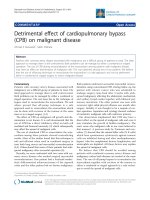
No oxygenation effect of ILA was observed before lung lavage
was initiated (Figure 4, left). The animals were ventilated at an
FiO
2
of 1.0, and consequently a high PaO
2
value of 505 (± 62)
Torr was found during the baseline conditions. During the
operation of ILA with one cannula open without and with gas
flow as well as with both cannulae open, the following PaO
2
values were determined: 526 (± 58) Torr, 542 (± 58) Torr and
519 (± 75) Torr, respectively. After the induction of severe
lung injury, a small but significant increase in PaO
2
was
observed when ILA was put into operation. Under the three
ILA settings studied, the arterial oxygenation rose from 64 (±
13) Torr to 71 (± 14) Torr (P < 0.001) and 74(± 16) Torr (P
< 0.001), respectively (Figure 4, right). The hemoglobin O
2
saturation at the ILA outlet was 100% with the sweep gas flow
turned on.
The O
2
delivery through ILA was significantly increased by the
higher extracorporeal shunt flow during operation with both
femoral arteries open when compared with the state when only
one femoral artery was open (Figure 5). The corresponding
mean volumes of O
2
delivered were 47 (± 17) ml/minute and
36 (± 14) ml/minute, respectively. These amounts of O
2
were
equal to 16.8 (± 6.0)% and 12.5 (± 5.0)% of the total O
2
con-
sumption, respectively. The lower the hemoglobin O
2
satura-
tion in the arterial blood entering the oxygenator, the higher the
amount of O
2
added by ILA. The O
2
delivery through ILA cor-
Figure 4
Arterial partial pressure of oxygen (PaO
2
) in animals with normal and lavaged lungsArterial partial pressure of oxygen (PaO
2
) in animals with normal and lavaged lungs. B, baseline; N-, normal lung, one arterial cannula open, no gas
flow; N+, normal lung, one arterial cannula open, gas flow of 2 lO
2
/minute; N++, normal lung, two arterial cannulae open, gas flow of 2 lO
2
/minute;
L-, lavaged lung, one arterial cannula open, no gas flow; L+, lavaged lung, one arterial cannula open, gas flow of 2 lO
2
/minute; L++, lavaged lung,
two arterial cannulae open, gas flow of 2 lO
2
/minute. ***P < 0.001.
Figure 5
Oxygen delivery through ILAOxygen delivery through ILA. One (circles) or two (triangles) arterial cannulae open.
Critical Care Vol 10 No 2 Zick et al.
Page 6 of 8
(page number not for citation purposes)
related linearly with the arterial hemoglobin O
2
saturation and
the oxygenator blood flow with the following equation: y = -
1.66 x
1
+ 24.88x
2
+ 148.83 (r
2
= 0.94), where y is the volume
of O
2
(ml) delivered per minute, x
1
is the hemoglobin O
2
satu-
ration and x
2
is the oxygenator blood flow.
Our study also revealed that both the systolic and diastolic pul-
monary artery pressures, which were significantly elevated
after the induction of acute lung injury, experienced a statisti-
cally highly significant fall during ILA operation with the 100%
O
2
gas flow of 2 l/minute when compared with ILA operation
without this sweep gas flow (Figure 6). The systolic pulmonary
artery pressure fell from 53 (± 12) mmHg to 48 (± 9) mmHg
(P < 0.01) and 47 (± 10) mmHg (P < 0.001), and the diastolic
pulmonary artery pressure from 32 (± 8) mmHg to 29 (± 7)
mmHg (P < 0.01) and 29 (± 7) mmHg (P < 0.001), respec-
tively.
Discussion
The application of extracorporeal respiratory support in ARDS
patients is currently recommended as a tool for minimizing the
invasiveness of mechanical ventilation [22,23]. Less aggres-
sive ventilator settings can be used in patients when extracor-
poreal membrane oxygenation is in operation because CO
2
is
removed not only by the lungs but also by the oxygenator. The
effective CO
2
removal has been well documented for the
pumpless arteriovenous extracorporeal oxygenators [16-
19,24,25] and has been confirmed in our study as well.
The primary aim of our experiments was to examine the oxy-
genation effect of the pumpless arteriovenous extracorporeal
assist device. At present there exist only few studies in which
the oxygenation effect of ILA was followed in a clinical setting.
Reng and colleagues claimed to have 'relevant oxygenation'
achieved in eight out of their 10 patients [26], Bein and col-
leagues demonstrated an improvement of oxygenation in 25
out of their 30 patients [27] and Liebold and colleagues found
a significant improvement of the oxygenation index after 24
hours of treatment in a study of 20 ARDS patients [28]. Zim-
mermann and colleagues studied retrospectively data from
eight patients with severe lung failure in whom ILA was applied
during interhospital transportation. An effective removal of
CO
2
and a moderate increase in oxygenation was found [29].
The experimental studies aimed at studying the O
2
delivery
with the pumpless arteriovenous extracorporeal assist were
partly performed in normal animals. For instance, Sussmane
and colleagues determined that only 19.5% of total O
2
con-
sumption were provided by the extracorporeal lung assist at
the lowest hemoglobin O
2
saturation of 60% induced in nor-
mal lambs by ventilation with a gas mixture with an FiO
2
of 0.1
[20].
Our intention was to study the ILA application under experi-
mental conditions that closely resembled the clinical situation
encountered in patients suffering from severe lung injury. We
therefore performed measurements in an animal model of
acute lung injury induced by bronchoalveolar lavage. This
experimental model enabled us to follow the effectiveness of
O
2
delivery through ILA during different, acutely modified
states of impaired pulmonary gas exchange as reflected by the
broad range of pulmonary venous admixture detected. During
the experiments we could also easily check the effect of differ-
ent blood flow rates through ILA using either one or both fem-
oral arteries for providing the inlet flow to the oxygenator.
Our findings correspond with the theoretical considerations
regarding the O
2
transport in blood. In general, the amount of
O
2
that can be added into the bloodstream is expected to
depend mainly on the flow rate and the degree of desaturation
of hemoglobin (as the amount of physically dissolved O
2
is
rather low). This means that the following two prerequisites
must be fulfilled if oxygenation is intended to be achieved by
Figure 6
Pulmonary artery pressures (PAP) (mean ± standard deviation) in animals with normal and lavaged lungsPulmonary artery pressures (PAP) (mean ± standard deviation) in animals with normal and lavaged lungs. sys, systolic; dias, diastolic; B, baseline; N-
, normal lung, one arterial cannula open, no gas flow; N+, normal lung, one arterial cannula open, gas flow of 2 lO
2
/minute; N++, normal lung, two
arterial cannulae open, gas flow of 2 lO
2
/minute; L-, lavaged lung, one arterial cannula open, no gas flow; L+, lavaged lung, one arterial cannula
open, gas flow of 2 lO
2
/minute; L++, lavaged lung, two arterial cannulae open, gas flow of 2 lO
2
/minute. **P < 0.01, ***P < 0.001.
Available online />Page 7 of 8
(page number not for citation purposes)
ILA: a sufficient blood pressure gradient must exist between
the inlet and outlet of ILA, and the pulmonary gas exchange
must be severely compromised so that the arterial blood enter-
ing the systemic circulation exhibits a substantial decrease in
hemoglobin O
2
saturation.
During our experiments, the hemodynamic status of the ani-
mals was sufficient to provide high and stable blood flow rates
through ILA even after the development of acute lung injury.
Cardiac output was relatively high due to vasopressor support
and adequate fluid supply. The relative flow rates equaled
approximately 16% and 22% of the cardiac output during
operation of ILA with either one or both femoral arteries,
respectively. The first prerequisite for O
2
transfer through ILA
stated earlier was therefore achieved and adequate ILA flow
was secured in spite of the small internal caliber of femoral
arteries in pigs. The second prerequisite was also fulfilled
because the acute lung damage induced by bronchoalveolar
lavage compromised the pulmonary arterialization of blood.
The mean hemoglobin O
2
saturation in the inlet blood was
87.6(± 8.8)% and 89.6(± 8.0)% during ILA operation with one
or both femoral arteries open. In spite of these conditions, the
O
2
delivery through ILA comprised only 12.5 (± 5.0)% and
16.8 (± 6.0)% of the total O
2
consumption. How relevant this
contribution is to the oxygenation might be judged differently.
In our opinion, the use of ILA is not justified if the oxygenation
effect is the major purpose of this therapy approach. As stated
by Pesenti and Patroniti [22], however, no clear recommenda-
tions on optimum PaO
2
and PaCO
2
values in ARDS patients
exist at present. In any case, the CO
2
elimination by ILA with
its additional small oxygenation effect facilitates lung-protec-
tive ventilator management.
Our experiments also showed the immediate effect of ILA
operation with an O
2
flow rate of 2 l/minute on pulmonary arte-
rial pressures after the development of acute lung injury. The
significant fall in pulmonary artery pressures, more pro-
nounced at higher shunt flows through ILA, is suggestive of a
decrease in hypoxic pulmonary vasoconstriction. In fact, the
O
2
content in the blood entering the lungs significantly rose
from 50 (± 14) ml/l to 61 (± 12) ml/l and 68 (± 13) ml/l with
ILA operating with the smaller and higher shunt flows, respec-
tively. Our data do not allow conclusions to be drawn regard-
ing the existence of this effect under the conditions of less
acute lung damage with possibly different pathogenetic mech-
anisms. The benefit of this influence of extracorporeal oxygen-
ation on pulmonary circulation cannot be judged
unambiguously at present. On the one hand, the lower pulmo-
nary arterial pressures may lead to diminished edema forma-
tion; on the other hand, the ventilation/perfusion matching may
deteriorate. To clarify these aspects of ILA use, measurements
using other experimental models of lung injury and studies on
patients with ARDS will be needed.
Conclusion
Our experimental study showed that pumpless arteriovenous
extracorporeal membrane oxygenator slightly improved oxygen-
ation in an animal model of severe acute lung injury. The volume
of O
2
delivered depended on the shunt flow rate and the
degree of hemoglobin desaturation and was rather small, on
average not exceeding 17% of the total O
2
consumption. The
operation of ILA significantly reduced pulmonary arterial pres-
sures, but the consequences of this effect on regional pulmo-
nary gas exchange remain to be determined in future studies.
Competing interests
The authors declare that they have no competing interests.
Authors' contributions
GZ participated in design of the study, carried out the study
and drafted the manuscript. IF performed the analysis and
interpretation of the data and revised the manuscript. DS car-
ried out the study and participated in the analysis of data. GS
participated in the analysis of data. SP carried out the study.
EC participated in the design of the study. FW carried out the
study. JS participated in design and coordination of the study.
NW conceived of the study and participated in design of the
study, analysis and interpretation of data and revision of the
manuscript. All authors read and approved the final manu-
script.
Acknowledgements
The authors acknowledge the partial financial support by Novalung,
Hechingen, Germany.
References
1. Rubenfeld GD, Caldwell E, Peabody E, Weaver J, Martin DP, Neff
M, Stern EJ, Hudson LD: Incidence and outcomes of acute lung
injury. N Engl J Med 2005, 353:1685-1693.
2. The Acute Respiratory Distress Syndrome Network: Ventilation
with lower tidal volumes as compared with traditional tidal vol-
umes for acute lung injury and the acute respiratory distress
syndrome. N Engl J Med 2000, 342:1301-1308.
3. Amato MB, Barbas CS, Medeiros DM, Magaldi RB, Schettino GP,
Lorenzi-Filho G, Kairalla RA, Deheinzelin D, Munoz C, Oliveira R, et
al.: Effect of a protective-ventilation strategy on mortality in the
Key messages
• ILA slightly improves oxygenation in an animal model of
severe acute lung injury. This effect is small, however,
and the use of ILA may not be justified if oxygenation is
the primary therapy goal.
• The amount of O
2
transferred depends on the blood
flow through ILA and on the degree of hemoglobin
desaturation in arterial blood.
• CO
2
elimination by ILA is pronounced, which makes ILA
beneficial in the treatment of acute lung injury by facili-
tating lung-protective ventilation strategies.
• ILA operation with an established sweep gas flow
reduces pulmonary artery blood pressures.
Critical Care Vol 10 No 2 Zick et al.
Page 8 of 8
(page number not for citation purposes)
acute respiratory distress syndrome. N Engl J Med 1998,
338:347-354.
4. Koh WJ, Suh GY, Han J, Lee SH, Kang EH, Chung MP, Kim H,
Kwon OJ: Recruitment maneuvers attenuate repeated dere-
cruitment-associated lung injury. Crit Care Med 2005,
33:1070-1076.
5. Suh GY, Koh Y, Chung MP, An CH, Kim H, Jang WY, Han J, Kwon
OJ: Repeated derecruitments accentuate lung injury during
mechanical ventilation. Crit Care Med 2002, 30:1848-1853.
6. Gattinoni L, Tognoni G, Pesenti A, Taccone P, Mascheroni D,
Labarta V, Malacrida R, Di Giulio P, Fumagalli R, Pelosi P, et al.:
Effect of prone positioning on the survival of patients with
acute respiratory failure. N Engl J Med 2001, 345:568-573.
7. David M, Weiler N, Heinrichs W, Neumann M, Joost T, Markstaller
K, Eberle B: High-frequency oscillatory ventilation in adult
acute respiratory distress syndrome. Intensive Care Med 2003,
29:1656-1665.
8. Derdak S, Mehta S, Stewart TE, Smith T, Rogers M, Buchman TG,
Carlin B, Lowson S, Granton J: High-frequency oscillatory venti-
lation for acute respiratory distress syndrome in adults: a ran-
domized, controlled trial. Am J Respir Crit Care Med 2002,
166:801-808.
9. Alpard SK, Zwischenberger JB: Extracorporeal gas exchange.
Respir Care Clin N Am 1998, 4:711-738.
10. Gattinoni L, Agostoni A, Pesenti A, Pelizzola A, Rossi GP, Langer
M, Vesconi S, Uziel L, Fox U, Longoni F, et al.: Treatment of acute
respiratory failure with low-frequency positive-pressure venti-
lation and extracorporeal removal of CO
2
. Lancet 1980,
2:292-294.
11. Gattinoni L, Kolobow T, Tomlinson T, Iapichino G, Samaja M,
White D, Pierce J: Low-frequency positive pressure ventilation
with extracorporeal carbon dioxide removal (LFPPV-ECCO2R):
an experimental study. Anesth Analg 1978, 57:Low470-477.
12. Alpard SK, Zwischenberger JB: Adult extracorporeal membrane
oxygenation for severe respiratory failure. Perfusion 1998,
13:3-15.
13. Fortenberry JD, Bhardwaj V, Niemer P, Cornish JD, Wright JA,
Bland L: Neutrophil and cytokine activation with neonatal
extracorporeal membrane oxygenation. J Pediatr 1996,
128:670-678.
14. Zwischenberger JB, Nguyen TT, Upp JRJ, Bush PE, Cox CSJ,
Delosh T, Broemling L: Complications of neonatal extracorpor-
eal membrane oxygenation. Collective experience from the
Extracorporeal Life Support Organization. J Thorac Cardiovasc
Surg 1994, 107:838-848. discussion 848–849
15. Totapally BR, Sussmane JB, Torbati D, Gelvez J, Fakioglu H, Mao
Y, Olarte JL, Wolfsdorf J: Cardiovascular stability during arteri-
ovenous extracorporeal therapy: a randomized controlled
study in lambs with acute lung injury. Crit Care 2004,
8:R495-R503.
16. Brunston RLJ, Zwischenberger JB, Tao W, Cardenas VJJ, Traber
DL, Bidani A: Total arteriovenous CO2 removal: simplifying
extracorporeal support for respiratory failure. Ann Thorac Surg
1997, 64:1599-1604. discussion 1604–1605
17. Conrad SA, Brown EG, Grier LR, Baier J, Blount J, Heming T,
Zwischenberger JB, Bidani A: Arteriovenous extracorporeal car-
bon dioxide removal: a mathematical model and experimental
evaluation. ASAIO J 1998, 44:267-277.
18. Frank BR, Tao W, Brunston RLJ, Alpard SK, Bidani A, Zwischen-
berger JB: High flow/low resistance cannulas for percutane-
ous arteriovenous carbon dioxide removal. ASAIO J 1997,
43:M817-M820.
19. Zwischenberger JB, Conrad SA, Alpard SK, Grier LR, Bidani A:
Percutaneous extracorporeal arteriovenous CO
2
removal for
severe respiratory failure. Ann Thorac Surg 1999, 68:181-187.
20. Sussmane JB, Totapally BR, Hultquist K, Torbati D, Wolfsdorf J:
Effects of arteriovenous extracorporeal therapy on hemody-
namic stability, ventilation, and oxygenation in normal lambs.
Crit Care Med 2001, 29:1972-1978.
21. Lachmann B, Robertson B, Vogel J: In vivo lung lavage as an
experimental model of the respiratory distress syndrome.
Acta Anaesthesiol Scand 1980, 24:231-236.
22. Pesenti A, Patroniti N: Therapeutic targets in acute respiratory
distress syndrome: role of the artificial lung. Crit Care Med
2001, 29:2034-2035.
23. Tao W, Brunston RLJ, Bidani A, Pirtle P, Dy J, Cardenas VJJ, Traber
DL, Zwischenberger JB: Significant reduction in minute ventila-
tion and peak inspiratory pressures with arteriovenous CO
2
removal during severe respiratory failure. Crit Care Med 1997,
25:689-695.
24. Conrad SA, Zwischenberger JB, Grier LR, Alpard SK, Bidani A:
Total extracorporeal arteriovenous carbon dioxide removal in
acute respiratory failure: a phase I clinical study. Intensive
Care Med 2001, 27:1340-1351.
25. Kolobow T, Gattinoni L, Tomlinson T, Pierce JE: An alternative to
breathing. J Thorac Cardiovasc Surg 1978, 75:261-266.
26. Reng M, Philipp A, Kaiser M, Pfeifer M, Gruene S, Schoelmerich J:
Pumpless extracorporeal lung assist and adult respiratory dis-
tress syndrome. Lancet 2000, 356:219-220.
27. Bein T, Prasser C, Philipp A, Muller T, Weber F, Schlitt HJ, Schmid
FX, Taeger K, Birnbaum D: Pumpless extracorporeal lung assist
using arterio-venous shunt in severe ARDS. Experience with
30 cases. Anaesthesist 2004, 53:813-819.
28. Liebold A, Reng CM, Philipp A, Pfeifer M, Birnbaum DE: Pumpless
extracorporeal lung assist – experience with the first 20 cases.
Eur J Cardiothorac Surg 2000, 17:608-613.
29. Zimmermann M, Bein T, Philipp A, Ittner K, Foltan M, Drescher J,
Weber F, Schmid FX: Interhospital transportation of patients
with severe lung failure on pumpless extracorporeal lung
assist. Br J Anaesth 2006, 96:63-66.