Báo cáo y học: "Variations in autologous neutralization and CD4 dependence of b12 resistant HIV-1 clade C env clones obtained at different time points from antiretroviral naïve Indian patients with recent infection" doc
Bạn đang xem bản rút gọn của tài liệu. Xem và tải ngay bản đầy đủ của tài liệu tại đây (862.42 KB, 15 trang )
RESEARC H Open Access
Variations in autologous neutralization and CD4
dependence of b12 resistant HIV-1 clade C env
clones obtained at different time points from
antiretroviral naïve Indian patients with
recent infection
Rajesh Ringe, Madhuri Thakar, Jayanta Bhattacharya
*
Abstract
Background: Limited information is available on HIV-1 Indian clade C sensitivities to autologous antibodies during
the course of natural infection. In the present study, a total of 37 complete envelope clones (Env) were amplified
at different time points predominantly from the plasma of five Indian patients with recent HIV-1 infection and
envelope-pseudotyped viruses were examined for their magnitude of sensitivity to autologous plasma antibodies
during natural course of infection.
Results: Variable low levels of neutralization were consistently detected with contemporaneous autologous plasma.
In contrast to clade B and African clade C HIV-1 envelopes, Env clones obtained from four patients were found to be
resistant to IgG1b12. The majority of the Env clones were resistant to 2G12 and 2F5 due to the absence of the
minimal motifs required for antibody recognition, but were sensitive to 4E10. Nonetheless, Env clones from one
patient were found to be sensitive to 2G12, atypical for clade C, and one Env clone exhibited unusual sensitivity to
17b, suggesting spontaneous exposure of CD4i epitopes. Phylogenetic analysis revealed that Env clones were closely
clustered within patients. Variation in the potential N-linked glycosylation pattern also appeared to be different in
patients over the course of infection. Interestingly, we found that the sensitivity of Envs to con temporaneous
autologous NAbs correlated positively with increased sensitivity to soluble CD4 and inversely with anti-CD4 antibody
and Envs with increased NAb sensitivity were able to efficiently infect HeLa cells expressing low CD4.
Conclusion: Our data showed considerable variations in autologous neutralization of these early HIV-1 clade C
Envs in each of these patients and indicate greater expo sure to CD4 of Envs that showed increased autologous
neutralization. Interestingly, Env clones obtained from a single patient at different time points were found to retain
sensitivity to b12 antibody that binds to CD4 binding site in Env in contrast to Envs obtained from other patients.
However, we did not find any association between increased b12 sensitivity of Envs obtained from this particular
patient with their degree of exposure to CD4.
Background
Induction of broadly neutralizing antibodies (NAbs)
against diverse strains of Human Immunodeficiency
Virus Type 1 (HIV-1) remains an important goal for
vaccine development [1-3]. Major obstacles are the
remarkable sequence variability of the envelope glyco-
proteins (Env) and the masking of critical neutralizing
epitopes by N-linked glycans and other structural and
steric constraints [4-6]. Most HIV-1 -infected individuals
mount a strong autologous NAb response within t he
first 6 to 12 months of infection that is highly specific
for the subject’s transmitted/founder virus. The response
generally broadens after several years of infection, where
in approximately 10-20 percent of cases the antibodies
* Correspondence:
Department of Molecular Virology, National AIDS Research Institute, Indian
Council of Medical Research, G-73 MIDC, Bhosari, Pune-411026, India
Ringe et al. Retrovirology 2010, 7:76
/>© 2010 Ring e et al; licensee BioMed Cent ral Ltd. This is an Open Access article distributed under the terms of the Creative Commons
Attribu tion License (http://cre ativecommons.org/licenses/by/2.0), which permits unrestricted use, distribution, and reproduction in
any medium, provided the original work is properly cited.
exhibit considerable b readth of neutralization against
diverse strains [7-15].
HIV-1 entry is media ted by bind ing of trimeric gp120
spikes to CD4 receptor that in turn exposes coreceptor
binding sites and facilitates fusion of viral and cell mem-
brane [16]. NAbs bind t o exposed epitopes on Env tri-
mers and thereby compromise HIV-1 entry [17,18,6,19].
The discovery of broadly neutralizing monoclonal anti-
bodies (MAbs) from HIV-1-infected patients with the
ability to neutralize diverse primary HIV-1 isolates
[20-23], suggested that there are indeed vulnerable epi-
topes on the functional Env trimer [24]. Thus, MAb
IgG1b12 binds the CD4-binding site (CD4bs) of gp120
[25] and neutralizes more than 50% of HIV-1 clade B
and approximately 30% of non-cla de B viruses [26,27].
Although many neutralization epitopes can be masked
by N-linked glycans, one M Ab, 2G12 [28,29], binds to
specific glycan residue and neutralizes many clade B iso-
lates but has limited breadth against non-clade B iso-
lates [26,3 0,31]. In addit ion, highly conserved sequences
[32] in the coreceptor binding site (also known as CD4-
induced or CD4i region) are potential targets for virus
neutralization [33-36]. Thus, antibodies mimicking pro-
totype MAb 17b show significant virus neutralization
after triggering gp120 with soluble CD4 (sCD4) [24].
Apart from epitopes in gp120 recognized by broadly
neutralizing MAbs, the membrane proximal e xternal
region (MPER) in gp41 is vulnerable to NAbs and found
to be a target of three well characterized MAbs 2F5,
4E10, and Z13 [37-39]. Antibodies targeting the MPER
of gp41 neutralize HIV-1 by blocking vi ral fusion with
thecellmembraneandtherebypreventingviralentry
[40]. 59). Interestingly, these types of antibodies are
rarely detected during natural infection [22,41,42].
Being highly variable, Env remains a major target of
the NAb response in HIV-1-infected individuals; thus,
Env-driven antibodies have been shown to neutralize
autologous virus variants moderately over time
[12,13,43,44], followed by rapid escape from neutraliza-
tion. Autologous NAbs appe ar to be directed to variable
regions of gp120 and are influenced by the pattern of
surface Env glycosylation that varies widely among HIV-
1 strains [9,10,44-52]. These data indicate that despite a
limited role for autologous NAbs in the control of vire-
mia, the antibodies exert selection pressure on Env early
in infection. In the case of HIV-1 clade B, the V1, V2
and V3 domains have also been shown to mediate CD4
independence, cellular tropism and receptor utilization
in addition to neutralization sensitivity [49,53-65].
HIV-1 clade C is the dominant genetic subtype circulat-
ing in India, Sub-Saharan Africa and China [66-70].
Though much information on autologous NAbs against
HIV-1 African clade C is available [9,10,42,49,50,52,71,72],
very limited information is available on the neutralization
properties of subtype C HIV-1 in India. Current evidence
suggests that sequences for the Indian HIV-1 clade C Env
and other genes such as gag and nef form a monophyletic
lineage and segregate separately as a sub clade within the
more diverse subtype C strains from Africa [69,73-77].
Recently, Kulkarni et al [27] demonstrated that newly
transmitted Indian Envs are antigenically complex despite
close genetic similarity. In this paper, we examined the
NAb response in subtype C HIV-1-infected individuals in
India by using Env clones amplified from uncultured per-
ipheral blood mononuclear cells (PBMC) at the baseline,
and plasma at the follow up visits of five recently infected
subjects and assessed autologous NAbs at different time
points for one year. We found that patient Envs varied
considerably in their sensitivities to their autologous
plasma antibodies and differed in their susceptibilities to
MAbs, indicating distinct mechanisms of autologous neu-
tralization and antibody specificities in these patients.
Results
Genetic properties of clade C env clones
Study s ubjects are described in Table 1. More than one
env clones was obtained from each of five recently
infected HIV-1 positive individuals from India at a base-
line visit and 6 and 12 months later except for subject
IVC5, for whom the last visit was at 24 mont hs (Table
2). Env clones from the baseline visit were obtained
from infected PBMC DNA whereas for follow up visits,
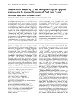
env was amplified from plasma viral RNA. Phylogenetic
analyses of the complete gp160 amino acid sequences
revealed that the Env clones were indeed subject specific
(Figure 1), with intra-clonal genetic divergences between
Table 1 Patient details
Plasma HIV-1 RNA (copies/ml) CD4 count (cells/mm3)
Patient ID Mode of Transmission Year of Infection Baseline F1 (moths) F2 (months) Baseline F1 (months) F2 (months)
NARI-IVC-2 Heterosexual 2008 8400 3070 (6) 17700 (12) 479 503 (6) 135 (12)
NARI-IVC-3 Heterosexual 2006 5380 29700 (6) 15700 (12) 592 499 (6) 477 (12)
NARI-IVC-4 Heterosexual 2006 37800 UD (6) UD (12) 328 374 (6) 402 (12)
NARI-IVC-5 Heterosexual 2006 1410 9040 (6) 48600 (24) 606 619 (6) 427 (24)
NARI-IVC-11 Heterosexual 2007 33400 11900 (6) 17300 (12) 552 693 (6) 590 (12)
UD: undetermined
Ringe et al. Retrovirology 2010, 7:76
/>Page 2 of 15
Env clones obtained from the same subject but at differ-
ent time points indicated ongoing viral evolution. All
Envs possessed low net V3 loop charge, a conserved
GPGQ motif (Additional file 1: Figure S1) and were
found to be CCR5 tropic (Table 2). Except for patients
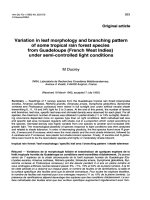
IVC 3 and IVC 4, no significant variation in total N-
linked glycosylation sites (PNLG) was found at the three
time points sampled (Figure 2); the number of PNLG
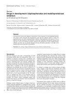
varied between 25-31 (Table 2). Median gp160 lengths
varied between patients; however they did not differ sig-
nificantly between clones obtained from the same
patient at different times (Figure 3). Although there
were no major differen ces between the variable loops of
the patient-specific envelope clones obtained at different
time points, Env clones 3-3.J9, 3-5.J25 and 5-4.J49, 5-4.
J16 a mplified from patients IVC 3 and IVC 5 were
Table 2 Genetic properties of patient Env clones
Patient ID Clone ID/Follow up Schedule† Source gp120 length gp41 length PNLG sites Net V3 loop charge CoR usage
2.J8/B PBMC 466 352 25 3 CCR5
2.J9/B PBMC 466 352 26 3 CCR5
2-3.J4/F1 PLASMA 465 352 30 3 CCR5
NARI-IVC2 2-3.J7/F1 PLASMA 466 352 29 3 CCR5
2-3.J17/F1 PLASMA 460 352 28 3 CCR5
2-3.J18/F1 PLASMA 465 352 30 3 CCR5
2-5.J3/F2 PLASMA 466 345 31 3 CCR5
2-5.J11/F2 PLASMA 465 352 29 2 CCR5
3.J16/B PBMC 466 352 27 5 CCR5
NARI-IVC3 3-3.J9/F1 PLASMA 459 352 28 5 CCR5
3-5.J25/F2 PLASMA 458 352 29 4 CCR5
3-5.J38/F2 PLASMA 463 352 31 3 CCR5
4.J2/B PBMC 462 352 30 3 CCR5
4.J22/B PBMC 462 352 30 3 CCR5
4.J27/B PLASMA 461 352 29 3 CCR5
4-2.J41/F1 PLASMA 458 352 27 2 CCR5
4-2.J45/F1 PLASMA 460 345 27 2 CCR5
NARI-IVC4 4-2.J42b/F1 PLASMA 464 345 27 2 CCR5
4-2.J45b/F1 PLASMA 459 345 26 2 CCR5
4-2.J46b/F1 PLASMA 464 345 28 2 CCR5
4-2.J47b/F1 PLASMA 459 345 27 2 CCR5
4-5.J5/F2 PLASMA 455 345 28 2 CCR5
5.J41/B PBMC 472 351 29 2 CCR5
5-3.J2/F1 PLASMA 461 351 26 3 CCR5
5-3.J4/F1 PLASMA 472 351 29 3 CCR5
5-3.J5/F1 PLASMA 461 362 30 3 CCR5
5-3.J9/F1 PLASMA 472 351 29 3 CCR5
NARI-IVC5 5-4.J16/F2 PLASMA 464 351 31 3 CCR5
5-4.J18/F2 PLASMA 475 351 30 4 CCR5
5-4.J22/F2 PLASMA 464 351 28 3 CCR5
5-4.J49/F2 PLASMA 475 351 30 3 CCR5
11.J25/B PBMC 461 352 27 4 CCR5
11.J28/B PBMC 461 352 27 4 CCR5
11-3.J3/F1 PLASMA 458 352 28 4 CCR5
NARI-IVC11 11-3.J9/F1 PLASMA 457 352 27 4 CCR5
11-3.J16/F1 PLASMA 457 352 26 4 CCR5
11-5.J12/F2 PLASMA 461 352 28 3 CCR5
† B = Baseline sample; F1 = First Follow up and F2 = Second follow up.
Ringe et al. Retrovirology 2010, 7:76
/>Page 3 of 15
found to have shorter V1 and V2 loops compared to the
contemporaneous Env clones (Additional file 1: Figure
S1).
Neutralization sensitivity of clonal Envs to autologous
plasma varied between study subjects
We next assessed the autologous neutralization of Env
clones amplified at three different time points from each
of five subjects. All five subjects mounted a moderate
NAb response against their early viruses obtained at the
baseline except patient IVC2; however this phenotype
var ied with respect to contemporaneous plasma antibo-
dies (Table 3). Surprisingly, only 1/8 clones from subject
IVC-2 was neutralized by the plasma samples obtained
at later time points, whereas a few (3/8) Env clones
were neutralized by the contemporaneous plasma. Thus,
while the autologous NAb response to early Env clones
improved over time in four subjects, it diminished over
time in one subject. This observation was correlated
with a gradual decline in CD4, indicating that autolo-
gous NAb possibly has selected the fittest Env variants
capable of f aster disease progre ssion in this particular
patient. The majority of the Envs obtained from follow
up visits were resistant to contemporaneous autologous
plasma antibodies indicating rapid escape of viral var-
iants. The persistence of a few sensitive Envs such as 3-
3.J9/F1, and 4-2.J45 during this period of infection
despite mounting hum oral immune pressure may indi-
cate that these Env v ariants had ad apted to sustain such
immune pressure possibly through certain compensatory
changes in Env sequence and retained their sensitivities
to autologous neutralizing antibodies.
Neutralization phenotype of the Envs as assessed with
common neutralizers
To test if the Envs obtained from patients at different
time points varied in their sensitivit ies to common
broadly neutralizing MAbs, pseudotyped viruses carrying
Figure 1 Phylogenetic relationships between inter and intra-patient Env gp160 a mino acid sequences used to study virus
neutralization as determined by Neighbor-Joining maximum likelihood tree using Mega 4.1. Bootstrapped values indicated that Env
sequences were patient specific and indicated monophyletic clustering of intra-patient Env.
Ringe et al. Retrovirology 2010, 7:76
/>Page 4 of 15
patient Envs were tested in neutralization assays with
sCD4 and five MAbs (b6, IgG1b12, 2G12 targeting
gp120 and 2F5, and 4E10 targeting gp41). As shown in
Table4themajorityofEnvclonesweresensitiveto
sCD4 at concentrations ranging from 0.1 to 6.66 μg/ml.
The pseudoviruses that required excess (>6.66 μg/ml)
sCD4 for 50% neutralization were considered as resis-
tant in our study. Consistent with the earlier report [27]
all Env variants were resistant to 2G12 except those
obtained from IVC-3 patient and this resistance was
associated with the absence of PNLG at position 295
(HXB2 numbering) at the N-terminal base of V3 loop.
The sensitivity of IVC-3 env clones was due to the pre-
sence of N295, atypical of clade C. In contrast to clade
B and Africa n clade C viruses [10,26], en velopes from
patient IVC 3, 4, 5, 11 were found resistant to IgG1b12.
This observation of b12 resistance of the India clade
Envs is in line with that reported by Kulkarni et al [27].
As with the MPER-specific MAbs, all the Envs were
resistant to 2F5 at the highest concentration tested
(Table 4). Interestingly, w hile 2F5 resistance was found
to be associated with the a bsence of DKW motif in
gp41 in most of the Envs, this motif was found to be
present in IVC3-3-9F1, IVC3-5-25F2, and all the Envs
obtained fr om IVC-11 and c onferred resistance as
shown in Additional file 2: Tab le S1. Our data indicate
that residues outside MPER domain possibly modulated
2F5 sensitivity despite the presence of a minimum DKW
motif in MPER for 2F5 sensitivity. The ability of 4E10 to
neutralize all the env clones was in a greement with the
presence of WFXI motif in gp41; however 4 Envs
(4-2_NEM.J46b, 4-5_NEM.J5, 5-3_NEM.J4 and 5-3_NEM.
J9) despite having WFXI motif (a minimum 4E10 recogni-
tion motif), they were found to be moderately resistant to
4E10 up to a concentration of 6.66 μg/ml (Additional file
1: Figure S1 and Additional file 2: Table S1).
Envs from one patient (NARI-IVC2) were moderately
sensitive to IgG1b12 but were resistant to
contemporaneous plasma antibodies
In contrast all others, Envs amplified from a patient
(NARI-IVC2) showed reasonable sensitivity to b12 MAb
that targets CD4bs in Env. As shown in Figure 4, these
Envs were found to provide a 50% reduction in infection
in TZM-bl cells at concentrations ranging from 0.2 to
2.23 μg/ml. The extent of b12 sensitivities of Envs
obtained from this particular patien t were found to be
much higher than the two b12-sensitive Indian clade C
Envs reported by Kulkarni et al [27]. The degree of b12
sensitivity of IVC Envs, however, did not correlate with
Figure 2 Variations of PNLGs in patient Envs at different time points during the course of infection. The bar represents median values.
Ringe et al. Retrovirology 2010, 7:76
/>Page 5 of 15
their sensitivity to sCD4 and contemporaneous plasma
antibodies. Thus, Envs 2-3.J18, 2-5.J3 and 2-5.J11 which
showed the highest neutralization sensitivity (IC
50
of 0.5,
0.29 and 0.21 μg/ml respectively) to b12 required more
sCD4 for 50% neutralization and except for 2-3.J18
showed neutralization resistance to contemporaneous
plasma antibodies (Tables 3 and 4). Our data indicated
that escape from contemporaneous NAbs in turn
mounted structural c onstraints in Env specificall y on
CD4 binding site. This feature therefore possibly con-
tributed in reduced sensitivity of NAb resistant IVC2
envelopes to sCD4, although all envelopes in this patient
surprisingly retained b12 sensitivity.
Sensitivity of Envs to contemporaneous autologous NAbs
correlated positively with increased sensitivity to sCD4
and inversely with anti-CD4 antibody
To assess whether the increased sensitivity of patient
envelopes to autologous NAbs could be due to greater
flexibilities of gp120 interactions with CD4, we next
compared the sensitiv ities of patient Envs to auto logous
plasmas, sCD4 and an anti-CD4 monoclonal antibody
(SIM.2) (hybridoma supernatant) that blocks gp120-CD4
binding. Interestingly, Envs that were resistant to con-
temporaneous plasmas were less sensitive to sCD4 and
required less anti-C D4 anti body (SIM.2) for 50% inhibi-
tion. Thus, as shown in Figure 5, a positive association
was seen between Env s ensitivity to contemporaneous
autologous plasma and an increased sens itivity to sCD4
and inverse corre lation between Env sensitivity to auto-
logous NAb anti-CD4 antibody, suggesting that Envs
with increased sensitivities to sCD4 exhibited greater
exposure of epitopes than are targeted by autologous
antibodies. The reduced sensitivity of Envs to SIM.2
suggests that Envs with more exposed epitopes for
sCD4requiremoreanti-CD4antibodyforoptimum
inhibition to entry. Overall, the sensitivities of Envs to
sCD4 varied and inversely correlated with their inhibi-
tion by SIM.2.
Increased sensitivity of patient Envs to contemporaneous
NAb and sCD4 correlated with reduced CD4 dependence
We next investigated if Envs with increased sensitivity to
autologous antibodies and sCD4 exhibited greater bind-
ing to cell surface CD4. Thus, He La cells expressing low
CD4 but high CCR5 (RC49 cell line) were infected with
Env-pseudotyped viruses and the degree of infection was
obtained by measuring the intracellular p24. As shown
in Figure 6, Envs with increased sensitivity to autologous
NAbs (such as 2-3. J18, 3-3.J9, 4.J2, 4 -2.J45, 5-4.J22 and
Figure 3 Variations in t otal gp160 lengths of Env clones obtained at different times in each patient durin g the course of infection.
Each bar represents median gp160 residues. Note that significant differences in median gp160 lengths of Envs between IVC 2 and 4, IVC 2 and
11, IVC 4 and 5 and IVC 5 and 11.
Ringe et al. Retrovirology 2010, 7:76
/>Page 6 of 15
5-4.J49) showed reduced CD4 dependence. However,
this phenomenon was found to be independent of the
patients and the follow up times examined here (Addi-
tional file 3: Figure S2). As expected, we found that
increased sensitivity of Envs to autologous NAbs was
correlated with reduced CD4 dependence (P < 0.0155)
and increased susceptibility to sCD4 (P < 0.0001) (Fig-
ure 7). Collectively, our data showed an inverse
Table 3 Neutralization sensitivity of patient envelopes to
autologous plasma antibodies
Env
clones
Baseline
plasma
Plasma First visit
(F1)
Plasma Second visit
(F2)
2.J8 601 228 <20
2.J9 522 240 <20
2-3.J4 350 <20 <20
2-3.J7 374 <50 <20
2-3.J17 300 <20 <20
2-3.J18 <20 540 652
2-5.J3 <50 <20 <20
2-5.J11 50 <20 <20
3.J16 195 696 2389
3-3.J9 349 554 1053
3-5.J25 <20 <20 184
3-5.J38 <20 <20 72
4.J2 421 2671 3848
4.J22 87 811 1172
4.J27 74 773 871
4-2.J41 103 98 406
4-2.J45 3375 6287 8307
4-2.J42b 60 <20 115
4-2.J45b 70 <50 500
4-2.J46b <50 <50 160
4-2.J47b 72 <50 340
4-5.J5 64 <20 244
5.J41 <20 110 1934
5-3.J2 <20 <20 1845
5-3.J4 <20 <20 1067
5-3.J5 <20 <20 1161
5-3.J9 <20 <20 1104
5-4.J16 <20 <20 <50
5-4.J18 <20 <20 <50
5-4.J22 <20 <50 223
5-4.J49 <20 <50 180
11.J25 66 2158 2830
11.J28 76 2008 2310
11-3.J3 <20 <50 1193
11-3.J9 <20 <20 148
11-3.J16 <20 <20 201
11-5.J12 <20 <20 <50
Values are reciprocal titer of patient plasma resulting 50% reduction in
relative luminescence unit (RLU) as an indicator of neutralization sensitivity in
TZM-bl cells following infection with pseudoviruses with 200TCID
50
. The ID
50
values are average of two independent assays wherein each assay was done
in duplicates.
Table 4 Neutralization sensitivity to monoclonal
antibodies, sCD4 and anti-CD4
Env clones b6 b12 2G12 17b 2F5 4E10 sCD4 SIM.2*
2.J8 >6.66 2.23 >6.66 >6.66 >6.66 0.34 3.66 120
2.J9 >6.66 2.16 >6.66 >6.66 >6.66 0.38 3.27 104
2-3.J4 >6.66 1.97 >6.66 >6.66 >6.66 3.36 >6.66 260
2-3.J7 >6.66 2.19 >6.66 >6.66 >6.66 5.85 >6.66 260
2-3.J17 >6.66 2.04 >6.66 >6.66 >6.66 4.85 >6.66 106
2-3.J18 >6.66 0.5 >6.66 5.1 >6.66 4.5 >6.66 37
2-5.J3 >6.66 0.29 >6.66 >6.66 >6.66 2.69 >6.66 201
2-5.J11 >6.66 0.21 >6.66 >6.66 >6.66 0.32 6.05 152
3.J16 >6.66 >6.66 4.20 >6.66 >6.66 0.23 0.54 103
3-3.J9 >6.66 >6.66 0.18 2.9 >6.66 0.3 0.1 76
3-5.J25 >6.66 >6.66 4.85 >6.66 >6.66 2.6 3.3 106
3-5.J38 >6.66 >6.66 4.30 >6.66 >6.66 2.22 >6.66 79
4.J2 >6.66 >6.66 >6.66 >6.66 >6.66 0.28 0.5 10
4.J22 >6.66 >6.66 >6.66 >6.66 >6.66 4 >6.66 138
4.J27 >6.66 >6.66 >6.66 >6.66 >6.66 5.28 >6.66 142
4-2.J41 >6.66 >6.66 >6.66 >6.66 >6.66 2.64 2.28 164
4-2.J45 >6.66 >6.66 >6.66 >6.66 >6.66 3.94 2.53 50
4-2.J42b >6.66 >6.66 >6.66 >6.66 >6.66 5 >6.66 224
4-2.J45b >6.66 >6.66 >6.66 >6.66 >6.66 6.2 >6.66 265
4-2.J46b >6.66 >6.66 >6.66 >6.66 >6.66 >6.66 >6.66 240
4-2.J47b >6.66 >6.66 >6.66 >6.66 >6.66 6.5 >6.66 212
4-5.J5 >6.66 >6.66 >6.66 >6.66 >6.66 >6.66 >6.66 334
5.J41 >6.66 >6.66 >6.66 >6.66 >6.66 0.29 0.5 114
5-3.J2 >6.66 >6.66 >6.66 >6.66 >6.66 5.6 >6.66 119
5-3.J4 >6.66 >6.66 >6.66 >6.66 >6.66 >6.66 >6.66 119
5-3.J5 5.9 >6.66 >6.66 >6.66 >6.66 5.66 >6.66 210
5-3.J9 >6.66 >6.66 >6.66 >6.66 >6.66 >6.66 >6.66 222
5-4.J16 >6.66 >6.66 >6.66 >6.66 >6.66 2.32 2.94 320
5-4.J18 >6.66 >6.66 >6.66 >6.66 >6.66 2.52 >6.66 157
5-4.J22 2.5 >6.66 >6.66 >6.66 >6.66 0.24 0.23 44
5-4.J49 5.9 >6.66 >6.66 >6.66 >6.66 0.52 0.53 121
11.J25 >6.66 >6.66 >6.66 >6.66 >6.66 0.34 3 99
11.J28 >6.66 >6.66 >6.66 >6.66 >6.66 0.32 2.4 88
11-3.J3 >6.66 >6.66 >6.66 >6.66 >6.66 5.64 >6.66 548
11-3.J9 >6.66 >6.66 >6.66 >6.66 >6.66 3.35 >6.66 555
11-3.J16 >6.66 6.05 >6.66 >6.66 >6.66 3.25 >6.66 585
11-5.J12 >6.66 >6.66 >6.66 >6.66 >6.66 2.67 >6.66 571
Values are concentrations resulting 50% reduction in relative luminescence
unit (RLU) as an indicator of neutralization sensitivity in TZM-bl cells following
infection with pseudoviruses with 200TCID
50
.TheIC
50
values are average of
two independent assays wherein each assay was done in duplicates. * The
values corresponding to anti-CD4 SIM.2 is hybridoma fluid are reciprocal
dilutions giving 50% reduction in relative luminescence unit (RLU).
Ringe et al. Retrovirology 2010, 7:76
/>Page 7 of 15
association of autologous neutralization sensitivity of
patient Envs with CD4 dependence.
Discussion
In contrast to the HIV-1 neutralization properties of
Afri can clade C, there is only one report on the neutra-
lization properties of HIV-1 clade C Env clones ampli-
fied from co-cultured PBMCs of acutely infected Indian
patients [27]. One of the disadvantages in obtaining Env
clones from co-culture is that it would potentially select
for virus variants that become adapted for favorable
replication in the absence of any immune pressure in
vitro. T his process would therefore fail to select viruses
growi ng in vivo which are respo nsible for the pathogen-
esis in the natural course of infection. In the present
study, we characterized for the first time the autologous
NAb response in subtype C HIV-1 infected Indian
patients using multiple molecul ar Env clones amplified
without culture from each study subject. We found that
while moderate NAb responses developed in three sub-
jects (IVC 3, 4 and 11), no significant NAb response
was detected at all three timepointsagainstcontem-
poraneous autologous virus in the remaining two sub-
jects (IVC 5 and I VC 11). In agreement with previous
reports, as with both subtype B and African subtype C
Envs, we found that in four patients (IVC3, 4, 5 and 11),
Envs obtained at baseline and earlier time points were
neutralized by plasma antibodies obtained at later time
points, indicating repeated cycles of escape [45,52]. Of
potential interest, Env clones obtained at all time points
from IVC2 patient were mo derately sensitive to
IgG1b12, whereas Env clones from the remaining sub-
jects were resistant to this MAb. Surprisingly NAb
response in this patient waned over the period of time
as plasma from later time points failed to neutralize
many contemporaneous as well as earlier envelopes.
Intriguingly, no correlation was observed between b12
sensitivity and sCD4 sensitivity as the b12 epitope over-
laps CD4 binding site. One plausible explanation for
this observation could be that this patient did not
develop b12 like antibodies and possibly the absence of
selective pressure on t he b12 binding site caused the
high sensitivity of these envelopes from IVC -2 towards
b12. It was also possible that due to lack of co- evolu-
tion of b12 and other CD4 binding sites in Env, we did
not find any association between b12 and sCD4 sensitiv-
ities to Env clones obtained from this p articular patient.
These observa tions indicate the presence of c ompensa-
tory a mino acid residues in the IVC-2 Env clones posi-
tioned either in the CD4bs or in the proximity that
favored enhanced neutral ization by b12 MAb. It would
be important to further investigate the Env sequence
that modulated b12 sensitivity in this patient.
Although we found repeated cycles of escape from
autologous NAbs in all the patients, one Env variant (4-
2_NEM.J45) obtained from patient NARI-IVC4 at the
Figure 4 Sensitivity of Env clones amplified from IVC2 patients to IgG1b12 antibody. Env-pseudotyped viruses were incubated with
IgG1b12 at indicated concentrations for 1 hour before TZM-bl cells were added as described in the Methods. The reduction of infectivity of
TZM-bl cells was measured as a function of the degree of IgG1b12 mediated neutralization of these Envs.
Ringe et al. Retrovirology 2010, 7:76
/>Page 8 of 15
first follow up retained unusually high sensitivity to con-
tempor aneous and earlier and follow-up plasmas with a
mean ID
50
of greater t han 1: 300 0. The persistence of
this sensitive Env against which high titer of N Ab was
developed for at least 6 months makes this envelope
interesting; in particular retention of neutralizing epi-
topes under immense humoral immune pressure prob-
ably indicates that this envelope might be more fit in
terms of CTL pressure or increased infectivity to com-
pensate for increased sensitivity to NA bs as previously
described by Moore et al [45,52]. When tested against
common HIV-1 neutralizing MAbs, most Envs obtained
at different time points f rom all the five participants
were resistant to IgGb6, IgG1b12, 2G12 and 2F5 and
sensitive to 4E10 only. Intri guingly, two Env variant s
each from subjects IVC4 and IVC5 despite containing
the minimum WFXI motif in gp41 MPER domain for
4E10 recognition, were found to require 4E10 antibody
in excess (>6.66 μg/ml)ofthatrequiredtoprovide50%
neutralization compared toallotherEnvs.Nakamura
et al [78] recently showed that while F673N and
W680G confers 4E10 resistance of HIV-1 envelopes,
W680R showed variable 4E10 resistance. In all cases,
IC
50
values were reported to be in the range of greater
than 50-100 μg/ml. In our study, we did not find any of
these substitutions in thesefourEnvs,suggestingthat
the relative resistance of these Envs over others tested
here are probably d ue to changes outside the MPER.
Nonetheless, these 4 Envs showed 30-40% sensitivity to
4E10 at a concentration of 6.66 μg/ml, indicating these
Envs required excess 4E10 for 50% neutralization but
certainl y not as much as that w ould require for W680 G
or F673N as shown by Nakamura et al [78]. One Env
variant each from subjects IVC2 and IVC 3 obtained at
firstfollowupvisitsthatshowedunusualsensitivityto
17b, indicating exposed CD4i epitopes. These two Env
variants in contrast to the majority of the Env clones
were also f ound to be efficient at infecting HeLa cells
Figure 5 Correlations between autologous neutralization sensitivities of patient Envs with their relative susceptibilities to sCD4 and
anti-CD4 antibody (SIM.2). Note that Envs that required sCD4 more than 6.66 μg/ml were given a value of 7 μg/ml for the benefit of
calculation. A strong correlation was observed between autologous neutralization and Env sensitivity to sCD4 (P < 0.0001) and SIM.2 (P = 0.0004)
and between Env susceptibilities to sCD4 and anti-CD4 (P =< 0.0001).
Ringe et al. Retrovirology 2010, 7:76
/>Page 9 of 15
expressing low levels of CD4 thereby indicating the pre-
sence of exposed CD4i epitopes on Env that enabled
them to productively infect HeLa cells expressing low
CD4. Nonethele ss, two Env variants (5.4.J22 and 5.4.J49)
obtained from IVC 5 patient at 2 years showed
increased infectivity to HeLa cells expressing low CD4
but were resistant to 17b, indicating that these Envs
evolved to conceal their coreceptor binding region on
gp120 without compromising low CD4 dependence in
the same way that most circulating variants do.
How NAbs drive the Env evolution that impacts on
CD4 affinity, tropism and sensitivity to NAbs is not v ery
clear in early HIV-1 clade C infection although two
groups using HIV-1 clade B Envs showed association of
R5 macrophage tropism with increased CD4 affinity
consistent with increased resistance to ant i-CD4
Figure 6 Variation in CD4-dependence of pseudoviruses carrying patient Envs. Pseudoviruses carrying distinct patient Envs were used to
infect HeLa cells (RC49 cell line) and the infectivity expressed as percentage infection of these pseudoviruses that infected HeLa cells expressing
high CD4 and high CCR5 (JC53 cell line).
Figure 7 Correlation between CD4 dependence of patient Envs with their sensit ivities to autologous plasma antibodies and sCD4.
Association of CD4 usage of Env-pseudotyped viruses with autologous plasma antibodies and (P < 0.0155) and sCD4 (P < 0.0001) indicated that
Env-pseudotyped viruses with low CD4 dependence tend to be more susceptible to autologous NAb in the patients tested here.
Ringe et al. Retrovirology 2010, 7:76
/>Page 10 of 15
monoclonal antibodies [79,80]. Although in general, the
majority of the Envs obtained from all the patients were
moderately sensitive to sCD4, we found a few Envs (5.
J41, 4-5.J5, 5-4.J16, 11.J25 and 11.J28) that showed auto-
logous antibody resistance but were moderately sensitive
to sCD4 indicating tha t these Envs evolved strategies in
escaping autologous neutralization however they
retained a very h igh affinity for the CD4 receptor. The
CD4 binding site (CD4bs) on Env experiences most
selective pressure as potent NAbs are directed to this
domain as document ed earlier [15,49]. Under this selec-
tive pressure exerted by humoral immunity, CD4bs is
compelled to acquire changes in Env sequences to
escape from NAb s that in turn would restrict Env b ind-
ing efficiently to CD4 receptors [81]. In our study w e
found that all the Envs that were sensitive to autologous
plasma antibodies were moderately susceptible to sCD4
indicating in this scenario, autologous NAbs were
mostly directed towards th e CD4 binding domain and
escape from NAb s possibly had compro mised Env bind-
ing with CD4. When tested for the extent of CD4 expo-
sure of gp120, Envs that were sensitive to autologous
antibodies as well as to sCD4 were found to require less
cell surface CD4 for efficient entry, indicating an inverse
correlation between Env sensitivity to autologous NAbs
and CD4 dependence. The relationship between sensi-
tivity of Envs to sCD4 and anti-CD4 antibodies with
var iable dependence to cell surface CD4 were described
previously by different investigators. Gorry et al [82]
showed that a neurotropic Env obtained from brain tis-
sue with higher affinity to CD4 was found to be increas-
ingly sensitive to CD4 mimetic, CD4-IgG2. Later,
Dunfee et al [83 ] showed that Envs with N283 substitu-
tion could productively infect cells expressing low cell
surface CD4 and show greater affinity to sCD4. Similar
observations were f ound by Vermeire et al [81], where
they showed that a NL4-3 variant that evolved to infect
cells expressing low CD4 in presence of the small mole-
cule CADA was found to be highly susceptible to het-
erologous sera and was concordant with increased
sensitivity and resi stan ce to sCD4 and anti-CD4 respec-
tively. In addition, Peters et al [79,84] demonstrated that
patient-derived Envs that were able to exploit low CD4
on cell surface were proportionately resistant and sensi-
tive to anti-CD4 antibody and sCD4 respectively.
In conclusion, in the present study, we have shown for
the first time the neutralization properties of HIV-1
India clade C Env clones obtained from patients fol-
lowed up with recent infection over time to their autolo-
gous antibodies during the natural course of infection
and investigated their genetic relatedness with sensitivity
to known broadly neutralizing monoclonal antibodies
and degree of exposure to CD4 for efficient entry.
While variations in autologous neutralization of viruses
are expected, a ll available data on the mechanisms of
resistance and sensitivity to ne utralizing antibodies of
geographically diversified HIV-1 clade C that contributes
to major global HIV-1 pandemic will help designing
strategies fostering vaccine discovery.
Methods
Patient details, PBMC and plasma samples
All five recently infected study subjects acquired HIV-1
through heterosexual contacts and were ART naïve at
the time of blood collection. The mean CD4 counts ran-
ged from 328-606 cells per cubic millimeter (mm
3
).
Based on detuned ELISA results [85-87] and history of
exposure within the last 6 to 8 months, these patients
were selected as recently infected patients f or further
characterization. Plasmas used for HIV-1 envelope
amplification and tested for antibody assays were
obtained at baseline, 6 and 12 months respectively.
Amplification and cloning of gp160
gp160 amplifi catio n from peripheral blood mononuclear
cell (PBMC) DNA and from reverse-transcribed plasma
viral RNA was carried out by nested PCR using 5’ -
TAGAGCCCTGGAAGCATCCAGGAAG-3’ as forward
and 5’ -TTGCTACTT GTGATTGCTCCATGT-3’ as
reverse primer in the first round and 5’ -CACCGGCT-
TAGGCATCTCCTATGGCAGGAAGAA-3’ as forward
and 5’ -TATCGGTACCAGTCTTGAGACGCTGCTCC-
TACTC-3’ as reverse primer in the second round by
using Platinum T aq proof reading polymerase (Invitro-
gen Inc.). Plasma viral RNA was purified by using a
nucleic acid isolation kit as described by the manufac-
turer (Roche Inc.). cDNA from d iluted viral RNA was
prepared using Superscript III first strand synthesis kit
(Invitr ogen Inc. ). gp160 was amplified by two rounds of
nested PCR gp160 amplicons were purified and ligated
into either pcDNA 3.1/V5-His-TOPO (Invit rogen Inc)
or pSVIIIenv [84].
DNA sequencing and phylogenetic analysis
Sequence analysis was performed using cycle sequencing
and big dye terminator methods by automated genet ic
analyzer (Applied Biosystems, Inc; Model 3730XL) as
described earlier [88]. Nucleotide and deduced amino
acid sequences w ere aligned using MEGA software and
phylogen etic tree was constructed by th e neighbor-join-
ing method [88].
Pseudovirion preparation and measurement of virus titer
Pseudotyped viruses carrying patient Envelope were pro-
duced by cotransfection of env
+
pSVIIIenv or env
+
pcDNA 3.1/V5-His-TOPO with env-defective HIV-1
backbone vector (pSG3ΔEnv) [44,89], into 293T cell s
during log gr owth phase in 6-well tissue cul ture trays
Ringe et al. Retrovirology 2010, 7:76
/>Page 11 of 15
(Corning Inc) using calcium phosphate (Promega Inc)
following manufacturer’ s protocol. Cell supernatants
car rying progeny pseudotyped viruses were harvested at
48 hours post-transfection, and stored at -152°C until
further usage. The infectivity assays were done in T ZM-
bl cells in 96-well microtiter plate and infectivity titers
determined by measuring the luciferase activity respec-
tively as described elsewhere [90].
Neutralization Assay
Patient plasma samples were evaluated for NAb activity
against Env pseudotyped viruses us ing a single round
reporter assay i n TZM-bl cells as described previously
with few modification [90]. Briefly, 200 TCID
50
of pseu-
dovirus was incubated with serial 3 fold dilutions of
plasma sample in duplicates in a total volume of 150 μl
for 1 hr at 37°C in 96-well flat-bottom culture plates.
Freshly trypsinized cells (10,000 cells in 100 μl of growth
medium containing 25 ug/ml DEAE Dextran) were
added to each well. One set of control wells received
cells plus pseudovirus (virus control) and another set
received cells only (backgr ound control). After 48 hours
of incubation, luciferase activity was measured by using
the Bright-Glo Luciferase Assay System (Promega Inc.).
The 50% inhibitory dose (ID
50
) was defined as either the
plasma dilution or sample concentration (in the case of
sCD4 and MAbs) that cause d a 50% reduction in rela-
tive luminescence un its (RLU) co mpared to virus con-
trol wells after subtraction of background RLU.
p24 antigen immunostaining
Immunostaining of HeLa cells infected with pseudo-
viruses was carried out as described earlier [84]. p 24
positive cells were regarded as foci of infection, and
virus infectivity was estimated as focus-forming units
(FFU) per milliliter.
Nucleotide sequence accession numbers
All env sequences have been submitted to GenBank
(accession numbers: [GenBa nk:EU908214] to [GenBank:
EU908221], [GenBank:EU908224] to [GenBank:
EU908225], and [GenBank:GU945306] to [GenBank:
GU945333]).
Statistical analyses
Correlations between NAb response and magnitude of
envelope binding to sCD4, RC49 cells and anti-CD4 anti-
body (SIM.2) were assessed by calculating Spearman’s
non-parametric 2-tailed correlat ion co-eff icient with 95%
confidence level using GraphPad Prism software. The
percent infectivity of Env clones in HeLa cells expressing
low CD4 (RC49) were plotted and compared by Mann-
Whitney and two-way ANNOVA tests using GraphPad
Prism software. Correlations were c onsidered significant
with P values less than 0.05. To avoid digression of corre-
lation, one Env clone (4.2J45) was not included during
assessing the correlation between Env sensitivity to NAb
and sCD4 (Figure 5) and between NAb and HeLa cell
(RC49) (Figure 6B) infection as the sensitivity of th is Env
clone to it autologous plasma was exceptionally higher
(ID
50
greater than 6000; see Table 3).
Additional material
Additional file 1: Figure S1. Alignments of deduced amino acids of
Indian clade C patient envelopes obtained at different course of
infection. Nucleotide sequences were translated and aligned using Mega
4.1. The residues were started from KpnI site in gp120 and did not
include signal peptide. While dashes denote sequence identity in Env,
dots indicate gaps. Letters in lowercase in the consensus sequence
indicate residues under represented at that position in Envs obtained
from all the patients. Residues that differed significantly at a particular
position were denoted as X in the consensus sequence. Potential N-
linked glycosylation sites were underscored and highlighted.
Additional file 2: Table S1. 2F5 and 4E10 minimum motifs in MPER
domain in patient Envs and their corresponding sensitivities to 2F5 and
4E10 monoclonal antibodies.
Additional file 3: Figure S2. Variations in CD4 dependence of patient
Envs obtained at different time points in each patient. Note that the bar
represents the median percentage infectivity of pseudoviruses to RC49
cells expressing low CD receptors.
Abbreviations
Env: (envelope); NAb: (neutralizing antibody); sCD4: (soluble CD4); MAb:
(monoclonal antibody)
Acknowledgements
This study was supported primarily by a research grant from the
Department of Biotechnology, Gove rnment of India (BT/PR7 829/MED/14/
1133/2006) to JB and in part by the Comprehensive Antibody-Vaccine
Imm une Monitoring Consortium through the Collaboration for A IDS
Vaccine Discovery, as funded by the Bill & Melinda Gates Foundation, and
a component of the Global HIV Vaccine Enterprise (#38619). We thank Dr
Paul Clapham, University of M ass achuset ts Medical School, Worcester,
USA for providing GHOST cell lines, JRCSF, JRFL, YU2 and SF162 Env c lones
and pSVIIIenv plasmid; and Dr David Kabat, University of Portland,
Oregon, USA for provid ing engineered HeLa cell li nes. We are grateful to
Dr David Montefiori for helpful advice on neutralization assay and critical
reading of the manuscript. We thank Dr James Robinson, University of
Tulane for 17b MAb, Dr Denn is Burton, Scripps Research Institute, La
Jolla,California for b6 MAb and NIH AIDS Rese arch Reagent and
Reference Program for making available many reagents used in this
study. RR is supported by a Junior Research Fellowship from t he Indian
Cou ncil of Medical Research, Govt of India. We thank Director, NARI for
support.
Authors’ contributions
JB conceptualized and planned the study; RR carried out molecular cloning,
neutralization assays and majority of the experiments; MT recruited patients
with recent infections, did detuned ELISA and provided essential patient
information including CD4 counts; RR and JB analyzed sequence analyses; JB
wrote the manuscript with the help of RR and MT. All the authors have read
and approved the final manuscript.
Competing interests
The authors declare that they have no competing interests.
Received: 31 May 2010 Accepted: 22 September 2010
Published: 22 September 2010
Ringe et al. Retrovirology 2010, 7:76
/>Page 12 of 15
References
1. Haynes BF, Montefiori DC: Aiming to induce broadly reactive neutralizing
antibody responses with HIV-1 vaccine candidates. Expert Rev Vaccines
2006, 5:347-363.
2. Hu SL, Stamatatos L: Prospects of HIV Env modification as an approach
to HIV vaccine design. Curr HIV Res 2007, 5:507-513.
3. Phogat S, Wyatt R: Rational modifications of HIV-1 envelope
glycoproteins for immunogen design. Curr Pharm Des 2007, 13:213-227.
4. Burton DR, Desrosiers RC, Doms RW, Koff WC, Kwong PD, Moore JP,
Nabel GJ, Sodroski J, Wilson IA, Wyatt RT: HIV vaccine design and the
neutralizing antibody problem. Nat Immunol 2004, 5:233-236.
5. Karlsson Hedestam GB, Fouchier RA, Phogat S, Burton DR, Sodroski J,
Wyatt RT: The challenges of eliciting neutralizing antibodies to HIV-1 and
to influenza virus. Nat Rev Microbiol 2008, 6:143-155.
6. Zhou T, Xu L, Dey B, Hessell AJ, Van Ryk D, Xiang SH, Yang X, Zhang MY,
Zwick MB, Arthos J, Burton DR, Dimitrov DS, Sodroski J, Wyatt R, Nabel GJ,
Kwong PD: Structural definition of a conserved neutralization epitope on
HIV-1 gp120. Nature 2007, 445:732-737.
7. Braibant M, Brunet S, Costagliola D, Rouzioux C, Agut H, Katinger H,
Autran B, Barin F: Antibodies to conserved epitopes of the HIV-1
envelope in sera from long-term non-progressors: prevalence and
association with neutralizing activity. Aids 2006, 20:1923-1930.
8. Donners H, Willems B, Beirnaert E, Colebunders R, Davis D, van der
Groen G: Cross-neutralizing antibodies against primary isolates in African
women infected with HIV-1. Aids 2002, 16:501-503.
9. Gray ES, Moore PL, Choge IA, Decker JM, Bibollet-Ruche F, Li H, Leseka N,
Treurnicht F, Mlisana K, Shaw GM, Karim SS, Williamson C, Morris L, CAPRISA
002 Study Team: Neutralizing antibody responses in acute human
immunodeficiency virus type 1 subtype C infection. J Virol 2007,
81:6187-6196.
10. Li B, Decker JM, Johnson RW, Bibollet-Ruche F, Wei X, Mulenga J, Allen S,
Hunter E, Hahn BH, Shaw GM, Blackwell JL, Derdeyn CA: Evidence for
potent autologous neutralizing antibody titers and compact envelopes
in early infection with subtype C human immunodeficiency virus type 1.
J Virol 2006, 80:5211-5218.
11. Moog C, Fleury HJ, Pellegrin I, Kirn A, Aubertin AM: Autologous and
heterologous neutralizing antibody responses following initial
seroconversion in human immunodeficiency virus type 1-infected
individuals. J Virol 1997, 71:3734-3741.
12. Pilgrim AK, Pantaleo G, Cohen OJ, Fink LM, Zhou JY, Zhou JT, Bolognesi DP,
Fauci AS, Montefiori DC: Neutralizing antibody responses to human
immunodeficiency virus type 1 in primary infection and long-term-
nonprogressive infection. J Infect Dis 1997, 176:924-932.
13. Richman DD, Wrin T, Little SJ, Petropoulos CJ: Rapid evolution of the
neutralizing antibody response to HIV type 1 infection. Proc Natl Acad Sci
USA 2003, 100:4144-4149.
14. Kraft Z, Strouss K, Sutton WF, Cleveland B, Tso FY, Polacino P, Overbaugh J,
Hu SL, Stamatatos L: Characterization of neutralizing antibody responses
elicited by clade A envelope immunogens derived from early
transmitted viruses. J Virol 2008, 82:5912-5921.
15. Li Y, Migueles SA, Welcher B, Svehla K, Phogat A, Louder MK, Wu X,
Shaw GM, Connors M, Wyatt RT, Mascola JR: Broad HIV-1 neutralization
mediated by CD4-binding site antibodies. Nat Med 2007, 13:1032-1034.
16. Wyatt R, Sodroski J: The HIV-1 envelope glycoproteins: fusogens,
antigens, and immunogens. Science 1998, 280 :1884-1888.
17. Parren PW, Mondor I, Naniche D, Ditzel HJ, Klasse PJ, Burton DR,
Sattentau QJ: Neutralization of human immunodeficiency virus type 1 by
antibody to gp120 is determined primarily by occupancy of sites on the
virion irrespective of epitope specificity. J Virol 1998, 72:3512-3519.
18. Ugolini S, Mondor I, Parren PW, Burton DR, Tilley SA, Klasse PJ, Sattentau QJ:
Inhibition of virus attachment to CD4+ target cells is a major
mechanism of T cell line-adapted HIV-1 neutralization. J Exp Med 1997,
186:1287-1298.
19. Labrijn AF, Poignard P, Raja A, Zwick MB, Delgado K, Franti M, Binley J,
Vivona V, Grundner C, Huang CC, Venturi M, Petropoulos CJ, Wrin T,
Dimitrov DS, Robinson J, Kwong PD, Wyatt RT, Sodroski J, Burton DR:
Access of antibody molecules to the conserved coreceptor binding site
on glycoprotein gp120 is sterically restricted on primary human
immunodeficiency virus type 1. J Virol 2003, 77:10557-10565.
20. Deeks SG, Schweighardt B, Wrin T, Galovich J, Hoh R, Sinclair E, Hunt P,
McCune JM, Martin JN, Petropoulos CJ, Hecht FM: Neutralizing antibody
responses against autologous and heterologous viruses in acute versus
chronic human immunodeficiency virus (HIV) infection: evidence for a
constraint on the ability of HIV to completely evade neutralizing
antibody responses. J Virol 2006, 80:6155-6164.
21. Doria-Rose NA, Klein RM, Manion MM, O’Dell S, Phogat A, Chakrabarti B,
Hallahan CW, Migueles SA, Wrammert J, Ahmed R, Nason M, Wyatt RT,
Mascola JR, Connors M: Frequency and phenotype of human
immunodeficiency virus envelope-specific B cells from patients with
broadly cross-neutralizing antibodies. J Virol 2009, 83:188-199.
22. Binley JM, Lybarger EA, Crooks ET, Seaman MS, Gray E, Davis KL, Decker JM,
Wycuff D, Harris L, Hawkins N, Wood B, Nathe C, Richman D, Tomaras GD,
Bibollet-Ruche F, Robinson JE, Morris L, Shaw GM, Montefiori DC,
Mascola JR: Profiling the specificity of neutralizing antibodies in a large
panel of plasmas from patients chronically infected with human
immunodeficiency virus type 1 subtypes B and C. J Virol 2008,
82:11651-11668.
23. Simek MD, Rida W, Priddy FH, Pung P, Carrow E, Laufer DS, Lehrman JK,
Boaz M, Tarragona-Fiol T, Miiro G, Birungi J, Pozniak A, McPhee DA,
Manigart O, Karita E, Inwoley A, Jaoko W, Dehovitz J, Bekker LG,
Pitisuttithum P, Paris R, Walker LM, Poignard P, Wrin T, Fast PE, Burton DR,
Koff WC: Human immunodeficiency virus type 1 elite neutralizers:
individuals with broad and potent neutralizing activity identified by
using a high-throughput neutralization assay together with an analytical
selection algorithm. J Virol 2009, 83:7337-7348.
24. Mascola JR, Montefiori DC: The role of antibodies in HIV vaccines. Annu
Rev Immunol 2010, 28:413-444.
25. Burton DR, Pyati J, Koduri R, Sharp SJ, Thornton GB, Parren PW, Sawyer LS,
Hendry RM, Dunlop N, Nara PL: Efficient neutralization of primary isolates
of HIV- 1 by a recombinant human monoclonal antibody. Science 1994,
266:1024-1027.
26. Binley JM, Wrin T, Korber B, Zwick MB, Wang M, Chappey C, Stiegler G,
Kunert R, Zolla-Pazner S, Katinger H, Petropoulos CJ, Burton DR:
Comprehensive cross-clade neutralization analysis of a panel of anti-
human immunodeficiency virus type 1 monoclonal antibodies. J Virol
2004,
78:13232-13252.
27. Kulkarni SS, Lapedes A, Tang H, Gnanakaran S, Daniels MG, Zhang M,
Bhattacharya T, Li M, Polonis VR, McCutchan FE, Morris L, Ellenberger D,
Butera ST, Bollinger RC, Korber BT, Paranjape RS, Montefiori DC: Highly
complex neutralization determinants on a monophyletic lineage of
newly transmitted subtype C HIV-1 Env clones from India. Virology 2009,
385:505-520.
28. Trkola A, Purtscher M, Muster T, Ballaun C, Buchacher A, Sullivan N,
Srinivasan K, Sodroski J, Moore JP, Katinger H: Human monoclonal
antibody 2G12 defines a distinctive neutralization epitope on the gp120
glycoprotein of human immunodeficiency virus type 1. J Virol 1996,
70:1100-1108.
29. Calarese DA, Scanlan CN, Zwick MB, Deechongkit S, Mimura Y, Kunert R,
Zhu P, Wormald MR, Stanfield RL, Roux KH, Kelly JW, Rudd PM, Dwek RA,
Katinger H, Burton DR, Wilson IA: Antibody domain exchange is an
immunological solution to carbohydrate cluster recognition. Science
2003, 300:2065-2071.
30. Trkola A, Pomales AB, Yuan H, Korber B, Maddon PJ, Allaway GP, Katinger H,
Barbas CF, Burton DR, Ho DD, et al: Cross-clade neutralization of primary
isolates of human immunodeficiency virus type 1 by human monoclonal
antibodies and tetrameric CD4-IgG. J Virol 1995, 69:6609-6617.
31. Li M, Salazar-Gonzalez JF, Derdeyn CA, Morris L, Williamson C, Robinson JE,
Decker JM, Li Y, Salazar MG, Polonis VR, Mlisana K, Karim SA, Hong K,
Greene KM, Bilska M, Zhou J, Allen S, Chomba E, Mulenga J, Vwalika C,
Gao F, Zhang M, Korber BT, Hunter E, Hahn BH, Montefiori DC: Genetic and
neutralization properties of subtype C human immunodeficiency virus
type 1 molecular env clones from acute and early heterosexually
acquired infections in Southern Africa. J Virol 2006, 80:11776-11790.
32. Decker JM, Bibollet-Ruche F, Wei X, Wang S, Levy DN, Wang W,
Delaporte E, Peeters M, Derdeyn CA, Allen S, Hunter E, Saag MS, Hoxie JA,
Hahn BH, Kwong PD, Robinson JE, Shaw GM: Antigenic conservation and
immunogenicity of the HIV coreceptor binding site. J Exp Med 2005,
201:1407-1419.
33. Wyatt R, Kwong PD, Desjardins E, Sweet RW, Robinson J, Hendrickson WA,
Sodroski JG: The antigenic structure of the HIV gp120 envelope
glycoprotein. Nature 1998, 393:705-711.
Ringe et al. Retrovirology 2010, 7:76
/>Page 13 of 15
34. Thali M, Moore JP, Furman C, Charles M, Ho DD, Robinson J, Sodroski J:
Characterization of conserved human immunodeficiency virus type 1
gp120 neutralization epitopes exposed upon gp120-CD4 binding. J Virol
1993, 67:3978-3988.
35. Xiang SH, Doka N, Choudhary RK, Sodroski J, Robinson JE: Characterization
of CD4-induced epitopes on the HIV type 1 gp120 envelope
glycoprotein recognized by neutralizing human monoclonal antibodies.
AIDS Res Hum Retroviruses 2002, 18:1207-1217.
36. Moulard M, Phogat SK, Shu Y, Labrijn AF, Xiao X, Binley JM, Zhang MY,
Sidorov IA, Broder CC, Robinson J, Parren PW, Burton DR, Dimitrov DS:
Broadly cross-reactive HIV-1-neutralizing human monoclonal Fab
selected for binding to gp120-CD4-CCR5 complexes. Proc Natl Acad Sci
USA 2002, 99:6913-6918.
37. Cardoso RM, Zwick MB, Stanfield RL, Kunert R, Binley JM, Katinger H,
Burton DR, Wilson IA: Broadly neutralizing anti-HIV antibody 4E10
recognizes a helical conformation of a highly conserved fusion-
associated motif in gp41. Immunity 2005, 22:163-173.
38. Zwick MB, Labrijn AF, Wang M, Spenlehauer C, Saphire EO, Binley JM,
Moore JP, Stiegler G, Katinger H, Burton DR, Parren PW: Broadly
neutralizing antibodies targeted to the membrane-proximal external
region of human immunodeficiency virus type 1 glycoprotein gp41. J
Virol 2001, 75:10892-10905.
39. Muster T, Steindl F, Purtscher M, Trkola A, Klima A, Himmler G, Ruker F,
Katinger H: A conserved neutralizing epitope on gp41 of human
immunodeficiency virus type. J Virol 1993, 67:6642-6647.
40. Binley JM, Cayanan CS, Wiley C, Schulke N, Olson WC, Burton DR: Redox-
triggered infection by disulfide-shackled human immunodeficiency virus
type 1 pseudovirions. J Virol 2003, 77:5678-5684.
41. Yuste E, Sanford HB, Carmody J, Bixby J, Little S, Zwick MB, Greenough T,
Burton DR, Richman DD, Desrosiers RC, Johnson WE: Simian
immunodeficiency virus engrafted with human immunodeficiency virus
type 1 (HIV-1)-specific epitopes: replication, neutralization, and survey of
HIV-1-positive plasma. J Virol 2006, 80:3030-3041.
42. Gray ES, Taylor N, Wycuff D, Moore PL, Tomaras GD, Wibmer CK, Puren A,
DeCamp A, Gilbert PB, Wood B, Montefiori DC, Binley JM, Shaw GM,
Haynes BF, Mascola JR, Morris L: Antibody specificities associated with
neutralization breadth in plasma from human immunodeficiency virus
type 1 subtype C-infected blood donors. J Virol 2009, 83:8925-8937.
43. Albert J, Abrahamsson B, Nagy K, Aurelius E, Gaines H, Nystrom G,
Fenyo EM: Rapid development of isolate-specific neutralizing antibodies
after primary HIV-1 infection and consequent emergence of virus
variants which resist neutralization by autologous sera. Aids 1990,
4:107-112.
44. Wei X, Decker JM, Wang S, Hui H, Kappes JC, Wu X, Salazar-Gonzalez JF,
Salazar MG, Kilby JM, Saag MS, Komarova NL, Nowak MA, Hahn BH,
Kwong PD, Shaw GM: Antibody neutralization and escape by HIV-1.
Nature 2003, 422:307-312.
45. Bunnik EM, Pisas L, van Nuenen AC, Schuitemaker H: Autologous
neutralizing humoral immunity and evolution of the viral envelope in
the course of subtype B human immunodeficiency virus type 1
infection. J Virol 2008, 82:7932-7941.
46. Moore PL, Gray ES, Choge IA, Ranchobe N, Mlisana K, Abdool Karim SS,
Williamson C, Morris L: The c3-v4 region is a major target of autologous
neutralizing antibodies in human immunodeficiency virus type 1
subtype C infection. J Virol 2008, 82
:1860-1869.
47. Davis KL, Gray ES, Moore PL, Decker JM, Salomon A, Montefiori DC,
Graham BS, Keefer MC, Pinter A, Morris L, Hahn BH, Shaw GM: High titer
HIV-1 V3-specific antibodies with broad reactivity but low neutralizing
potency in acute infection and following vaccination. Virology 2009,
387:414-26.
48. Salazar-Gonzalez JF, Salazar MG, Keele BF, Learn GH, Giorgi EE, Li H,
Decker JM, Wang S, Baalwa J, Kraus MH, Parrish NF, Shaw KS, Guffey MB,
Bar KJ, Davis KL, Ochsenbauer-Jambor C, Kappes JC, Saag MS, Cohen MS,
Mulenga J, Derdeyn CA, Allen S, Hunter E, Markowitz M, Hraber P,
Perelson AS, Bhattacharya T, Haynes BF, Korber BT, Hahn BH, Shaw GM:
Genetic identity, biological phenotype, and evolutionary pathways of
transmitted/founder viruses in acute and early HIV-1 infection. J Exp Med
2009, 206:1273-1289.
49. Rong R, Bibollet-Ruche F, Mulenga J, Allen S, Blackwell JL, Derdeyn CA: Role
of V1V2 and other human immunodeficiency virus type 1 envelope
domains in resistance to autologous neutralization during clade C
infection. J Virol 2007, 81:1350-1359.
50. Rong R, Gnanakaran S, Decker JM, Bibollet-Ruche F, Taylor J, Sfakianos JN,
Mokili JL, Muldoon M, Mulenga J, Allen S, Hahn BH, Shaw GM, Blackwell JL,
Korber BT, Hunter E, Derdeyn CA: Unique mutational patterns in the
envelope alpha 2 amphipathic helix and acquisition of length in gp120
hypervariable domains are associated with resistance to autologous
neutralization of subtype C human immunodeficiency virus type 1. J
Virol 2007, 81:5658-5668.
51. Moore PL, Ranchobe N, Lambson BE, Gray ES, Cave E, Abrahams MR,
Bandawe G, Mlisana K, Abdool Karim SS, Williamson C, Morris L: Limited
neutralizing antibody specificities drive neutralization escape in early
HIV-1 subtype C infection. PLoS Pathog 2009, 5:e1000598.
52. Rong R, Li B, Lynch RM, Haaland RE, Murphy MK, Mulenga J, Allen SA,
Pinter A, Shaw GM, Hunter E, Robinson JE, Gnanakaran S, Derdeyn CA:
Escape from autologous neutralizing antibodies in acute/early subtype
C HIV-1 infection requires multiple pathways. PLoS Pathog 2009, 5:
e1000594.
53. Bouma P, Leavitt M, Zhang PF, Sidorov IA, Dimitrov DS, Quinnan GV Jr:
Multiple interactions across the surface of the gp120 core structure
determine the global neutralization resistance phenotype of human
immunodeficiency virus type 1. J Virol 2003, 77:8061-8071.
54. Carrillo A, Ratner L: Cooperative effects of the human immunodeficiency
virus type 1 envelope variable loops V1 and V3 in mediating infectivity
for T cells. J Virol 1996, 70:1310-1316.
55. Cheng-Mayer C, Brown A, Harouse J, Luciw PA, Mayer AJ: Selection for
neutralization resistance of the simian/human immunodeficiency virus
SHIVSF33A variant in vivo by virtue of sequence changes in the
extracellular envelope glycoprotein that modify N-linked glycosylation. J
Virol 1999, 73:5294-5300.
56. Gram GJ, Hemming A, Bolmstedt A, Jansson B, Olofsson S, Akerblom L,
Nielsen JO, Hansen JE: Identification of an N-linked glycan in the V1-loop
of HIV-1 gp120 influencing neutralization by anti-V3 antibodies and
soluble CD4. Arch Virol 1994, 139:253-261.
57. Koito A, Harrowe G, Levy JA, Cheng-Mayer C: Functional role of the V1/V2
region of human immunodeficiency virus type 1 envelope glycoprotein
gp120 in infection of primary macrophages and soluble CD4
neutralization. J Virol 1994, 68:2253-2259.
58. Kolchinsky P, Kiprilov E, Bartley P, Rubinstein R, Sodroski J: Loss of a single
N-linked glycan allows CD4-independent human immunodeficiency
virus type 1 infection by altering the position of the gp120 V1/V2
variable loops. J Virol 2001, 75 :3435-3443.
59. Korber B, Gaschen B, Yusim K, Thakallapally R, Kesmir C, Detours V:
Evolutionary and immunological implications of contemporary HIV-1
variation. Br Med Bull
2001, 58:19-42.
60. Morikita T, Maeda Y, Fujii S, Matsushita S, Obaru K, Takatsuki K: The V1/V2
region of human immunodeficiency virus type 1 modulates the
sensitivity to neutralization by soluble CD4 and cellular tropism. AIDS Res
Hum Retroviruses 1997, 13:1291-1299.
61. Pincus SH, Messer KG, Nara PL, Blattner WA, Colclough G, Reitz M:
Temporal analysis of the antibody response to HIV envelope protein in
HIV-infected laboratory workers. J Clin Invest 1994, 93:2505-2513.
62. Edwards TG, Hoffman TL, Baribaud F, Wyss S, LaBranche CC, Romano J,
Adkinson J, Sharron M, Hoxie JA, Doms RW: Relationships between CD4
independence, neutralization sensitivity, and exposure of a CD4-induced
epitope in a human immunodeficiency virus type 1 envelope protein. J
Virol 2001, 75:5230-5239.
63. Dumonceaux J, Goujon C, Joliot V, Briand P, Hazan U: Determination of
essential amino acids involved in the CD4-independent tropism of the
X4 human immunodeficiency virus type 1 m7NDK isolate: role of
potential N glycosylations in the C2 and V3 regions of gp120. J Virol
2001, 75:5425-5428.
64. Joliot V, Goujon C, Dumonceaux J, Renard A, Briand P, Hazan U: A human
immunodeficiency virus Env inducible transcription system to examine
consequences of gp120 expression. J Virol Methods 2001, 98:145-151.
65. Rossi F, Querido B, Nimmagadda M, Cocklin S, Navas-Martin S, Martin-
Garcia J: The V1-V3 region of a brain-derived HIV-1 envelope
glycoprotein determines macrophage tropism, low CD4 dependence,
increased fusogenicity and altered sensitivity to entry inhibitors.
Retrovirology 2008, 5:89.
Ringe et al. Retrovirology 2010, 7:76
/>Page 14 of 15
66. Esparza J, Bhamarapravati N: Accelerating the development and future
availability of HIV-1 vaccines: why, when, where, and how? Lancet 2000,
355:2061-2066.
67. Moore JP, Parren PW, Burton DR: Genetic subtypes, humoral immunity,
and human immunodeficiency virus type 1 vaccine development. J Virol
2001, 75:5721-5729.
68. Osmanov S, Pattou C, Walker N, Schwardlander B, Esparza J: Estimated
global distribution and regional spread of HIV-1 genetic subtypes in the
year 2000. J Acquir Immune Defic Syndr 2002, 29:184-190.
69. Shankarappa R, Chatterjee R, Learn GH, Neogi D, Ding M, Roy P, Ghosh A,
Kingsley L, Harrison L, Mullins JI, Gupta P: Human immunodeficiency virus
type 1 env sequences from Calcutta in eastern India: identification of
features that distinguish subtype C sequences in India from other
subtype C sequences. J Virol 2001, 75:10479-10487, 75.
70. Peeters M: Recombinant HIV sequences: their role in the global
epidemic. In HIV sequence compendium 2000 Theoretical Biology and
Biophysics Group. Edited by: Kuiken FB, Hahn CB, Marx P, McCutchan F,
Mellors J, Mullins J, Sodroski J, Wolinksy S, Korber B. Los Alamos National
Laboratory, Los Alamos, N Mex; 2000:39-54.
71. Gray ES, Moore PL, Bibollet-Ruche F, Li H, Decker JM, Meyers T, Shaw GM,
Morris L: 4E10-resistant variants in a human immunodeficiency virus
type 1 subtype C-infected individual with an anti-membrane-proximal
external region-neutralizing antibody response. J Virol 2008, 82:2367-2375.
72. Gray ES, Moore PL, Pantophlet RA, Morris L: N-linked glycan modifications
in gp120 of human immunodeficiency virus type 1 subtype C render
partial sensitivity to 2G12 antibody neutralization. J Virol 2007,
81:10769-10776.
73. Agnihotri KD, Tripathy SP, Jere AP, Kale SM, Paranjape RS: Molecular
analysis of gp41 sequences of HIV type 1 subtype C from India. J Acquir
Immune Defic Syndr 2006, 41:345-351.
74. Jere A, Tripathy S, Agnihotri K, Jadhav S, Paranjape R: Genetic analysis of
Indian HIV-1 nef: subtyping, variability and implications. Microbes Infect
2004, 6:279-289.
75. Kurle S, Tripathy S, Jadhav S, Agnihotri K, Paranjape R: Full-length gag
sequences of HIV type 1 subtype C recent seroconverters from Pune,
India. AIDS Res Hum Retroviruses 2004, 20:1113-1118.
76. Agnihorti K, Tripathy S, Jere A, Jadhav S, Kurle S, Paranjape R: gp120
sequences from HIV type 1 subtype C early seroconverters in India. AIDS
Res HumRetroviruses 2004, 20:889-894.
77. Khan IF, Vajpayee M, Prasad VV, Seth P: Genetic diversity of HIV type 1
subtype C env gene sequences from India. AIDS Res Hum Retroviruses
2007, 23:934-940.
78. Nakamura K, Gach JS, Jones L, Semrau K, Walter J, Bibollet-Ruche F,
Decker JM, Heath L, Decker WD, Sinkala M, Kankasa C, Thea D, Mullins J,
Kuhn L, Zwick MB, Aldrovand GM: 4E10-Resistant HIV-1 Isolated from Four
Subjects with Rare Membrane-Proximal External Region Polymorphisms.
PLoS One 2010, 5(3):e9786.
79. Peters PJ, Duenas-Decamp MJ, Sullivan WM, Brown R, Ankghuambom C,
Luzuriaga K, Robinson J, Burton DR, Bell J, Simmonds P, Ball J, Clapham PR:
Variation in HIV-1 R5 macrophage-tropism correlates with sensitivity to
reagents that block envelope: CD4 interactions but not with sensitivity
to other entry inhibitors. Retrovirology 2008, 5:5.
80. Dunfee RL, Thomas ER, Gabuzda D: Enhanced macrophage tropism of HIV
in brain and lymphoid tissues is associated with sensitivity to the
broadly neutralizing CD4 binding site antibody b12. Retrovirology 2009,
6:69.
81. Vermeire K, Van Laethem K, Janssens W, Bell TW, Schols D: Human
immunodeficiency virus type 1 escape from cyclotriazadisulfonamide-
induced CD4-targeted entry inhibition is associated with increased
neutralizing antibody susceptibility. J Virol 2009, 83:9577-9583.
82. Gorry PR, Taylor J, Holm GH, Mehle A, Morgan T, Cayabyab M, Farzan M,
Wang H, Bell JE, Kunstman K, Moore JP, Wolinsky SM, Gabuzda D:
Increased CCR5 affinity and reduced CCR5/CD4 dependence of a
neurovirulent primary human immunodeficiency virus type 1 isolate. J
Virol 2002, 76:6277-6292.
83. Dunfee RL, Thomas ER, Gorry PR, Wang J, Taylor J, Kunstman K,
Wolinsky SM, Gabuzda D: The HIV Env variant N283 enhances
macrophage tropism and is associated with brain infection and
dementia. Proc Natl Acad Sci USA 2006, 103:15160-15165.
84. Peters PJ, Bhattacharya J, Hibbitts S, Dittmar MT, Simmons G, Bell J,
Simmonds P, Clapham PR: Biological analysis of human
immunodeficiency virus type 1 R5 envelopes amplified from brain and
lymph node tissues of AIDS patients with neuropathology reveals two
distinct tropism phenotypes and identifies envelopes in the brain that
confer an enhanced tropism and fusigenicity for macrophages. J Virol
2004, 78:6915-6926.
85. Parekh BS, Kennedy MS, Dobbs T, Pau CP, Byers R, Green T, Hu DJ,
Vanichseni S, Young NL, Choopanya K, Mastro TD, McDougal JS:
Quantitative detection of increasing HIV type 1 antibodies after
seroconversion: a simple assay for detecting recent HIV infection and
estimating incidence. AIDS Res Hum Retroviruses 2002, 18:295-307.
86. Parekh BS, Pau CP, Kennedy MS, Dobbs TL, McDougal JS: Assessment of
antibody assays for identifying and distinguishing recent from long-term
HIV type 1 infection. AIDS Res Hum Retroviruses 2001, 17:137-146.
87. McDougal JS, Pilcher CD, Parekh BS, Gershy-Damet G, Branson BM, Marsh K,
Wiktor SZ: Surveillance for HIV-1 incidence using tests for recent
infection in resource-constrained countries. Aids 2005, 19(Suppl 2):S25-30.
88. Lakhashe S, Tripathy S, Paranjape R, Bhattacharya J: Characterization of B/C
recombinants of near full-length HIV type 1 from northeastern India
with mosaics identical to ARE195FL but with a different ancestral origin.
AIDS Res Hum Retroviruses 2008, 24:92-99.
89. Wei X, Decker JM, Liu H, Zhang Z, Arani RB, Kilby JM, Saag MS, Wu X,
Shaw GM, Kappes JC: Emergence of resistant human immunodeficiency
virus type 1 in patients receiving fusion inhibitor (T-20) monotherapy.
Antimicrob Agents Chemother 2002, 46:1896-1905.
90. Li M, Gao F, Mascola JR, Stamatatos L, Polonis VR, Koutsoukos M, Voss G,
Goepfert P, Gilbert P, Greene KM, Bilska M, Kothe DL, Salazar-Gonzalez JF,
Wei X, Decker JM, Hahn BH, Montefiori DC: Human immunodeficiency
virus type 1 env clones from acute and early subtype B infections for
standardized assessments of vaccine-elicited neutralizing antibodies. J
Virol 2005, 79:10108-10125.
doi:10.1186/1742-4690-7-76
Cite this article as: Ringe et al.: Variations in autologous neutralization
and CD4 dependence of b12 resistant HIV-1 clade C env clones
obtained at different time points from antiretroviral naïve Indian
patients with recent infection. Retrovirology 2010 7:76.
Submit your next manuscript to BioMed Central
and take full advantage of:
• Convenient online submission
• Thorough peer review
• No space constraints or color figure charges
• Immediate publication on acceptance
• Inclusion in PubMed, CAS, Scopus and Google Scholar
• Research which is freely available for redistribution
Submit your manuscript at
www.biomedcentral.com/submit
Ringe et al. Retrovirology 2010, 7:76
/>Page 15 of 15