Báo cáo y học: "HIV-1 subtype distribution in the Gambia and the significant presence of CRF49_cpx, a novel circulating recombinant form" pdf
Bạn đang xem bản rút gọn của tài liệu. Xem và tải ngay bản đầy đủ của tài liệu tại đây (2.38 MB, 14 trang )
RESEARC H Open Access
HIV-1 subtype distribution in the Gambia and the
significant presence of CRF49_cpx, a novel
circulating recombinant form
Thushan I de Silva
1,2
, Roxanne Turner
1
, Stéphane Hué
2
, Roochi Trikha
1
, Carla van Tienen
1
, Clayton Onyango
1
,
Assan Jaye
1
, Brian Foley
4
, Hilton Whittle
1
, Sarah L Rowland-Jones
3
, Matthew Cotten
1*
Abstract
Background: Detailed local HIV-1 sequence data are essential for monitoring the HIV epidemic, for maintaining
sensitive sequence-based diagnostics, and to aid in designing vaccines.
Results: Reported here are full envelope sequences derived from 38 randomly selected HIV-1 infections identified
at a Gambian clinic between 1991 and 2009. Special care was taken to generate sequences from circulating viral
RNA as uncloned products, either by limiting dilution or single genome amplification polymerase chain reaction
(PCR). Within these 38 isolates, eight were subtyped as A and 18 as CRF02_AG. A small number of subtype B, C, D
viruses were identified. Surprising, however, was the identification of six isolates with subtype J-like envelopes, a
subtype found normally in Central Africa and the Democratic Republic of the Congo (DRC), with gag p24 regions
that clustered with subtype A sequences. Near full-length sequence from three of these isolates confirmed that
these represent a novel circulating recombinant form of HIV-1, now named CRF49_cpx.
Conclusions: This study expan ds the HIV-1 sequence database from the Gambia and will provide important data
for HIV diagnostics, patient care, and vaccine development.
Background
Current data on the HIV epidemic in the Gambia are
lacking. The most recent p ublished data on HIV preva-
lence in the general population are from a nationwide
perinatal clinic survey in 2000-2001 and indicate a low,
but possibly increasing prevalence of HIV-1 infection in
the country [1]. More recent data from the Medical
Research Council Laboratories Genitourinary medicine
(GUM) clinic indicate that although HIV-2 infection fre-
quency is declining in patients attending t he clinic, the
HIV-1prevalencerosefrom4.2%in1988to17.5%in
2003 [2]. Information on the genetic diversity of the
local HIV-1 subtypes and genetic variety is also not
abundant. The Los Alamos HIV Database (LAHDB) [3]
currently lists only 31 sequence entries reporting sub-
type information from the Gambia, while the surround-
ing country Senegal has 840 reports, neighboring Mali
has 392, a nd Guinea Bissau has 290. Detailed seque nce
data are required to correctly document the AIDS epi-
demic, to trace the infection history, monitor change s in
infection patterns and to maintain sensitive and accurate
viral diagnostics. Furthermore, whether future HIV-1
vaccine strategy is based on immunogens optimized for
local strains, or recently described ‘global’ mosaic vac-
cines that maximize coverage across HIV-1 strains
worldwide [4,5], ongoing documentation of HIV-1
sequence diversity is crucial. The current study was an
attempt to improve the local HIV-1 sequence database.
Reported here are the full envelope gene (env)
sequences derived from 3 8 HIV-1 infe ctions identified
at a Gambian clinic between 1991 and 2009, as well as
three near full-genome sequences from a novel complex
circulating recombinant form (CRF) identified in the
study. The length of env se quence derived from each
patient (approximately 2500 bp) allowed a robust deter-
mination of HIV-1 subtype.
* Correspondence:
1
Medical Research Council (UK) Laboratories, Atlantic Road, PO Box 273,
Fajara, The Gambia
Full list of author information is available at the end of the article
de Silva et al. Retrovirology 2010, 7:82
/>© 2010 de Silva et al; licensee BioMed Central Ltd. This is an Open Access article distributed under the terms of the Creative Commons
Attribution License ( which permits unrestricted use, distribution, and reproduction in
any medium, pro vided the original work is properly cited.
Methods
Patient selection
The v iral sequences were obta ined from patients attend-
ing the Genito-Urinary Medicine (GUM) clinic in Fajara,
the Gambia, who had archived plasma samples available.
Patient selection was based on two criteria (see below)
and PCR was attempted on a total of 53 patient sam-
ples: the first group of 33 patients were selected at ran-
dom from all those enrolled in the cohort with a CD4
count of ≥ 28% at diagnosis (these criteria were applied
in order to use the amplified products for a concurrent
study). The second group of five patients were selected
at random from individuals who had recently been diag-
nosed with advanced HIV infection and starte d on anti-
retroviral therapy ( ART); these patients therefore had
lower CD4 counts (median CD4% of 13 for the ART
group, 35 for the non-ART group). Additional patient
details are given in Table 1. For this second group of
patients, the last blood sample before initiating ART
was used as the source of virus.
Viral RNA Extraction
Viral RNA was extracted from 200 μ l of plasma diluted
in 800 μl of RNase free water using the QIAamp Ultra-
sens Viral RNA Extraction Kit (QIAGEN) with final elu-
tion into 60 μl. Each sample was loaded on a single
column and washed according to the manufacturer’s
protocol.
Amplification of full-length HIV-1 env
Reverse transcription and the first round of a nested
PCR reaction were performed in single reaction. Each
25 μl RT-PCR reaction con tained the foll owing mix : 1 ×
PCR buffer Titan One Tube System (Roche Applied
Science), 2.5 mM MgCl2, 400 nM dNTP mix, 0.1 μMof
primers O_envf and O_envr, 0.208 U/μl RNase inhibitor,
1 μl of the Titan One Tube enzyme mix and 5 μlof
extracted RNA. Reverse transcription proceed at 45°C
for 45 min. followed by 95°C for 3 min, 10 cycles of 94°
C (30 sec), 56°C (30 sec), 68°C (3 min), followed by 30
cycles of 94°C (30 sec), 56°C 30 sec), 68°C (3 min) plus
5 sec time extension at 68°C after each round and a
final extension of 7 min at 68°C. The inner (nested)
PCR reactions used 1 μl of the first-round RT-PCR pro-
duct in 50 μl containing: 1 × Buffer (with 1.5 mM
MgCl
2
final concentration), 0.05U/μlExpandHiFiPlus
polymerase (Roche Applied Science), 400 nM dNTP
mix, 0.25 μM of primers MO130 and M O147. Amplifi-
cation was conducted at 95°C for 3 min followed by 40
cycles of 94°C (15 sec), 56°C (30 sec), 72°C (3 min), and
a final extension of 7 min at 72°C. The PCR products
were resolved on a 1% agarose (Tris- Borate EDTA,
TBE) gel, DNA was visualized by ethidium bromide
staining and the 2.5 kb product purified using the
MinElute Gel Extraction Kit (QIAGEN).
Amplification of HIV-1 p24
Reverse transcription and the first round of a nested PCR
reaction were performed i n single reaction. Each 50 μl
RT-PCR reaction contained the following m ix: 1 × PCR
Table 1 Cohort Summary
ID Sex Age at
diagnosis
Ethnicity ART Year of
diagnosis
Subtype
N006909 F 27 Wolof no 1997 A
N009845 F 42 Jola no 2000 A
N040736 F 29 Mandinka no 2005 A
N057856 F 25 Mandinka no 2009 A
N058579 M 49 Mandinka yes 2009 A
N059096 F 35 Jola yes 2009 A
N33456 M 52 Fula no 2005 A
N75698 F 50 Manjago no 1994 A
N004445 M 37 Jola no 1999 CRF02_AG
N010897 M 25 Mandika no 1999 CRF02_AG
N011064 F 34 Mandinka no 2000 CRF02_AG
N016805 F 35 Jola no 2002 CRF02_AG
N017561 F 32 Mandinka no 2002 CRF02_AG
N018622 M 18 Mandinka no 2000 CRF02_AG
N022314 F 32 Mandinka no 2003 CRF02_AG
N041366 M 40 Mandinka no 2006 CRF02_AG
N047046 F 60 Mandinka no 2006 CRF02_AG
N056537 F 24 Mandinka no 2008 CRF02_AG
N058521 M 64 Wolof no 2008 CRF02_AG
N058628 F 26 Wolof yes 2009 CRF02_AG
N059677 F 30 Other yes 2009 CRF02_AG
N180032 F 30 Fula no 1995 CRF02_AG
N32458 F 24 Mandinka no 2003 CRF02_AG
N32468 F 26 Wolof no 2004 CRF02_AG
N36165 F 25 Jola no 2005 CRF02_AG
N73487 F 25 Fula no 1993 CRF02_AG
N059733 M 39 Wolof yes 2009 B
N005312 F 22 Mandinka no 1991 C
N025015 F 30 Mandinka no 2003 C
N025567 M 34 Fula no 1996 C
N001823 F 20 Jola no 1998 D
N73603 F 23 Serahuli no 1993 D
N001605 F 22 Jola no 1998 CRF49_cpx
N005284 F 20 Mandinka no 1999 CRF49_cpx
N018380 M 29 Manjago no 2002 CRF49_cpx
N024017 F 29 Mandinka no 1998 CRF49_cpx
N026677 F 37 Manjago no 2002 CRF49_cpx
N28353 F 29 Serahuli no 1996 CRF49_cpx
de Silva et al. Retrovirology 2010, 7:82
/>Page 2 of 14
buffer Titan One Tube System (Roche Applied Science),
2.5 mM MgCl
2
, 200 nM dNTP mix, 0.5 μMofprimers
MO042 or MO024 (alternate outer forward) and MO044,
0.208 U/μlRNaseinhibitor,1μl of the Titan One Tube
enzyme mix and 10 μl of extracted RNA. Reverse t ran-
scription proceed at 50°C for 30 min, followed by 95°C
for 3 min, 40 cycles of 94°C (30 sec), 54°C ( 30 sec), 72°C
(1 min) and a final extension of 7 min at 72°C. The inner
(nested) PCR reactions used 1 μl of the first-round
RT-PCR product in 50 μl containing: 1 × Buffer (with 1.5
mM MgCl2 final concentration), 0.05U/μlExpandHiFi
Plus polymerase (Roche Applied Science), 400 nM dNTP
mix, 0.5 μM of primers MO043 and M O045. Amplifica-
tion was conducted at 95°C for 3 min followed by 40
cycles of 94°C (30 sec), 56°C (30 sec), 72°C (1 min), and a
final extension of 7 min at 72°C. The PCR products were
resolved on a 1% agarose (Tris-Borate EDTA, TBE) gel,
DNA was visualized by ethidium bromide staining and
the product was puri fied using the MinElute Gel Extrac-
tion Kit (QIAGEN).
Amplfication of near full-length HIV-1 genomes
In addition to env and p24 fragments, near full-length
genome sequence was obtained by amplifying three
further fragments: (A) 5’ LTR to gag p24, (B) gag p24 to
env and (C) env to 3’ LTR. For fragment (A), reverse
transcription and the first round of a nested PCR reac-
tion were performed in single reaction. Each 25 μl
RT-PCR reaction contained the following m ix: 1 × PCR
buffer Titan One Tube System (Roche Applied Science),
2.5 mM MgCl2, 400 nM dNTP mix, 0.5 μMofprimers
MO034 and MO191, 0.208 U/μlRNaseinhibitor,1μlof
theTitanOneTubeenzymemixand5μlofextracted
RNA. Reverse transcription proceed at 50°C for 30 min,
followed by 95°C for 3 min, 40 cycles of 94°C (30 sec),
54°C (30 sec), 72°C (1 min) and a final extension of 7 min
at 72°C. The inner (nested) PCR reactions used 1 μl
of the first-round RT-PCR product in 50 μl containing:
1 × Buffer (with 1.5 mM MgCl2 final concentration),
0.05U/μl Expand HiFi Plus polymerase (Roche Applied
Science), 400 nM dNTP mix, 0.5 μM of primers MO024
and MO192. Amplification was conducted at 95°C for
3 min followed by 40 cycles of 94°C (30 sec), 56°C
(30 sec), 72°C (1 min), and a final extension of 7 min at
72°C. Fragment (C) was amplfied with a nested PCR on
products obtained with primers O_envf and O_envr as
described above. The inner (nested) PCR reactions and
conditions were identical to those used above for frag-
ment (A), but using primers MO193 and MO194. For
fragment (B), reverse transcription was performed in a
20 μl reaction containing 1× Qiagen LongRange RT buf-
fer, 1 mM mix of each dNTP, 1 μMofprimerMO187,
0.04 U/μl RNase inhibitor, 1 μl LongRange Reverse Tran-
script ase (Qiagen) and 10 μl of extract ed RNA. Reactions
were incubated at 42°C for 90 minutes followed by 85°C
for 5 minutes. Each 50 μl first round PCR contained the
following: 1× Expand Long Template (Roche Applied
Science) buffer 1 (with 1.75 mM MgCl2 final concentra-
tion), 400 nM dNTP mix, 0.3 μM of primers MO186 and
MO187, 0.75 μl of the Expand Long Template enzyme
mix and 5 μl of cDNA template. PCR conditions were as
follows: 94°C for 2 min, 10 cycles of 94°C (10 sec), 56°C
(30 sec), 68°C (4 min), followed by 30 cycles of 94°C
(10 sec), 56°C (30 sec), 68°C (4 min) plus 20 sec time
extension at 68°C after each round and a final extension
of 7 min at 68°C. The inner (nested) PCR used 1 μl of the
first round PCR product in 50 μl containin g 1 × Expand
Long Template (Roche Applied Science) buffer 1 (with
1.75 mM MgCl2 final concentration), 400 nM dNTP
mix, 0.5 μMofprimersMO188andMO189and0.75μl
of the Expand Long Template enzyme mix. Amplification
was conducted using the same conditions as descri bed
above for the first round PCR.
Limiting dilution PCR and Single Genome Amplification
All env fragments were initially amplified using bulk PCR
conditions on undiluted template and sequencing was
carried out as described below for the highly variable
V1/V2 region, followed by the entire env fragment if no
double peaks were observed. In those samples showing
multiple peaks in the V1/V2 region, the cDNA was then
amplified using two different dilution methods in order
to obtain amplification from single genomes. Both meth-
ods involved diluting the cDNA and running a standard
PCR. First, three-fold limiting d ilution of a single cDNA
sample (reverse transcribed using the Titan One Tube
RT-PCR reaction mix, for 45 min at 45°C) was carried
out (from 1:3 to 1:243), followed by the standard first
round and nest PCR conditions as described above. The
highest dilution at which the env fragment amplification
was successful was chosen for sequencing. If the V1/V2
region still contained multiple sequences, single geno me
amplification was carried out with a modified protocol to
that described in the literature [6]. Briefly, three-fold
dilution of cDNA was carried out with nine replicates per
dilution (starting at the highest dilution at which the sin -
gle sample limiting dilution PCR was successful), fol-
lowedbythestandardfirstroundandnestPCR
conditions as described above. An ampl ified env from the
dilution where only one or two replicates yielded a posi-
tive PCR reaction (i.e. <30% of replicates positive [6]) was
selected for sequencing and purified using the MinElute
Gel Extraction Kit (QIAGEN).
Sequencing strategy
The full-length env products were sequenced using a set of
overlapping reactions. The internal nested primers,
MO130 and MO147, were used as the 5’ most and 3’ most
de Silva et al. Retrovirology 2010, 7:82
/>Page 3 of 14
primers for sequencing. An additional six primers were
designed to generate eight contigs covering the full env
sequence (see Table 2 for details). Sequencing primers
were designed to hybridize to conserved regions ca. 600-
800 bp apart using a collection of 30 West African
sequences from the LAHDB plus the reference HIV-1
HXB2. The p24 PCR products were sequenced using
internal nested primers MO043 and MO045. Additional
fragments re quired to assemble near full-genome sequence
were sequenced as follows: fragments (A) and (C) were
sequenced with internal nested primers MO024/MO192
and MO193/MO194 respectively. For f ragment (B), i nternal
nested primers, MO188 and MO189 were used as the 5’
most and 3’ most primers, along with 1 3 additional primers
designed as described above to span the entire region from
gag to env (see Table 2 for details). A ll primers for PCR and
Table 2 Primers used in this work
Name Function Position in HXB2
2
Sequence (5’ to 3’)
O_envf env PCR OF
1
5964-5984 TYTCCTATGGCAGGAAGAAGC
O_envr env PCR OR 9096-9074 TAACCCWTCCAGTCCCCCCTTTT
MO130 env PCR IF 6207-6228 GAGCAGAAGACAGTGGCAATGA
MO147 env PCR IR 8834-8810 CATCCMACTATRCTRCTTTTTGACC
MO150 env sequencing 6976-6955 ATTCCATGTGTACYTTGTACTG
MO151 env sequencing 6859-6880 CAATTCCCATACATTATTGTGC
MO152 env sequencing 7668-7647 CACTTCTCCAATTGTCCRTCAT
MO153 env sequencing 7516-7537 GACAAGCAATGTATGCCCCTCC
MO154 env sequencing 8241-8220 ACCAATTCCACAYACTTGCCCA
MO155 env sequencing 8050-8072 CTGGAACKCTAGTTGGAGTAAT
MO042 gag p24 OF-1 890-909 TAGTATGGGCAAGCAGGGAG
MO024 gag p24 OF-2 508 - 527 AACCCACTGCTTAAGCCTCA
MO044 gag p24 OR 2272-2252 TGCCAAAGAGTGATTTGAGGG
MO043 gag p24 IF 1048-1067 TGYGTRCATCAAARGATAGA
MO045 gag p24 IR 2118-2101 CCCCTTGYTGGAAGGCCA
MO034 5’ LTR to gag p24 OF 478 - 479 TGAGCCTGGGAGCTCTCTG
MO186 p24 to env OF 1958 - 1985 TTAARTGTTTCAACTGTGGCAAAGAAGA
MO187 p24 to env OR 6420 - 6445 CAAGCATGKGTAGCCCAGAYATTATG
MO188 p24 to env IF 2034 - 2060 ATGTGGGAARGARGGACACCAAATGAA
MO189 p24 to env IR 6335 - 6360 TCCACACAGGTACCCCATAATAGACT
MO191 5’ LTR to gag p24 OR 832 - 859 AATGCTGWRAACATGGGTATTACTTCTG
MO192 5’ LTR to gag p24 IR 786 - 814 TCTATTACTTTYACCCATGCATTTAAAGT
MO193 env to 3’ LTR IF 7922 - 7944 CAGACCCTTATCCCAAACCCAAC
MO194 env to 3’LTR IR 8606 - 8629 CCCCCCTTTTCTTTTAAAAAGWRGC
AJB-1R p24 to env sequencing 2239 - 2262 TATGGATTTTCAGGYCCAATTYTTG
AJB-2F p24 to env sequencing 2036 - 2058 GCCCAAARGTTAAACAATGGCCA
AJB-3R p24 to env sequencing 2846 - 2871 TTCTGTATRTCATTGACAGTCCAGCT
AJB-4F p24 to env sequencing 2741 - 2765 ACACCAGAYAARAARCATCAGAAAG
AJB-5R p24 to env sequencing 3585 - 3610 GATTCCTAATGCATACTGTGAGTCTG
AJB-6F p24 to env sequencing 3585 - 3610 CAGACTCACAGTATGCATTAGGAATC
AJB-7R p24 to env sequencing 3722 - 3750 ACTAATTTATCTACTTGTTCATTTCCGCC
AJB-8R p24 to env
sequencing 4357 - 4383 ATGTCTAYTATTCTTTCCCCTGCACTG
AJB-9F p24 to env sequencing 4196 - 4219 ATTCCCTACAATCCCCAAAGMCARG
AJB-10F p24 to env sequencing 4609 - 4633 TGATTGTGTGGCARGTAGACAGGAT
AJB-11R p24 to env sequencing 4830 - 4854 TCCATTCTATGGAGACYCCMTGACC
AJB-12R p24 to env sequencing 5498 - 5521 TGCCATAGGARATGCCTAAGCCYTT
AJB-13F p24 to env sequencing 5498 - 5521 AARGGCTTAGGCATYTCCTATGGCA
1
Abbreviations:OF, outer forward;, OR, outer reverse; IF, inner forward, IR, inner reverse.
2
HXB2 numbering is based on sequence with accession number K03455.
de Silva et al. Retrovirology 2010, 7:82
/>Page 4 of 14
sequencing were synthesized by Metabion ( Metabion Inter-
national AG, Lena-Christ-Str. 44/I, 82152 Martinsried, Ger-
many, [7]). Sequencing reactions w ere carried out by
Macrogen [8].
Assembling full-length env, p24 and near full-genome
sequences
For all samples, the sequencing chromatograms were
carefully inspected for sites of ambiguous sequence. All
reliable sequence data were assembled using the BioEdit
Sequence Alignment Editor [9,10] and aligned using the
Cap Contig Assembly program. For each assembled
sequence, the open reading frame (ORF) was established
using alignments with HXB2 env and the ORF finder in
the Sequence Manipulation Suite [11,12]. In areas where
premature stop codons appeared, the sequence chroma-
tograms were re-examined to determine if miscalled
nucleotides in the region could a ccount for the loss of
the open reading frame. Such errors were manually cor-
rected to give full reads of the respective sequence.
All sequences described in t his manuscript have been
deposited in GenBank with the following accession
numbers: Envelopes (n = 35): HQ385442 - HQ385476;
CRF49 genomes (n = 3): HQ385477 - HQ385479; 3
extra p24 sequences from presumed CRF49 isolates
(n = 3): HQ385480 - HQ385482.
HIV-1 subtyping and phylogenetic analyses
HIV-1 subtype was assigned to each completed
sequence in the following manner. Env DNA sequences
from each subje ct, along with the HIV-1 subtype refer-
ence set (2005) obtained from the LAHDB, additional
CRF02_AG sequences DJ263 (Djibouti), MP1211 (Sene-
gal), MP1213 (Senegal) (accession nu mbers AB485634,
AJ251056 and AJ251057 respectively) and additional A3
env sequences from Senegal (DD1579, DDJ360, DDJ362
and DDJ364; ac cession numbers AY521629, AY521630,
AY521632 and AY521633 respectively) [13,14] were
aligned using CLUSTALW2 [15,16]. All alignments were
inspected and edited manually using Se-Al (Sequence
Alignment editor, v2.0a11, Rambaut, A. Department of
Zoology, University of Oxford, UK), and ambiguous
regions with multiple indels were deleted. Phylogenetic
trees were constructed with the program PAUP* version
4.0b10 [17] using a maximum likelihood (ML) ap proach
[18]. The trees were reconstructed under the General
Time Reversible model of nucleotide substitution [19],
with proportion of invariable sites and substitution rate
heterogeneity. The statistical r obustness of the ML
topologies was assessed by bootstrapping with 1000
replicates using the neighbour-joining method. The soft-
ware Inkscape [20] was used to color code and label the
trees.
Phylogenies of env, p24 and near full-length sequences
from CRF49_cpx isolates
Env fragments from six individuals designated as sub-
type J-like using the phylogenetic analyses described
above were further aligned with all available subtype J
env sequence of approximately 1200 bp or above in
length in the LAHDB: SE92809 (AF082394), SE9173
(AF082395), MBTB4 (AJ401046), KTB147 (AJ401041),
MBS41 (AJ4010145), VLGCJ1 (AY669766), VLGCJ2
(AY669767), 98BW21.17 (AF192135), GM4 (U33099),
GMB22 (AJ276694) and GMB24 (AJ276695). All
sequences were trimmed to the length of the shortest
sequence, thus an alignment c ontaining 1125 bp frag-
ments were used to build a subtype J env phylogenetic
tree using the methodology described above.
The p24 sequence from these six individuals were also
aligned with HIV-1 subtype A and CRF02_AG reference
isolates from the LAHDB (2005) subtype reference set [3],
additional gag sequence from three CRF02_AG isolates
SE7812 (AF107770), MP1211 (AJ251056), MP1213
(AJ251057), t hree A3 Senegalese isolates DDJ3 60
(AY521630), DDI579 (AY521629), DDJ369 (AY521631)
[13,14], additional subtype A1 isolates SE7535 (AF069671),
SE8891 (AF069673), SE8131 (AF107771), SE8538
(AF069669). and the DRC isolates MBTB4 (AJ404293),
KCC2 (AM000053), KTB13 (AM000054) and KTB035
(AM000055). A phylogenetic tree was reconstructed with
the methodology described above.
Near full-genome sequences obtained from three of
these i solates were aligned with the 2008 LAHDB s ub-
type reference set and isolates 98 BW21.17 (AF192135),
DDJ360 (AY521630), DDI579 (AY521629) and DDJ369
(AY521631). Bayesian Markov chain Monte Carlo
(MCMC) phylogenies were estimated under the General
Time Reversible model of nucleotide substitution with
gamma-distributed rate h eterogeneity, using the pro-
gram MRBAYES version 3.1.2. [21]. The Bayesian
MCMC search was set to 1,500,000 iterations with trees
sampled every 100 th generations. A maximum clade
credibility tree ( MCCT) was selected from the sampled
posterior distribution with the programTreeAnnotator
version 1.5.2 a fter discarding
trees corresponding to a 10% burnin. The MCCT Tree
was edited with the program FigTree version 1.1.2.
Characterization of subtype recombination in CRF49_cpx
Simplot and bootscan analyses of near full-genome iso-
lates N18380_GM, N26677_GM and N 28353_GM were
performed using Simplot [22]. Pure subtypes A through
K were included (and in a second analysis, isolate
98BW21.17 added) and the alignment was globally gap
stripped. Sliding w indow was set to 400 bp and incre-
ments set to 50 bp. Bootscanning was performed using
de Silva et al. Retrovirology 2010, 7:82
/>Page 5 of 14
the neighbour-joining method, using the Kimura (two-
parameter) distance model and 100 bootstrap replicates
for each sliding window. The transition/traversion ratio
was set to 2.0. For each CRF49_cpx sequence, markers
were placed at breakpoints between subtypes and an
alignment of each fragment used to construct phyloge-
netic trees using the maximum likelihood methodology
(and bootstrapping with 1000 replicates using the neigh-
jour-joining method) described above. The HIV
Sequence Locator tool at the LAHDB was used to assign
HXB2 numbering to each fragment and the Recombi-
nant HIV-1 Drawing Tool (also at the LAHDB) utilised
to construct a recombinant map of CRF49_cpx repre-
senting a consensus of breakpoints across the three full
genomes.
Results and Discussion
Description of the Cohort
The majority of the subjects was female (n = 28, 74%); a
higher percentage of women attending the GUM clinic
in Gambia has been reported and may be due to
changes in referral policies and sex-specific differences
in health-care seeking behaviour [2]. The median age at
diagnosis was 29.5 years. The ethnic composition of the
cohort was largely similar to the Gambian general popu-
lation with Mandinka 42% (42% in general population),
Fula 11 (18), Wolof 13 (16), Jola 18 (10), Serahuli 5 (9),
Manjago 8 (not listed) and other groups 3 (4). The
numbers in parentheses are from the 2003 census data
[23]. The number of Jola subjects (18.4%) was noticeably
higher than the general population (10%).
Virus subtyping
The subtype assignment of the 38 env sequences was
obt ained by aligning the sequences with LAHDB HIV-1
(2005) subtype reference sequences (which includes
approximately four reference sequences from each rele-
vant subtype), along with an additional three CRF02_AG
and four A3 sequences (two from A3/CRF02_AG
recombinants) as described above and constructing a
maxi mum likelihood tree. As no ne of the new Gambian
env sequences clustered with currently known recombi-
nant forms other than CRF02_AG, for clarity Fig. 1 dis-
plays reference isolates from pure subtypes and
CRF02_AG only.
Five of the new Gambian sequences (N057856_GM,
N059096_GM, N9845_GM, N75698_GM and
N040736_GM) clustered with the Senegalese A3
(DDJ360, DD1579) and A3/CRF02_AG recombinant
(DDJ364, DDJ362) sequences [13,14] with a bootstrap
support of 81% (see Fig. 1 cluster denoted by ∞).
Given the regional frequency of A3-like viruses, their
occurrence in Gambia is not unexpected. Four isolates
(N59677_GM, N058521_GM, N22314_GM and
N018622_GM) clustered with reference and Gambian
CRF02_AG sequences (bootstrap support 83%),
although it can be difficult to distinguish subtype A
(A1, A2, A3) from CRF02_AG isolates based in env
alone as this region is largely subtype A derived in
CRF02_AG [24]. An additional four isolates did not
form significant clusters (N32458_GM, N47046_GM,
N058628_GM and N006909_GM). Thus these data do
not support the existence of a Gambian-specific AG
sub-subtype. From this analysis, it a ppears that the
heterogeneity within the global CRF02_AG subgroup is
equally reflected within the Gambian AG viruses. It is
clear that the subtype A env se quences from circulat-
ing Gambian strains are distinct from both A1 and A2
referenceisolatesintheLAHDB,andmoreclosely
related t o Senegalese A3 or CRF02_AG isolates.
In addition to the A and AG like isolates, the novel
viruses include a single subtype B (N059733_GM), three
subtype C isolates (N005312_GM, N25667_GM,
N025015_GM) and two subtype D isolates
(N73603_GM, N001823_GM) clustering with high boot-
strap values within the reference isolate clusters for
these subtypes (Fig. 1). Of special interest were six iso-
lates (N18380_GM, N001605_GM, N24017_GM,
N28353_GM, N005284_GM and N26677_GM) forming
a monophyletic cluster within the subtype J branch
(bootstrap value of 100%, see Fig. 1 and below).
An additional consideration was raised by the recent
analysis concluding that CRF02_AG is more likely to be a
pure subtype and the precursor to subtyp e G, which may
in turn be a recombinant derived from subtypes
CRF02_A G and J [25]. This history could account for the
high prevalence of CRF02_AG in West Africa and m ay
account for loca l differences (for example between Sene -
gal and Gambia) in the prevalence of subtype G and J
viruses. A more recent analysis has however questioned
these claims and suggested that CRF02_AG did indeed
arise as a result of recombination events that occurred
early in the divergence between subtype A and G [26].
Isolates with subtype J-like env have subtype A gag
regions
Three previous Gambian HIV-1 samples, GM4 (U33099),
GM5 and GM7, were reported to be distinct from the
pure HIV-1 subtypes A to G known at the time [27]
when the J subtype had not yet been defined. GM4 is
described in the LAHDB as a subtype CGJ mosaic,
although phylogenetic analyses suggest that it is subtype
J-like in env [28]. Since t hat time, two ad ditional Gam-
bian J-like env sequences were reported (GMB22,
GMB24 [28]). GenBank was searched for sequences with
genetic similarity to either the GMB22 or the N28353
sequences and additional subtype J env sequences were
identified: VLGC-J1 (env from a virus identified in
de Silva et al. Retrovirology 2010, 7:82
/>Page 6 of 14
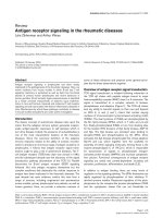
Figure 1 Phylogenetic classification of 38 new Gambian HIV-1 full-lengt h env sequences (highlighted in red), along with reference
subtypes and additional subtype A sequences (CRF02_AG and Senegalese A3 variants). The full Los Alamos HIV Database (2005) subtype
reference set was initially used to construct the tree, but all CRFs other than CRF02_AG have been omitted here for clarity. The phylogenetic
tree was constructed using a maximum likelihood method [18], under the General Time Reversible model of nucleotide substitution [19], with
proportion of invariable sites and substitution rate heterogeneity. Bootstrap percentiles above 70% from 1000 replications (using the neighbor-
joining method) are shown at the corresponding branches defining major grouping of sequences. Five of the new Gambian sequences cluster
with the Senegalese A3 variant sequences with a bootstrap support of 81 (∞). Branch lengths represent the number of substitutions per
nucleotide sites.
de Silva et al. Retrovirology 2010, 7:82
/>Page 7 of 14
Germany), VLGC-J2 (of unknown origin) [29], the 98
BW21.17 isolate from Botswana [30] and the MBTB4,
KTB147 and MBS41 isolates from DRC [31]. A phyloge-
netic tree was constructed as described above with these
isolates, along with the six subtype J-like env samples
from the current study (Fig. 2). All nine subtype J-like
env sequences from the Gambia form a monophyletic
cluster (with a bootstrap support of 92%) and are distinct
from the DRC isolates (Fig. 2).
The Botswana isolate was reported as a novel subtype
A/J recombinant [30], although it has since been reclas-
sified by the LAHDB as an AGJ recombinant, as parts
of the genome are said to be more closely related to
CRF06_AJGK than to any one isolate of subtype A or J
[3]. The G MB22 and GMB24 isolates are also reported
as having subtype A gag regions, although only gag
sequence from GMB22 is available [28]. To test the idea
that a novel recombinant is circulating in the Gambia,
the gag p24 regions from the six novel J-like env isolates
were sequenced and all were found to be subtype A.
Furthermore the gag regions from t he Botswana isolate
98BW21.17, GMB22 and five of the new A/J isolates
form a monophyletic cluster with a bootstrap support of
94% (Fig. 3). These gag isolates are distinct from sub-
subtype A1, A2, A3 sequences, as well as those derived
from CRF02_AG isolates. One new recombinant isolate
(N5284_GM) gag region clustered with A3 [13,14] iso-
lates reported in surrounding Senegal, which may indi-
cate further recombination between the novel
recombinant with circulating local A3 strains. One addi-
tional isolate described in the literature, MBTB4 from
DRC, is reported to have a subt ype A gag and subtype J
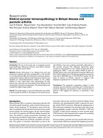
Figure 2 Phylogenetic tree wit h all available subtype J-like env Gambian isolates (red), including the three older isolates GM4, GM22
and GM24, and other subtype J env sequences from the Los Alamos HIV Database. MBTB4 and 98BW21.17 (in purple) are subtype A gag
/J env recombinants described from outside the Gambia (DRC and Botswana respectively). The Gambian subtype J-like env monophyletic cluster
is boxed. SE92809 and SE9173 are the two subtype J reference strains (From DRC, isolated in Sweden). The phylogenetic tree was reconstructed
as in Fig. 1 and bootstrap percentiles above 70% from 1000 replications (using the neighbour-joining method) are shown. The tree is rooted by
outgroups formed by subtype A1 and CRF02_AG env fragments from the Gambia (N75698A1_GM and N16805_GM). Branch lengths are
expressed as the number of substitutions per nucleotide sites.
de Silva et al. Retrovirology 2010, 7:82
/>Page 8 of 14
env region [31]. The subtype A gag phylogenetic tree
was re-built including this isolate, along with three
further DRC subtype A sequences (KCC2, KTB13 and
KTB035), which required use of a shorter fragment
length as described above. The MTBT4 isolate gag
appears to be more closely related to subtype A gag
regions from gag A/env J-like recombinants than other
subtype A sequences (with a bootstrap support of 76%),
including those from DRC (Fig. 3). Of note, the env
region from MTBT4 clusters with the two reference J
envs SE9173 (from an individual known to be infected
in DRC) and SE92809 (bootstrap support of 98), rather
than the other env JisolateswithsubtypeAgag regions
(Fig. 2).
CRF49_cpx, a novel circulating recombinant form
Near full-genome sequences from three of the gag A/env
J-like isolates (N18380_GM, N28353_GM and
N26677_GM) were generated and a ph ylogenetic tree
constructed as described above (Fig. 4), which provided
confirmation that these viruses represent a novel CRF,
now named CRF49_cpx in the LAHDB. The three iso-
lates clearly form a new cluster, separate from any cur-
rently known pure subtypes o r recombinants (with a
posterior probability of 1) and appear to be closely
related to the Botswanan isolate 98BW21.17. Analyses
of subtype recombination (as described above) revealed
a complex, but consistent pattern across the three iso-
lates (see Figs. 5, S1 and S2). In addition to the largely
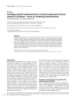
Figure 3 Phylogenetic tree constructed using alignments of gag sequence from subtype A reference strains (denoted by prefix ‘Ref’),
additional subtype A1 isolates, A3 isolates from Senegal, CRF02_AG isolates and subtype A gag sequence from isolates with subtype
J-like env regions. Gambian isolates are in red, which includes an older isolate GMB22. Sequence from the non-Gambian gagA/envJ
recombinants 98BW21.17 and MTBT4 are highlighted in purple. The cluster formed by gag A sequence from isolates with J-like env regions is
boxed. One Gambian isolate (N5284_GM) falls outside this cluster. The tree was reconstructed as in Fig. 1 and bootstrap percentiles above 70%
from 1000 replications (using the neighbour-joining method) are shown. The trees are rooted by outgroups formed by subtype J and C
reference isolates from the Los Alamos HIV Database (2005) subtype reference set (SE7887 and 95IN21068). Branch lengths represent the number
of substitutions per nucleotide sites. The tree includes the DRC isolates MTBT4, KCC2, KTBT13 and KTB035 which required the sequences to be
trimmed to 623 bp. A similar tree lacking these sequences but reconstructed with a 951 bp length alignment confirmed the clustering (for the
remaining sequences) although with higher bootstrap support.
de Silva et al. Retrovirology 2010, 7:82
/>Page 9 of 14
subtype A gag region and J-like env, a significant sub-
type C fragment is present in a portion of pol, extending
through vif to vpr (which is absent in 98BW21.17),
where a breakpoint with the subtype J-like fragment is
found. The pol gene is mosaic and contains regions with
similarity to subtypes A, J, K and C, as well a fragment
which is not clearly defined by currently known pure
subtype sequences. A phylogenetic tree constructed with
this pol fragment (not resolved through Simplot boo t-
scanning analysis), suggested that this region was sub-
type F-like (Fig. 5). Simplot and bootscan analysis [22]
clearly showed a similar pattern of subtype recombina-
tion a cross the three isolates, a lthough there was varia-
tion in where the exact breakp oints are (Supplementary
Fig. S1 and S2), especially in the highly mosaic pol gene.
The diversity between the three CRF49_cpx sequences
may suggest that they are derived from a virus that
recombined decades ago and as a great deal of evolution
may have occurred since that time, many of the
recombination breakpoints cannot be clearly defined.
The Simplot and bootscan analysis [22] was repeated for
each sequence, with inclusion of the Botswanan isolate
98BW21.17 in the reference set. This suggested that
apart from the subtype C-like fragment, the CRF49_cpx
sequences are more similar to 98BW21.17 than to most
pure reference subtyp es representing ea ch recombinant
fragment (Supplementary Fig. S3). I t is possible, there-
fore, that CRF49_cpx originated via further recombina-
tion between a 98BW21.17-like strain and a subtype C
isolate.
A careful examination of patient records was per-
formed to determine social factors that might be asso-
ciated with the CRF49_cpx viruses. There was no
evidence that any of these subjects were related and
there was no exclusive association with an ethnic group
in this set of subjects (two Mandinka, two Manjago, one
Jola and one Serahuli - see Table 1). None of these sub-
jects were reported commercial sex workers (CSWs),
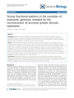
Figure 4 Midpoint rooted Bayesian tree using Los Alamos 2008 subtype reference set HIV-1 full g enomes, additio nal A3 sequenc es,
98BW21.17 and 3 new Gambian CRF49_cpx isolates. Pure subtype sequences represented in the new Gambian complex recombinant are
shown in color (A (red), J (turquoise), C (brown), K (purple)). Relevant nodes to the new complex recombinant, with a posterior probability of 1,
are marked with *.
de Silva et al. Retrovirology 2010, 7:82
/>Page 10 of 14
one reported a blood transfusion and there were no
reports of travel to the DRC or Botswana for any of the
patients.
HIV-1 subtype distribution relative to Senegal
The m ost recent survey from Senegal show a high pre-
valence of subtype C (40%), followed by CRF 02_AG
(24.3%), then subtype B (18.6%) in a Senegalese cohort
of men who have sex with men [32]. This distribution
was different from female sex workers (FSWs) and from
the general population where CRF02_AG was reported
to predominate [33]. In the Senegalese FSW cohort,
despite large sample numbers (328), only 2 subtype J
isolates (in env) were reported. Because a small (385 bp)
C2-V3 env fragment was used for subtyping [32,33],
there is a concern that this might have missed detecting
subtypes Js. However when the G ambian 38 samples
plus the Los Alamos reference set are trimmed to the
385 bp C2-V3 regi on used in the Senegal ese study, the
six new Gambian subtype J-like env sequences still clus-
ter with the reference J sequences with high bootstrap
values (results not shown). If J subtypes or CRF49_cpx
isolates were present in the Senegalese cohort, they
would have been detected by the 385 bp C2-V3 analysis,
therefore the high frequency of CRF49_cpx isolates
observed in the Gambia may not extend to neighboring
Senegal.
The geographical and subtype information in the
LAHDB are gathered from investigator-supplied infor-
mation. Different levels of rigor can be used to define
HIV-1 subtype (e.g. the REGA HIV subtyp ing algorithm
[34] requires a minimum of 800 bp of sequence whereas
many of th e LAHDB subtype designations are provided
for sequences of less than 300 bp). Furthermore, of sub-
type designations, there can be multiple listings for the
same patient and this may result in over-reporting of
some subtypes. For example, for CRF02_AG, when the
840 Senegalese entries in LAHDB with reported subtype
Figure 5 Recombinant map of CRF49_cpx and phylogenetic trees constructed from each non-recombinant fragment. Recombinant map
of CRF49_cpx (top) drawn using the Recombinant HIV-1 Drawing Tool at LAHDB and depicting HXB2 numbering at breakpoints estimated via
bootscan analysis in Simplot [22]. Below are maximum likelihood trees constructed with each non-recombinant fragment (1 to 8) showing the
relationship to pure subtypes. Only bootstrap values (from 1000 replications using the neighbour-joining method) at relevant nodes to the
CRF49_cpx isolates and closest pure subtype are shown for ease of presentation.
de Silva et al. Retrovirology 2010, 7:82
/>Page 11 of 14
are screened for entries 800 bp or larger and the known
multiple patient entr ies are remov ed, a set of 183
sequence entries remain. These 183 sequences were ana-
lysed phylogenetically, using maximum likelihood me th-
ods as described above, to generate a more stringent
subtype distribution (Fig. 6, left pie). Similar criteria
were applied to the 38 novel Gambia n sequences from
this study plus the four Gambian LAHDB entries >800
bp (Fig. 6, right pie). In this analysis, there are large dif-
ferences in the frequency of the HIV-1 subtypes
between the two countries (Fig 6). This could be due to
cultural differences, or to differences in the age and
extent of the epidemic in each country. In addition, the
Senegalese data are dominated by sequences derived
from specific cohorts (MSM, CSW) while the Gambia
data (mostly deriv ed from the current study) come from
random selection of patients attending a GUM clinic;
such differences in the patient composition could results
in the large differences in the subtype distribution.
Conclusions
InformationonthediversityofHIV-1intheGambiais
currently lacking and the current study has attempted to
address this gap by generating full-length HIV-1 env
sequences from 38 local HIV-1 isolates. Documentation
of the ongoing HIV-1 epidemic and sequence data from
West Africa is important for several reasons. In a region
where HIV-1 diversity is higher than in many other
parts of sub-Saharan Africa, such information is
required to maintain accurate viral diagnostics and
sensitive viral load assays. HIV-1 subtypes may differ
biologically in areas such as viral fitness [35,36] and
co-receptor usage (e.g. likelihood of switch from R5 to
X4 usage) [37,38]. These may in turn translate into
higher risk of disease progression in certain subtypes
and recombinant viruses could also have certain advan-
tages over their parent strains. Studies in East Africa,
using both prevalent and incident infections, have
shown a higher risk of progression to AIDS and AIDS-
related death in subtype D (and inter-subtype recombi-
nant) -infected individuals when compared to subtype
A-infected patients [39,40]; even following adjustment
for baseline viral load [41]. A Senegalese study supports
the notion that non-A subtype infections progress faster
than subtype A infections [42], although outcomes in
CRF02_AG infected in dividuals appear to be no worse
compared to non-AG infections [43]; despi te the rise of
this circulating recombinant form (CRF) in West Africa
and in vitro data suggesting enhan ced viral fitness [35].
With the increasing availability of anti-retroviral therapy
(ART) in West Africa, it is also important to consider
potential differences between HIV-1 subtypes in drug
resistance pathways and the ease with which resistance
appears due to naturally occurring polymorphisms (e.g.
the development of K65R in subtype C infections)
[44,45]. Such findings would clearly have implications
for local ART regimes and choice of 2
nd
line drugs.
Finally, local sequence data are important in the design
of potential immunogens for future prophylactic and
therapeutic HIV-1 vaccines, although the greater
Figure 6 HIV-1 subtype distribution in Seneg al compared to Gambia. The left chart shows the distribution in the 183 LAHDB sequences
from Senegal >800 bp. The right chart shows the distribution in all LAHDB subtyped HIV-1 sequences from Gambia >800 bp (4 entries) plus the
38 sequences from the current work (42 entries total). Note that the CRF49_cpx viruses identified in this study were included in the J category.
The frequency of selected subtypes with 95% confidence intervals (calculated using the modified Wald method) were Gambia J 21% (11.2-
35.4%), Gambia CRF 02_AG 49% (34.6-63.3%); Senegal J 0.6% (< 0.01 to 3.3%), Senegal CRF 02_AG 31% (24-38%).
de Silva et al. Retrovirology 2010, 7:82
/>Page 12 of 14
diversity in West Africa makes this daunting task even
more challenging in this subregion. Mosaic vaccine stra-
tegies [4,5] may overcome this barrier and document a-
tion of new CRFs and accurate representation of global
sequence diversity is essential for these strategies.
While the majority prevalence of subtype A and
CRF02_AG in the new set of HIV-1 isolates is consistent
with data from other West African countries, the identifi-
cation of 6 isolates of a novel recombinant, CRF49_cpx, in
the 38 isolates was surprising and unique to the Gambia.
These six infected individuals w ere epidemiologically
unlinked and env sequence from these viruses cluster with
three previously described Gambian subtype J-like env
sequences. Thus, all nine isolates are likely t o represent
the novel HIV-1 CRF49_cpx. Full genome sequence from
the Botswanan isolate (98BW21.17) [30] is closely linked
to the Gambian isolates in phylogenetic analyses (more so
than to any other virus currently in the LAHDB). Due to
the limited number of patients examined, it is difficult to
predict the importance of CRF49_cpx in the Gambian
HIV-1 epidemic. Although some criteria were imposed in
sample selection, within both patient group s (CD4 >/=
28% at first presentation and recently diagnosed and com-
menced antiretroviral therapy) selection was randomized.
There is good reason to believe therefore, that CRF49_cpx
may represent a reasonable proportion of the HIV-1 infec-
tions in the Gambia. Further studies are important to clar-
ify its prevalence (including changes over time), the
contribution to new infections in recent years and the dis-
ease potential relative to other local subtypes.
Additional material
Additional file 1: Figure S1 - Bootscan analyses of CRF49_cpx
isolates N18380_GM (a), N26677_GM (b) and N28353_GM (c)
performed with Simplot [22] and including HIV-1 subtypes A
through K. Alignment was gap stripped. Sliding window was set to 400
bp with increments set to 50 bp. Bootscanning was performed by
neighbour-joining tree construction model, using the Kimura (two-
parameter) distance model and 100 bootstrap replicates for each sliding
window. Transition/traversion ratio was set to 2.0.
Additional file 2: Figure S2 - Simplot analyses of CRF49_cpx isolates
N18380_GM (a), N26677_GM (b) and N28353_GM (c) [22] and
including HIV-1 subtypes A through K. Alignment was gap stripped.
Sliding window was set to 400 bp with increments set to 50 bp.
Bootscanning was performed by neighbour-joining tree construction
model, using the Kimura (two-parameter) distance model and 100
bootstrap replicates for each sliding window. Transition/traversion ratio
was set to 2.0.
Additional file 3: Figure S3 - Simplot analyses of CRF49_cpx isolates
N18380_GM (a), N26677_GM (b) and N28353_GM (c) [22] and
including HIV-1 subtypes A through K and Botswana isolate
98BW21.17. Alignment was gap stripped. Sliding window was set to 400
bp with increments set to 50 bp. Bootscanning was performed by
neighbour-joining tree construction model, using the Kimura (two-
parameter) distance model and 100 bootstrap replicates for each sliding
window. Transition/traversion ratio was set to 2.0.
Acknowledgements
TdS is supported by a Medical Research Council (UK) Clinical Research
Training Fellowship. We would like to thank the patients and staff at the
MRC (UK) Fajara Genitourinary Medicine (GUM) clinic for their participation in
the HIV cohort, which made this study possible.
Author details
1
Medical Research Council (UK) Laboratories, Atlantic Road, PO Box 273,
Fajara, The Gambia.
2
MRC/UCL Centre for Medical Molecular Virology,
Division of Infection and Immunity, University College London, UK.
3
Weatherall Institute of Molecular Medicine, Medical Research Council
Human Immunology Unit, John Radcliffe Hospital, Oxford, UK.
4
Theoretical
Biology & Biophysics, Los Alamos National Laboratory, Los Alamos, NM
87545, USA.
Authors’ contributions
TdS participated in all parts of the study, co-supervised the work, was
responsible for env and gag sequencing, participated in the sequence and
phylogenetic analysis and participated in writing the manuscript, RT was
responsible for amplifying and sequencing most of the envs and helped
with the initial analysis of the sequences, SH provided detailed phylogenetic
support and assisted in writing the manuscript, RTr assisted in generating
near full-genome sequence from the three described CRF49_cpx isolates,
CvT provided database analysis, organized the patient data and participated
in writing the manuscript, CO participated in designing the sequencing
strategy and methods and assisted in the initial analysis of the sequences,
BF helped interpret the recombination and phylogenetic analyses of
CRF49_cpx and assisted in writing the manuscript, HW participated in the
design of the study, provided virological support and participated in writing
the manuscript, SR-J participated in the design of the study, provided
virological support and participated in writing the manuscript, AJ provided
virological support and participated in writing the manuscript, MC
participated in all parts of the study, co-supervised the work, assisted in the
sequence analysis and phylogenetic analysis and participated in writing the
manuscript.
Competing interests
The authors declare that they have no competing interests.
Received: 21 April 2010 Accepted: 9 October 2010
Published: 9 October 2010
References
1. Schim van der Loeff MF, Sarge-Njie R, Ceesay S, Awasana AA, Jaye P,
Sam O, Jaiteh KO, Cubitt D, Milligan P, Whittle HC: Regional differences in
HIV trends in The Gambia: results from sentinel surveillance among
pregnant women. Aids 2003, 17:1841-1846.
2. van der Loeff MF, Awasana AA, Sarge-Njie R, van der Sande M, Jaye A,
Sabally S, Corrah T, McConkey SJ, Whittle HC: Sixteen years of HIV
surveillance in a West African research clinic reveals divergent epidemic
trends of HIV-1 and HIV-2. Int J Epidemiol 2006, 35:1322-1328.
3. Los Alamos HIV Database. [ />4. Barouch DH, O’Brien KL, Simmons NL, King SL, Abbink P, Maxfield LF,
Sun YH, La Porte A, Riggs AM, Lynch DM, et al: Mosaic HIV-1 vaccines
expand the breadth and depth of cellular immune responses in rhesus
monkeys. Nat Med 16:319-323.
5. Santra S, Liao HX, Zhang R, Muldoon M, Watson S, Fischer W, Theiler J,
Szinger J, Balachandran H, Buzby A, et al: Mosaic vaccines elicit CD8+ T
lymphocyte responses that confer enhanced immune coverage of
diverse HIV strains in monkeys. Nat Med 16:324-328.
6. Salazar-Gonzalez JF, Bailes E, Pham KT, Salazar MG, Guffey MB, Keele BF,
Derdeyn CA, Farmer P, Hunter E, Allen S, et al: Deciphering human
immunodeficiency virus type 1 transmission and early envelope
diversification by single-genome amplification and sequencing. J Virol
2008, 82:3952-3970.
7. Metabion. [ />8. Macrogen. [ />9. Hall TA: BioEdit: a user-friendly biological sequence alignment editor and
analysis program for Windows 95/98/NT. Nucleic Acids Symposium Series
1999, 41:95-98.
de Silva et al. Retrovirology 2010, 7:82
/>Page 13 of 14
10. BioEdit. [ />11. Stothard P: The sequence manipulation suite: JavaScript programs for
analyzing and formatting protein and DNA sequences. Biotechniques
2000, 28:1102, 1104.
12. Sequence Manipulation Suite. [ />orf_find.html].
13. Meloni ST, Kim B, Sankale JL, Hamel DJ, Tovanabutra S, Mboup S,
McCutchan FE, Kanki PJ: Distinct human immunodeficiency virus type 1
subtype A virus circulating in West Africa: sub-subtype A3. J Virol 2004,
78:12438-12445.
14. Meloni ST, Sankale JL, Hamel DJ, Eisen G, Gueye-Ndiaye A, Mboup S,
Kanki PJ: Molecular epidemiology of human immunodeficiency virus
type 1 sub-subtype A3 in Senegal from 1988 to 2001. J Virol 2004,
78:12455-12461.
15. Larkin MA, Blackshields G, Brown NP, Chenna R, McGettigan PA,
McWilliam H, Valentin F, Wallace IM, Wilm A, Lopez R, et al: Clustal W and
Clustal × version 2.0. Bioinformatics 2007, 23:2947-2948.
16. EBI ClustalW2. [ />17. Phylogenetic analysis using parsimony and other methods. Version
4.0b10. [ />18. Felsenstein J: Maximum-likelihood estimation of evolutionary trees from
continuous characters. Am J Hum Genet 1973, 25:471-492.
19. Yang Z: Estimating the pattern of nucleotide substitution. J Mol Evol
1994, 39:105-111.
20. Inkscape. [ />21. Huelsenbeck JP, Ronquist F: MRBAYES: Bayesian inference of phylogenetic
trees. Bioinformatics 2001, 17:754-755.
22. Lole KS, Bollinger RC, Paranjape RS, Gadkari D, Kulkarni SS, Novak NG,
Ingersoll R, Sheppard HW, Ray SC: Full-length human immunodeficiency
virus type 1 genomes from subtype C-infected seroconverters in India,
with evidence of intersubtype recombination. J Virol 1999, 73:152-160.
23. Gambian 2003 Census Data. [ />world-factbook/fields/2075.html].
24. Carr JK, Salminen MO, Albert J, Sanders-Buell E, Gotte D, Birx DL,
McCutchan FE: Full genome sequences of human immunodeficiency
virus type 1 subtypes G and A/G intersubtype recombinants. Virology
1998, 247:22-31.
25. Abecasis AB, Lemey P, Vidal N, de Oliveira T, Peeters M, Camacho R,
Shapiro B, Rambaut A, Vandamme AM: Recombination confounds the
early evolutionary history of human immunodeficiency virus type 1:
subtype G is a circulating recombinant form. J Virol 2007, 81:8543-8551.
26. Zhang M, Foley B, Schultz AK, Macke JP, Bulla I, Stanke M, Morgenstern B,
Korber B, Leitner T:
The role of recombination in the emergence of a
complex and dynamic HIV epidemic. Retrovirology 7:25.
27. Bobkov A, Cheingsong-Popov R, Salminen M, McCutchan F, Louwagie J,
Ariyoshi K, Whittle H, Weber J: Complex mosaic structure of the partial
envelope sequence from a Gambian HIV type 1 isolate. AIDS Res Hum
Retroviruses 1996, 12:169-171.
28. Cham F, Heyndrickx L, Janssens W, Van der Auwera G, Vereecken K, De
Houwer K, Coppens S, Whittle H, van der Groen G: Study of HIV type 1
gag/env variability in The Gambia, using a multiplex DNA polymerase
chain reaction. AIDS Res Hum Retroviruses 2000, 16:1915-1919.
29. Binley JM, Wrin T, Korber B, Zwick MB, Wang M, Chappey C, Stiegler G,
Kunert R, Zolla-Pazner S, Katinger H, et al: Comprehensive cross-clade
neutralization analysis of a panel of anti-human immunodeficiency virus
type 1 monoclonal antibodies. J Virol 2004, 78:13232-13252.
30. Novitsky VA, Gaolekwe S, McLane MF, Ndung’u TP, Foley BT, Vannberg F,
Marlink R, Essex M: HIV type 1 A/J recombinant with a pronounced pol
gene mosaicism. AIDS Res Hum Retroviruses 2000, 16:1015-1020.
31. Vidal N, Peeters M, Mulanga-Kabeya C, Nzilambi N, Robertson D, Ilunga W,
Sema H, Tshimanga K, Bongo B, Delaporte E: Unprecedented degree of
human immunodeficiency virus type 1 (HIV-1) group M genetic diversity
in the Democratic Republic of Congo suggests that the HIV-1 pandemic
originated in Central Africa. J Virol 2000, 74:10498-10507.
32. Ndiaye HD, Kane CT, Vidal N, Niama FR, Niang-Diallo PA, Dieye T, Gaye-
Diallo A, Wade AS, Peeters M, Mboup S: Surprisingly High Prevalence of
Subtype C and Specific HIV-1 Subtype/CRF Distribution in Men Having
Sex With Men in Senegal. J Acquir Immune Defic Syndr 2009.
33. Hamel DJ, Sankale JL, Eisen G, Meloni ST, Mullins C, Gueye-Ndiaye A,
Mboup S, Kanki PJ: Twenty years of prospective molecular epidemiology
in Senegal: changes in HIV diversity. AIDS Res Hum Retroviruses 2007,
23:1189-1196.
34. Abecasis AB, Wang Y, Libin P, Imbrechts S, de Oliveira T, Camacho RJ,
Vandamme AM: Comparative performance of the REGA subtyping tool
version 2 versus version 1. Infect Genet Evol 10:380-385.
35. Njai HF, Gali Y, Vanham G, Clybergh C, Jennes W, Vidal N, Butel C, Mpoudi-
Ngolle E, Peeters M, Arien KK: The predominance of Human
Immunodeficiency Virus type 1 (HIV-1) circulating recombinant form 02
(CRF02_AG) in West Central Africa may be related to its replicative
fitness. Retrovirology 2006, 3:40.
36. Abraha A, Nankya IL, Gibson R, Demers K, Tebit DM, Johnston E,
Katzenstein D, Siddiqui A, Herrera C, Fischetti L, et al: CCR5- and CXCR4-
tropic subtype C human immunodeficiency virus type 1 isolates have a
lower level of pathogenic fitness than other dominant group M
subtypes: implications for the epidemic. J Virol 2009, 83:5592-5605.
37. Tscherning C, Alaeus A, Fredriksson R, Bjorndal A, Deng H, Littman DR,
Fenyo EM, Albert J: Differences in chemokine coreceptor usage between
genetic subtypes of HIV-1. Virology 1998, 241:181-188.
38. Ping LH, Nelson JA, Hoffman IF, Schock J, Lamers SL, Goodman M,
Vernazza P, Kazembe P, Maida M, Zimba D,
et al: Characterization of V3
sequence heterogeneity in subtype C human immunodeficiency virus
type 1 isolates from Malawi: underrepresentation of X4 variants. J Virol
1999, 73:6271-6281.
39. Kaleebu P, French N, Mahe C, Yirrell D, Watera C, Lyagoba F, Nakiyingi J,
Rutebemberwa A, Morgan D, Weber J, et al: Effect of human
immunodeficiency virus (HIV) type 1 envelope subtypes A and D on
disease progression in a large cohort of HIV-1-positive persons in
Uganda. J Infect Dis 2002, 185:1244-1250.
40. Kiwanuka N, Laeyendecker O, Robb M, Kigozi G, Arroyo M, McCutchan F,
Eller LA, Eller M, Makumbi F, Birx D, et al: Effect of human
immunodeficiency virus Type 1 (HIV-1) subtype on disease progression
in persons from Rakai, Uganda, with incident HIV-1 infection. J Infect Dis
2008, 197:707-713.
41. Baeten JM, Chohan B, Lavreys L, Chohan V, McClelland RS, Certain L,
Mandaliya K, Jaoko W, Overbaugh J: HIV-1 subtype D infection is
associated with faster disease progression than subtype A in spite of
similar plasma HIV-1 loads. J Infect Dis 2007, 195:1177-1180.
42. Kanki PJ, Hamel DJ, Sankale JL, Hsieh C, Thior I, Barin F, Woodcock SA,
Gueye-Ndiaye A, Zhang E, Montano M, et al: Human immunodeficiency
virus type 1 subtypes differ in disease progression. J Infect Dis 1999,
179:68-73.
43. Laurent C, Bourgeois A, Faye MA, Mougnutou R, Seydi M, Gueye M,
Liegeois F, Kane CT, Butel C, Mbuagbaw J, et al: No difference in clinical
progression between patients infected with the predominant human
immunodeficiency virus type 1 circulating recombinant form (CRF)
02_AG strain and patients not infected with CRF02_AG, in Western and
West-Central Africa: a four-year prospective multicenter study. J Infect Dis
2002, 186:486-492.
44. Invernizzi CF, Coutsinos D, Oliveira M, Moisi D, Brenner BG, Wainberg MA:
Signature nucleotide polymorphisms at positions 64 and 65 in reverse
transcriptase favor the selection of the K65R resistance mutation in HIV-
1 subtype C. J Infect Dis 2009, 200:1202-1206.
45. Martinez-Cajas JL, Pai NP, Klein MB, Wainberg MA: Differences in resistance
mutations among HIV-1 non-subtype B infections: a systematic review
of evidence (1996-2008). J Int AIDS Soc 2009, 12:11.
doi:10.1186/1742-4690-7-82
Cite this article as: de Silva et al.: HIV-1 subtype distribution in the
Gambia and the significant presence of CRF49_cpx, a novel circulating
recombinant form. Retrovirology 2010 7:82.
de Silva et al. Retrovirology 2010, 7:82
/>Page 14 of 14