Báo cáo y học: "Subtype-associated differences in HIV-1 reverse transcription affect the viral replication" ppt
Bạn đang xem bản rút gọn của tài liệu. Xem và tải ngay bản đầy đủ của tài liệu tại đây (2.13 MB, 18 trang )
RESEARC H Open Access
Subtype-associated differences in HIV-1 reverse
transcription affect the viral replication
Sergey Iordanskiy
1,2,3*
, Mackenzie Waltke
1
, Yanjun Feng
2
, Charles Wood
1
Abstract
Background: The impact of the products of the pol gene, specifically, reverse transcriptase (RT) on HIV-1
replication, evolution, and acquisition of drug resistance has been thoroughly characterized for subtype B. For
subtype C, which accounts of almost 60% of HIV cases worldwide, much less is known. It has been reported that
subtype C HIV-1 isolates have a lower replication capacity than B; however, the basis of these differences remains
unclear.
Results: We analyzed the impact of the pol gene products from HIV-1 B and C subtypes on the maturation of HIV
virions, accumulation of reverse transcription products, integration of viral DNA, frequency of point mutations in
provirus and overall viral replication. Recombinant HIV-1 viruses of B and C subtypes comprising the pol fragments
encoding protease, integrase and either the whole RT or a chimeric RT from different isolates of the C and B
subtypes, were used for infection of cells expressing CXCR4 or CCR5 co-receptors. The viruses carrying different
fragments of pol from the isolates of B and C subtypes did not reveal differences in Gag and GagPol processing
and viral RNA incorporation into the virions. However, the presence of the whole RT from subtype C, or the
chimeric RT containing either the polymerase or the connection and RNase H domains from C isolates, caused
significantly slower viral replication regardless of B or C viral backbone. Subtype C RT carrying viruses displayed
lower levels of accumulation of strong-stop cDNA in permeabilized virions during endogenous reverse
transcription, and decreased accumulation of both strong-stop and positive strand reverse transcription products in
infected cells and in isolated reverse transcription complexes. This decreased accumulation correlated with lower
levels of viral DNA integration in cells infected with viruses carrying the whole RT or RT domains from subtype C
isolates. The single viral genome assay analysis did not reveal significant differences in the frequency of point
mutations between the RT from B or C subtypes.
Conclusions: These data suggest that the whole RT as well as distinct polymerase and connection-RNase H
domains from subtype C HIV-1 confer a lower level of accumulation of reverse transcripts in the virions and reverse
transcription complexes as compared to subtype B, resulting in a lower overall level of virus replication.
Background
Almost 60% of HIV-positive individuals (more than 22
million people) are infected with HIV-1 subtype or clade
C. Subtype C is the most rapidly expanding HIV-1 sub-
type, which predominates in Eastern and Southern Africa
and India, and is increasing in frequency in China, Brazil,
Uruguay, and nearby countri es (reviewed in [1]). In spite
of intensive global expansion, no significant differences
were observed in the disease progression or pathogenicity
of infection in individuals infected by subtype C versus
patients infected by other group M subtypes [2]. The epi-
demic success of subtype C viruses relative to other HIV-
1 strains nevertheless suggests that there are factors
which may affect the transmission and/or replication of
this group of viruses [3]. Although the o verall genomic
organization is similar among HIV-1 subtypes, sequence
diversity between HIV-1 clades may range from 5 to 35%
for different genes [4,5]. Indeed, a number of f actors
related to viral entry and pathogenesis have been indi-
cated as distinct for subtype C HIV-1. They include the
predominant use of CCR5 co-receptor by subtype C
* Correspondence:
1
Nebraska Center for Virology, School of Biological Sciences, University of
Nebraska - Lincoln, 4240 Fair Street, Ken Morrison Life Sciences research
Center, East Campus, Lincoln, NE 68583-0900 USA
Full list of author information is available at the end of the article
Iordanskiy et al. Retrovirology 2010, 7:85
/>© 2010 Iordanskiy et al; licensee BioMed C entral Ltd. This i s an Open Access article distributed under the terms of the Creative
Commons Attribution License ( s/by/2.0), which permits unrestricte d use, distribution, and
reproduction in any m edium , provided the original work is properly cited.
strains, even in late infection [6,7], and relatively high
transmission fitness in dendritic cells, which may
increase the frequencies of vaginal shedding and mother-
to-child transmission [8,9]. In addition, most subtype C
isolates are non-syncyti um-inducing which may decrease
their cytopathogenicity and hence contribute to the
spread of this group of viruses [8,10]. At the viral geno-
mic level, the long terminal repeats have three NF-B
binding sites and a truncation of the Rev protein [11],
which may both influence viral replication by enhancing
gene expression. There is also a 5-amino-acid insertion
in the Vpu polypeptide which may affect the virulence of
subtype C viruses through modulation of the Vpu func-
tions, such as CD4 degradation or enhancement of virion
release from the cells [12]. Despite these molecular char-
acteristics which may determine enhanced viral replica-
tion,thesubtypeCviruseswerefoundtohavelower
replication fitness in primary CD4+ T ce lls and periph-
eral blood mononuclear cells when compared to all other
group M subtypes [8,13,14]. These data suggest there are
some viral components of clade C viruses which may
decrease the overall r eplication level or increase the vul-
nerability of the virus to host restriction factors, but do
not alter an enhanced capacity of these viruses to
transmit.
The HIV gene po l encodes th e viral enzymes protease,
reverse transcriptase (RT), and integrase and represents
the most conserved region of the HIV genome. Never-
theless, differences in the pol sequences inherent to cer-
tain HIV-1 subtypes have been identified. They include
different consensus amino acid (AA) residues in the
non-catalytic regions of the protease, RT and integrase.
Some of these differences are considered to be subtype-
specific signature sequences [15-17], which may poten-
tially affect drug resistanc e acquisition and probably
replicative capacities of the subtypes, as reviewed earlier
[18,19]. The protease of subtype C is highly conserved
and has differences in the AA sequence when compared
to subtypes A, B, and D [3,20]. The subtype C protease
has been shown catalytically more efficient than the pro-
tease from B subtype, and capable of recognizing more
diverse cleavage sites in its substrates [21].
Bioinformatic analysis of the integrase sequences
showed that twelve of fourteen subtype C-specific con-
sensus AAs are variable within the subtypes. These con-
sensus residues are loc ated beyond the catalytic triad
and functionally important zinc binding motif, LEDGF
p75 binding region, and the nuclear localization signal
[19,22,23]. Recent investigation of the 3’ processing and
strand transfer activities of the integrase from subtypes
B and C, in the presenc e and absence of the strand
transfer inhibitors, did not reveal any differences in
these activities and in susceptibility of these enzymes to
the inhibitors [24].
RT is an essential enzyme responsible for HIV replica-
tion and determination of the viral variability/poly-
morphism. T he reverse transcription and related events
of the virus life cycle have been thoroughly character-
ized for s ubtype B viruses (reviewed in [25,26]), while
much less information is available for subtype C
[5,27,28]. Despite relative conservation of the RT
sequence among the HIV-1 subtypes, differences in the
effect of RT on virus replication [29], on frequency and
location of background polymorphisms [16], and on the
developme nt of different resistance patterns in response
to treatment with RT inhibitors have been observed
between subtypes B and C [15,30,31]. These differences
may reflect the functional diversity of RT between sub-
types. However, the mechanisms contributing to these
differences remain to be determined.
In this study, we hypothesize that RT is the major fac-
tor within the pol-encoding proteins responsible for sub-
type-specific diff erences in the replication of HIV-1. To
test this hypothesis, we generated chimeric subtype B
and C viruses carrying fragments of the pol gene encod-
ing the whole RT, distinct domains of RT, and the pro-
tease or integrase sequences from different subtype C
and B isolates. In this report we analyzed the basic func-
tions of the Pol-derived proteins in these virus strains,
including Gag and GagPol polyprotein processing, accu-
mulation of reverse transcription products in virions
and reverse transcription complexes (RTCs), viral DNA
integration, the frequency of point mutations in the pro-
virus, and the overall viral replication rates. We did not
observe significant differences in the viral protease and
integrase activities in viruses carrying the Pol products
from B and C subtypes, but found that RT affected
replication of the viruses in a subt ype- depende nt man-
ner. Specifically we showed that viruses carrying RT
from subtype C isolates, as well as RT chimeras contain-
ing either the subtype C RT polymerase domain or con-
nection a nd RNase H domains, had decreased levels of
viral cDNA accumulation, which correlated with
reduced integration and lower levels of viral replication.
The frequencies of nucleotide substitutions in the pro-
viral DNA were found to be similar.
Results
Characterization of subtype C HIV-1 pol genes
The pol gene s of three subtype C HIV-1 isolates were
characterized. The viruses were isolated from three peri-
natally-infected, anti-retroviral naïve Zambian infants.
Isolates 1084i and 1984i were obtained from patients
with slow disease progression, characterized by a pro-
longed clinically asymptomatic period (more than four
years), whereas isolate 2669i was associated with fast
disease progression and a lethal outcome of the infected
infant within the first year of life [32]. We also selected
Iordanskiy et al. Retrovirology 2010, 7:85
/>Page 2 of 18
two wild-type subtype B strains, NL4-3 (T cell tropic X4
virus) [33] and YU-2 (macrophage tropic R5 isolate)
[34] as comparisons. The Pol sequences of these viruses
are similar to the s ubtype B consensus and have only
1.29 (NL4-3) and 1.36% (YU-2) of AA differences from
consensus sequence (Lo s Alamos HIV sequence data-
base ). The RT f ragments within
the Pol are rel ativel y more variable and differ from sub-
type B c onsensus by 1.6 and 2.3% respectively (Figure
1A and 1B). In contrast, the available subtype C variants
are more heterogene ous. The differences of RT AA
sequences from subtype C consensus are ranging from
2.4 to 2.9%. Sequences of the RT polymerase domain
from analyzed subtype C isolates 2669i, 1984i, and 1084i
have from 3.6 to 5.6% diversity among them, whereas
the difference between homologous sequences of NL4-3
and YU-2 isolates is 2.6% (Figure 1A). Comparison of
the AA clusters in RT, which are distinct between
selected isolates and consensus sequences of subtypes B
and C indicates that the varying amino acids are not
located in the motifs which are critical for the RT enzy-
matic activity.
The presence of gag-pol or pol fragments from HIV-1
subtype C correlates with decreased level of virus
replication independently of viral backbone and the cell
types
It has been demonstrated that subtype C viruses do not
replicate as well as subtype B and display lower replica-
tion fitness in primary CD4+ T cells and peripheral
blood mononuclears [8,13,14]. To determine whether
the pol gene products have a subtype-specific effect on
the viral replication, we compared the replication
dynamics of a subtype B s train, NL4-3, and a chimeric
NL4-3-based virus NL-pol(1084), which carried the
1084i pol gene without its protease domain (Figure 2A
and 2C), in Sup-T1 cells. Virus replication was moni-
tored by measuring p24
CA
. We found the NL-pol(1084)
displayed a much lower level of replication in Sup-T1
cells than the parental NL4-3 virus (Figure 3A, solid
lines), as well as less cytopathic effects (Figure 3A, dash
lines) and less syncytia in infected SupT1 cell cultures
(Figure 3E).
To determine which region of the subtype C pol gene
affects viral replication, several more chimeric viruses
between subtypes B and C were designed, and their
replicative capacities and cytho pathic effects were tested.
We analyzed the replication of two clones NL-RTpd
(YU2) and NL-RTpd(1084), which contain sequences
encoding the RT polymerase domain only from subtype
B isolate YU-2 or subtype C isolate 1084 in the NL4-3
backbone (Figure 2D) (RT domains are indicated
according to [35-37]). Another two chimeras carrying
the connection domain and RNaseH domain of RT, the
integrase, the Vif and the N-terminal portion of Vpr
from either the subtype B YU-2, NL-polR(YU2), or from
subtype C 1084i isolates, NL-polR(1084), in the NL4-3
backbone were also studied (Figure 2E). All recombinant
viruses expressed the backbone NL4-3 Env glycoprotein
and were tested on SupT1 cells. The presence of either
the polymerase domain (pd), or the connection and
RNase H domains of RT, integrase and Vif (R) from
subtype C 1084i isolate, led to slower viral replication as
compared to parental NL4-3 and chimeric viruses carry-
ing homologous frag ments from subtype B YU-2 isolate
(Figure 3B, left panel). Cytopathic effects of the viruses
containing RT fragments from 1084i were proportional
to their replicative dynamics, and were reflected in cell
killing (Figure 3B, right panel) and formation of syncytia
in the infe cted cell cultures (Figure 3E). To detect
whether these differences are subtype- dependent or iso-
late-dependent, similar chimeric constructs were gener-
ated from the other two subtype C isolates: 1984i
isolated from a slow disease progressing patient and
2669i from a fast progressor (Figure 2D and 2G). The
results were found to be similar to 1084i (data not
shown).
Comparison of the replication of the viral strain NL-
pol(1084), which carries the subtype C Pol without the
protease domain (Figure 3A, grey solid line), with the
chimeric viruses NL-RTpd(1084) and NL-polR(1084),
containing either the subtype C polymerase domain of
RT or the connection and RNase H domains (Figure 3
B, dash lines), shows that after 21 days of infection the
first virus displays approximately three logs lower repli-
cation level than the other two chimeric viruses. This
difference suggests that the N-terminal portion of RT
together with the C-terminal Pol domains, the Vif and
probably the Vpr proteins may contribute to the lower
replication level of the subtype C viruses.
To further determine whether the observed negative
effect of the subtype C pol gene products on viral repli-
cation is independent of the virus backbone, we gener-
ated a chimeric virus 1084-polL(NL) containing the
protease, RT polymerase domains, and 52 AA residues
from the connection domain of subtype B NL4-3 isolate
in the 1084i backbone (Figure 2F). In parallel, we gener-
ated the NL-based virus carry ing a similar fragment of
the pol gene from subtype C 1084i isolate, encodi ng the
protease and RT polymerase domains without the part
of connection domain (Figure 2G). Since 1084i Env is
R5 tropic, we then tested the replication dynamics of
subtype C-based viruses in U87.CD4.CCR5 cells,
whereas the infection with NL4-3 and NL4-3- based chi-
meric virus was performed in Sup-T1 cells. The chi-
meric subtype C-based strain carrying the pol gene
fragment from NL4-3, 1084-polL(NL), demonstrated
productive infection with increasing p24
CA
level and a
Iordanskiy et al. Retrovirology 2010, 7:85
/>Page 3 of 18
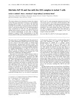
Figure 1 Comparison of RT sequences of experimental subtype B and C isolates. All sequences of polymerase domain (AA residues 1-315)
(A), connection (AA residues 316-437) and RNase H (AA residues 438-560) domains (B) are aligned with HIV-1 subtype B consensus (upper line).
Functionally important RT regions are indicated by the colored boxes: grey - conservative regions: K65, R72 - coordinate triphosphate moiety of
dNTPs; LPQG (149-152) - provide proper positioning of incoming dNTPs; LWMGYELH (228-235) - polymerase primer grip; GAH (359-361) - RNase
H primer grip; pink - YMDD box: residues 183-186, essential for polymerase activity of RT; orange - catalytic Asp (polymerase and RNase H
domains) and Glu (RNase H) residues; yellow - areas of high variability within subtypes. All conservative regions are indicated according to Coté
and Roth, 2008 [25].
Iordanskiy et al. Retrovirology 2010, 7:85
/>Page 4 of 18
high cytopathic effect, in contrast to the control wild-
type 1084i isolate which resulted in poor viral replica-
tion and low cytopathogenicity (Figure 3C). The NL-
polL(1084) viral strain containing subtype C pol frag-
ment in the subtype B backbone displayed an overall
threefold lower p24
CA
level than the wild- type NL4-3
isolate (Figure 3D). The tested chimeric virus strains
were not absolutely ide ntical. The presence of 52 AA
sequence of RT connection domain from NL4-3 in sub-
type C-based virus 1084-pol(NL) could affect the overall
level of virus replication. However, the data that both
subtype B- and C-based viruses containing the pol gene
sequences from the subtype C displayed decreased repli-
cation level indicate that the subtype C Pol domains to
poor viral replication regardless of the subtype B or C
viral backbones.
Taken together, our results indicate that the presence
of the polymera se domain or t he connection and RNase
H domains of RT, integrase and Vif from subtype C iso-
lates correlates with slower or low-efficiency replication
of chimeric virus es. The presence of both the whole RT
and integrase products of pol gene from subtype C iso-
lates in subtype B backbone virus strongly decreases the
level of viral replication (Figure 3A). This lower replica-
tion suggests that the polymerase and C-terminal
domains of RT, and likely the integrase protein all con-
tributed to th e slow er replicative kineti cs of the subtype
C viruses. On the other hand, the presence of the pro-
tease and RT polymerase domain from subtype C isolate
1084i in NL4-3 virus led to a three-fold decrease in viral
replication by the 27
th
day of infection (Figure 3D).
Whereas the clone NL-RTpd(1084), containing the same
Figure 2 Generation of recombinant HIV-1 proviral clones comprising fragments of pol gene from subtype B and C isolates. Schematic
presentation of the pol gene region of subtype B backbone NL4-3 (panel A) and subtype C backbone1084i (B) viruses, recombinant NL-based
viruses (C-E and G), and recombinant 1084i-based construct (F). The indicated fragments of the gag-pol or pol genes from subtype B (isolates
NL4-3 and YU-2) or subtype C (isolates 1084i, 1984i and 2669i) proviral DNA were PCR-amplified with primers containing sites of the indicated
restriction endonucleases, and inserted into the linearized NL4-3 or HIV1084i proviral vectors to replace the homologous fragments. Selected
molecular clones were used for transfection of 293T/17 cells to generate infectious recombinant virus strains.
Iordanskiy et al. Retrovirology 2010, 7:85
/>Page 5 of 18
Figure 3 Presence of the Gag and Pol domains from HIV-1 subtype C correlates with decreased level of virus replication. A - Kinetics of
replication (solid lanes) and cytopathicity (dash lanes) of the backbone NL4-3 and chimeric NL-pol(1084) viruses in Sup-T1 cells. The cells (1 ×
10
6
) were incubated with virus suspensions (0.01 pg of p24
CA
per cell) and then cultured in a fresh culture media. Ninety percent of the volume
of cell suspensions were harvested every 3 to 4 days, and replaced with uninfected cells. HIV-1 p24
CA
levels were detected in culture
supernatants at the indicated days after infection. Cell viability was measured in cell suspensions using trypan blue staining. Each curve
indicating p24
CA
concentration in the culture media represents the mean data of two independent experiments. Error bars show the standard
error. Each curve indicating cell viability represents data of one experiment. B - Kinetics of replication (left panel) and cytopathic effect (right
panel) of the indicated NL4-3-based viruses in Sup-T1 cells. Infection with virus clones and cultivation of infected cells were performed as
described in A. The p24
CA
curves represent the mean data ± SE from two independent experiments. The curves indicating cell viability represent
data from one experiment. C - Kinetics of replication and cytopathic effect of the backbone 1084i and chimeric 1084ipolL(NL) viruses in U87.CD4.
CCR5 cells. Each viral inoculum (MOI = 0.05) was added to 0.25 × 10
6
cells. HIV-1 p24
CA
concentrations and cell viability were monitored at the
indicated days. Each point represents mean p24
CA
level from two independent experiments. Error bars show the standard error. Each point
indicating cell viability represents data of one experiment. D - Kinetics of replication (solid lanes) and the cytopathicity (dash lines) of the
backbone NL4-3 and chimeric NL-polL(1084) viruses in Sup-T1 cells. Infection with virus clones and cultivation of infected cells were performed
as in A. Each curve indicating p24
CA
concentration represents the mean data ± SE of two independent experiments. Each curve indicating cell
viability represents data of one experiment. E - Syncytia formation by the Sup-T1 cells infected with the indicated virus strains. Live cells from
the experiment described in A and B, maintained in 1 ml of culture medium, were subjected to phase-contrast microscopy on the indicated
days after infection. One of ten representative images for each time point is shown.
Iordanskiy et al. Retrovirology 2010, 7:85
/>Page 6 of 18
RT sequence without subtype C protease, displayed only
slower replication kinetics and reached a similar p24
CA
level to the NL4-3 backbone by the 21
st
day of infection
(Figure 3B). These data suggest that subtype C protease
may also affect the replication of the recombinant
viruses.
The presence of GagPol domains from HIV-1 subtype C
does not affect incorporation of viral genomic RNA and
maturation of the virions
We quantitatively analyzed the incorporation of viral
RNA into the virions and processing of GagPol polypro-
tein-precursor in the virus particles to test the potential
effect of the subtype C protease and C-terminal domains
of Gag in GagPol chimeras on the precursor protein sta-
bility and processing, Gag-RNA binding, and compatibil-
ity between the pol sequences. Virus particles were
harvested from 297T/17 cells transfected with the pro-
viral clones, DNase I-treated, and purified through a 30%
sucrose cushion. To quantify viral RNA in the particles,
we performed real-time RT-PCR using a primer set
recognizing U5-Ψ region of HIV-1 LTR. The results did
not reveal any significant differences in viral RNA copy
numbers between subtype B and C control viruses and
the recombinant viral strains (Figure 4A). Since the pro-
tease from B and C subtypes may affect GagPol polypro-
tein processing (differences are shown in [21]), viral
release, dimerization, and total RT count in mature vir-
ions differently, we examined the ratio of the products of
GagPol processing in the virus particles generated by dif-
ferent viral clones. The Western blot analysis of the puri-
fied virus particles was performed with antibodies against
p24
CA
, inte grase, and RT and with human HIV immuno-
globulin which recognizes the Pr160
GagPol
precursor (Fig-
ure 4B). Quantification of Western blotting results
relative to p24
CA
levels for each virus sample did not
reveal substantial differences among different viruses
(Figure 4C). Collectively, these data demonstrate that the
C-terminal domains of Gag and protease from subtype C
viruses do not affect incorporation of RNA and the
maturation of different recombinant viruses significantly.
The presence of RT functional domains from HIV-1
subtype C leads to decreased cDNA accumulation in the
virions and reverse transcription complexes
To determine why viruses c arrying the sub type C RT
domains confer lower viral replication than virus strains
containing subtype B RT and whether this is due to a dif-
ference in reverse transcription, we analyzed the accumu-
lation of reverse transcription products in permeabilized
virions, in isolated reverse transcription complexes
(RTCs), and in the cytoplasm of cells infected with paren-
tal subtype B or C viruses or with chimeric viruses. As
reported earlier, reverse transcription of HIV-1 can be
initiated within the intact virions [38], and initial steps of
endogenous reverse transcription (ERT) taken place
before infection can increase HIV-1 replication in some
target cells [39]. Therefore, we employed the ERT assay
to test the various intact viral particles normalized by p24
ELISA as described earlier [40,41]. The basic level of the
early DNA products (negative-strand strong-stop DNA)
was found to be very low in all viral particles. In contrast
quantitative real-time PCR analysis of the strong-stop
cDNA purified from ERT samples after incubation with
dNTPs displayed a significant increase in early reverse
transcription product only in NL4-3 virions (Figure 5A).
Chimeric viruses contain ing the RT polymerase domain,
the connection and RNase H domains, or the whole RT
from subtype C 1084i isolate demonstrated an increase of
strong-stop cDNA level for the first 1.5 h of incubation,
fol lowed by a gradual reduction for the subsequent 3.5 h
of incubation.
We analyzed the accumulation of the reverse tran-
scription products in the cytoplasm at 24 h post-infec-
tion to identify the effects of the RT from subtypes B
and C on reverse transcription in infected cells. To
exclude the possibility that differences in viral DNA
content in the cytoplasm can be caused by natural ERT
and to assess the ratio of DNA synthesized only i n the
cytoplasm, we synchronously infected Sup-T1 cells by
different viruses in the presence or absence of 10 μM
non-nucleoside RT inhibitor nevirapine. We then deter-
mined the amount of HIV-1 DNA by quantitative real-
time PCR. The amount of strong-stop cDNA from the
cytoplasm of nevirapine-treated cells due to natural ERT
was subtracted so that only DNA synthesized within th e
infected cells was measured. We found an approximately
twofold lower count of the strong-stop DNA in the cells
infected with NL-1084 recombinants (Figure 5B). We do
not believe that this difference is due to the ability of
nevrapine to inhibit subtype B and C RT di fferently,
because it has been shown that in vitro 10 μM nevira-
pine inhibited wild-type RTs from both subtype B and C
viruses by over 100-fold [28].
Analysis of the cDNA accumulation in Sup-T1 cells
infected with recombinant viruses carrying C-terminal
Gag products, protease, and RT polymerase domains
from different subtype C isolates (1084i, 2669i and
1984i) displayed a significantly decreased level of both
early (strong-stop DNA) and l ate (positive strand DNA)
reverse transcription products at 24 h post-infection
(Figure 5C). This result shows the similar effect of the
Pol fragment containing RT p olymerase domain from
three different isolates of subtype C virus on the reverse
transcription, in spite of individual polymorphism of the
AA sequences of RT (Figure 1) and different dynamics
in disease progression in patients infected with these
viruses. Our findings suggest that observed differences
Iordanskiy et al. Retrovirology 2010, 7:85
/>Page 7 of 18
in reverse transcription efficiency are dependent on the
viral subtype.
Since RTCs are undergoing proteasome-mediated
degradation in the cytoplasm and two thirds of them
have been shown to be degraded by several hours post-
infection [42], the ratio of the reverse transcription pro-
ducts in cells infected with different virus strains shown
in previous experiments, could be affected by intracyto-
plas mic degradation of RTCs. To minimize the effect of
host cell-mediated degradation of RTCs on reverse
Figure 4 Recombinant viruses containing the Gag and Pol domains from HIV-1 subtypes B and C do not have differences in RNA
incorporation and GagPol processing. A - Quantitation of viral genomic RNA in virus particles. Virus particles were purified from the culture
media of 293T/17 cells transfected with molecular clones of viruses at 48 h post-transfection, treated with DNase I RNase free for 2 h and
concentrated by centrifugation through 30% sucrose. RNA was isolated from p24
CA
-normalized virus particles, subjected to the reverse
transcription with oligo-dT primer and then to quantitative real-time PCR with the primer set specific for positive-strand HIV-1 DNA. The data of
analysis of three independent viral preparations were quantified. Each point represents mean RNA copy number ± SD per 1 ng of p24
CA
in virus
sample. B - Processing of Pr160
GagPol
polyprotein-precursor in the virus particles. The virus particles harvested from culture media of transfected
293T/17 cells and purified as in A were analyzed by Western blotting using the antibodies indicated in Materials and Methods. C - Quantification
of Western blotting results. Western blotting data from two independent experiments were quantified using ImageJ software. Results show
mean grey values of the bands ± SE and are presented as percentage of p24
CA
in each virus sample.
Iordanskiy et al. Retrovirology 2010, 7:85
/>Page 8 of 18
transcription, we quantitatively analyzed the cDNA in
RTCs isolated from the cytoplasm during the first five
hours after infection with subtype B NL4-3, subtype C
1084i, or with chimeric viruses NL-polL(1084) a nd
1084-polL(NL). Since NL and 1084 viral vectors have
different tropism, all viruses were pseudotyped with Env
glycoprotein of the amphotropicmurineleukemiavirus
(MLV). To ensure similar levels of viruses have entered
regardless of the virus b ackbone and source of the
inserted fragment, we measured p24
CA
content in the
RTCs isolated at 1 h after infection, since capsid protein
was shown to remain associated with the viral core for
hours after infection until completion of the reverse
transcription [43,44]. We found that the p24
CA
level was
similar in early RTCs within virus strains of the same
backbone. Differences in p24
CA
levels between control
backbone and chimeric viruses did not exceed 20% (data
are not shown). However, analysis of the accumulation
of reverse transcription products in the R TCs revealed
significant differences between viruses containing the
protease and RT polymerase domains from the NL4-3
and 1084i isolates regardless of the backbone vector
(Figure 6). The RTCs of viruses carrying the subtype B
RT polymerase domain, harvested at 1 h post-infection
displayed a 2.5- (NL backbone) and 5-fold (1084i back-
bone) higher relative amount of strong-stop cDNA with
respect to those carrying the 1084i RT po lymerase
domain (Figure 6A a nd 6C). The ratios of early cDNA
between these strains, measured at 5 h after infection,
were about 2x for NL backbone and 2.5x for 1084i
backbone viruses. Similar results were observed in accu-
mulation of the positive-strand DNA (Figures 6B and
6D) measured at 5 h post-infection, suggesting that the
difference in cDNA accumulation between the viruses
with RTs from B and C subtypes are dependent on the
initial steps of the reverse transcription.
Taken together our data indicate that the presence of
the RT, as well as only the polymerase, or the connec-
tion and RNase H domains of RT from subtype C
viruses leads to a lower level of accumulation of strong-
stop cDNA and late reverse transcription products, in
bot h intact virio ns and intracytoplasmic RTCs indepen-
dent of the virus backbone. The difference in viral DNA
accumulation between viruses carrying RT from subtype
B and C isolates may eventually determine the overall
level of viral replication, that is consistent with the pub-
lished data on subtype-associatedeffectofRTonviral
replicative fitness [29].
Figure 5 The presence of RT functional domains from HIV-1 subtype C leads to decreased cDNA accumulation. A - Endogenous reverse
transcription (ERT) in permeabilized virions. Purified and p24
CA
-normalized virus particles of either the backbone NL4-3 or NL-based chimeric
viruses were subjected to ERT with addition of dNTPs and permeabilizing agent melittin. Samples without dNTPs were used as a control. DNA
was harvested after the indicated time of incubation. The relative amounts of negative-strand strong-stop DNA were measured using
quantitative real-time PCR. Data from the control samples were subtracted. Levels of cDNA are shown as percentages of the peak accumulation
detected in virions of NL4-3 at 5 h after initiation of incubation. Error bars show the standard deviation from three independent viral
preparations. B - Accumulation of early or strong-stop viral DNA in Sup-T1 cells at 24 h p.i. Untreated or treated with 10 μM nevirapine cells
were infected with backbone NL4-3 or the chimeric viruses, containing pol fragments from subtype C 1084i isolate using spinoculation. Relative
amounts of reverse transcription products were measured using quantitative real-time PCR analysis of DNA from infected cells after incubation
with or without 10 μM nevirapine. Data from nevirapine-treated samples were subtracted. Levels of cDNA are shown as percentages of the
maximal accumulation detected for cDNA in cells infected with NL4-3 virus strain. Error bars show the standard deviation from three
independent viral preparations. C - Accumulation of early and late reverse transcription products in Sup-T1 cells infected with recombinant
viruses carrying protease and RT polymerase domain from 1084i, 2669i, and 1984i isolates of subtype C at 24 h p.i. The cells were infected with
the indicated viruses as described in B. Harvested DNA was measured using quantitative real-time PCR analysis. Levels of cDNA are shown as
percentages of the maximal accumulation detected for negative strand strong-stop cDNA in cells infected with NL4-3. Error bars indicate the
standard deviation from three independent viral preparations.
Iordanskiy et al. Retrovirology 2010, 7:85
/>Page 9 of 18
Cells infected with viruses carrying RT functional domains
from HIV-1 subtype C isolates display decreased viral
DNA integration
Lower levels of accumulation of reverse transcription
products in viruses carrying subtype C pol products may
correlate with the level of viral DNA integration into
the host chromosomes. We then analyzed integration of
these viruses using a two-step Alu-based nested PCR
assay [45,46]. Quantitative analysis of the cellular DNA
showed that viruses carrying protease and RT polymer-
ase domains from different subtype C isolates, NLpolL
(1084), NLpolL(2669) and NLpolL(1984), displayed
between three- to fifty-fold fewer proviruses than sub-
type B NL4-3 (Figure 7A). To further confirm that this
difference is due to the functional domains of RT, we
compared various recombinant viruses that carry only
the polymerase domain from subtype B [NL-RTpd
(YU2)] or subtype C [NL-RTpd (1084) and NL-RTpd
(2669)] isolates with virus strains carrying the whole Pol
fragment without protease, or the connection, RNase H,
and the integrase sequences from subtype B and C iso-
lates . As expected, sub type B NL-RTpd (YU2) had simi-
lar levels of integrated provirus as NL4-3 (Figure 7B, left
two pairs of columns). The two viruses carrying subtype
C RT polymerase domain had 2-2.5-fold lower levels of
integration at 24 h and 3- and 4-fold lower at 48 h
post-infection (Figure 7B, 5
th
and 6
th
pairs of columns
vs 1
st
and 2
nd
). These findings are consistent with our
data on cDNA accumulation in the virions and RTCs.
Our results also showed that the integrase from B and
C subtypes did not significantly affect th e integration
rate of the viruses containing B and C RT domains
(Figure 7B, 2
st
and 3
rd
sets of bars vs. 5
th
and 7
th
). Ana-
lysis performed at 48 h post-infection showed a mean of
threefold higher levels of integration than at 24 h post-
infection. Taken together, our data suggest that differ-
ences in the kinetics of cDNA accumulation in the
RTCs are reflected in the levels of viral DNA
integration.
Viruses carrying RT polymerase domain from isolates of B
and C subtypes do not show differences in the
mutational rate
Differences in cDNA accumulation between viral stains
carrying pol gene fragments from B and C subtypes are
likely to be dependent on the in vivo RT enzymatic
activity. To test whether these differences correlate with
the fidelity of reverse transcription, we analyzed the fre-
quencies of point mutations in the RT sequences of
wild-type NL4-3 and chimeric NL -polL(1084) viruses
after 27 days of infection in H9 cells. We analyzed a
total of 28 individual sequences of the 750 base RT
encoding fragment (codons 16-266) from NL4-3 and 43
sequences from NL-polL(1084) provirus using single
viral genome PCR and sequenceanalysis[47].Changes
were observed when compared to the initial viral
sequences. However, comparison of the RT encoding
fragment sequences with the parental isolates did not
show a s ignificant difference in the frequencies of the
nucleotide substitutions in this r egion of pol between
NL4-3 and NL-polL(1084) viruses (Table 1, column 2).
To test for the potential impact of deamination on
mutation frequency in both virus strains, we separately
determined the ratio of G-to-A substitutions, which may
Figure 6 The presence of RT polymerase domain from HIV-1 subtype C leads to decreased cDNA accumulation in reverse
transcription complexes. Accumulation of strong-stop (A and C) and positive-strand (B and D) viral DNA in RTCs isolated at 1 and 5 h p.i. Sup-
T1 cells were synchronously infected with MLV Env-pseudotyped backbone NL4-3 or chimeric NLpolL(1084) (A and B), and backbone HIV1084i or
chimeric 1084polL(NL) viruses (C and D). RTCs were purified from cell lysates. DNA was isolated from RTCs and subjected to quantitative real-
time PCR. Levels of cDNA are shown as percentages of the maximal accumulation detected for strong-stop cDNA in RTCs. Error bars show the
standard deviation from three independent viral preparations.
Iordanskiy et al. Retrovirology 2010, 7:85
/>Page 10 of 18
be a result of editing by APOBEC cytidine deaminases
(reviewed in [48]). The detected G-to-A sub stitutions
werelocatedintheknownpositionswhichwere
described earlier for RT domain [49]. However, we did
not detect significant differences in the frequency and
proportion of G-to-A mutations between NL4-3 and
NL-polL(1084), and both viruses demonstrated a similar
G-to-A substitution rate of about 2 × 10
-4
(Table 1, col-
umn 3). Alignments of the RT encoding region revealed
similar synonymous mutation rates for both virus strains
of about 1.5 × 10
-4
(Table 1, column 4). H owever, the
rate of non-synonymous substitutions (dN) was approxi-
mately fourfold higher than the rate of synonymous
mutations (dS) (Table 1, column 5), indicating a high
potential for positive selection for both viruses [50].
Discussion
Genetic diversity of the pol gene among HIV-1 clade s
has been reported primarily in the context of drug resis-
tance manifestation [5,15,27,30,51-53], and reviewed
previously [2,54]. In this study, we have demonstrated a
correlation between the presence of either the whole
RT, or o nly the N-terminal (polym erase), or C-terminal
(connection and RNase H) domains of RT from the
HIV-1 subtype C and a decreased level of viral replica-
tion, cDNA accumulation in virions or cytoplasmic
RTCs, and integration. The C-terminal Gag region (part
of NC, sp1, and p6
Gag
), the protease, as well as the inte-
grase and Vif protein of subtype C viruses did not seem
to play a substantial role in lower levels of cDNA accu-
mulation, integration, and the overall virus replication
when compared to subtype B viruses.
Our data indicat e that the RT polymerase domain
from subtype C alone significantly affected the accumu-
lation of negative strand strong-stop DNA and late
DNA products, demonstrating the importance of this
domain for subtype-specific differences in reverse t ran-
scription.However,theviruseswiththechimericRT,
Figure 7 Presence of RT functional domains from HIV-1 subtype C isolates correlates with decreased level of viral DNA integration. A -
Integration of cDNA of NL4-3 or NL-based viruses carrying protease and RT polymerase domains from subtype C isolates in Sup-T1 cell DNA at
24 h p.i. Cells were infected as described in the legend to Figure 5B. Total DNA was harvested and relative amounts of proviral DNA were
measured using two-step Alu-based nested PCR assays as described in Materials and methods. Levels of provirus are shown as percentages of
the maximum levels of integration detected in cells infected with NL4-3. Error bars show the standard deviation of three independent viral
preparations. B -Integration of the backbone NL4-3 and chimeric viruses in Sup-T1 cells at 24 and 48 h p.i. DNA from the infected cells was
harvested and subjected to quantitative real-time PCR as described in A. Levels of proviral DNA are shown as percentage of those detected in
cells infected with NL4-3 at 48 h p.i. Results are mean ± SD of three independent experiments.
Table 1 The substitution rate, frequency of G-to-A and
silent mutations, and the ratios of non-synonymous to
synonymous mutations in RT-encoding fragment (codons
25-250) of NL4-3 and NLpolL(1084) viruses
Substitution rate
a
Viral
Strain
Total
mutations
×10
-4
per
nucl.
G-to-A
mutations
×10
-4
per
nucl.
Silent
mutations
×10
-4
per
nucl.
dN/
dS
b
NL4-3 6.39 ± 1.88 2.35 ± 1.10 1.19 ± 0.82 4.28
NLpolL
(1084)
7.83 ± 1.75 1.97 ± 0.76 1.64 ± 0.70 3.62
a
calculated as a mean frequency of mutations per nucleotide for each viral
strain on the base of single genome PCR and sequence analysis of proviral
genomes.
b
ratio of non-synonymous to synonymous mutations per site.
Iordanskiy et al. Retrovirology 2010, 7:85
/>Page 11 of 18
which contains t he connection and RNase H domains
from clade C and polymerase domain from clade B, also
demonstrate decreased levels of accumulation of early
cDNA products in permeabilized virions (Figure 5A),
even though RNase H enzymatic activity is not required
for the minus-strand strong-stop DNA synthesis [25].
Since the RNase H domain has been shown to pro-
foundly affect the functions of the polymerase domain
[55,56], our findings suggest that the C-terminal part of
RT from subtype C viruses influences the polymerase
domain of subtype B RT in the chimeric constructs.
This effect results in a decreased efficiency of reverse
transcription in the virions and RTCs of recombinant
viruses. Therefore, the observed high level of cDNA
accumulation in subtype B virus probably involves a
cooperative effect of both the N- and C-terminal ends
of the RT molecule, whereas the presence of the whole
RT from subtype C virus, as well as chimeric B-C RT
resulted in low level of cDNA accumulation (Figure 5
and 6).
Our results also showed that the efficiency of DNA
integration for viruses carrying subtype C pol fragments
is always lower than those with pol from subtype B iso-
lates, even though the integrase gene were identical.
This observation, together with published data demon-
strating the similarity between the integrase of B and C
subtypes [24], suggests that t he differences in the level
of integration may be an outcome of the differences in
the accumulation of integration-competent reverse tran-
scription products. The RT may still be playing a major
role in contributing to the differences observed in early
replication events and the overall level of replication
between subtype B and C viruses. Moreover, we expect
that the delayed reverse transcription, related viral
uncoating or other pre-integration events of subtype C
viruses may extend the presence of the RTCs in the
cytoplasm. Since RTCs undergo proteasome-mediated
degradation in the cytoplasm [42], an extended presence
of subtype C RTCs in this compartment may increase
the risk of their degradation in t he proteasoms, thereby
decreasing the level of viral DNA integration and overall
viral replicative capacity.
Our analysis of the RT sequences of clade B and C
viruses d id not reveal any clade-specific AA differences
in their functionally important regions. The AA motifs
of the polymerase domain, responsible for polymerase
activity, primer grip, proper dN TP positioning, and
coordination o f triphosphate moiety, as well as catalyti-
cally important residues in the RNase H domain are
identical in all the studied isolates from both subtypes
(Figure 1). However, the distinct subtype-specific AA
changes in functionally non-important regions may
indirectly affect the RT function. Quan and colleagues
suggested th at typical for subtype C viruses T39K/E and
Q207E/R substitutions located in the middle of the aA
and aF helices can potentially disturb structures in the
finger subdomain of RT [28]. Our analysis of the poten-
tial effect of the detected AA changes in the RT poly-
merase domains of B and C subtypes on the secondary
structure of the p66 subunit of RT, performed using
Network Protein Sequence Analysis software [57], also
indicated that there are some differences located in the
regions between AA 128 to 168 and 214 to 246 (data
not shown). Si nce these regions inc lude functionall y
important LPQG (149-152) and LWMGYELH (228-235)
motifs (Figure 1, grey boxes), and are located near the b
strand of YMDD motif, we anticipate that subtype-spe-
cific AA difference s may a ffect the net charge at their
positions and hereby facilitate the conformational
changes of the functionally important RT regions. We
expect that our observed subtype-specific AA differences
in the region of RT polymerase domain, surrounding
catalytic Asp185 and Asp186 residues, as well as AA
changes in the variable regions of the RNase H domain
in clade C viruses may eventually influence the RT
activity, resulting in slower kinetics of accumulation of
the DNA products.
Earlier studies of the DNA polymerase activity and RT
inhibitor susceptibilities of the recombinant RTs from
different subtypes of HIV-1, performed using synthetic
RNA or DNA substrates [28,58,59], did not reveal differ-
ences in basic RT activity between subtypes. However,
since the RT kinetics and processivity have been shown
to be dependent on the sequence of the RNA template
[60,61] and affected by the viral NC protein, which is
essential for proper tRNA binding [62], strand transfer
[63,64], and RNase H activity modulation [65], the bio-
chemical analysis of recombinant RT enzymes with syn-
thetic substrates in vitro may not necessarily reflect
their activities in vivo during virus infection. Identifica-
tion of the molecular determinants of subtype-specific
differences in RT function in vivo will be the focus of
our future studies.
Taken together, our results show t hat RTs of B and C
subtypes display functional difference in HIV-1 infec-
tion, suggesting that this difference is one of the impor-
tant factors affecting replication capacity and lower
cytopathogenicity of subtype C isolates. These data pro-
vide new insight into the functional diversity of HIV-1
subt ypes. Our findings may also contribute to optimiza-
tion of HIV-1 subtype-specific therapy, and would facili-
tate the development of new ART strategies.
Materials and methods
Plasmid Constructs
The HIV-1 proviral clones NL [33] and HIV1084i [66]
were used as the source of reference viruses and vectors
for cloning of the HIV-1 pol gene fragments (Figure 2A
Iordanskiy et al. Retrovirology 2010, 7:85
/>Page 12 of 18
and 2B). To create the backbone subtype C vector for
recombinant clones, complete 1084i provirus was
excised from the parental pCR2 .1 Topo c loning vector
(Invitrogen, Carlsbad, CA) by NotI (all restr iction
enzymes were from New England Biolabs) and sub-
cloned into the same vector, previously cleaved with
NotI and PspOMI to provide compatible ends and to
remove the 28 base fragment of the multicloning region.
Fragments of the HIV-1 DNA genome, encoding 26 C-
terminal amino acids (AA) of the nucleocapsi d (p7 NC)
and p 6 protein of Gag, complete protease, a nd 312 (for
subtype C) or 367 (for subtype B) N-terminal AAs of
RT were amplified from NL and HIV1084i clones or
from 1984i and 2669i proviral DNA of subtype C HIV-1
primary isolates. Primers B1339p7F (5’ -AAATTG-
CAGGGCCCCTAGGAAAAAGGGCTGTTG-3’ ), con-
taining a PspOMI restriction enzyme site, and 2992p51R
(5’ -GCCTCTGT TAACTGTTTTACATCATTAGTG
TGG-3’ ) with an introduced HpaI site, were used for
PCR amplification of the NL4-3 DNA fragment. For-
ward primer C1339p7F (5’ -AAATTGCAGGGCCCCC
AGGAAAAAGGGCTGTTG-3’ ), also containing the
PspOMI site, and reverse primer C3478p51R (5’ -
CCATGTACCGGTTCTTTTAAAATTTCCCT G-3’ )
with an introduced AgeI site were used for PCR amplifi-
cation of the homologous fragments from 1084i, 1984i
and 2669i DNA. The fragments were first subc loned
into the pGEM-T Easy v ector (Promega, Madison, WI),
and the inserts were then used to replace the homolo-
gous fragments in the HIV-1 proviral clones HIV1084i
or NL4-3 (Figure 2F and 2G). For cloning of the pol
gene fragment encoding the RT polymerase domain, the
DNA sequence containing 124 nt from the protease
enco ding region and RT polymerase domain was ampli-
fied by PCR from subtype B YU-2 molecular clone with
forward primer F-NLpr-BclI (5’ -ACAGTATGATCAGA-
TACTCATAGAAATCTGCGG-3’ )containingBclI
restriction enzyme site and reverse primer polCR2 (5’-
ATACTCCATGTACCGGTTCTTTTAGAA-3’)withan
introduced AgeI site. Identical fragments from subtype
C molecular clone HIV1084i and primary provirus 2669i
were PCR ampli fied with forward primer F-Cpr-BclI (5’-
ACAGTATGATCAGATACTTATAGAAATTTGTGG-
3’ ), which also contains the BclI site, and polCR2 pri-
mer. T he fragments were then subcloned into the
pGEM-T Easy vector and transformed into dam
-
/dcm
-
Competent E. coli (New England BioLabs) since BclI is
susceptible to the dam methylation. The DNA frag-
ments after digestion with respective restriction enzymes
were then ligated with the linearized HIV-1 NL4-3 pro-
viral clones to replace the host gene fragments (Figure
2D). To clone the DNA fragments encoding the RT
connection and RNase H domains, the integrase, and
the Vif into the NL vec tor, the fragments were PCR
ampli fied from YU-2 and HIV1084i proviral clones with
forward primer RTage1F (5’-TAAAAGAACCGGTA-
CATGGAGT-3’ )withanintroducedAgeIsiteand
reverse primer po lEcoR1R (5’-TTGTTGCAGAATTCT-
TATTAT-3’) containing the EcoRI restriction enzyme
site. After subcloning into the pGEM-T Easy vector, the
fragments were ligated either into NL4-3 proviral clone
(Figure 2E), or into the recombinat NL-based vectors
containing the RT polymerase domain, encoding the pol
gene segment from 1084i isolate, to generate the chi-
meric s ubtype B virus carrying the entire RT from sub-
type C isolate (Figure 2C).
Cells and Viruses
293T/17 and H 9 cells were purchased from ATCC
(Manassas, VA). Sup-T1 (from James Hoxie), MAGI
(from Michael Emerman), and TZM-bl (from John
Kappes and Xiaoyun Wu) were provided by the NIH
AIDS Research & Reference Reagent Program. U87.
CD4.CCR5 cells were kindly provided by Lee Ratner
from Washington University. All cell cultures were
maintained under conditions recommended by the
providers.
HIV-1 backbone and recombinant virus stocks were
prepared by transfecting 293T/17 cells with provirus-
encoding plasmids using Metafectene (Biontex, Planegg,
Germany). The DMEM media was replaced with RPMI-
1640 about 18 h after transfection. At about 30 h the
supernatants were harvested and filtered through a 0.45-
μm filter. The 50% tissue culture infective dose of each
virus stock was determined by single infection cycle
assa y using 10
5
HeLa-CD4-LTR/b-gal (MAGI) indic ator
cells [67] for the NL4-3 b ackbone viruses, or TZM-bl
cell s for the 1084i backbone virus es, with fourfold ser ial
dilutions of viruses as described previously [68].
To generate HIV-1 viruses for analysis of reverse tran-
scription, nuclear import, and integration, 293T/17 cells
were transfected with d ifferent HIV-1 proviral clones
alone or with the pcDNA-Env(MLV) plasmid (kindly
provided by Nathaniel Landau) at a 4:1 ratio using
Metafectene as described earlier (23). The resulting
viruses were then incubated for 2 h at 37°C in a buffer
containing 10 mM MgCl
2
and 50 U/ml of RNase-free
DNase I (Roche, Indianapolis, IN). Virus particles were
further concentrated by centrifugation through a 30%
sucrose cushion in PB S at 24,000 RPM in a Beckman
SW-28 rotor for 2 h at 4°C. Virus pellets were resus-
pended either in RPMI medium containing 20 mM
HEPES pH 7.4 (for infection) or in PBS for RNA isola-
tion and Western blot analysis. For infection viral titers
were normalized to 0.01 or 0.1 pg of p24
CA
per cell,
using a p24 ELISA kit (PerkinElmer, Waltham, MA).
Infection of Sup-T1 cells was performed in 12-well
plates (3 × 10
6
cells per well) by spinoculation at 1000 ×
Iordanskiy et al. Retrovirology 2010, 7:85
/>Page 13 of 18
g for 2 h at 18°C, according t o the published protocol
[69]. The cells were washed twice with PBS at room
temperature and incubated in culture medium at 37°C
for 0-48 h. For infection with nevirapine, the cells were
pre-treated overnight with 10 μM nevirapine (AIDS
Research and Reference Reagent Program) and then cul-
tivated for 24 h after infection in the fresh culture
media containing 10 μM nevirapine.
Western blot analysis
The suspensions of virus particl es in PBS were mixed
with equal volumes of Laemmli Sample Buffer (Bi oRad),
heated in boiling water for 2 min and then subjected to
SDS-PAGE.ProteinsweretransferredtoPVDFmem-
branes, and detected using anti-HIV-1 p24 (24-3) or
anti-HIV-1 integrase (2C11) mouse monoclonal antibo-
dies from NIH AIDS Research & Reference Reagent
Program, or anti-HIV-1 RT (ab9066) monoclonal anti-
body from Abcam. The HIV -1 GagPol polyprotein was
identified using human HIV immunoglobulin (HIV-IG)
also from NIH AIDS Research & Reference Reagent
Program. Specific bands were visualized by ECL
(Thermo Scientific, Rockford, IL). Quantification of the
Western blotting results wasperformedusingImageJ
software.
Endogenous reverse transcription (ERT) in viral particles
Preparations of viral particles containing 100 ng of
p24
CA
were used for ERT assay. The virus particles were
incubated with o r without (control) dNTP mixture (1
mM) for 1.5, 2, 3, and 5 h at 37°C in ERT buffer (5 mM
MgCl
2
, 1 mM DTT, and 15 μg/ml melittin in PBS) as
previously described [40,41]. Samples were collected and
DNA was purified with 25 μg of glycogen using Iso-
Quick DNA Extraction Kit (ISC BioExpress, Kaysville,
UT). RT products were analyzed by real-time P CR with
primer sets specific for strong-stop viral DNA as
described below.
RNA purification and RT reaction
RNA was purified from suspensions of virus particles
containing 250 ng of p24
CA
using RNA STAT-50LS
RNA isolation solution (Tel-Test, Friendswood, TX)
according to manufacturer’s protocol. Reverse transcrip-
tion of isolated RNA to cDNA for subsequent quantita-
tive real-time PCR analysis was performed using
GeneAmp RNA PCR Kit components (Applied Biosys-
tems, Foster City, CA) and the oligo-dT primer accord-
ing to manufacturer’s protocol.
Reverse transcription complex (RTC) isolation and
purification of DNA from RTCs and cell lysates
Approximately 5 × 10
6
infected Sup-T1 cells were col-
lected and washed twice with 40 ml cold PBS. For
quantitative analysis of cDNA and proviral DNA, total
DNA was purified using the IsoQuick DNA Isolation kit
andthenanalyzedbyreal-time quantitative PCR. Frac-
tionation of cells and isolation of the RTCs was per-
formed according to F assati and Goff [70] with
modifications as described previously [71]. Briefly, har-
vested cells were washed with cold PBS and homoge-
nized in cold hypotonic buffer supplemented with
0.025% Brij 96 using EZ-Grind kit (G Biosciences, St.
Louis, MO). Viral RTCs were purified from total cell
homogenates by centrifugation through a 50% sucrose
cushion in hypotonic buffer at 100 ,000 × g in a Beck-
man MLS-50 rotor for 3 h at 4°C. Pellete d HIV-1 RTCs
were resuspended in 200 μl of buffer K [20 mM HEPES,
pH 7.3, 150 mM KCl, 5 mM MgCl
2
, 1 mM dithiothrei-
tol, and 0.01 volume of Halt protease inhibitor cocktail
(Pierce, Rockford, IL)] and stored at -80°C [72]. DNA
from RTC suspensions containing about 500 pg p24
CA
(as detected by p24
CA
ELISA) was extracted using the
IsoQuick DNA Isolation kit with an addition of 25 μgof
glycogen (Invitrogen, Carlsbad, CA) in each RTC
sample.
Quantitative PCR
DNA from purified viral RTCs was analyzed by real-
time PCR using two sets of primers. The first set detects
the negative-strand “strong-stop” DNA (the early reverse
transcription product) and consists of forward primer
M667 (5’-GGCTAACTAGGGAACCCACTG-3’), reverse
primer AA55 (5’ -CTGCTAGAGATTTTCCACACT-
GAC- 3’), and probe Er-LTR (5’-FAM-GTCACACAACA
GACGGGCACACACTA-TAMRA-3’) specific for the R-
U5 region of the HIV-1 LTR. The second set reco gnizes
the positive-strand DNA (late reverse transcription pro-
duct) and consists of prime rs: FOR-LATE (5’-TGTGTG
CCCGTCTGTTGTGT-3’), REV-LATE (5’-GAGTCCTG
CGTCGAGAGATC-3’ ), and probe Lt-LTR-Prb (5’ -
FAM-CAGTGGCGCCCGAACAGGGA-TAMRA-3’ )
specific for the U5-Ψ LTR region [45]. PCR reactions
were performed with PerfeCTa qPCR FastMix, UNG
(Quanta Biosciences, Gaithersburg, MD) using 300 nM
of each primer and 200 nM prob e. The conditions used
were: one cycle at 45°C for 2 min, and at 95°C for 4
min, then 15 sec at 95°C, and 30 sec at 60°C for 45
cycles. Serial dilutions of DNA from 8E5 cells (CEM cell
line containing a single copy of HIV-1 LAV provirus per
cell) were used as the quantitative standards [73]. Quan-
titative analysis of 2-LTR circles was performed accord-
ing to published protocol [45]. The 2- LTR standard was
kindly provided by Michael Bukrinsky. The Real-time
PCR assay was performed with f orward primer MH535
(5’ -AACTAGGGAACCCACTGCTTAAG-3’ ), re verse
primer MH536 (5’-TCCACAGATCAAGGAT ATCTTG
TC-3’), and probe MH603 (5’-FAM-ACACTACTTGA
Iordanskiy et al. Retrovirology 2010, 7:85
/>Page 14 of 18
AGCACTCAAGGCAAGCTTT-TAMRA-3’). Viral 2-
LTR circles were detected from 500 n g total cellular
DNA with PerfeCTa qPCR FastMix, UNG. Reaction
conditions were the same as described above. Two-step
nested PCR assays were used for quantitative HIV-1
DNA integration analysis. The first round PCR was per-
formed in a 25 μl reaction mix as described previously
[46]. Briefly, 100 nM of the genomic Alu forward pri-
mer, Alu-F (5’-GCCTCAATAAAGCTTGCCTTGA-3’),
600 nM of HIV-1 gag reverse primer, Gag-R ( 5’-
GCTCTCGCACCCAT CTCTCTCC-3 ’ ), and 100 ng of
cellular genomic DNA were mixed with 1.5 mM MgCl
2
,
0.25 mM dNTPs, 0.05 U of Platinum Taq DNA poly-
merase (Invitrogen) and Taq polymerase reacti on buffer
(Invitrogen). The conditions were 2 min hot start at 94°
C, then 30 sec at 93°C, 1 min at 50°C, and 2 min at 70°
C for 20 cycles. The second round was performed with
5 μl of the material from the first round in 20 μlof
reaction mix. The prime r set and react ion conditions
were the same a s for quantitative detectio n of the posi-
tive-strand HIV-1 DNA described above. Serial dilutions
of DNA from 8E5 cells were used to calculate the rela-
tive copy numbers of integrated DNA. To normalize
integration data relative to target cell DNA, a quantita-
tive real-time PCR of b-globin DNA was performed
using the forward primer BGF1 (5’ -CAACCTCAAA-
CAG ACACCATGG-3’), reverse primer BGR1 (5’-TCC
ACG TTCACCTTGCCC-3’), and probe BGX1 (5’-FAM-
CTCCTGAGGAGAAGTCTGCCGTTACTGCC-TAMR
A-3’). Real-time P CR reactions were carried out at least
in triplicate using the iCycler with iQ Multicolor Real-
time PCR Detection System (BioRad) and iCycler
software.
In vitro virus replication
Experiments were performed using procedures described
previously [74]. Sup-T1 cells (1 × 10
6
in 1 ml of culture
medium) w ere exposed to backbone NL4-3 (X4 isolate)
or recombinant NL-based viruses normal ized to 0.01 pg
of p24
CA
per cell for 4 h. The cultures were subse-
quently m aintained in 1 ml of growth medium. Every 3
to 4 days, 100 μl of cultures were placed into 900 μlof
medium containing 0.9 × 10
6
uninfected Sup-T1 cells.
The remaining culture medium and cell pellets were
collected. Virus replication was monitored by syncytium
formation and then quantitated using p24
CA
ELISA of
the culture supernatants. Harvested cells were used for
isolation of total cellular DNA. Cell viability was mea-
sured using Vi-Cell cell viability analyzer (Beckman
Coulter). Cellul ar DNA was purified with IsoQuick
DNA Isolation kit for sequence analysis.
Similar experimental procedures were performed to
analyze infection by HIV1084i and chimeric 1084polB
(NL) viruses in U 87.CD4.CCR5 cells. Two hundred fifty
thousand cells were infected by spinoculation with
2.5 ml of virus suspension per well (MOI = 0.05) in a
6-well tissue culture plate. The cells were lifted
mechanically every 3-4 days using cell lifters (Corning,
Lowell, MA) and resuspended in the culture medium by
pipetting. Two hundred microliters of the suspension
with 0.25 × 10
6
cells were incubated in 1.8 ml of fresh
growth medium for subsequent cultivation. Virus repli-
cation was monitored by p24
CA
measurement.
Single-Genome Amplification and DNA Sequencing
SGA of the 750 base RT encoding fragment (codons 16-
266) was performed from individual provirus sequence
according to the described methods [47]. Samples of cel-
lular genomic DNA were harvested from cultured cells
on 27
th
day of infection with NL and NLpolC(10 84)
virus strains The samples containing from 500 to 500
000 copies/μl of HIV-1 DNA (ac cording to real-time
PCR measurement) were diluted until approximately
30% of the PCR reactions yielded DNA product. The
RT region of the provirus was amplified from diluted
cellular DNA samples by nested PCR and used for
sequence analysis. The first PCR round was performed
with primers B1339p7F and 2992p51R. First-round PCR
products (1 μl) were used for se cond round PCR for 25
cycles at 56°C annealing temperature with primers
881MF (5’-TGT AAA ACG ACG GCC AGT CCC GGG
ATG GAT GGC CCA AAA GTT AAA CAA-3’ )and
891MR (5’-CAG GAA ACA GCT ATG ACC GCT AGC
CCA ATT CAA TTT TCC CAC TAA-3’ ), containing
the 17 nt M13 sequence at 5’-ends [75,76]. Second-
round PCR products were purified with Perfectprep
PCR Cleanup 96 kit (Eppendorf, Hamburg, Germany)
and sequenced directly using both M13 forward (5’ -
GTAAAACGACGGCCAGT-3’ )andM13reverse(5’ -
CAGGAAACAGCTATGAC-3’) primers in BigDye Ter-
minator v3.1 Cycle Sequencing master mix (Applied
Biosystems, Foster City, CA). Sequences were analyzed
with the 3100-Avant automated DNA sequenc er
(Applied Biosystems/Hitachi). Sequence data were
manually edited with Sequencher, version 4.6, and
CodonCode Aligner software (Gene Codes Corporation).
From 25 to 43 individual sequences were obtained from
each sample. F requency of polymorphisms was calcu-
lated as a mean of the number of mutations per nucleo-
tide for each viral genome. Analysis of synonymous
versus nonsynonymous mutations relative to the initial
HIV-1 RT reference sequences was performed using
Highlighter software tool />tent/sequence/HIGHLIGHT/highlighter.html.
Acknowledgements
The following reagents were obtained through the AIDS Research and
Reference Reagent Program, Division of AIDS, NIAID, NIH: Nevirapine, Sup-T1
Iordanskiy et al. Retrovirology 2010, 7:85
/>Page 15 of 18
cells from James Hoxie, MAGI cells from Michael Emerman, TZM-bl cell line
from John Kappes and Xiaoyun Wu, anti-HIV-1 p24 mouse monoclonal
antibody from Michael Malim, anti-HIV-1 IN monoclonal antibody from Dag
E. Helland, and human HIV immunoglobulin from Luiz Barbosa. The HIV-1
proviral clone NL and U87.CD4.CCR5 cells were a gift from Dr. Lee Ratner;
pcDNA-Env(MLV) was kindly provided by Dr. Nathaniel Landau; the 8E5 cells
were granted by Dr. Michael Bukrinsky. The HIV-1 subtype C primary isolates
1084i, 1984i and 2669i were from the University of Nebraska collection of
samples from Zambian patients. Authors are also grateful to Sandra
Gonzalez-Ramirez for optimized single genome sequencing protocol and
technical support in sequence analysis. We thank Danielle Shea for excellent
technical assistance. This work was partially supported by the PHS award
CA75903, NCRR P20 COBRE grant RR015635 and P20RR15635-10S1 to C.W.
M.W. was supported by NIH NIAID Kirschstein National Research Service
Award 1T32AIO60547.
Author details
1
Nebraska Center for Virology, School of Biological Sciences, University of
Nebraska - Lincoln, 4240 Fair Street, Ken Morrison Life Sciences research
Center, East Campus, Lincoln, NE 68583-0900 USA.
2
The George Washington
University Medical Center, Department of Microbiology, Immunology and
Tropical Medicine, 2300 I Street NW, Ross Hall Rm. 735A, Washington, DC
20037, USA.
3
The D.I. Ivanovsky Institute of Virology, Russian Academy of
Medical Sciences, Moscow, Russia.
Authors’ contributions
SI designed and carried out most of the experiments, analyzed data and
prepared the manuscript; SI and MW performed the experiments on virus
replication, isolation of RTCs and DNA sequence reactions; YF assisted in the
preparation of recombinant viruses and Western blot analysis; CW oversaw
the entire project and preparation of the manuscript. All authors read and
approved the final manuscript.
Competing interests
The authors declare that they have no competing interests.
Received: 26 July 2010 Accepted: 12 October 2010
Published: 12 October 2010
References
1. Requejo HI: Worldwide molecular epidemiology of HIV. Rev Saude Publica
2006, 40:331-345.
2. Taylor BS, Sobieszczyk ME, McCutchan FE, Hammer SM: The challenge of
HIV-1 subtype diversity. N Engl J Med 2008, 358:1590-1602.
3. de Oliveira T, Engelbrecht S, Janse van Rensburg E, Gordon M, Bishop K, zur
Megede J, Barnett SW, Cassol S: Variability at human immunodeficiency
virus type 1 subtype C protease cleavage sites: an indication of viral
fitness? J Virol 2003, 77:9422-9430.
4. Korber BT, Allen EE, Farmer AD, Myers GL: Heterogeneity of HIV-1 and HIV-
2. AIDS 1995, 9(Suppl A):S5-18.
5. Loemba H, Brenner B, Parniak MA, Ma’ayan S, Spira B, Moisi D, Oliveira M,
Detorio M, Wainberg MA: Genetic divergence of human
immunodeficiency virus type 1 Ethiopian clade C reverse transcriptase
(RT) and rapid development of resistance against nonnucleoside
inhibitors of RT. Antimicrob Agents Chemother 2002, 46:2087-2094.
6. Chen Z, Huang Y, Zhao X, Skulsky E, Lin D, Ip J, Gettie A, Ho DD: Enhanced
infectivity of an R5-tropic simian/human immunodeficiency virus
carrying human immunodeficiency virus type 1 subtype C envelope
after serial passages in pig-tailed macaques (Macaca nemestrina). J Virol
2000, 74:6501-6510.
7. Cilliers T, Nhlapo J, Coetzer M, Orlovic D, Ketas T, Olson WC, Moore JP,
Trkola A, Morris L: The CCR5 and CXCR4 coreceptors are both used by
human immunodeficiency virus type 1 primary isolates from subtype C.
J Virol 2003, 77:4449-4456.
8. Ball SC, Abraha A, Collins KR, Marozsan AJ, Baird H, Quinones-Mateu ME,
Penn-Nicholson A, Murray M, Richard N, Lobritz M, et al: Comparing the ex
vivo fitness of CCR5-tropic human immunodeficiency virus type 1
isolates of subtypes B and C. J Virol 2003, 77:1021-1038.
9. Renjifo B, Gilbert P, Chaplin B, Msamanga G, Mwakagile D, Fawzi W,
Essex M: Preferential in-utero transmission of HIV-1 subtype C as
compared to HIV-1 subtype A or D. AIDS 2004, 18:1629-1636.
10. Peeters M, Vincent R, Perret JL, Lasky M, Patrel D, Liegeois F, Courgnaud V,
Seng R, Matton T, Molinier S, Delaporte E: Evidence for differences in MT2
cell tropism according to genetic subtypes of HIV-1: syncytium-inducing
variants seem rare among subtype C HIV-1 viruses. J Acquir Immune Defic
Syndr Hum Retrovirol 1999, 20:115-121.
11. Rodenburg CM, Li Y, Trask SA, Chen Y, Decker J, Robertson DL, Kalish ML,
Shaw GM, Allen S, Hahn BH, Gao F: Near full-length clones and reference
sequences for subtype C isolates of HIV type 1 from three different
continents. AIDS Res Hum Retroviruses 2001, 17:161-168.
12. McCormick-Davis C, Dalton SB, Singh DK, Stephens EB: Comparison of Vpu
sequences from diverse geographical isolates of HIV type 1 identifies
the presence of highly variable domains, additional invariant amino
acids, and a signature sequence motif common to subtype C isolates.
AIDS Res Hum Retroviruses 2000, 16:1089-1095.
13. Abraha A, Nankya IL, Gibson R, Demers K, Tebit DM, Johnston E,
Katzenstein D, Siddiqui A, Herrera C, Fischetti L, et al
: CCR5- and CXCR4-
tropic subtype C HIV-1 isolates have lower pathogenic fitness as
compared to the other dominant group M subtypes: Implications for
the epidemic. J Virol 2009.
14. Arien KK, Abraha A, Quinones-Mateu ME, Kestens L, Vanham G, Arts EJ: The
replicative fitness of primary human immunodeficiency virus type 1
(HIV-1) group M, HIV-1 group O, and HIV-2 isolates. J Virol 2005,
79:8979-8990.
15. Kantor R, Katzenstein DA, Efron B, Carvalho AP, Wynhoven B, Cane P,
Clarke J, Sirivichayakul S, Soares MA, Snoeck J, et al: Impact of HIV-1
subtype and antiretroviral therapy on protease and reverse transcriptase
genotype: results of a global collaboration. PLoS Med 2005, 2:e112.
16. Rhee SY, Kantor R, Katzenstein DA, Camacho R, Morris L, Sirivichayakul S,
Jorgensen L, Brigido LF, Schapiro JM, Shafer RW: HIV-1 pol mutation
frequency by subtype and treatment experience: extension of the
HIVseq program to seven non-B subtypes. AIDS 2006, 20:643-651.
17. Myers RE, Pillay D: Analysis of natural sequence variation and covariation
in human immunodeficiency virus type 1 integrase. J Virol 2008,
82:9228-9235.
18. Kantor R: Impact of HIV-1 pol diversity on drug resistance and its clinical
implications. Curr Opin Infect Dis 2006, 19:594-606.
19. Ceccherini-Silberstein F, Malet I, D’Arrigo R, Antinori A, Marcelin AG,
Perno CF: Characterization and structural analysis of HIV-1 integrase
conservation. AIDS Rev 2009, 11:17-29.
20. Gordon M, De Oliveira T, Bishop K, Coovadia HM, Madurai L, Engelbrecht S,
Janse van Rensburg E, Mosam A, Smith A, Cassol S: Molecular
characteristics of human immunodeficiency virus type 1 subtype C
viruses from KwaZulu-Natal, South Africa: implications for vaccine and
antiretroviral control strategies. J Virol 2003, 77:2587-2599.
21. Velazquez-Campoy A, Todd MJ, Vega S, Freire E: Catalytic efficiency and
vitality of HIV-1 proteases from African viral subtypes. Proc Natl Acad Sci
USA 2001, 98:6062-6067.
22. Rhee SY, Liu TF, Kiuchi M, Zioni R, Gifford RJ, Holmes SP, Shafer RW: Natural
variation of HIV-1 group M integrase: implications for a new class of
antiretroviral inhibitors. Retrovirology 2008, 5:74.
23. Armon-Omer A, Graessmann A, Loyter A: A synthetic peptide bearing the
HIV-1 integrase 161-173 amino acid residues mediates active nuclear
import and binding to importin alpha: characterization of a functional
nuclear localization signal. J Mol Biol 2004, 336:1117-1128.
24. Bar-Magen T, Sloan RD, Faltenbacher VH, Donahue DA, Kuhl BD, Oliveira M,
Xu H, Wainberg MA: Comparative biochemical analysis of HIV-1 subtype
B and C integrase enzymes. Retrovirology 2009, 6:103.
25. Cote ML, Roth MJ: Murine leukemia virus reverse transcriptase: structural
comparison with HIV-1 reverse transcriptase. Virus Res 2008,
134:186-202.
26. Sluis-Cremer N, Tachedjian G: Mechanisms of inhibition of HIV replication
by non-nucleoside reverse transcriptase inhibitors. Virus Res 2008,
134:147-156.
27. Diallo K, Brenner B, Oliveira M, Moisi D, Detorio M, Gotte M, Wainberg MA:
The M184V substitution in human immunodeficiency virus type 1
reverse transcriptase delays the development of resistance to
amprenavir and efavirenz in subtype B and C clinical isolates. Antimicrob
Agents Chemother 2003, 47:2376-2379.
28. Quan Y, Brenner BG, Marlink RG, Essex M, Kurimura T, Wainberg MA: Drug
resistance profiles of recombinant reverse transcriptases from human
immunodeficiency virus type 1 subtypes A/E, B, and C. AIDS Res Hum
Retroviruses 2003, 19:743-753.
Iordanskiy et al. Retrovirology 2010, 7:85
/>Page 16 of 18
29. Armstrong KL, Lee TH, Essex M: Replicative capacity differences of
thymidine analog resistance mutations in subtype B and C human
immunodeficiency virus type 1. J Virol 2009, 83:4051-4059.
30. Grossman Z, Vardinon N, Chemtob D, Alkan ML, Bentwich Z, Burke M,
Gottesman G, Istomin V, Levi I, Maayan S, et al: Genotypic variation of HIV-
1 reverse transcriptase and protease: comparative analysis of clade C
and clade B. AIDS 2001, 15:1453-1460.
31. Novitsky V, Wester CW, DeGruttola V, Bussmann H, Gaseitsiwe S, Thomas A,
Moyo S, Musonda R, Van Widenfelt E, Marlink RG, Essex M: The reverse
transcriptase 67N 70R 215Y genotype is the predominant TAM pathway
associated with virologic failure among HIV type 1C-infected adults
treated with ZDV/ddI-containing HAART in southern Africa. AIDS Res Hum
Retroviruses 2007, 23:868-878.
32. Zhang H, Hoffmann F, He J, He X, Kankasa C, West JT, Mitchell CD,
Ruprecht RM, Orti G, Wood C: Characterization of HIV-1 subtype C
envelope glycoproteins from perinatally infected children with different
courses of disease. Retrovirology 2006, 3:73.
33. Westervelt P, Trowbridge DB, Epstein LG, Blumberg BM, Li Y, Hahn BH,
Shaw GM, Price RW, Ratner L: Macrophage tropism determinants of
human immunodeficiency virus type 1 in vivo. J Virol 1992, 66:2577-2582.
34. Li Y, Kappes JC, Conway JA, Price RW, Shaw GM, Hahn BH: Molecular
characterization of human immunodeficiency virus type 1 cloned
directly from uncultured human brain tissue: identification of
replication-competent and -defective viral genomes. J Virol 1991,
65:3973-3985.
35. Kohlstaedt LA, Wang J, Friedman JM, Rice PA, Steitz TA: Crystal structure at
3.5 A resolution of HIV-1 reverse transcriptase complexed with an
inhibitor. Science 1992, 256:1783-1790.
36. Rodgers DW, Gamblin SJ, Harris BA, Ray S, Culp JS, Hellmig B, Woolf DJ,
Debouck C, Harrison SC: The structure of unliganded reverse
transcriptase from the human immunodeficiency virus type 1. Proc Natl
Acad Sci USA 1995, 92:1222-1226.
37. Jacobo-Molina A, Ding J, Nanni RG, Clark AD Jr, Lu X, Tantillo C, Williams RL,
Kamer G, Ferris AL, Clark P, et al: Crystal structure of human
immunodeficiency virus type 1 reverse transcriptase complexed with
double-stranded DNA at 3.0 A resolution shows bent DNA. Proc Natl
Acad Sci USA 1993, 90:6320-6324.
38. Trono D: Partial reverse transcripts in virions from human
immunodeficiency and murine leukemia viruses. J Virol 1992,
66:4893-4900.
39. Zhang H, Dornadula G, Orenstein J, Pomerantz RJ: Morphologic changes in
human immunodeficiency virus type 1 virions secondary to intravirion
reverse transcription: evidence indicating that reverse transcription
may not take place within the intact viral core. J Hum Virol 2000,
3:165-172.
40. Zhang H, Dornadula G, Pomerantz RJ: Endogenous reverse transcription
of human immunodeficiency virus type 1 in physiological
microenviroments: an important stage for viral infection of nondividing
cells. J Virol 1996, 70:2809-2824.
41. Bishop KN, Verma M, Kim EY, Wolinsky SM, Malim MH:
APOBEC3G inhibits
elongation of HIV-1 reverse transcripts. PLoS Pathog 2008, 4:e1000231.
42. Schwartz O, Marechal V, Friguet B, Arenzana-Seisdedos F, Heard JM:
Antiviral activity of the proteasome on incoming human
immunodeficiency virus type 1. J Virol 1998, 72:3845-3850.
43. Arhel NJ, Souquere-Besse S, Munier S, Souque P, Guadagnini S,
Rutherford S, Prevost MC, Allen TD, Charneau P: HIV-1 DNA Flap formation
promotes uncoating of the pre-integration complex at the nuclear pore.
EMBO J 2007, 26:3025-3037.
44. Campbell EM, Hope TJ: Live cell imaging of the HIV-1 life cycle. Trends
Microbiol 2008, 16:580-587.
45. Butler SL, Hansen MS, Bushman FD: A quantitative assay for HIV DNA
integration in vivo. Nat Med 2001, 7:631-634.
46. O’Doherty U, Swiggard WJ, Jeyakumar D, McGain D, Malim MH: A sensitive,
quantitative assay for human immunodeficiency virus type 1 integration.
J Virol 2002, 76:10942-10950.
47. Kearney M, Palmer S, Maldarelli F, Shao W, Polis MA, Mican J, Rock-Kress D,
Margolick JB, Coffin JM, Mellors JW: Frequent polymorphism at drug
resistance sites in HIV-1 protease and reverse transcriptase. AIDS 2008,
22:497-501.
48. Holmes RK, Koning FA, Bishop KN, Malim MH: APOBEC3F can inhibit the
accumulation of HIV-1 reverse transcription products in the absence of
hypermutation. Comparisons with APOBEC3G. J Biol Chem 2007,
282:2587-2595.
49. Kieffer TL, Kwon P, Nettles RE, Han Y, Ray SC, Siliciano RF: G–>A
hypermutation in protease and reverse transcriptase regions of human
immunodeficiency virus type 1 residing in resting CD4+ T cells in vivo. J
Virol 2005, 79:1975-1980.
50. Rambaut A, Posada D, Crandall KA, Holmes EC: The causes and
consequences of HIV evolution. Nat Rev Genet 2004, 5:52-61.
51. Grossman Z, Istomin V, Averbuch D, Lorber M, Risenberg K, Levi I,
Chowers M, Burke M, Bar Yaacov N, Schapiro JM: Genetic variation at
NNRTI resistance-associated positions in patients infected with HIV-1
subtype C. AIDS 2004, 18:909-915.
52. Grossman Z, Paxinos EE, Averbuch D, Maayan S, Parkin NT, Engelhard D,
Lorber M, Istomin V, Shaked Y, Mendelson E, et al: Mutation D30N is not
preferentially selected by human immunodeficiency virus type 1
subtype C in the development of resistance to nelfinavir. Antimicrob
Agents Chemother 2004, 48:2159-2165.
53. Brenner BG, Oliveira M, Doualla-Bell F, Moisi DD, Ntemgwa M, Frankel F,
Essex M, Wainberg MA: HIV-1 subtype C viruses rapidly develop K65R
resistance to tenofovir in cell culture. AIDS
2006, 20:F9-13.
54. Martinez-Cajas JL, Pant-Pai N, Klein MB, Wainberg MA: Role of genetic
diversity amongst HIV-1 non-B subtypes in drug resistance: a systematic
review of virologic and biochemical evidence. AIDS Rev 2008, 10:212-223.
55. Seckler JM, Howard KJ, Barkley MD, Wintrode PL: Solution structural
dynamics of HIV-1 reverse transcriptase heterodimer. Biochemistry 2009,
48:7646-7655.
56. Talele TT, Upadhyay A, Pandey VN: Influence of the RNase H domain of
retroviral reverse transcriptases on the metal specificity and substrate
selection of their polymerase domains. Virol J 2009, 6:159.
57. Combet C, Blanchet C, Geourjon C, Deleage G: NPS@: network protein
sequence analysis. Trends Biochem Sci 2000, 25:147-150.
58. Kissel JD, Held DM, Hardy RW, Burke DH: Single-stranded DNA aptamer
RT1t49 inhibits RT polymerase and RNase H functions of HIV type 1, HIV
type 2, and SIVCPZ RTs. AIDS Res Hum Retroviruses 2007, 23:699-708.
59. Xu HT, Martinez-Cajas JL, Ntemgwa ML, Coutsinos D, Frankel FA,
Brenner BG, Wainberg MA: Effects of the K65R and K65R/M184V reverse
transcriptase mutations in subtype C HIV on enzyme function and drug
resistance. Retrovirology 2009, 6:14.
60. Huber HE, McCoy JM, Seehra JS, Richardson CC: Human immunodeficiency
virus 1 reverse transcriptase. Template binding, processivity, strand
displacement synthesis, and template switching. J Biol Chem 1989,
264:4669-4678.
61. Coutsinos D, Invernizzi CF, Xu H, Moisi D, Oliveira M, Brenner BG,
Wainberg MA: Template usage is responsible for the preferential
acquisition of the K65R reverse transcriptase mutation in subtype C
variants of human immunodeficiency virus type 1. J Virol 2009,
83:2029-2033.
62. Brule F, Marquet R, Rong L, Wainberg MA, Roques BP, Le Grice SF,
Ehresmann B, Ehresmann C: Structural and functional properties of the
HIV-1 RNA-tRNA(Lys)3 primer complex annealed by the nucleocapsid
protein: comparison with the heat-annealed complex. RNA 2002, 8:8-15.
63. Allain B, Lapadat-Tapolsky M, Berlioz C, Darlix JL: Transactivation of the
minus-strand DNA transfer by nucleocapsid protein during reverse
transcription of the retroviral genome. EMBO J 1994, 13:973-981.
64. Guo J, Wu T, Anderson J, Kane BF, Johnson DG, Gorelick RJ, Henderson LE,
Levin JG: Zinc finger structures in the human immunodeficiency virus
type 1 nucleocapsid protein facilitate efficient minus- and plus-strand
transfer. J Virol 2000, 74:8980-8988.
65. Wisniewski M, Chen Y, Balakrishnan M, Palaniappan C, Roques BP, Fay PJ,
Bambara RA: Substrate requirements for secondary cleavage by HIV-1
reverse transcriptase RNase H. J Biol Chem 2002, 277:28400-28410.
66. Grisson RD, Chenine AL, Yeh LY, He J, Wood C, Bhat GJ, Xu W, Kankasa C,
Ruprecht RM: Infectious molecular clone of a recently transmitted
pediatric human immunodeficiency virus clade C isolate from Africa:
evidence of intraclade recombination. J Virol
2004, 78:14066-14069.
67. Kimpton J, Emerman M: Detection of replication-competent and
pseudotyped human immunodeficiency virus with a sensitive cell line
on the basis of activation of an integrated beta-galactosidase gene. J
Virol 1992, 66:2232-2239.
68. Marozsan AJ, Fraundorf E, Abraha A, Baird H, Moore D, Troyer R, Nankja I,
Arts EJ: Relationships between infectious titer, capsid protein levels, and
Iordanskiy et al. Retrovirology 2010, 7:85
/>Page 17 of 18
reverse transcriptase activities of diverse human immunodeficiency virus
type 1 isolates. J Virol 2004, 78:11130-11141.
69. O’Doherty U, Swiggard WJ, Malim MH: Human immunodeficiency virus
type 1 spinoculation enhances infection through virus binding. J Virol
2000, 74:10074-10080.
70. Fassati A, Goff SP: Characterization of intracellular reverse transcription
complexes of human immunodeficiency virus type 1. J Virol 2001,
75:3626-3635.
71. Iordanskiy SN, Bukrinsky MI: Analysis of viral and cellular proteins in HIV-1
reverse transcription complexes by co-immunoprecipitation. Methods Mol
Biol 2009, 485:121-134.
72. Bushman FD, Miller MD: Tethering human immunodeficiency virus type 1
preintegration complexes to target DNA promotes integration at nearby
sites. J Virol 1997, 71:458-464.
73. Desire N, Dehee A, Schneider V, Jacomet C, Goujon C, Girard PM,
Rozenbaum W, Nicolas JC: Quantification of human immunodeficiency
virus type 1 proviral load by a TaqMan real-time PCR assay. J Clin
Microbiol 2001, 39:1303-1310.
74. Zhang Z, Walker M, Xu W, Shim JH, Girardet JL, Hamatake RK, Hong Z:
Novel nonnucleoside inhibitors that select nucleoside inhibitor
resistance mutations in human immunodeficiency virus type 1 reverse
transcriptase. Antimicrob Agents Chemother 2006, 50:2772-2781.
75. Kavlick MF, Wyvill K, Yarchoan R, Mitsuya H: Emergence of multi-
dideoxynucleoside-resistant human immunodeficiency virus type 1
variants, viral sequence variation, and disease progression in patients
receiving antiretroviral chemotherapy. J Infect Dis 1998, 177:1506-1513.
76. Kosalaraksa P, Kavlick MF, Maroun V, Le R, Mitsuya H: Comparative fitness
of multi-dideoxynucleoside-resistant human immunodeficiency virus
type 1 (HIV-1) in an In vitro competitive HIV-1 replication assay. J Virol
1999, 73:5356-5363.
doi:10.1186/1742-4690-7-85
Cite this article as: Iordanskiy et al.: Subtype-associated differences in
HIV-1 reverse transcription affect the viral replication. Retrovirology 2010
7:85.
Submit your next manuscript to BioMed Central
and take full advantage of:
• Convenient online submission
• Thorough peer review
• No space constraints or color figure charges
• Immediate publication on acceptance
• Inclusion in PubMed, CAS, Scopus and Google Scholar
• Research which is freely available for redistribution
Submit your manuscript at
www.biomedcentral.com/submit
Iordanskiy et al. Retrovirology 2010, 7:85
/>Page 18 of 18