Báo cáo y học: " The cellular source for APOBEC3G’s incorporation into HIV" pdf
Bạn đang xem bản rút gọn của tài liệu. Xem và tải ngay bản đầy đủ của tài liệu tại đây (623.93 KB, 10 trang )
RESEARC H Open Access
The cellular source for APOBEC3G’s incorporation
into HIV-1
Jing Ma
1,3,4†
, Xiaoyu Li
1†
, Jian Xu
1†
, Quan Zhang
1
, Zhenlong Liu
1
, Pingping Jia
1
, Jinming Zhou
1
, Fei Guo
2
,
Xuefu You
1
, Liyan Yu
1
, Lixun Zhao
1
, Jiandong Jiang
1
, Shan Cen
1,3,4*
Abstract
Background: Human APOBEC3G (hA3G) has been identified as a cellular inhibitor of HIV-1 infectivity. Viral
incorporation of hA3G is an essential step for its antiviral activity. Although the mechanism underlying hA3G virion
encapsidation has been investigated extensively, the cellular source of viral hA3G remains unclear.
Results: Previous studies have shown that hA3G forms low-molecu lar-mass (LMM) and high-molecular-mass (HMM)
complexes. Our work herein provides evidence that the majority of newly-synthesized hA3G interacts with
membrane lipid raft domains to form Lipid raft-associated hA3G (RA hA3G), which serve as the precursor of the
mature HMM hA3G complex, while a minority of newly-synthesized hA3G remains in the cytoplasm as a soluble
LMM form. The distribution of hA3G among the soluble LMM form, the RA LMM form and the mature forms of
HMM is regulated by a mechanism involving the N-terminal part of the linker region and the C-terminus of hA3G.
Mutagenesis studies reveal a direct correlation between the ability of hA3G to form the RA LMM complex and its
viral incorporation.
Conclusions: Together these data suggest that the Lipid raft-associated LMM A3G compl ex functions as the
cellular source of viral hA3G.
Background
Human APOBEC3G (hA3G) has been identified as one
of anti-HIV-1 host factors [1]. hA3G belongs to an
APOBEC superfamily contain ing at least 11 members,
which share a cytidine deaminase motif (a conserved
His-X-Glu and Cys-X-X-Cys zinc coordination motif)
[2]. The APOBEC superfamily in humans includes APO-
BEC1, APOBEC2, APOBEC3A-H (hA3A-H), APOBEC4
and activation-induced cytidine deaminase (AID). The
viruscountershA3G’ s anti-viral activity through the
viral p rotein Vif (virion infectivity factor), which inter-
acts with cytoplasmic hA3G as a part of Vif-Cul5-SCF
complex, resulting in the ubiquitinat ion and degradation
of hA3G [3,4].
Viral encapsidation of hA3G is an essential step for its
antiviral activity. Only if hA3G is encapsidated into the
virions, can it exert its antiviral activity on the
replication of progeny virons in the infectious target
cells. This encapsidation of hA3G is facilitated by HIV-1
Gag. The nucleocapsid (NC) domain of Gag mediat es
the interaction of Gag with hA3G [5-9]. Although the
Gag/hA3G interaction has been investigated extensively
[10-12], the cellular source of viral hA3G remains
unclear. It was found that hA3G in the HIV-1 virion
wasnotreducedasmuchasthecellularhA3Ginthe
presence of Vif. Furthermore, our previous w ork has
also shown that the removal of the C-terminal region of
hA3G results in a significant decrease in its cellular con-
centration without a corresponding decrease in its
incorporation into viral particles [6]. These observations
suggest t hat viruses may recruit hA3G from a particular
intracellular pool, and the decrease in total cellular
hA3G does not reflect any change occurring in this pool
which acts as cellular source of viral hA3G.
The main cytoplasmic form of hA3G in H9 and
293T cells has been reported to be an enzymatically
inactive, high-molecular-mass (HMM) ribonucleopro-
tein complex [13]. RNase treatment converts this com-
plex to an enzymatic ally active, low-molecular-mass
* Correspondence:
† Contributed equally
1
Institute of Medicinal Biotechnology, Chinese Academy of Medical Science,
Beijing, PR China
Full list of author information is available at the end of the article
Ma et al. Retrovirology 2011, 8:2
/>© 2011 Ma et al; licensee BioMed Central Ltd. This is an Open Access article distributed under the terms of the Creative Commons
Attribution License ( which permits unrestricted use, distribution, and reproduction in
any medium, provided the original work is properly cited.
(LMM) form [13]. Biochemical studies have demon-
strated the HMM hA3G complex associates with sev-
eral cellular RNA binding proteins, as well as certain
mRNAs and small non-coding RNAs [14-16]. hA3G
has been shown to dynamically associate with various
RNPs including ribosomes, miRNA-induced silencing
complexes, RoRNPs, proc essing bodies, stress granule s,
and Staufen granules [14,16].
Recent work suggests that HIV-1 recruits hA3G from
the cellular pool of newly-synthesized enzymes prior to
its assembly into the HMM RNA-protein complexes,
because of the appearance of viral hA3G shortly after
its synthesis [17]. In favor of this hypothesis, most
components of the HMM hA3G complex have not
been found in virions containing hA3G. In addition,
Khan et al. reported that encapsidation of hA3G into
HIV-1 virions involves lipid raft association and does
not correlate with hA3G ol igomerization [18]. Never-
theless, another group showed that hA3G mutants
failing to form the HMM complex were poorly incor-
porated into the HIV-1 particle, suggesting that the
HMM hA3G may act as the cellular sourc e for virion
encapsidation [19].
The purposes of this study are to better characterize
cellular distribution of hA3G, a nd provide insigh t into
the cellular source for hA3G encapsidation into HIV-1.
Our work herein shows that the majority of newly-
synthesized hA3G interacts with lipid rafts, and acts as
both the precursor of mature HMM hA3G complex and
the cellular source of hA3G in HIV-1.
Results
The subcellular distribution of hA3G in P100 and S100
fractions
We first analyzed the cytoplasmic distribution of
hA3G, using a subcellular fractionation assay. H9 cells,
a human T-cell line expressing endogenous hA3G,
were lysed by dounce homogenization in hypotonic TE
buffer in the presence of RNase inhibitor and protease
inhibitor. Simil arly, 293T cells that do not express
endogenous hA3G were transfected with a plasmid
coding for HA (hemagglutinin) tagged hA3G, and then
lysed 48 hours post-transfection. Following centrifuga-
tion of the cell homogenate at low speed to remove
nuclei and unbroken cells, the resultant supernatant
(S1) was ultra-centrifuged at 100,000 × g, resulting in
pellet (P100) and supernatant (S100). Western blots of
the P100 and S100 fractions were probed with either
anti-hA3G or anti-HA for the samples derived from
H9 cells or 293T cells, respectively (Figure 1A).
Approximate 85% of total endogenous hA3G in H9
cells presented in t he P100 (lanes 1 to 3), and a similar
pattern was also obtainedfromhA3Gtransiently
expressing in 293T cells (lane 4 to 6). Next, we
analyzed the S1, P100 and S100 fractions prepared
from 293T cells expressing hA3G, using a 4-35% dis-
continuous Opti-prep velocity gradient. Nine fractions
were collected from the top to the bottom of the gra-
dient, and then subjected to Western blot. In these
gradients, hA3G in the S1 was found in both LMM
fractions (including fractions 3 and 4) and HMM frac-
tions (including fractions 7 and 8), as shown in the
top panel of Figure 1B. hA3G in the P100 was solely
detected in fractions 7 and 8 (middle panel,
Figure 1B), and co-sediments with the HMM form of
hA3G found in the S1, while h A3G in the S100 was
only found in fractions 3 and 4 (bottom panel, Figure
1B). These results suggest that the majority of hA3G
in the P100 and S100 fractions represented the HMM
and LMM forms of hA3G respectively.
Steady state hA3G in the cytoplasm appears in three
different forms
hA3G has been shown to localize to lipid rafts, which
are specialized membrane domains enriched in certain
lipids, cholesterol and a specific set of proteins [5].
hA3G
S100
P100
S1
hA3G
S100
P100
S1
A
H9
293T
B
S100
P100
S1
1
2
3
4
5
6
7
8
9
Opti-prep gradient
4% 35%
1
2
3
4
5
6
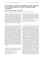
Figure 1 The cellular distribution of hA3G in P100 and S100
fractions. H9 cells and 293T cells expressing HA tagged hA3G were
lysed in hypotonic TE buffer, and the resultant S1 was ultra-
centrifuged, resulting in the P100 and the S100 fractions. The S1,
P100 and S100 fractions prepared from 293T cells were analyzed by
using a 4-35% discontinuous Opti-prep velocity gradient, as
described in Methods. A. Western blots of the S1, P100, and S100
fractions were probed with either anti-hA3G (left) or anti-HA (right)
for the samples derived from H9 cells or 293T cells, respectively.
B. Nine fractions were collected from the top to the bottom of the
gradient, then subjected to Western blot probed with anti-HA. The
fraction numbers increase from the top to the bottom of the
gradient.
Ma et al. Retrovirology 2011, 8:2
/>Page 2 of 10
We reasoned that some of the HMM form of hA3G
might result from association of soluble hA3G with lipid
rafts. To examine this, P100 was f urther analyzed by
floatation assay. After ultra-centrifugation at 100,000 × g
overnight in sucrose gradient, all the collected fractions
were subjected to Western Blot probed with anti-Caveo-
lin-1 (lipid raft marker), anti-membrane transferrin
receptor (TFR, a cytoplasm membrane bound protein)
and anti-HA. As shown in Figure 2A, total HMM hA3G
was fractionated into raft (lane 3 and 4) and non-raft
(lane 7 to 9) fractions. Approximately 30% of the HMM
form of hA3G associated with lipid rafts. Following
treatment with mild nonionic detergent octyl glucoside,
both the raft and non-raft fractions from HMM hA3G
were subjected to the velocity gradient analysis. It shows
that raft-associated h A3G was found to shift from the
HMM fraction to the LMM fracti ons, while non-raft
hA3G presented in fraction 8 at the bottom of the gra-
dient and represented the HMM compl ex reported pre-
viously (Figure 2B). These data clearly demonstrate that
a proportion (approximately 30%) of pelletable HMM
hA3G is detergent-sensitive, which represents a LMM
form of hA3G associated with lipid rafts, and the major-
ity of pelletable hA3G appears to be mature HMM
complexes.
A similar result was obtained by using cellular frac-
tionation. We treated the S1 fraction containing
hA3G with 0.5% Triton X-100 (TX-100) at 37°C,
which will also resulted in solubilization of lipid rafts
[20,21]. Following the ultra-centrifugation of the S1
fraction, Western blots of the P100 and S100 fraction s
were then probed with antibody specific for HA and
caveolin-1 respectively. As shown in Figure 2C, the
detergent treatment resulted in total release of caveo-
lin-1 from the P100 fraction to the S100 fraction.
Simultaneously, approximately 30% of pelletable
HMM hA3G (lane 6) was reduced with a correspond-
ingincreaseinthesolubleLMMhA3G(lane5).In
contrast, incubation of the S1 fraction with TX-100
at a low temperature (4°C), a condition that only
resolves cytoplasm membrane but not lipid rafts, did
not affect the distribution of either hA3G or caveolin-
1. A similar result was also obtained when H9 cells
were used in the same experiment as described above
(data not shown).
Together these data indicate that steady state hA3G in
the cytoplasm appears as three different forms: LMM
hA3G (or soluble hA3G), raft-associated LMM hA3G
(RA LMM hA3G) and HMM hA3G complex. Consis-
tent with the conclusion, a co-immunoprecipitation ana-
lysis shows that Staufen and RNA helicase A (RHA),
two components found in the HMM complex [14], on ly
associated with the HMM hA3G, but not with the
LMM or RA LMM hA3G (Figure 2D).
hA3G
TFR
Caveolin-1
S
P
TX-100
-
S
P
S
P
B
1
2
3
4
5
6
7
8
9
Opti-prep gradient
4% 35%
A
37°C4°C
C
hA3G
Co-IP with anti-HA
Staufen
RHA
1
2
3
1
2
3
4
5
6
Caveolin-1
hA3G
hA3G
123456789
TFR
raft non-raft
raft
non-raft
D
RA LMM
HMM
LMM
Octyl glucoside
-
+
-
-
+
+
TFR
Caveolin-1
+
+
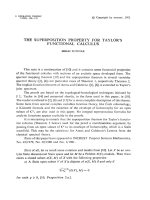
Figure 2 Steady state hA3G in the cytoplasm appears in thr ee
different forms. A. 293T cells expressing HA tagged hA3G were
lysed in hypotonic TE buffer. As described in Methods, P100 was
prepared and treated with or without nonionic detergent octyl
glucoside as indicated, then resolved by the sucrose floatation assay
into the raft and non-raft proteins. Each fraction was analyzed by
Western blot for the presence of hA3G, Caveolin-1 and TFR. B. The
raft and non-raft fractions of hA3G were collected and treated with
octyl glucoside, then resolved in the Opti-prep velocity gradient.
Western blots of each fraction were probed with anti-HA. C. 293T
cells expressing HA tagged hA3G were lysed in hypotonic TE buffer,
and the S1 fractions were either untreated (lane 1 and 2) or treated
with 0.5% Triton X-100 at 4°C (lane 3 and 4) and 37°C (lane 5 and
6), respectively. Following the ultra-centrifugation of the S1 fraction,
Western blots of the P100 and S100 fractions were then probed
with antibody specific for HA (top), TFR (middle), and caveolin-1
(bottom). S and P represent the S100 and P100 fractions,
respectively. D. Fractions that respectively contain the soluble LMM
(lane 1), RA LMM (lane 2) and HMM (lane 3), were subjected to
immunoprecipitation with anti-HA, followed by Western blots of the
immunoprecipitates probed with anti-HA, anti-RHA, and anti-
Staufen, respectively.
Ma et al. Retrovirology 2011, 8:2
/>Page 3 of 10
The RA LMM hA3G acts as the precursor of the HMM
hA3G complex
In an attempt to make a dynamic analysis of these three
forms of hA3G, 293T cells expressing HA-tagged hA3G
were labeled with [
35
S]methionine-[
35
S]cysteine for 10
min at 36 h posttransfection, followed by a chase period
with cold methionine-cysteine. Aliquots of the cells were
taken during the chase up to 3 hours, and then lysed
hypotonically as previously described. The resultant S1
supernatant was firstly fractionated into S100 and P100
fractions. The P100 fractions were further treated with
TX-100 at 37°C, and th en separated by 100,000 × g cen-
trifugation into s upernatant and pellet. These fractions,
which respectively contain the LMM, RA LMM, and
HMM hA3G, were subjected to immunoprecipitation
with anti-HA, followed by analysis of the distribution of
radioactive hA3G using one-dimensional (1-D) PAGE
(Figure 3A). The relative amount of radio-labeled hA3G
in each fraction was determined by autoradiography and
presented graphically in Figure 3B. T he total amount of
hA3G in each fraction was set as 100%. Results show that
radio-labeled hA3G was present in the LMM a nd RA
LMM, but not HMM fractions at 0-min of chase, i.e.,
after a 10-min pulse. The LMM hA3G decreased rapidly
over the first 30 minutes of chase and remained stably
thereafter, then reduced gradually after 2 hours post-
radiolabel. In contrast, the amount of RA LMM hA3G
increased during the first 30 minutes, and underwent a
significant decrease thereafter. The radio-labeled hA3G
in the HMM fraction increased gradually during the early
time of chase, reached a peak by 1 to 2 hours and then
declined. During the first 30 min chase period, the
decrease of hA3G in LMM and the simultaneous increase
of hA3G in RA LMM probably reflect a rapid movement
of newly-synthesized hA3G to the lipid rafts. The distinct
dynamics of newly-synthesized hA3G in RA LMM and
HMM indicate that the RA LMM hA3G found here is
not a breakdown product of the HMM hA3G complex,
rather it is a distinct LMM form of hA3G. After 30 min-
utes chase, the amount of radio-labeled hA3G in the
HMM compl ex increased significantly accompanied by a
great reduction in newly-synthesized hA3G in RA LMM
and the amount of soluble LMM hA3G remained stable.
It is worthy to note that total hA3G was only reduced
approximately 30% over 3 hours of chase, consistent with
previous reports [22,23]. Althou gh some LMM hA3G
were degraded during the chase, the majority of LMM
undergoing a significant decrease was most likely con-
verted into the HMM form. This suggests that the RA
LMM hA3G, inst ead of the LM M form, acts as the pre-
cursor of the HMM hA3G complex. All this reflects a
dynamic course am ong LMM, R A LMM and HMM
forms of hA3G, including a rapid movement of newly-
synthesized hA3G from the LMM form to the lipid rafts,
which serve as a precursor to the HMM hA3G complex.
The correlation between the cellular distribution and viral
incorporation of hA3G
Attempting to identify the cellular source of viral hA3G,
we first determined if a correlation existed between the
cellular distribution and viral incorporation of hA3G.
We investigated the effect of a set of truncated muta-
tions, which were described previously [6] and shown
graphically in Figure 4A, upon the overall distribution of
hA3G among the LMM form, the RA LMM form and
the mature form of HMM hA3G complex, as described
60
50
40
30
10
0
20
0
0.5
1
2
3
0
0.5
1
2
3
0
0.5
1
2
3
LMM
HMM
RA LMM
Time (hour)
relative amounts to total
radiolabeled hA3G (%)
0
0.5
1
2
3
Time (hour)
LMM
RA LMM
HMM
A
B
Total
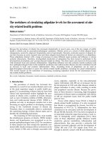
Figure 3 The RA LMM hA3G acts as the precursor of the HMM complex. 293T cells expressing hA3G were radiolabeled and chased.
Aliquots of the cells were collected, and then lysed hypotonically, as described in Methods. A. Total cell lysate and three fractions containing
LMM, RA LMM and HMM form of hA3G were subjected to immunoprecipitation with anti-HA, respectively, followed by analysis of the
distribution of radioactive hA3G using one-dimensional (1-D) PAGE. B. The relative amount of radio-labeled hA3G in each fraction was
determined by autoradiography and presented graphically. The bar graphs represent the means of results of experiments performed at least
three times, and the error bars represent standard deviations.
Ma et al. Retrovirology 2011, 8:2
/>Page 4 of 10
in Figure 2. The amounts of hA3G in these three forms
were determined by Western blot (Fig ure 4B) and gra-
phically shown in Figure 4C. It shows that hA3G miss-
ing amino acids 1-156 resulted in its failure to assemb le
into either the R A LMM or the matu re form of the
HMM complex. It is worth noting that the majority of
two C-terminal deletions of hA3G resided in the RA
LMM, i.e., approximately 70% of Δ157-384 formed the
RA LMM and only 25% assembled into the mature
HMM complex. This data suggest that the removal of
the C-terminus of hA3G may impair its ability to con-
vert the RA LMM into the mature HMM hA3G
complex.
Next, we co-transfected 293T cells with plasmids
coding for hGag and either wild-type or mutations of
hA3G described above. The expression and viral
A
1
64 101
256 298 384
Δ1-104
Δ1-156
Δ157-384
Δ246-384
hA3G
B
C
Δ1-104
Δ1-156
Δ157-384
Δ246-384
hA3G
Δ1-104
Δ1-156
Δ157-384
Δ246-384
hA3G
Δ1-104
Δ1-156
Δ157-384
Δ246-384
hA3G
LMM RA LMM HMM
0
100
20
40
60
80
Percentage of total hA3G (%)
F
Δ1-104
Δ1-156
Δ157-384
Δ246-38
4
hA3G
0
1
2
3
4
5
6
R
elative viral/cellular hA3G ratio
Δ1-104
Δ1-156
Δ157-384
Δ246-384
D
E
Cell
Gag VLP
hA3G
Δ1-104
Δ1-156
Δ157-384
Δ246-384
hA3G
Δ1-104
Δ1-156
Δ157-384
Δ246-384
hA3G
LMM
RA LMM
HMM
Figure 4 The correlat ion between the cellular distribution and viral incorporation of hA3G. A.WildtypeandmutanthA3G.Thefilled
rectangles represent the two zinc coordination units. The numbers represent the amino acid positions. B. 293T cells were co-transfected with
hGag and either wild-type or mutated forms of hA3G, and the S1 fractions of the cell lysates were subjected to ultra-centrifugation and octyl
glucoside treatment, resulting in the LMM, RA LMM and HMM forms of hA3G. The amounts of hA3G in the three forms were determined by
Western blot. C. The cellular distributions of wild type and mutant hA3G are graphically shown. D. Western blot of cell lysates were probed with
anti-HA. E. Western blot of virus like particle lysates probed with anti-HA. F. The relative amounts of mutated hA3G in the cell or viral lysates
were normalized to wild-type hA3G, and then a ratio of viral to cellular hA3G was determined and used to measure its ability to be packaged
into virions. The bar graphs in panel C and F represent the means of results of experiments performed at least three times, and the error bars
represent standard deviations.
Ma et al. Retrovirology 2011, 8:2
/>Page 5 of 10
incorporation of the hA3G variants were assessed by
Western blots of cell and virion lysates, respectively
(Figure 4D and 4E). Consistent with our previous
report [6], Western blot analysis shows that hA3G
missing amino acids 1-156 exhibited reduced incor-
poration into Gag VLPs, while the removal of the C-
terminal portion of hA3G r esulted in more efficient
viral incorporation compared to wild type hA3G. The
relative amounts of mutated hA3G in the cell or viral
lysates were normalized to wild-type hA3G, and then a
ratio of viral to cellular hA3G was determined and
used to measure its ability to be packaged into virions
(Figure 4F). A comparison of Figure 4F and Figure 4C
indicates that the amount of hA3G residing in the RA
LMM directly correlates with its ability to be incorpo-
rated into HIV-1. A similar quantitative change in the
amounts of hA3G in the RA LMM and the virions
provides further supporting evidence that the RA
LMM represents the cellular source of viral hA3G.
The ability of hA3G to bind to Gag is insufficient for its
incorporation into HIV-1
Several amino acid residues (i.e., W127) within the
N-terminal part of the linker region play an important
role in mediating the hA3G/Gag interaction. hA3G
missing in this region will not likely b ind to Gag, thus
abolishing its incorpora tion into the virions [6,18,24,25].
While another mutation hA3G, Y124A, has been
reported to posses s the ability to bind to Gag but not to
be packaged into virions [24]. To better define the role
of the RA LMM in viral i ncorporation of hA3G, we
further investigated the cellular distribution of hA3G
Y124A. As shown in Figure 5A, hA 3G Y124A was
expressed at a similar level as wild type hA3G (left
panel), whereas viral incorporation of hA3G Y124A was
reduced by 3-4 folds (central panel), which is consistent
with a previous report [24]. By co-immunoprecipitation
analysis, similar amounts of hA3G and hA3G Y124A
were detected in anti-p24 immunoprecipitates from the
cell lysates (right panel), indicating that this mutant is
able to interact with HIV-1 Gag as efficiently as wild-
type hA3G. In contrast, viral inefficient encapsidation of
W127A was mainly attributable to the loss of its inter-
action with HIV-1 Gag (Figure 5B). So, we further
determined the distribution of wild type hA3G and
hA3G Y124A among the LMM form, the RA LMM
form and the mature form of HMM hA3G complex. In
Figure 5C showed that the Y124A mutation, similar to
the N-terminal hA3G truncations, caused a significant
reductionintheRALMMandHMMcomplex.This
data suggest that, in addition to the ability to bind to
HIV-1 Gag, the cellular distribution of hA3G is also cri-
tical for its viral incorporation.
Discussion
The purposes of this study are to better characterize cel-
lular distribution of hA3G and to provide insight into
the cellular source for hA3G encapsidation into HIV-1.
In this work, we found that the majority of either endo-
genous hA3G in H9 cells, or hA3G transiently expressed
in 293T cells, resided in the P100 fraction and were
solely detected in the HMM fraction in a Opti-prep
velocity gradient. App roximately 15% of the total hA3G
appeared in the cytoplasm as a soluble form that was
found i n the S100 fraction after ultracentrifugation and
in the LMM fraction of the velocity gradient. Using the
criteria of sensitivity to the nonionic detergent octyl glu-
coside, we determined that the pelletable hA3G
A
C
Cell
hA3G
Gag
β-actin
hA3G
hA3G
Y124A
hA3G
Gag
hA3G
Y124A
hA3G
Y124A
Virion Co-IP
hA3G
Y124A
hA3G
Y124A
hA3G
Y124A
LMM RA LMM HMM
0
10
20
30
40
50
60
70
percentage of total hA3G (%)
B
Cell
hA3G
Gag
β-actin
hA3G
hA3G
W127A
hA3G
Gag
hA3G
W127A
hA3G
W127A
Virion Co-IP
Figure 5 The ability of hA3G to bind Gag is insufficient for its
incorporation into HIV-1. 293T cells were co-transfected with
plasmids coding for hGag and either hA3G Y124A (A) and W127A
(B). Left panel: Western blot of cell lysates were probed with either
anti-HA (top) or anti-b-actin (bottom). Middle panel: Western blot of
virion lysates were probed with either anti-HA (top) or anti-p24
(bottom). Right panel: Western blots of rabbit anti-p24
immunoprecipitates were probed with either anti-HA (top) or
mouse anti-p24 (bottom). C. Relative amounts of hA3G Y124A in the
LMM, RA LMM and HMM hA3G complex were determined by
Western blot and graphically presented. The bar graphs represent
the means of results of experiments performed at least three times,
and the error bars represent standard deviations.
Ma et al. Retrovirology 2011, 8:2
/>Page 6 of 10
consisted of two distinct forms: RA LMM hA3G which
were associated with lipid rafts and hA3G in the mature
HMM complex. The HMM complex contains both
Staufen and RNA helixase A, which is consistent with
previous characterization of the mature HMM hA3G
complex [14]. The results of a pulse-chase radiolabeli ng
experiment revealed that the RA LMM hA3G represents
the majority of newly-synthesized hA3G that associates
with membrane lipid raft domains, and serves as the
precursor of the HMM hA3G complex.
Although LMM hA3G can be converted to HMM
complex when CD4 T cells are activated with various
mitogens and cytokines [26,27], the mechanism by
which hA3G is regulated to assemble into different
complexes is largely unknown. Our work herein sug-
gests two essential steps during the assembly of the
HMM complex: 1) formation of the RA LMM precursor
at lipid rafts and 2) conversion of this precursor into the
mature HMM complex. For the first step, mutagenesis
studies of hA3G revealed that the removal of amino
acids 105 to 156, the linker region of hA3G, inhibited
its localization at lipid raft domains to assemble the RA
LMM and subsequent mature form of the HMM com-
plex, thus resulting in a predominantly soluble LMM
form of hA3G. Investigation of hA3G Y124A provides
further evidence supporting the importance of the linker
region for the assembly of the RA LMM and the distri-
bution of hA3G. These data together suggest that hA3G
amino acids 105-156 are required for its localization at
lipid rafts where hA3G assemble into the RA LMM, the
precursor of the HMM complex. A recent work has
identified a novel cytoplasmic retention signal (CRS)
within the linker region of hA3G [28]. The CRS residing
within amino acids 113-128 is necessary and sufficient
to retain hA3G in the cytoplasm. We reason that the
CRS may be involved in the lipid rafts localization of
hA3G and thereby restricts hA3G to the cytoplasm.
Further studies are still needed to fine-map the motif
within hA3G required for its cytoplasmic retention and
lipid rafts localization, and to determine if a correlation
exists between these two parameters.
It is worthy to note that all of the hA3G C-terminal
deletions tested become pelletable and no soluble LMM
form was detected. One explanation for this observation
is a r apid conversion of the mutants from LMM to
HMM. Alternatively, other groups and we have reported
that N-terminal fragments of hA3G are inherently
unstable [6,29], it is thus possible that the LMM form of
these mutants has been degraded rapidly and association
with lipid raft may increase their stability. In addition,
significant accumulation of the RA LMM fo rm of
mutants suggests that the assembly of the mature HMM
hA3G complex may involve sequences further down
stream of hA3G 105-156; the removal of the C-terminus
of hA3G may therefore impair its ability to convert the
RA LMM into the HMM complex.
Previous works have shown that reduced cellular
expression of the hA3G C-terminal truncations did not
result in a corresponding decrease in its viral incorpora-
tion [6]. This suggests that viruses may recruit hA3G
from a particular intracellular pool, i.e., the cellular dis-
tribution of hA3G may strongly influence its viral incor-
poration. Indeed, the fact that hA3G Y124A is able to
bind to Gag in vitro, but fails to be packaged into vir-
ions, suggests that some other properties of this pro tein,
such as specific cellula r localization, are also required
for the interacti on of these two molecule s occurring in
vivo. Khan et al. reported that inability of the mutant to
be packaged may result from its failure to associate with
lipid rafts [10] . Studies of vi ral incorporation of trun-
cated hA3G show that its ability to be packaged into vir-
ions directly corre lates with its concentration in the RA
LMM hA3G co mplex (Figure 4), suggesting that the RA
LMM acts as a cellular source for hA3G virion encapsi-
dation. In agreement with this hypothesis, hA3G Y124A
that is deficient in its ability to form the RA LMM and
HMM complexes (the precursor and mature forms), is
unable to be packaged into virions, even though it is
able to interact with Gag as efficiently as wi ld type
hA3G (Figure 5). Furthermore, the movement of newly-
synthesized hA3G to lipid raft domains to rapidly form
the RA LMM is consistent with a previous finding that
hA3G is incorporated into the virion shortly after its
synthesis in cytoplasm [17]. These data together indicate
that the RA LMM hA3G complex acts as the cellular
source for its virion encapsidation.
Since HIV-1 Gag c oncentrates in the mult ivesicular
bodies (MVB)/late endosomal compartments enriched
in lipid rafts during v irion assembly, one explanation for
the role of the RA LMM hA3G in the incorporation is
the localization in lipid rafts, where both Gag and hA3G
concentrate, thereby interacting with each other. In
agreement with the hypothesis, hA3G has been shown
to associate with intracellular memb rane rafts, and more
specifically, late endosomal vesicles [5].
Conclusions
This work thus provides the first evidence for the exis-
tence of RA LMM hA3G complex and leads toward a
better understanding of the regulation of hA3G regard-
ing its antiviral and cellular functions. The potential
implications of this work for the development of anti-
HIV therapeutics i nclude either enhanced viral incor-
poration of hA3G by accumulation of the RA LMM
complex, or increased accumulation of the LMM com-
plex by blocking its localization at lipid rafts, which may
produce a Vif-resistant post-entry inhibition on HIV-1
replication found in resting T cells.
Ma et al. Retrovirology 2011, 8:2
/>Page 7 of 10
Methods
Plasmid construction
The hGag pla smid, which encodes the HIV-1 Gag
sequence, produces mRNA whose codons have been
optimized f or mammalian codon usage, and was a kind
gift from G Nabel, NIH [30]. The construction of wild-
type and mutant forms of hA3G has been previously
described [6]. hA3G Y124A was constructed using a
site-directed mu tagenesis kit (Stratagene) and confirmed
by sequencing.
Cell, transfection and virus purification
The culture and transfection of HEK-293T cells with
these plasmids using Lipofectamine 2000 (Invitrogen,
Carlsbad, California), and the isolation of virions 48 h
posttransfection from the cell supernatant, were done as
previously described [6,31]. Unless stated otherwise,
293T cells were transfected with 1 μgofhGagand1μg
of plasmid coding for wild type or mutant forms of
hA3G. The total amount of plasmid DNA used for
transfection was kept constant in contro ls by replacing
plasmid coding for hA3G with the empty vector,
pcDNA3.1.
Protein Analysis
Cellular and viral proteins were extracted with RIPA
buffer ( 10 mM Tris, pH 7.4, 100 mM NaCl, 1% sodium
deoxycholate, 0.1% SDS, 1% NP40, 2 mg/ml aprotinin, 2
mg/ml leupeptin, 1 mg/ml pepstatin A, 100 mg/ml
PMSF). Equal amounts of protein (determined by a
Bio-Rad assay) were analyzed by SDS PAGE (10% acry-
lamide), followed by blotting onto nitrocellulose mem-
branes (Amersham Pharmacia). Western b lots were
probed with the following antibodies that are specifically
reactive with: HIV-1 capsid ( Zepto Metrocs Inc.), HA,
TFR (Invitrogen) and caveolin-1 (Santa Cruz Biotechnol-
ogy Inc.), b-actin (Sigma), RNA helicase (a gift from
Chen Liang [32]), Staufen (a gift from Andrew Mouland
[33]). Detection of proteins was performed by enhanced
chemiluminescence (NEN Life Sciences Products), using
as secondary antibodies anti-mouse and anti-rabbit,
both obtained from Amersham Life Sciences. Bands in
Western b lots were quantitated using the ImageJ 1.35 s
automated digitizing system (NIH).
Subcellular fractionation
293T cells were lysed 48 h post-transfection at 4°C in
hypotonic medium; lysis was done by Dounce homoge-
nization in 1. 0 ml of hypotonic TE buffer (20 mM Tris-
HCl, pH 7.4, 1 mM EDTA, 0.01% ß-mercaptoethanol)
supplemented with protease inhibitor cocktail (Com-
plete; Boehringer Mannheim) and RNase inhibitors
(Amb ion). The cell homogenate was then centrifuged at
1,500 × g for 30 minutes to remove nuclei and unbro-
ken cells. The supern atant (S1) was then centrifuged for
1 h at 100,000 × g in an SW55Ti rotor (Beckman,
Columbia, Md.) at 4°C, resulting in the supernatant
(S100) and the pellet (P100). To resolve cytoplasmic
membrane or lipid rafts, the S100 and the P100 sus-
pended in 1 ml of h ypotonic TE buffer was incubated
with 0.5% Triton X-100 at 4°C or 37 °C for 1 5 minutes,
respectively.
Resolution of hA3G into the LMM and HMM forms
was performed, using a 4-35% discontinuous Opti-prep
velocity gradient. This gradient was prepared in advance
by layering 0.5 ml of 35%, 0.5 ml of 30%, 0.5 ml of 25%,
1.5 ml of 20%, 0.5 ml of 15%, 0.5 ml of 10%, and 0.5ml of
4% Opti-prep from bottom to top. 0.5 ml of the S1, S100
or the P100 re-suspended in hypotonic TE buffer was
layered on top of the gradient, and then centrifuged at
100,000 × g in a Beckman SW55Ti rotor for 1 h at 4°C.
Nine fractions (0.5 ml) were collected and diluted with
an equal volume of 2× TNT, an d each fraction was sub-
jected to Western blot or immunoprecipitation analysis.
Memberane floatation assay (raft association)
293T cells expressing HA tagged hA3G were lysed and
fractionated into S100 and P100 as described above. The
pellet P100 was resuspended in TNE buffer (100 mM
Tris-HCl, 600 mM NaCl, 16 mM EDTA, supplemented
with protease inhibitor cocktail and RNase inhibitors) con-
taining either 0.5% Triton X-100 or 0.5% nonionic deter-
gent n-octyl- b -D-glucopyranoside (octyl glucoside).
Following mixed with 85.5% sucrose (w/v) in TNE lysis
buffer to obtain 73% sucrose (w/v), samples were placed at
the bottom of ultracentrifuge tubes, and overlaid with 65%
(w/v) sucrose and 10% sucros e (w/v) in TNE lysis buffer.
Then samples were centrifuged at 4°C in a SW55 rotor for
16 hours at 35,000 rpm to obtain raft and non-raft asso-
ciated fractions [18,34]. Nine equal fractions were col-
lected from the top, followed by analysis of Western blot.
Immunoprecipitation assay
293T cells from 100 mm plates were collected 48 hours
post transfection, and lysed in 500 μl TNT buffer
(20 mM Tris-HCl pH 7.5, 200 mM NaCl, 1% Triton X-
100). Insoluble material was pelleted at 1800 × g for 30
minutes. Equal amounts of protein were incubated with
5 μl HA (or p24)-specific antibody for 16 hours at 4°C,
followed by the incubation with protein A-Sepharose
(Pharmacia) for two hours. The immunoprecipitate was
then washed three times with TNT buffer and twice
with phosphate-buffered saline (PBS). After the final
supernatant was removed, 30 μlof2Xsamplebuffer
(120 mM Tris HCl, pH 6.8, 20% glycerol, 4% SDS, 2%
b-mercaptoethanol, and 0.02% bromphenol blue) was
Ma et al. Retrovirology 2011, 8:2
/>Page 8 of 10
added, and the precipitate was then boiled for 5 min-
utes. After microcentrifugation, the resulting superna-
tant was analyzed using Western blots, a s previously
described [35].
Pulse-chase radiolabeling experiments
Transfected 293T cells were labeled 36 h posttransfec-
tion with 400 μCi of [
35
S]methionine-[
35
S]cysteine for
15 minutes and then chased for various lengths of time
in Dulbecco modified Eagle medium containing 10%
fetal bovine serum and 100 μM cysteine and methio-
nine. After being washed, the cells were lysed hypotoni-
cally by Dounce homogenization in 1 ml of hypotonic
TE buffer at 4°C, and the cell lysates were centrifuged at
1,500 × g for 30 min to remove nuclei and unbroken
cells. The resulting S1 supernatant (1 ml) was fractio-
nated into S100 and P100 fractions by centrifuging for 1
h at 100,000 × g in SW55Ti rotor at 4°C. The P100 frac-
tions were further treated with octyl glucoside, and then
separated by 100,000 × g centrifugation into a superna-
tant and pellet which contain RA LMM and HMM
hA3G, respectively. The immunoprecipitation with an ti-
HA was performed, followed by analys is of the distribu-
tion of radioactive hA3G using one-dimensional (1-D)
PAGE and autoradiography.
Acknowledgements
This work was supported in part by Nature Science Foundation of China
30973569 (S.C.), The National S&T Major Special Project on Major New Drug
Innovation 2009ZX09103-138 (S.C.) and 2009ZX09303-005 (X.F.Y . and C.S.),
and the Canadian Institutes for Health Research grant (S. C.).
Author details
1
Institute of Medicinal Biotechnology, Chinese Academy of Medical Science,
Beijing, PR China.
2
State Key Laboratory for Molecular Virology and Genetic
Engineering, Institute of Pathogen Biology, Chinese Academy of Medical
Science, Beijing, PR China.
3
Lady Davis Institute for Medical Research and
McGill AIDS Centre, Jewish General Hospital, Montreal, Quebec, Canada.
4
Microbiology & Immunology, McGill University, Montreal, Quebec, Canada.
Authors’ contributions
First three authors contributed equally to this work. JM, XYL and JX have
made equal contributions to conception and design, acquisition of data, and
analysis and interpretation of data. JM was involved in drafting the
manuscript. QZ, ZLL and PPJ carried out the mutation construction. FG, LYY
and XFY participated in the design of the study and performed the
statistical analysis. LXZ and JDJ were involved in revising the manuscript and
helping to draft the manuscript. SC supervised the project and commented
on the manuscript. All authors read and approved the final manuscript.
Competing interests
The authors declare that they have no competing interests.
Received: 6 September 2010 Accepted: 6 January 2011
Published: 6 January 2011
References
1. Sheehy AM, Gaddis NC, Choi JD, Malim MH: Isolation of a human gene
that inhibits HIV-1 infection and is suppressed by the viral Vif protein.
Nature 2002, 418:646-650.
2. Jarmuz A, Chester A, Bayliss J, Gisbourne J, Dunham I, Scott J,
Navaratnam N: An anthropoid-specific locus of orphan C to U RNA-
editing enzymes on chromosome 22. Genomics 2002, 79:285-296.
3. Yu X, Yu Y, Liu B, Luo K, Kong W, Mao P, Yu XF: Induction of APOBEC3G
ubiquitination and degradation by an HIV-1 Vif-Cul5-SCF complex.
Science 2003, 302:1056-1060.
4. Stopak K, de Noronha C, Yonemoto W, Greene WC: HIV-1 Vif blocks the
antiviral activity of APOBEC3G by impairing both its translation and
intracellular stability. Mol Cell 2003, 12:591-601.
5. Alce TM, Popik W: APOBEC3G is incorporated into virus-like particles by a
direct interaction with HIV-1 Gag nucleocapsid protein. J Biol Chem 2004,
279:34083-34086.
6. Cen S, Guo F, Niu M, Saadatmand J, Deflassieux J, Kleiman L: The
interaction between HIV-1 Gag and APOBEC3G. J Biol Chem 2004,
279:33177-33184.
7. Khan MA, Kao S, Miyagi E, Takeuchi H, Goila-Gaur R, Opi S, Gipson CL,
Parslow TG, Ly H, Strebel K: Viral RNA is required for the association of
APOBEC3G with human immunodeficiency virus type 1 nucleoprotein
complexes. J Virol 2005, 79:5870-5874.
8. Schafer A, Bogerd HP, Cullen BR: Specific packaging of APOBEC3G into
HIV-1 virions is mediated by the nucleocapsid domain of the gag
polyprotein precursor. Virology 2004, 328:163-168.
9. Zennou V, Perez-Caballero D, Gottlinger H, Bieniasz PD: APOBEC3G
incorporation into human immunodeficiency virus type 1 particles. J
Virol 2004, 78:12058-12061.
10. Strebel K, Khan MA: APOBEC3G encapsidation into HIV-1 virions: which
RNA is it? Retrovirology 2008, 5:55.
11. Friew YN, Boyko V, Hu WS, Pathak VK: Intracellular interactions between
APOBEC3G, RNA, and HIV-1 Gag: APOBEC3G multimerization is
dependent on its association with RNA. Retrovirology 2009, 6:56.
12. Bach D, Peddi S, Mangeat B, Lakkaraju A, Strub K, Trono D: Characterization
of APOBEC3G binding to 7SL RNA. Retrovirology 2008, 5:54.
13. Chiu YL, Greene WC: APOBEC3G: an intracellular centurion. Philos Trans R
Soc Lond B Biol Sci 2009, 364:689-703.
14. Gallois-Montbrun S, Holmes RK, Swanson CM, Fernandez-Ocana M,
Byers HL, Ward MA, Malim MH: Comparison of cellular ribonucleoprotein
complexes associated with the APOBEC3F and APOBEC3G antiviral
proteins. J Virol 2008,
82:5636-5642.
15.
Wang T, Tian C, Zhang W, Luo K, Sarkis PT, Yu L, Liu B, Yu Y, Yu XF: 7SL
RNA Mediates Virion Packaging of the Antiviral Cytidine Deaminase
APOBEC3G. J Virol 2007, 81:13112-13124.
16. Kozak SL, Marin M, Rose KM, Bystrom C, Kabat D: The anti-HIV-1 editing
enzyme APOBEC3G binds HIV-1 RNA and messenger RNAs that shuttle
between polysomes and stress granules. J Biol Chem 2006,
281:29105-29119.
17. Soros VB, Yonemoto W, Greene WC: Newly synthesized APOBEC3G is
incorporated into HIV virions, inhibited by HIV RNA, and subsequently
activated by RNase H. PLoS Pathog 2007, 3:e15.
18. Khan MA, Goila-Gaur R, Kao S, Miyagi E, Walker RC, Strebel K: Encapsidation
of APOBEC3G into HIV-1 virions involves lipid raft association and does
not correlate with APOBEC3G oligomerization. Retrovirology 2009, 6:99.
19. Wang X, Dolan PT, Dang Y, Zheng YH: Biochemical differentiation of
APOBEC3F and APOBEC3G proteins associated with HIV-1 life cycle. J
Biol Chem 2007, 282:1585-1594.
20. Yu J, Fischman DA, Steck TL: Selective solubilization of proteins and
phospholipids from red blood cell membranes by nonionic detergents. J
Supramol Struct 1973, 1:233-248.
21. Halwani R, Khorchid A, Cen S, Kleiman L: Rapid localization of Gag/GagPol
complexes to detergent-resistant membrane during the assembly of
HIV-1. J Virol 2003, 77:3973-3984.
22. Marin M, Rose KM, Kozak SL, Kabat D: HIV-1 Vif protein binds the editing
enzyme APOBEC3G and induces its degradation. Nat Med 2003,
9:1398-1403.
23. Sheehy AM, Gaddis NC, Malim MH: The antiretroviral enzyme APOBEC3G
is degraded by the proteasome in response to HIV-1 Vif. Nat Med 2003,
9:1404-1407.
24. Huthoff H, Malim MH: Identification of amino acid residues in APOBEC3G
required for regulation by human immunodeficiency virus type 1 Vif
and Virion encapsidation. J Virol 2007, 81:3807-3815.
25. Zhang KL, Mangeat B, Ortiz M, Zoete V, Trono D, Telenti A, Michielin O:
Model structure of human APOBEC3G. PLoS One 2007, 2:e378.
Ma et al. Retrovirology 2011, 8:2
/>Page 9 of 10
26. Kreisberg JF, Yonemoto W, Greene WC: Endogenous factors enhance HIV
infection of tissue naive CD4 T cells by stimulating high molecular mass
APOBEC3G complex formation. J Exp Med 2006, 203:865-870.
27. Stopak KS, Chiu YL, Kropp J, Grant RM, Greene WC: Distinct patterns of
cytokine regulation of APOBEC3G expression and activity in primary
lymphocytes, macrophages, and dendritic cells. J Biol Chem 2007,
282:3539-3546.
28. Bennett RP, Presnyak V, Wedekind JE, Smith HC: Nuclear Exclusion of the
HIV-1 host defense factor APOBEC3G requires a novel cytoplasmic
retention signal and is not dependent on RNA binding. J Biol Chem 2008,
283:7320-7327.
29. Conticello SG, Harris RS, Neuberger MS: The Vif protein of HIV triggers
degradation of the human antiretroviral DNA deaminase APOBEC3G.
Curr Biol 2003, 13:2009-2013.
30. Huang Y, Kong W-P, Nabel GJ: Human immunodeficiency virus type I-
specific immunity after genetic immunization is enhanced by
modification of Gag and Pol expression. J Virol 2001, 75:4947-4951.
31. Guo F, Cen S, Niu M, Saadatmand J, Kleiman L: The inhibition of tRNA
Lys3
-
primed reverse transcription by human APOBEC3G during HIV-1
replication. J Virol 2006, 80:11710-11722.
32. Roy BB, Hu J, Guo X, Russell RS, Guo F, Kleiman L, Liang C: Association of
RNA helicase a with human immunodeficiency virus type 1 particles. J
Biol Chem 2006, 281:12625-12635.
33. Mouland AJ, Mercier J, Luo M, Bernier L, DesGroseillers L, Cohen EA: The
double-stranded RNA-binding protein Staufen is incorporated in human
immunodeficiency virus type 1: evidence for a role in genomic RNA
encapsidation. J Virol 2000, 74:5441-5451.
34. Ono A, Freed EO: Plasma membrane rafts play a critical role in HIV-1
assembly and release. Proc Natl Acad Sci USA 2001, 98:13925-13930.
35. Khorchid A, Halwani R, Wainberg MA, Kleiman L: Role of RNA in facilitating
Gag/Gag-Pol Interaction. J Virol 2002, 76:4131-4137.
doi:10.1186/1742-4690-8-2
Cite this article as: Ma et al.: The cellular source for APOBEC3G’s
incorporation into HIV-1. Retrovirology 2011 8:2.
Submit your next manuscript to BioMed Central
and take full advantage of:
• Convenient online submission
• Thorough peer review
• No space constraints or color figure charges
• Immediate publication on acceptance
• Inclusion in PubMed, CAS, Scopus and Google Scholar
• Research which is freely available for redistribution
Submit your manuscript at
www.biomedcentral.com/submit
Ma et al. Retrovirology 2011, 8:2
/>Page 10 of 10