Báo cáo y học: "No evidence of XMRV in prostate cancer cohorts in the Midwestern United States" potx
Bạn đang xem bản rút gọn của tài liệu. Xem và tải ngay bản đầy đủ của tài liệu tại đây (766.63 KB, 11 trang )
RESEARCH Open Access
No evidence of XMRV in prostate cancer cohorts
in the Midwestern United States
Toshie Sakuma
1
, Stéphane Hué
2
, Karen A Squillace
1
, Jason M Tonne
1
, Patrick R Blackburn
1
, Seiga Ohmine
1
,
Tayaramma Thatava
1
, Greg J Towers
2
and Yasuhiro Ikeda
1*
Abstract
Background: Xenotropic murine leukemia virus (MLV)-related virus (XMRV) was initially identified in prostate cancer
(PCa) tissue, particularly in the prostatic stromal fibroblasts, of patients homozygous for the RNASEL R462Q
mutation. A subsequent study reported XMRV antigens in malignant prostatic epithelium and association of XMRV
infection with PCa, especially higher-grade tumors, independently of the RNASEL polymorphism. Further studies
showed high prevalence of XMRV or related MLV sequences in chronic fatigue syndrome patients (CFS), while
others found no, or low, prevalence of XMRV in a variety of diseases including PCa or CFS. Thus, the etiological link
between XMRV and human disease remains elusive. To address the association between XMRV infection and PCa,
we have tested prostate tissues and human sera for the presence of viral DNA, viral antigens and anti-XMRV
antibodies.
Results: Real-time PCR analysis of 110 PCa (Gleason scores >4) and 40 benign and normal prostate tissues
identified six positive samples (5 PCa and 1 non-PCa). No statistical link was observed between the presence of
proviral DNA and PCa, PCa grades, and the RNASEL R462Q mutation. The amplified viral sequences were distantly
related to XMRV, but nearly identical to endogenous MLV sequences in mice. The PCR positive samples were also
positive for mouse mitochondrial DNA by nested PCR, suggesting contamination of the samples with mouse DNA.
Immuno-histochemistry (IHC) with an anti-XMRV antibody, but not an anti-MLV antibody that recognizes XMRV,
sporadically identified antigen-positive cells in prostatic epithelium, irrespectively of the status of viral DNA
detection. No serum (159 PCa and 201 age-matched controls) showed strong neutralization of XMRV infection at
1:10 dilution.
Conclusion: The lack of XMRV sequences or strong anti-XMRV neutralizing antibodies indicates no or very low
prevalence of XMRV in our cohorts. We conclude that real-time PCR- and IHC-positive samples were due to
laboratory contamination and non-specific immune reactions, respectively.
Background
Prostate cancer (PCa) is the most frequently diagnosed
noncutaneous malignancy among men in industrialized
countries [1]. Although early detection using tests for
prostate-specific antigen and improved treatment have
emerged as important interventions for decreasing PCa
mortality, there is potential for improved prognosis
through detection of genetic risk factors. Indeed, a posi-
tive family history is among the strongest epidemiologi-
cal risk factors for PCa, and a number of genetic
mutations have been implicated in PCa. For example, an
R462Q polymorphism in the R Nase L protein, which
impairs the catalytic activity of an important effector of
the innate antiviral response, has been implicated in up
to 13% of unselected PCa cases [2].
Xenotropic murine leukemia virus (MLV)-related virus
(XMRV) was first identified in PCa tissues, particularly
those with the homozygous RNASEL R462Q mutation
[3]. Genetic analysis identified XMRV as a xenotropic
gammaretrovirus, closely related to those found in mice
[4,5]. This suggested that XMRV represented a zoonotic
transmission from mice to humans. When compared
with exogenous and endogenous MLV sequences,
XMRV appeared to have a unique, conserved 24 bp
deletion in the gag leader region [3]. However, this
* Correspondence:
1
Department of Molecular Medicine, Mayo Clinic, Rochester, MN 55905 USA
Full list of author information is available at the end of the article
Sakuma et al. Retrovirology 2011, 8 :23
/>© 2011 Sakuma et a l; licensee BioMed Central Ltd. This is an Open Access article distributed under the terms of the Cr eative Commons
Attribu tion License ( which permits unrestricted use, distribution, and repro duction in
any medium, provided the original work is properly cited.
deletion has recently been found in endogenous MLV
proviruses in a variety of mice [6]. Initially, immuno-his-
tochemistry (IHC) and FISH analyses suggested that
only prostatic stromal fibroblasts were infected with
XMRV [3]. Subsequently, Schlaberg, S ingh and collea-
gues reported the expression of XMRV antigens in 23%
of PCa and an association of XMRV infection with
higher grade tumors [7]. Contrary to the initial study,
Singh’s study found viral antigen-positive cells primar ily
in malignant prostatic epithelium, independently of the
RNASEL polymorphism [7]. It is notable that this study
found many immuno-histochemistry-positive samples
which did not have detectable XMRV DNA [7]. Another
study found 11 (27.5%) of 40 PCa patients with XMRV
neutralizing antibodies [8]. Importantly, there were cor-
relations between serum positivity and nested PCR
results, FISH, or the R462Q RNASE L mutation [8]. In
sharp contrast, several recent reports found no or very
low prevalence of XMRV (DNA, RNA or antibodies) in
PCa samples [9-12].
If the role of XMRV in PCa is confirmed, detection
and prevention of XMRV infection could provide a
novel intervention strategy for ea rly diagnosis and treat-
ment of PCa. However, the conflicting epidemiological
data have made it unclear whether XMRV plays a role
in PCa and have questio ned whether the virus is truly a
human pathogen. In this study we have sought to
address the association between XMRV infection and
PCa, PCa grades and RNASEL R462Q polymorphism by
testing prostate tissues for the presence of XMRV. In
additio n, to determine the correlation between PCa and
seroprevalence of XMRV, serum sa mples from pat ients
with PCa were compare d with age-mat ched controls for
detectable anti-XMRV antibodies. Our s tudy found no
XMRV sequences and no XMRV-neutralizing antibodies
in 150 prostate tissues (110 PCa and 40 benign/normal)
and serum samples (159 PCa and 201 age-matched con-
trols), respectively, indicating no or very low prevalence
of XMRV in our cohorts. We did detect MLV sequences
in 6 samples, but these samples were also PCR positive
for mouse mitochondrial DNA suggesting DNA contam-
ination as a source of the MLV. We were therefore
unable to confirm the links between XMRV infection
with PCa, PCa grades or RNASEL mutation.
Results
Prevalence of XMRV proviral DNA in PCa
We have previously developed a real-time PCR assay
for detection of XMRV gag sequences [13,14]. Tests
using the XMRV infectious molecular clone plasmid,
pcDNA3.1(-)/VP62, could detectasinglecopyofthe
XMRV genome in 1.0 μg of total cellular DNA (approxi-
mately 1.4 × 10
5
cell s). The primers and the pr obe used
in this assay were designed to detect most MLV-related
sequences from mice. Using this sensitive real-time PCR
assay, we screened DNA from 150 prostate tissues (110
PCa and 40 benign/normal controls). One out of 40
high grade PCa (Gleason score 8-10), 4 out of 70 inter-
mediate grade PCa (Glea son score 5-7), and 1 out of 40
benign/normal p rostate tissues (Gleason score <4) were
repeatedly positive by this assay (Table 1). The viral
DNA copy n umbers ranged from 0.5 to 11 copies per
1.0 μg DNA (average of 4 reactions). As one diploid cell
contains approximately 7.1 pg of DNA, we estimate that
PCR-positive clinical samples had 0.5 to 11 copies of
proviral DNA in 1.4 × 10
5
cells.
To confirm the real-time PCR results, we screened the
same DNA samples by nested PCR for XMRV/MLV gag
sequences. In order to establish consistency and to
minimize the risk of contamination during the proce-
dure, three individuals independently performed the
nested PCR experiments using independently aliquoted
DNA samples. Four out of 6 real-time PCR po sitive
samples (#15, 51, 52 and 112) were consistently positive
by the nested PCR analysis, while the other two positive
samples from intermediate grade PCa (#53 and 103)
were shown to be nested PCR-positive twice in the first
three attempts. Further analysis confirmed that these
two samples were nested PCR-positive for viral DNA.
The 144 real-time PCR-negative samples were also
found to be negative by nested PCR.
No statistical link between the presence of viral DNA and
prostate cancer or higher tumor grade
We then sought a corre lation between viral DNA detec-
tion and the presence of PCa. There was no statistical
difference between the frequency of PCR positivity in
PCa and in benign/normal controls (Table 2). We also
examined a link between PCR positivity and tumor
grade as measured by the Gleason score. Using total of
110 DNA samples from PCa, 4 out of 70 intermediate
grade (Gleason score 5-7) and 1 out of 40 high grade
(Gleason score 8-10) were positive by real time PCR
(Table 3). These data were not statistically significant by
Table 1 Prevalence of XMRV and tumor grade
No.
a
Positive
b
Low grade
c
40 1
Gleason 5 2 0
Gleason 6 22 3
Gleason 7 46 1
Gleason 8 16 1
Gleason 9 23 0
Gleason 10 1 0
a
Total number of samples tested from each Gleason score.
b
Number of PCR positive samples from each Gleason score.
c
Low grade includes Gleason score 1 through 4.
Sakuma et al. Retrovirology 2011, 8 :23
/>Page 2 of 11
chi-square (x
2
) as indicated in Table 3, suggesting no
correlation between the prevalence of viral DNA and
higher tumor grade in our samples.
No correlation between viral DNA detection and RNASEL
R462Q mutation
In order to consider the association between RNASEL
mutation and viral infection, we amplified part of the
RNASEL gene by PCR and determined the status of the
R462Q RNASEL polymorphism. Of 150 prostate tissues,
20 cases were found to be homozygous for RNASEL
R462Q (Table 4). However, these samples were all nega-
tive for vi ral DNA by real-time PCR. Thus there was no
linkage between viral DNA detection and RNASEL
R462Q in our clinical samples (Table 5).
Phylogenetic analyses of MLV-like sequences in prostate
tissue DNA
XMRV has been PCR amplified from prostate cancer
samples in a number of studies [3,8,12] as well as in
blood samples from patients with chronic fatigue syn-
drome (CFS) [15]. Furth ermore, a recent study reported
a high frequency of MLV that was distinct from XMRV
by PCR in patients with chronic fatigue syndrome [16].
To examine the viral sequences identified in our PCa
samples, we cloned the PCR-amplified DNA bands from
four viral DNA-positive pat ien t samples (#15 [GenBank
no. JF288880, JF288881], #5 1 [GenBank no. JF2888 78,
JF288879], #52 [GenBank no. JF288 882, JF288883] and
#112 [GenBank no. JF288884]) and determine d their
nucleotide sequences. We were able to identify two
independent sequences from each of patients #15, #51
and #52 and a single sequence from patient #112. To
compare these sequences to XMRV and to previously
published MLV sequences from mice and patient sam-
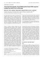
ples, w e reconstructed Bayesian phylogenies (Figure 1).
None of the gag gene sequences amplified f rom our
clinical samples belonged to the clade formed by pre-
viously reported XMRV sequences; instead, they clus-
tered with known polytropic murine leukemia virus
(PMLV), modified polytropic murine leukemia virus
(MPMLV) or xenotropic murineleukemiavirus(MLV-
X) endogenous sequences of mice (Figure 1). Impor-
tan tly, one of the patients (#52) appeared to be infected
with two independent MLVs, one from the modified
polytropic MLV clade and one from the xenotropic
MLV clade. A similar result was seen when a maximum
likelihood phylogeny was constructed using the software
RAxML [17] (not shown). In e ach case, BLAST analys is
of the amplified sequences identified at least one endo-
genous MLV sequence in the mouse genome with very
high (>99%) similarity (Table 6). Two of the five frag-
ments were identical to known endogenous proviruses
and t he other three were greater than 99% similar.
These proviruses ex ist in multiple locations within the
mouse genome.
Because the sequences we amplified were similar to
the MLV sequences detected in CFS patients [16], we
also analyzed the sequences reported in that study. The
sequences amplified from CFS patients also fell into
both polytropic and modified polytropic clades of endo-
genous MLVs (Figure 1). They were also very similar
(98-100%) to known endogenous MLV proviruses in
mice (Table 7). In fact, the differences between the
amplified sequences and the endogenous sequences are
consistent with known error rates of Taq polymerase or
could also be explained by polymorphisms between
mice [18-20].
Table 2 Statistical analysis of XMRV positivity in controls
and PCa
No.
a
Positive
b
x
2c
Non-PCa
d
40 1 0
PCa
e
110 5 0.319
a
Total number of samples tested from each Gleason score.
b
Number of PCR positive samples from each Gleason score.
c
Statistical results from chi-square (x
2
) tests.
d
Samples from benign/normal cancer patients.
e
Samples from prostate cancer patients.
Table 3 Statistical analysis of XMRV prevalence and
tumor grade
No.
a
Positive
b
x
2c
Low grade 40 1 0
Intermediate 70 4 0.606
High grade 40 1 0
a
Total number of samples tested from each Gleason score.
b
Number of PCR positive samples from each Gleason score.
c
Statistical results from chi-square (x
2
) tests.
Table 4 RNASEL genotyping and tumor grade
Normal/benign
a
Intermediate
b
High
c
Total
d
RNASEL RR 11 28 15 54
RNASEL RQ 21 36 19 76
RNASEL QQ 8 6 6 20
a
Samples with Gleason score 1 through 4.
b
Samples with intermediate Gleason score.
c
Samples with high Gleason score.
d
Total numbers of each RNASEL genotypes.
Table 5 Statistical analysis of XMRV prevalence and
RNASEL genotyping
XMRV+
a
XMRV-
b
x
2c
RNASEL RR+RQ 6 124 0
RNASEL QQ 0 20 0.962
a
XMRV positive samples from real time PCR.
b
XMRV negative samples from real time PCR.
c
Statistical results from chi-square (x
2
) tests.
Sakuma et al. Retrovirology 2011, 8 :23
/>Page 3 of 11
0.53
0.63
0.80
1.00
0.91
0.81
0.78
0.99
0.73
0.65
0.87
1.00
0.98
0.89
1.00
0.92
1.00
0.97
1.00
0.88
1.00
1.00
MLV-
X
XMRV
MPMLV
PMLV
0.73
1.00
Figure 1 Bayesian maximum clade credibility phylogeny of endogenous murine MLV sequences, 22Rv1 cell line and patient derived
MLV gag gene sequences. Sequences derived from PCa samples in this study are colored red. Sequences from [16] are colored blue. The tree
is rooted against the Moloney MLV sequence. Bayesian posterior probabilities above 0.50 are indicated on the corresponding branches. The scale
bar represents the number of nucleotide substitutions per site.
Sakuma et al. Retrovirology 2011, 8 :23
/>Page 4 of 11
The similarity between the patient amplified sequences
and known endogenous MLV provirus sequence in mice
suggests that the MLVs may have been amplified from
samples t hat had been inadvertently contaminated with
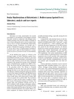
mouse DNA. To examine this possibility further in our
samples we tested each positive PCa sample for the pre-
sence o f mouse mitochondrial DNA by PCR. Strikingly,
all of the clinical samples that were po sitive for MLV
were also positive for mouse mitochondrial DNA
(Figure 2). When the amplified DNA fragments were
cloned and sequenced, they were 100% identical to Mus
musculus cytochrome b gene sequence. Thus, the MLV
sequences detected by sensitive PCR methods in patient
samples likely originated from contaminating mouse
DNA encoding endogenous MLV proviruses.
Detection of XMRV antigens in PCa tissues
Previous IHC studies found XMRV antigen-positive cells
in prostatic stromal fibroblasts [3] or in malignant pro-
static epithelium [7]. Importantly, Schlaberg, Singh and
colleagues also showed frequent detection of viral anti-
gen-positive cells in PCR-negative tissues [7]. In order
to seek viral antigen-positive cel ls in our clinical sam-
ples, we prepared prostate tissue sections and performed
IHC analysis. We used the rabbit anti-XMRV antibody,
which was used in the previous study by Schlaberg et al.
[7]. We also used a goat anti-MLV p30/gp70 antibody,
which can detect XMRV precursor Gag, CA, and Env
proteins in XMRV transfected cells [13,14]. Both
antisera showed clear and reproducible staining of 293T
cells transfected with the infe ctious XMRV clone VP62
(Figure 3A) or XMRV-producing 22Rv1 cells (data not
shown). No specific staining was seen when uninfected
control 293T cells were stained with these antisera
(Figure 3A). We prepared tissue sections of four PCR-
positive tissues (Gleason scores 6 and 8) as well as two
PCR negative tissues (Gleason scores 6 and 8, real-time/
nested PCR-double negative), and analyzed them with
the two antisera. The anti-XMRV antibody sporadically
detected antigen-positive cells, exclusively in prostatic
epithelium, in the sections of tissues (Figure 3B, upper
middle panel with FITC). Similar results were observed
with a different secondary antibody conjugated with
Texas Red (Figure 3C). In contrast, no signal was
detected with the anti-MLV p30/gp70 in any of the tis-
sue sections (Figure 3B). Importantly, the anti-MLV
serum did not stain the cells, which were shown to be
IHC-positive by the anti-XMRV serum, in the serial sec-
tions of the same tissue (Figure 3B, upper panels). It was
also notable that the anti-XMRV serum found antigen-
positive cells in PCR-negative tissue sections (Figure 3C),
suggesting that this serum also recognizes a non-viral
protein. Similar results were recently reported by Switzer
et al [21]. Considering the data obtained using the anti-
MLV serum, we conclude that we cannot detect XMRV
in prostate cancer tissues and that the antibody described
by Schlaberg, Singh and colleagues recognizes non-viral
proteins in addition to XMRV.
Table 6 Comparison of MLV sequences amplified from patient samples with mouse genomic sequences
Sequence (GenBank no.) Length (nt) Closest relative GenBank no. Similarity Nucleotide difference
51_PCR_LF2_GagR (JF288878) 608 Mus musculus BAC clone RP23-457E5 AC121813 100% 0/608
51_PCR_LF3_GagR (JF288879) 250 Mus musculus chrom 7, clone RP24-220N8 AC167466 99% 1/250
15_PCR_LF2_GagR (JF288880) 608 Mus musculus BAC clone RP23-152O2 AC163634 100% 0/608
15_PCR_LF3_GagR (JF288881) 271 Mus musculus BAC clone RP23-152O2 AC163634 >99% 1/271
52_PCR_GagF_GagR (JF288882) 525 Mouse DNA sequence, clone CH29-187G15 CU407131 100% 0/525
52_PCR_LF2_GagR (JF288883) 540 Mus musculus chrom 5, clone RP23-280N22 AC123679 >99% 1/540
112_PCR_LF2_GagR (JF288884) 691 Mouse DNA sequence, CH29-187G15 CU407131 >99% 6/691
NB: Gaps are treated as mismatches .
Table 7 Comparison of MLV sequences amplified from patient samples [16] with mouse genomic sequences
Sequence Length (nt) Closest relative GenBank no. Similarity Nucleotide difference
HM630557 319 Mouse endogenous retrovirus M26006 99% 4/310
HM630558 698 Mus musculus BAC clone RP23-115O21 AC163617 99% 7/698
HM630559 698 Mouse DNA sequence from clone RP23-131N18 AL772224 99% 1/697
HM630560 697 Mouse endogenous retrovirus M26005 99% 3/698
HM630561 339 Mouse DNA sequence, clone CH29-187G15 CU407131 99% 6/339
HM630562 698 Mus musculus BAC clone RP23-115O21 AC163617 99% 5/697
Patient amplified MLV sequences were used as a BLAST query to identify their closest relative. The accession numbers of the mouse genomic sequences
identified are shown as are the number of nucleotide differences between the patient amplified sequence and their nearest relatives in the mouse genome.
Sakuma et al. Retrovirology 2011, 8 :23
/>Page 5 of 11
Absence of XMRV antibodies in patients with PCa and
age-matched controls
Serological testing was performed with a recently devel -
oped XMRV neutralizing assay which measures vi ral
neutralizing activity using a GFP-encoding XMRV and
flow cytometry [14]. Positive seroreactivity was defined
as 100% block of XMRV-GFP transduction with a
10-fold diluted serum sample. We randomly sampled
159 PCa cases out of 933 patients who are consenting,
age 50-70 and have a clinical Gleason Score of 6 or
7 (most common) in the Mayo Clinic Prostate SPORE
Biospecimen files. 201 sera from age-matched patients
without PCa or any known urological disorders were
included as non-PCa controls. As positive controls, we
used anti-XMRV sera from XMRV-infected wild mice,
Mus pahari [14]. Sera from XMRV-infected mice
diluted 10-fold completely blocked XMRV-GFP infec-
tion (Figure 4A) . In contrast , none of the clinical sam-
ples showed strong anti-XMRV act ivity at 10-fold
dilution (Figure 4B). Two out of 159 PCa (Figure 4C)
and five out of 201 non-PCa (Figure 4D) sera marginally
reduced the XMRV infectivity (over 80% block of
XMRV infectivity at a 10-fold dilution). However, by
Western blot probing cell lysates from XMRV-infected
and uninfected cells, these patients’ sera failed to detect
XMRV Env, Gag or p30 Capsid (data not shown). To
rule out the possibility that these patients’ sera cannot
detect denatured XMRV proteins by Western blotting,
we also performed the indirect immunofluorescent assay
using HeLa cells (control) and XMRV-infected HeLa
cells as antigens. None of the sera could detect XMRV
antigens in HeLa cells at 50- a nd 200-fold dilutions
(data not shown). We, therefore, conclude that XMRV
antibodies are absent from our patient population.
Discussion
In this study, we have examined the prevalence of
XMRV in patients with or without PCa at Mayo Clinic.
We were unable to find XMRV sequences or anti-
XMRV antibodies in our patients, most of whom are
from the mid-west area of the USA, indicating that
there is no or very low prevalence of XMRV in this
region. Moreover, we were unable to confirm the corre-
lation between XMRV infection and PCa, higher tumor
grade or RNASEL R462Q mutation.
A high prevalence of XMRV has been reported in
patients with PCa and chronic fatigue syndr ome (CFS)
in the USA [3,7,8], but similar studies in Europe have
failed to detect XMRV [10-12]. It has been suggested
that geographical differences might explain this striking
variationinXMRVprevalence[11]butourresults,as
well as recent U S studies that also find no evidence for
XMRV [9,21], appear to rule this explanation out. In
this regard, it is notable that previous studies to identify
XMRV in patients with PCa or chronic fatigue syn-
drome have relied on very sensitive PCR detection
methods. Because of the high similarity between
patient associated XMRV/MLV and endogenous MLV
sequences and the str iking discordance between studies,
it has been suggested that PCR-positive results might be
attributed to unintentional detection of contaminating
mouse DNA in human specimens [6,22-24]. It is notable
that Lo et al. [16] detected polytropic and modified
polytropic MLV sequences, but not XMRV, in blood
samples from chronic fatigue patients (Figure 1). These
authors were unable to ident ify the samples as contami-
nated using mouse mitochondrial PCR. In our study,
real-time PCR and nested PCR identified 6 of 150 sam-
ples as positive for MLV. H owever, the amplified
sequences were closely related to known endogenous
MLV proviruses, rather than XMRV. In fact one patient
sample (#52) contained two independent MLV sequ-
ences. This might be interpreted as evidence for evolu-
tion of the virus in the patient but closer analysis
reveals that one of the sequences is identical to a known
endogenous modified polytropic sequence whilst the
other is a single nucleotide different from a known
mouse endogenous xenotropic MLV. This, therefore,
suggests either infection of this patient with two inde-
pendent MLVs or PCR contamination with mouse DNA
as a source. As all of the MLV PCR-positive samples
contained detectable levels of mouse mitochondrial
DNA, we conclude that the amplified sequences origi-
nated from mouse DNA that somehow contaminated
the study samples.
In order to confirm that the viral sequences w ere
amplified fro m endogenous MLV in mouse genomic
DNA, but not replicat ing MLV in human tissue, we
attempted to determine viral integration sites. We first
used the protocol described by Kim et al. [25] but failed
to amplify DNA sequences containing the partial XMRV
LTR. We then designed universal primers to recognize
LTRs from XMRV and endogenous and exogenous
MLVs [26], as well as a series of primers specific fo r the
viral sequences identified in our clinical samples.
(bp)
#15 #51 #52 #53 #103#112
2
000-
Cont
-153 b
p
200-
100-
1000-
Figure 2 PCR for mouse mitochondrial DNA.qPCRpositive
samples (#15, 51, 52, 53, 103, 112) were PCR amplified for mouse
mitochondrial DNA. Positive samples yielded PCR products at 153
bp [16]. Water was used as a control.
Sakuma et al. Retrovirology 2011, 8 :23
/>Page 6 of 11
MRV-positive
Anti-XMRV Anti-MLV
A
XXMRV-negative
B
5
1 (GS 8)
Anti-XMRV Anti-MLVH&E
PCR-positive
#
103 (GS 6) #
5
#47 (GS 8)#112 (GS 6)
PCR-negative
PCR-positive
#
C
Anti-XMRV
Figure 3 Detection of XMRV in prostate cancer tissues. (A) Specificity of anti-XMRV antiserum and anti-MLV antibody. 293T cells transfected
with XMRV infectious plasmid (pcDNA3.1(-)/VP62) were stained with either rabbit anti-XMRV or goat anti-MLV. No positive staining was observed
in control uninfected 293T cells. (B) Serial tissue sections from qPCR positive samples, including #51 (Gleason score (GS) 8) and #103 (GS 6) were
immunostained with either anti-XMRV or anti-MLV antibody. H&E staining from each sample is also shown. (C) Serial tissue sections from qPCR
positive (#112, GS 6) and negative (#47, GS 8) samples were immunostained with anti-MLV antibody, followed by TexasRed-conjugated donkey
anti-rabbit antibody (Jackson ImmunoResearch Laboratories, Inc., 1:200).
Sakuma et al. Retrovirology 2011, 8 :23
/>Page 7 of 11
Unfortunately, we were not successful, likely due to low
viral copy numbers in the clinical samples. Very
recently, Robinson et al. [23] and Oakes et al. [22]
reported similar o bservations; all XMRV PCR-positive
specimens contained detectable levels of mouse mito-
chondrial or endogenous retroelements (IAPs). Together
with our data, these findi ngs highlight the difficulty of
avoiding DNA contamination in clinical samples and
the risk of testing contaminated samples as XMRV-posi-
tive by sensitive PCR detection assays. As a possible
source of contamination, Sato et al. [24] demonstrated
that a commercially available hot-start PCR enzyme
contained mouse DNA. We used several enzymes and
obtained similar results. Thus, it is unlikely that t he
contaminating mouse genome originated from a PCR
kit.Sincewecouldamplifytheviralsequencesfrom
multiple aliquoted DNA samples, they a ppeared to be
contaminated before or during the DNA isolation step,
most likely during tissue sectioning on a microtome.
XMRV antigen-positive cells have been detected in
prostatic stromal fibroblasts [3] or in malignant prostatic
epithelium [7]. Our IHC study using two different anti-
sera showed conflicting results. The goat anti-MLV anti-
body found no viral antigens in clinical samples, while
the rabbit anti-XMRV antibody used in the study by
Schlaberg, Singh and colleagues [7] detected antigen-
posit ive cells in pro static epithelium. Strikingly, the goat
anti-MLV serum did not stain the cells, which were
IHC-positive by the anti-XMRV rabbit serum, in serial
sections of the same tissue. The rabbit antiserum also
found antigen-positive cells in PCR-negative sections,
confi rming the observations of Schlaberg and colleagues
who reported frequent detection of IHC-positive sam-
ples in PCR-negative tissues [7]. Importantly, both the
rabbit and g oat antibodies detected XMRV in experi-
mentally infected cells with high sensitivity (Figure 3).
Together, these observations strongly suggest that the
rabbit antiserum is detecting a non-viral antigen spora-
dically expressed by tumor cells in the t issue section.
We conclude that our PCa samples do not have XMRV
antigen-expressing cells that are detectable by IHC.
We recently reported t hat Mus pahari mice elicit
potent XMRV-specific humoral immune response upon
XMRV infection [14]. At a serum dilution of 1:640, anti-
sera from infected animals almost completely blocked
XMRV infection [14]. Similarly, an animal study using
XMRV-infected rhesus macaques and sensitive ELISA
detection assays showed that infected animals rapidly
develop antibodies against XMRV proteins, including
gp70 (Env), p15E (transmembrane), and p30 (CA) [27].
These results indicate that XMRV i s strongly immuno-
genic in these animals. In contrast, we were unable to
detect strong XMRV-specific neutralizing antibodies i n
our 360 patients, age 50-70, with o r without PCa. This
observation further su ggests a lack of XMRV in our
cohorts. It is possible, although less likely, that XMRV is
not immunogenic in humans or that XMRV-specific
immune response might have disappeared in these rela-
tively elderly patients.
Conclusion
In our study population of patients with or without PCa
from the USA, we found no evidence o f infection with
XMRV using PCR, IHC and serological tests. Our nega-
tive results are in accordance with previous studies using
sensitive PCR, ELISA and Western blot assays, which
failed to detect PCR or seropositive samples in a large
number of blood donors, HTLV- and HIV-infected, or
patients with or without CFS [9-12,21,27-31]. Our results
indicate the possible false-positive detection of XMRV/
MLV-related sequences or antigen-positive cells through
XMRV-
Control
XMRV+
anti-XMRV
XMRV+
Control
PCa-1
NonPCa-1
AB
0 11.9
11.3
14 0
PCa-2 PCa-3
C
10
0
10
4
10
0
10
4
10
0
10
4
10
0
10
4
10
0
10
4
0% 100% n.a. 19.3% 15%
2.5 2.5
NonPCa
-
2
NonPCa
-
3
NonPCa
-
4
NonPCa
-
5
NonPCa
-
6
D
10
0
10
4
10
0
10
4
82.1% 82.1%
2.0
2.4
2.3 1.7
2.0
NonPCa
2
NonPCa
3
NonPCa
4
NonPCa
5
NonPCa
6
10
0
10
4
10
0
10
4
10
0
10
4
10
0
10
4
10
0
10
4
82.9
%
83.6
%
87.9
%
85.7
%
85.7
%
Figure 4 Neutralization activity of patient sera.(A)XMRV
infected 293T cells (XMRV+ control) and XMRV-infected and treated
with anti-XMRV sera at a dilution of 1:10 [14] are shown. (B) Data
from non-XMRV infected 293T cells is shown as a control. Patients
samples which did not show positive neutralization reaction
(Patients with non-prostate cancer (NonPCa)-1, Patients with
prostate cancer (PCa)-1) are shown. (C) Two samples that showed
positive reaction from patients with prostate cancer (PCa-2 and -3)
are shown. (D) Six samples that showed positive neutralization
reaction from patients with non-prostate cancer (NonPCa-2 to -6)
are shown. 1:10 dilution of sera were applied for all the
experiments. Percent GFP positive and percent neutralization are
indicated within the gated areas and below the flow data,
respectively. The percent neutralization was calculated as the
reciprocal of infectivity, with a maximum infectivity being
determined by incubation of the virus with an uninfected mouse
serum. n.a., not applicable.
Sakuma et al. Retrovirology 2011, 8 :23
/>Page 8 of 11
laboratory contamination or non-specific immune reac-
tion respectively, and underscore the need for careful
validation of previous and future studies.
Materials and methods
Prostate tissues and plasma samples from patients
Prostate tissues and plasma samples were obtained from
Mayo Clinic Biospecimen Core with an approval from
the Institutional Review Boards. Frozen sections of pros-
tate cancer tissues (10 μm) were identified as 1 through
150 in duplicates. These samples included 40 normal/
low grade Gleason score, 70 intermediate (Gleason score
5-7), and 40 high grade (Gleason score 8-10) with men
aged between 50-70 years old. For plasma analysis, total
of 360 plasma samples from 50-70 year old male
patients including 159 prostate patients (Gleason score
5-7) and 201 patients with no prostate cancer or urolo-
gical disorders were used in this study.
TaqMan qPCR
Total cellular DNA was extracted by PureLink Genomic
DNA Mini Ki t accordi ng to the manufacturer’s protocol
(Invitrogen). All samples were eluted in 50 μlofelution
buffer, and the concentration and quality of the DNA
were determined by a NanoDrop Spectrophotometer.
For the real-time PCR assay, TaqMan Universal PCR
Master Mix (Roche) was used along with 2 μlofeach
sample. Primers were used at a range of 230 nM to
300 nM final concentration. TaqMan probe #51 from
Roche Universal Prob e Library was used for XMRV-gag
at 100 nM final concentration. A standard curve was cre-
ated by using serially diluted XMRV plasmid (pcDNA3.1
(-)/VP62). The assay was analyzed by the ABI 7300 Real-
Time PCR System using the default thermal cycling
conditions for the two-step RT-PCR method and FAM
reporter [13].
Genotyping
RNASEL genotype was determined by nested PCR amplifi-
cation using outer primers 5’ -CTGGGGTTCTATGA-
GAAGCAAG-3’ and 5’ -TGAGCTTTCAGATCCTC
AAATG-3’ , and inner primers 5’-GAGAGAACAGT-
CACTTGGTGAC-3’ and 5’-CAGCCCACTTGATGCTC
TTATC-3’ with pfx polymerase (Invitrogen). Final PCR
products were purified with QIAquick PCR Purification
Kit (Qiagen) before sequence analysis.
Neutralization assay
The neutralization assay was carried out using GFP-
encoding XMRV as described previously [13,14]. Briefly,
293T cells were transfected with pcDNA3.1(-)/VP62 and
a GFP-encoding retroviral vector using FuGene 6
(Roche). Serum samples were heat inactivated at 56°C
for 30 min. A mixture of plasma samples and 2.5 × 10
4
infectious units of GFP-ca rrying XMRV were incubated
at 37°C for 30 min before infecting 293T cells (5 × 10
4
).
Three days post-infection, cells were resuspended, fixed
with 4% paraformaldehyde and analyzed by flow cyto-
metry (BD FACScan). The percentages of GFP-positive
cells were measured using CellQuestPro software [14].
Western blot analysis
For Western blot analysis of XMRV proteins, cell lysates
of prostate cancer (PC-3) cell line (ATCC) and PC-3
infected with XMRV were harvested in 1.0 ml of RIPA
lysis buffer. Cell debris was removed by centrifuga-
tion, and the supernatant was diluted with Laemmli
sample buffer containing b-mercaptoethanol. After heat-
denaturation at 95°C for 5 min, 10 μlofproteinswere
subjected to SDS-PAGE with a 4-15% gradient gel
(Bio-Rad), and transferred to a polyvinylidene diflour ide
membrane at 0.7 mA/cm
2
for 40 min. Membranes were
blocked in 5% milk/PBS, then stained with patient’ s
plasma samples diluted to 1:250, followed by anti-human
IgG (1:1000, Jackson ImmunoResearch Laboratories, Inc.).
Immuno-histochemistry
Immunohistochemistry was performed on tissue samples
from patie nts with or without prostate cancer. Section s
were fixed with 4% paraformaldehyde for 20 min and
treated with 0.3% Triton X100 for 15 min at room tem-
perature. T hey were then blocked with 5% FBS/PBS for
30 min and immunostain ed with rabbit-anti XMRV
(kindly provided by Dr. Ila Singh, University of Utah) or
goat-anti p30/gp70 (NCI H D625 CAT No. 04-0109,
LOT No. 81S000262, Quality Biotech, kindly provided
by Dr. Yasuhiro Takeuchi, UCL) at a dilution of 1:500
for 4 h at room temperature . FITC-conjugated anti-rab-
bit IgG (1:500; Amersham) or DyLight 488-conjugated
anti-goat IgG (1:500; Jackson ImmunoResearch Lab)
were applied for 2 h at room temperature. Nuclei were
then counter-stained with 4’-6-Diamidino-2-phenylin-
dole (DAPI), and analyzed by confocal microscopy
(Zeiss).
Nested PCR and sequence analysis of proviral DNA
Sequence analysis was performed as previously described
[14]. Briefly, DNA was extracted by PureLink Genomic
DNA Mini Kit (Invitrogen). Nested-PCR was performed
for XMRV gag (primers for outer gag:5’ -ACGAGTT
CGTATTCCCGGCCGCA-3’ and 5’ -CCGCCTCTTCT
TCATTGTTC-3’, primers for inner gag:5’ -GCCCATT
CTGTATCAGTTAA- 3’ and 5’ -AGAGGGTAAGGG-
CAGGGTAA-3’ ) with platinum Taq polymerase ( Cat.
no. 10966-034, Invitrogen). The resulting PCR produ cts
from a total of 4 patient samples (#15, #51, #52 and
#112) were cloned into the TOPO vector (Invitrogen).
Sequences from the two patient samples #53 and #103
Sakuma et al. Retrovirology 2011, 8 :23
/>Page 9 of 11
were not analyzed. From patients #15, 51, 52, 112, we
sequenced2,2,4,1clones,andgot2,2,2,1different
sequences, respectively. They were analyzed by DNADy-
namo (BlueTractorSoftware).
Phylogenetic analyses
Seven unique gag gene sequences (255 to 528 nt), ampli-
fied from our clinical samples (GenBank no. JF288878,
JF288879, JF288880, JF288881, JF288882, JF288883,
and JF288884), were manually aligned with previously
described murine leukemia virus gag gene sequences
(n = 79), 22Rv1 cell line derived gag sequences (1605 nt;
n = 15), XMRV gag sequences apparently amplified from
prostate cancer and CFS samples (n = 7) [6], as well as
6 MLV virus gag sequences isolated from chronic fatigue
syndrome samples [16]. Bayesian phylogenies were recon-
structed as pre viously described [6]. The Markov chain
Monte Carlo search was set to 10,000,000 iterations, with
trees sampled every 1000th generation, and with a 20%
burn in. The phylogeny of the aforementioned sequences
was also reconstructed by maximum likelihood (ML)
inference under the general time reversible model of
nucleotide substitution, with gamma-distributed rate het-
erogeneity and proportion o f invariable sites, using the
program RAxML (data not shown) [32]. The ML topol-
ogy was assessed by neighbor joining bootstrapping with
1000 replicates using the program PAUP*.
A semi-nested mouse-specific mtDNA PCR
WeusedaPCRassayformousemitochondrialDNA
reported to be able to detect 2.5 fg of mouse DNA in
thepresenceof35nghumanbackgroundDNA[16].
Using this assay, we tested whether our samples were
contaminated with mouse DNA. DNA from PCR posi-
tive samples were PCR amplified with KOD Hot Start
DNA Polymerase following the manufactures instruction
(Novagen) as described [16]. The resulting PCR frag-
ments were further c loned into the T OPO vector and
the sequences were confirmed to be identical to the
mouse cytochrome b gene by DNA BLAST.
Acknowledgements
Rabbit-anti XMRV and goat-anti p30/gp70 were kindly provided by Dr. Ila
Singh and Dr. Yasuhiro Takeuchi respectively. This work was supported by
the National Institute of Health (AI093186), Mayo Clinic Career Development
Project in Prostate SPORE grant CA91956-080013, the Mayo Foundation (YI),
Wellcome Trust senior fellowship WT090940 (GJT) European Community’s
Seventh Framework Programme (FP7/2007-2013) under the project
‘Collaborative HIV and Anti-HIV Drug Resistance Network (CHAIN)’, grant
agreement no. 223131 (SH) and the National Institute of Health Research
UCL/UCLH Comprehensive Biomedical Research Centre (GJT).
Author details
1
Department of Molecular Medicine, Mayo Clinic, Rochester, MN 55905 USA.
2
Department of Infection and Immunity, MRC Centre for Medical Molecular
Virology, University College London, 46 Cleveland St, London W1T 4JF, UK.
Authors’ contributions
TS, SH, KAS, JMT, and PRB performed experiments. TS, SH, GT and YI
designed the experiments, analyzed the data and wrote the paper. All
authors read and approved the final manuscript.
Competing interests
The authors declare that they have no competing interests.
Received: 2 February 2011 Accepted: 29 March 2011
Published: 29 March 2011
References
1. Simard J, Dumont M, Soucy P, Labrie F: Perspective: prostate cancer
susceptibility genes. Endocrinology 2002, 143:2029-2040.
2. Casey G, Neville PJ, Plummer SJ, Xiang Y, Krumroy LM, Klein EA,
Catalona WJ, Nupponen N, Carpten JD, Trent JM, et al: RNASEL Arg462Gln
variant is implicated in up to 13% of prostate cancer cases. Nat Genet
2002, 32:581-583.
3. Urisman A, Molinaro RJ, Fischer N, Plummer SJ, Casey G, Klein EA, Malathi K,
Magi-Galluzzi C, Tubbs RR, Ganem D, et al: Identification of a novel
Gammaretrovirus in prostate tumors of patients homozygous for R462Q
RNASEL variant. PLoS Pathog 2006, 2:e25.
4. Dong B, Kim S, Hong S, Das Gupta J, Malathi K, Klein EA, Ganem D,
Derisi JL, Chow SA, Silverman RH: An infectious retrovirus susceptible to
an IFN antiviral pathway from human prostate tumors. Proc Natl Acad Sci
USA 2007, 104:1655-1660.
5. Baliji S, Liu Q, Kozak CA: Common inbred strains of the laboratory mouse
that are susceptible to infection by mouse xenotropic
gammaretroviruses and the human-derived retrovirus XMRV. J Virol
84:12841-12849.
6. Hue S, Gray ER, Gall A, Katzourakis A, Tan CP, Houldcroft CJ, McLaren S,
Pillay D, Futreal A, Garson JA, et al: Disease-associated XMRV sequences
are consistent with laboratory contamination. Retrovirology 2010, 7:111.
7. Schlaberg R, Choe DJ, Brown KR, Thaker HM, Singh IR: XMRV is present in
malignant prostatic epithelium and is associated with prostate cancer,
especially high-grade tumors. Proc Natl Acad Sci USA 2009,
106:16351-16356.
8. Arnold RS, Makarova NV, Osunkoya AO, Suppiah S, Scott TA, Johnson NA,
Bhosle SM, Liotta D, Hunter E, Marshall FF, et al: XMRV infection in patients
with prostate cancer: novel serologic assay and correlation with PCR
and FISH. Urology 2010, 75:755-761.
9. Aloia AL, Sfanos KS, Isaacs WB, Zheng Q, Maldarelli F, De Marzo AM, Rein A:
XMRV: a new virus in prostate cancer? Cancer Res 2010, 70:10028-10033.
10. Verhaegh GW, de Jong AS, Smit FP, Jannink SA, Melchers WJ, Schalken JA:
Prevalence of human xenotropic murine leukemia virus-related
gammaretrovirus (XMRV) in dutch prostate cancer patients. Prostate
2010, 71(4):415-20, Epub 2010 Sep 28.
11. Hohn O, Krause H, Barbarotto P, Niederstadt L, Beimforde N, Denner J,
Miller K, Kurth R, Bannert N: Lack of evidence for xenotropic murine
leukemia virus-related virus(XMRV) in German prostate cancer patients.
Retrovirology 2009, 6:92.
12. Fischer N, Hellwinkel O, Schulz C, Chun FK, Huland H, Aepfelbacher M,
Schlomm T: Prevalence of human gammaretrovirus XMRV in sporadic
prostate cancer. J Clin Virol 2008, 43:277-283.
13. Sakuma R, Sakuma T, Ohmine S, Silverman RH, Ikeda Y: Xenotropic murine
leukemia virus-related virus is susceptible to AZT.
Virology 2010, 397:1-6.
14.
Sakuma T, Tonne JM, Squillace KA, Ohmine S, Thatava T, Peng KW,
Barry MA, Ikeda Y: Early events in retrovirus XMRV infection of the wild-
derived mouse Mus pahari. J Virol 2011, 85:1205-1213.
15. Lombardi VC, Ruscetti FW, Das Gupta J, Pfost MA, Hagen KS, Peterson DL,
Ruscetti SK, Bagni RK, Petrow-Sadowski C, Gold B, et al: Detection of an
infectious retrovirus, XMRV, in blood cells of patients with chronic
fatigue syndrome. Science 2009, 326:585-589.
16. Lo SC, Pripuzova N, Li B, Komaroff AL, Hung GC, Wang R, Alter HJ:
Detection of MLV-related virus gene sequences in blood of patients
with chronic fatigue syndrome and healthy blood donors. Proc Natl Acad
Sci USA 2010, 107:15874-15879.
17. Stamatakis A: RAxML-VI-HPC: maximum likelihood-based phylogenetic
analyses with thousands of taxa and mixed models. Bioinformatics 2006,
22:2688-2690.
Sakuma et al. Retrovirology 2011, 8 :23
/>Page 10 of 11
18. Malet I, Belnard M, Agut H, Cahour A: From RNA to quasispecies: a DNA
polymerase with proofreading activity is highly recommended for
accurate assessment of viral diversity. J Virol Methods 2003, 109:161-170.
19. Bracho MA, Moya A, Barrio E: Contribution of Taq polymerase-induced
errors to the estimation of RNA virus diversity. J Gen Virol 1998, 79(Pt
12):2921-2928.
20. Cline J, Braman JC, Hogrefe HH: PCR fidelity of pfu DNA polymerase and
other thermostable DNA polymerases. Nucleic Acids Res 1996,
24:3546-3551.
21. Switzer WM, Jia H, Hohn O, Zheng H, Tang S, Shankar A, Bannert N,
Simmons G, Hendry RM, Falkenberg VR, et al: Absence of evidence of
xenotropic murine leukemia virus-related virus infection in persons with
chronic fatigue syndrome and healthy controls in the United States.
Retrovirology 2010, 7:57.
22. Oakes B, Tai AK, Cingoz O, Henefield MH, Levine S, Coffin JM, Huber BT:
Contamination of human DNA samples with mouse DNA can lead to
false detection of XMRV-like sequences. Retrovirology 2010, 7:109.
23. Robinson MJ, Erlwein OW, Kaye S, Weber J, Cingoz O, Patel A, Walker MM,
Kim WJ, Uiprasertkul M, Coffin JM, McClure MO: Mouse DNA
contamination in human tissue tested for XMRV. Retrovirology 2010, 7:108.
24. Sato E, Furuta RA, Miyazawa T: An Endogenous Murine Leukemia Viral
Genome Contaminant in a Commercial RT-PCR Kit is Amplified Using
Standard Primers for XMRV. Retrovirology 2010, 7:110.
25. Kim S, Kim N, Dong B, Boren D, Lee SA, Das Gupta J, Gaughan C, Klein EA,
Lee C, Silverman RH, Chow SA: Integration site preference of xenotropic
murine leukemia virus-related virus, a new human retrovirus associated
with prostate cancer. J Virol 2008, 82:9964-9977.
26. Tomonaga K, Coffin JM: Structures of endogenous nonecotropic murine
leukemia virus (MLV) long terminal repeats in wild mice: implication for
evolution of MLVs. J Virol 1999, 73:4327-4340.
27. Qiu X, Swanson P, Luk KC, Tu B, Villinger F, Das Gupta J, Silverman RH,
Klein EA, Devare S, Schochetman G, Hackett J Jr: Characterization of
antibodies elicited by XMRV infection and development of
immunoassays useful for epidemiologic studies. Retrovirology 2010, 7:68.
28. Groom HC, Boucherit VC, Makinson K, Randal E, Baptista S, Hagan S,
Gow JW, Mattes FM, Breuer J, Kerr JR, et al: Absence of xenotropic murine
leukaemia virus-related virus in UK patients with chronic fatigue
syndrome. Retrovirology 2010, 7:10.
29. Hong P, Li J, Li Y: Failure to detect Xenotropic murine leukaemia virus-
related virus in Chinese patients with chronic fatigue syndrome. Virol J
2010, 7:224.
30. Hohn O, Strohschein K, Brandt AU, Seeher S, Klein S, Kurth R, Paul F,
Meisel C, Scheibenbogen C, Bannert N: No evidence for XMRV in German
CFS and MS patients with fatigue despite the ability of the virus to
infect human blood cells in vitro. PLoS One 2010,
5:e15632.
31. Erlwein O, Kaye S, McClure MO, Weber J, Wills G, Collier D, Wessely S,
Cleare A: Failure to detect the novel retrovirus XMRV in chronic fatigue
syndrome. PLoS One 2010, 5:e8519.
32. Swofford DL: PAUP*. Phylogenetic analysis using parsimony (* and other
methods). Sinauer Associates, Sunderland, MA; 19984, versions.
doi:10.1186/1742-4690-8-23
Cite this article as: Sakuma et al .: No evidence of XMRV in prostate
cancer cohorts in the Midwestern United States. Retrovirology 2011 8:23.
Submit your next manuscript to BioMed Central
and take full advantage of:
• Convenient online submission
• Thorough peer review
• No space constraints or color figure charges
• Immediate publication on acceptance
• Inclusion in PubMed, CAS, Scopus and Google Scholar
• Research which is freely available for redistribution
Submit your manuscript at
www.biomedcentral.com/submit
Sakuma et al. Retrovirology 2011, 8 :23
/>Page 11 of 11