Báo cáo y học: "WFDC1 expression identifies memory CD4 Tlymphocytes rendered vulnerable to cell-cell HIV-1 transfer by promoting intercellular adhesive junctions" pptx
Bạn đang xem bản rút gọn của tài liệu. Xem và tải ngay bản đầy đủ của tài liệu tại đây (2.43 MB, 14 trang )
RESEARC H Open Access
WFDC1 expression identifies memory CD4 T-
lymphocytes rendered vulnerable to cell-cell
HIV-1 transfer by promoting intercellular adhesive
junctions
Raymond A Alvarez, Georgina Thorborn, James L Reading, Shalini Kamu Reddy and Annapurna Vyakarnam
*
Abstract
Background: Elucidating mechanisms that promote HIV-1 transfer between CD4
+
T-lymphocytes and their
subsequent loss is of importance to HIV-1 pathogenesis. We recently reported that whey acidic protein, ps20,
promotes cell-free HIV-1 spread through ICAM-1 modulation. Since ICAM-1 is pivotal in cell conjugation and
intercellular HIV-1 transfer, this study examines ps20 effects on HIV-1 spread between T lymphocy tes.
Results: We demonstrate intrinsic ps20 variability in primary CD4
+
T-lymphocyte clonal populations and a significant
positive correlation between endogenous ps20 levels and virus transfer involving fusion resulting in a spreading
infection that could be reversed by the addition of reverse transcriptase inhibitors. Blocking anti-ps20 antibody or
siRNA mediated ps20 knockdown, significantly reduced virus transfer. Conversely, virus transfer was promoted by
ectopic ps20 expression or by exogenous addition of recombinant ps20. A higher frequency of virological synapse
formation was evident in cocultures of HIV-1 infected donor T-cells with ps20
high
v ps20
low/intermediate
targets.
Blocking ps20 inhibited T-lymphocyte conjugate formation and ICAM-1 expression, and was as potent as ICAM-1 in
inhibiting HIV-1 transfer.
Conclusions: Therefore ps20 is a novel marker of CD4
+
T-cells rendered vulnerable to HIV-1 infection by regulating
the fundamental biologic process of intercellular conjugate formation and consequently of potential importance in
HIV-1 pathogenesis.
Background
Understanding the mechanisms by which retroviruses
spread from o ne cell to another is of central importance
to disease pathogenesis as this process enables viruses to
effectively escape immune responses. Three modes of
cell contact have been described which are capable of
transmitting retrovi ruses. One mode is through the for-
mation of filopodial bridges, which are protrusions that
originate from uninfected target cells that become teth-
ere d to infected donor cell s thro ugh the surface expres-
sion of viral ENV proteins [1]. After tethering, both
MLV and HIV-1 were shown to travel along the outside
of these bridge structures onto the surface of target cells
[1]. A similar mode of retroviral transfer involves thin
elongated structures called nanotubes, which form when
two T cells come into contact and begin to move apart,
independent of virus protein expression and described
in HIV-1 transmission [2]. Lastly, a highly prevalent
mode of v irus transfer, occurs through the close apposi-
tion of infected and uninfected cells which form cellular
conjugates [3,4] leading to the formation of virological
synapses (VS). A VS forms when CD4 and HIV-1 Env
and Gag polarize to conjugate interfaces in a microtu-
bule- and actin- dependent manner, allowing for the
rapid and direct transfer of virus from infected to unin-
fected cells [3-10]. A recent study demonstrated conju-
gate formation preceding and leading to Gag
redistribution/polarization with VS formation detected
in 80% of conjugates formed [11]. Similarly, the forma-
tion of multiple conjugates precedes the formation of
multiple VS termed “ polysynapses” [12] and is
* Correspondence:
Department of Infectious Diseases, King’s College London, U.K
Alvarez et al. Retrovirology 2011, 8:29
/>© 2011 Alvarez et al; licensee BioMed Central Ltd. T his is an Open Access a rticle distri buted un der the terms of the Creati ve Commons
Attribution License ( which permits unrestricted use, distribution, and reproduction in
any medium, provide d the original work is properly cited.
postulated as an efficient mode of virus dissemination in
vivo, enabling a single infected cell to infect multiple
target cells, as observed in the cervix and lymph nodes
of SIV
+
Macaques [12].
Several host factors bey ond the HIV-1 receptor/core-
ceptor complex can regulate the process of cell-cell
HIV-1 transfer depending on whether the conjugates
formed are between CD4
+
T cells or between CD4
+
T
cells and dendritic cells. These include adhesion mole-
cules, lipid raft components, signalling molecules and
the tetraspanins [6,13-22]. More recently, our laboratory
identified a novel HIV-1 enhancing pathway, namely the
whey acidic pro tein, ps20, in memory CD4
+
Tlympho-
cytes that promotes cell-free HIV-1 replication through
the modulation of ICAM-1 surface expression [23].
Blocking endogenous ps20 suppressed HIV-1 replica-
tion, while the exogenous addition of recombi nant ps20
promoted infection. Furthermore, blocking anti-ps20 Ab
suppressed ICAM-1 surface expression [23]. Cell adhe-
sion antigens like ICAM-1 and integrins (e.g. like LFA-1
and a4b7 [17,18,24-27]), can be exploited by viruses like
HIV-1 to promote spreading infection. Specifically, bud-
ding cell-free HIV-1 particles that incorporate ICAM-1
bind target cells better through cognate LFA-1 binding
[24-27]. Additionally, ICAM-1 can promote cell-to-cell
HIV spread by stabilising virus fusion to target cells and
VS formation [17,26,27] and anti-ICAM-1 blocking anti-
body can reduce VS formation by ~30% [17]. Together,
these observations prompted us to test the hypothesis
that ps20 can promote cell-cell HIV transfer by modu-
lating ICAM-1 expression.
WFDC1/ps20 is a member of the extended whey
acidic protein (WAP) family, identified by a highly con-
served 4-disulphide core domain, which includes a num-
ber of small, secreted proteins found within mucosal
secretions [28,29]. Of the 18 human members, only
three, namely secretory lymphocyte protease inhibitor
(SLPI), Elafin and more recently ps20, have ascribed
functions. All three proteins appear multifunctional;
SLPI and Elafin possess anti-microbial activity, including
anti-HIV-1 activity, as well anti-protease and anti-
inflammatory activity [28-30]. Consequently, these pro-
teins are implicated in innate immunity by providing
broad anti-microbial cover and by negating the dama-
ging effects of host and pathogen proteases and limiting
immune activation [28-30]. To date, ps20 has not been
ascribed with anti-microbial activity or anti-protease
activity, and in contrast to SLPI and Elafin [30], ps20
promotes HIV-1 infection [23]. A previous study high-
lighted the ability of ps20 to promote wound healing,
cell migration and angiogenesis [31]. Al l these processes
require the modulation of adhesion molecules [32,33],
and therefore ps20 function is postulated to involve cell-
extracellular matrix or cell-cell interactions [31,34]. In
this paper, we provide data in support of this contention
by demonstrating that HIV-1 exploits ps20-mediated
regulation of the quality and quantity of T lymphocyte-
T lymphocyte (T-T) conjugate f ormation and ICAM-1
expression in the process of cell-cell virus transfer and
ps20 to be a novel marker of CD4+ T cells that are
highly vulnerable to HIV-1 infection.
Results
Jurkat CD4
+
T cells stably transduced to express ps20, are
rendered more susceptible to T-T HIV-1 transfer
Screening steady state ps20 mRNA in ten primary
clones from multiple donors confirmed profound het-
erogeneity in ps20 levels spanning 5 logs (Additional
file 1 figure S1A) and confirmed ps20 expression, in
the transduced J-ps20
high
cells, falls within the range
seen in primary clones. As ps20 expression in this
panel segregated naturally into three distinct clusters,
we arbitrarily assigned populations to be ps20
high
(RCN above 0.1), ps20
Intermediate
(ps20
inter
)(RCN 0.001-
0.1) and ps20
low
(RCN below 0.001). Ps20 mRNA
expression in J-ps20
high
cells was 3-logs higher than J-
ps20
inter
cells; accordingly, J-ps20
high
cultures were
clearly ps20 protein positive (Additional file 1 figure
S1B). A 23-fold higher level of infection in J-ps20
high
vs. J-ps20
inter
cells was noted i n a spreading infection
assay (Additional file 1 figure S1C). Blocking anti-ps20
Ab reduced single-cycle infection by 2.8-fold in the J-
ps20
high
population (Additional file 1 figure S1D).
These data extend previous observation that human
ps20 promotes cell-free HIV-1 infection [23].
We next probed the role of ps20 in cell-to-cell HIV
transfer using a flow cytometry assay [10,12,15] (see Fig-
ure 1A). HIV-infected WT Jurkat cells (Jwt-ps20
inter
)
served as infected donor cells. J-ps20
high
and empty vec-
tor transduced J-ps20
inter
target cells were co-cultured
with donor cells that were 40% Gag
+
following infection
with NL4-3 virus at 1:1 or 1:0.2 target:donor (T:D) cell
ratios and the percentage of Gag
+
target cells enumer-
ated at 4 (Figure 1B) and 24 hours (Figure 1C) post co-
culture. At both time points and ratios tested, a higher
proportion of Gag
+
cells were detected in J-ps20
high
cells. However, a significant 2-fold difference between
the J-ps20
high
vs. J-ps20
inter
population was only
observed at the lower T:D ratio of 1:0.2, similar to our
previous study that highlighted ps20-dependency of
HIV-1tobemostmarkedatlowviruschallengedoses
[23].
We next tested the ps20-dependency of an R5 HIV-1
strain (YU2) and additionally used a PCR-based assay to
verify infection levels. Following co-culture with YU2
infected donor cells at a 1:0.2 T:D ratio, J-ps20
high
tar-
gets had a 3-fold higher level of Gag transfer, after 4
hours compared to J-ps20
inter
target cells (Figure 1C). In
Alvarez et al. Retrovirology 2011, 8:29
/>Page 2 of 14
(
A
)
1:1 1:0.2
0.01
0.1
1
10
100
J-ps20
inter
J-ps20
high
(B)
*
uninfected target:infected cell ratio
1:1 1:0.2
0.01
0.1
1
10
100
J-ps20
inter
J-ps20
high
(C)
*
uninfected target:infected cell rati
o
J-ps20inter J-ps20hi
g
h
0.01
0.1
1
10
100
(D)
*
*
*
J-ps20inter J-ps20hi
g
h
0.01
0.1
1
10
100
(E)
*
*
*
Figure 1 Jurkat CD4
+
T cells stably transduced to express full-length human ps20 are rendered more susceptible to T-T HIV-1 transfer.
(A) Representative dot plots of dye labelled target cells co-cultured with uninfected (transfer control) or infected donor cells at 4 and 24 hours
post co-culture. (B) Mean percentage of Gag
+
J-ps20
inter
vs. J-ps20
high
target cells at 4 hours post co-culture with 36% NL4-3 Jwt-ps20
inter
donor
cells at T:D ratio of 1:1 and 1:0.2. Data represent mean of three replicate assays. (C) Mean percentage of Gag
+
J-ps20
inter
vs. J-ps20
high
target cells
at 24 hours post co-culture with 36% NL4-3 infected Jwt-ps20
inter
donor cells at T:D ratio of 1:1 and 1:0.2. Data represent mean of three replicate
assays. (D) Mean percentage of Gag
+
J-ps20
inter
vs. J-ps20
high
target cells at 4 hours post co-culture with YU2 infected Jwt-ps20
inter
donor cells at
T:D ratio of 1:0.2. Data represent mean of three replicate assays. (E) Target cells co-cultured with 40% YU2 infected donor cells were sorted for
dye-positive single cells based on both FSC height vs. width followed by SSC height vs. width, on a BD FACS Aria II cell sorter. DNA extracted
from these sorted singlet cells was subject to qDNA PCR for HIV-1 LTR. The level of HIV-1 LTR in J-ps20
inter
vs. J-ps20
high
target cells is shown
relative to b-actin expression and normalized against DNA isolated from 8E5 cells. Asterisks denotes statistically significant data as calculated
using an unpaired t-test (*P ≤0.05; **P ≤0.01; ***P ≤0.001).
Alvarez et al. Retrovirology 2011, 8:29
/>Page 3 of 14
parallel, the co-cultured populations were FACS sorted
for dye-positive single target cells and HIV-1 DNA mea-
sured in the sorted population. This sorting procedure
ensured that infection levels were determined in single
target cells, excluding possible target-target or donor-
target conjugates [35], thereby providing an accurate
estimation of infection in the infected target cells. qPCR
on these samples showed a 6 -fold higher level of HIV-1
LTR in the J-ps20
high
vs. J-ps20
inter
target cells (Figure
1D).
HIV-1 transfer into J-ps20
high
cells is fusion dependent
and leads to productive infection
Evidence exists for fusion -dependent and -independent
T-T transfer of HIV-1 [6,36,37]. To probe this in the
context of ps20, target cells were cultured with Jwt-
ps20
inter
donor cells productively infected with NL4-3 at
a T:D ratio of 1:0:2 for 4 hours in the presence or
absence of the T-20 fusion inhibitor. T-20 addition
reduced virus transfer significantly by 3-fold and 2.4-
fold in the J-ps20
inter
vs. J-ps20
high
cells, respectively
(Figure 2A). To determine productive infection [38], tar-
get cells were cultured with reverse transcription RT
inhibitors prior to co-culturing with Jwt-ps20
inter
infected donor cells at a T:D ratio of 1:0.2 and Gag
+
cell s enumerated at 4, 24, and 72 hours post co-culture.
J-ps20
high
target cells had higher infection with evidence
of progressive increase in Gag
+
cells from the 4 to 72
hour time point, whereas there was no significant virus
spread in the J-ps20
inter
population (Figure 2B). The
addition of RT inhibitors did not inhibit virus transfer
in either population at 4 hours (Figure 2B). However, a
significant reduction was observed in the J-ps20
high
population with a 1.6-fold and 3-fold reduction between
the J-ps20
high
RT-inhibitor treated and untreated popu-
lations at 24 and 72 hours respectively (Figure 2B). RT-
inhibitors have been noted not to influence HIV-1
transfer, but can inhibit Gag accumulation in prolonged
co-cultures [37]. Our findings c orroborate these o bser-
vations. We next tested if increasing the virus challenge
Figure 2 HIV-1 transfer into J-ps20
high
cellsisdependentonvirusfusionandleadstohigherlevelsofproductiveinfection.(A)J-
Ps20
high
and J-ps20
inter
target cells stained with DDAO SE vital dye were seeded at 1 × 10
5
cells per well of a 24 well plate in the presence or
the absence of 5 μg/ml of T-20 for 1 hour prior to co-cultured with 18% Jwt ps20
inter
NL4-3-infected donor cells at a T:D ratio 1:0.2. Mean
percentage of Gag
+
J-ps20
inter
vs. J-ps20
high
target cells 4 hours post co-culture is shown. Data represent mean of three replicate assays. (B) The
dye-labelled J-Ps20
high
and J-ps20
inter
target cells were seeded at 1 × 10
5
cells in the presence or the absence of 5 μM of RT-inhibitors (AZT
+Lamimidine) for 1 hour prior to co-culture with 25% NL4-3-infected donor cells at a T:D ratio of (B) 1:0.2 or (C) 1:1. The percentage of Gag
+
J-
ps20
inter
vs. J-ps20
high
target cells +/- RT inhibitors were assessed at 4, 24 and 72 hours post co-culture. Data represent the mean of three
replicate assays. Asterisks denotes statistically significant data as calculated using a paired t-test (*P ≤0.05; **P ≤0.01).
Alvarez et al. Retrovirology 2011, 8:29
/>Page 4 of 14
dose to 1:1 T:D ratio promoted virus spread in the J-
ps20
inter
cells. Figure 2C shows increas e of Gag
+
cell s at
the 1:1 ratio from the 24-72 hour time point to be
1.83% (± 0.36) to 3.43% (± 0.78) respectively in J-ps20
in-
ter
cells, versus 3.84% (± 0.45) to 9.34% (± 0.79) respec-
tively in J-ps20
high
targets . At the lower T:D ratio, Gag
+
stainingincreasedfrom1.82%(±0.13)to4.3%(±0.28)
in J-ps20
high
cells between 24-72 hours versus 0.66% (±
0.11) to 0.77% (± 0.05) in J-ps20
inter
cells (Figure 2B).
These data confirm J-ps20
inter
cells require a higher
virus challenge dose than J-ps20
high
for efficient virus
spread to be achieved in these cells.
HIV-1 transfer correlates directly with ps20 expression in
primary CD4
+
T cell clones
A panel of six CD4
+
T cell clones from multiple donors
(Additional file 1 figure S1D)wereexamined.Clones1
(ps20
low
)and6(ps20
inter
) were gut-derived isogenic
clones. Clones 3 (ps20
inter
), 7 (ps20
high
), 4 (ps20
inter
) and 8
(ps20
high
) were all blood-derived, with clones 3 and 7
being isogenic (Figure 3A). Cells were co-cultured for 4
hours with Jwt-ps20
inter
donor cells that were 60%
productively infected with the X4-HIV-1 strain, 2044 and
in each case, ps20
high
clones had a higher frequency of
Gag
+
cells as compared to the ps20
inter
or ps20
low
counter-
parts. Differences between these clone pairs were as fol-
lows: 1.6-fold between clone 2 and clone 6, 16-fold
between clone 3 and clone 7 and 5-fold between clone 4
and clone 8 (Figure 3A). Furthermore, comparison of all
the ps20 low and intermediate clones (C1, C3, C4, C6)
versus the ps20 high clones (C 7, C8) highli ghted stat isti-
cally higher virus infection of the ps20 high clones (Mann-
Whitney p = 0.0009) (Figure 3A). Indeed, a significant
positive correlation was noted between HIV-1 transfer and
ps20 mRNA expression in these clones (Two-tailed non-
parametric Spearman’s correlation, p < 0.0001, Figure 3B).
Virus transfer into primary clones was next confirmed
to be fusion-dependent resulting in spreading infection.
Representative Clone 7 (ps20
high
) was treated with either
5 μM RT-inhibitors or 5 μg/ml T-20 for 1 hour prior to
co-culturing with 2044 infected Jwt-ps20
inter
donor cells
at a T:D ratio of 1:0.2 for 48 hours. The presence of RT
inhibitors reduced Gag accumulation by 43-fold (Figure
3C). In the presence of the T-20 fusion inhibitor,
C1 C3 C6 C4 C8 C7
0.1
1
10
ps20 mRNA
Mean 0.0003 0.005 0.012 0.033 0.269 0.319
SD 0.0003 0.0001 0.007 0.004 0.039 0.081
(A)
**
*
*
***
0.0001
0.001
0.01
0.1
1
0.1
1
10
p<0.0001
r
2
=0.9685
(B)
Relative expression of ps20 mRN
A
u
ntr
ea
t
ed
RT-inhi
b
it
o
r
s
T-2
0
0.1
1
10
100
(C)
Untreated
RT-inhibitors
T-20
*
****
****
Figure 3 HIV-1 transfer correlates directly with ps20 expression levels in primary CD4
+
T cell clones. (A) Mean percentage of Gag
+
dye-
labelled ps20
low
, ps20
inter
and ps20
high
primary target CD4
+
clones 4 hours post co-culture with 40% Jwt ps20
inter
2044-infected donor cells at a
T:D ratio of 1:0.2. Mean relative copy number of ps20 mRNA of each clone is given along the x-axis. (B) Correlation coefficient comparing the
relative expression of ps20 in Clones 1,3,4,6,7,8 with their corresponding level of HIV-1 transfer 4 hours post co-culture with 40% Jwt ps20
inter
2044-infected donor cells at a T:D ratio of 1:0.2. (C) Clone 7 (ps20
high
) was used as the target population and seeded at 1 × 10
5
cells in the
presence or the absence of 5 μg/ml of T-20 or 5 μM of RT-inhibitors (AZT+Lamimidine) for 1 hour prior to co-culture with 40% 2044-infected
donor cells at T:D ratio of 1:0.2. The percentage of Gag
+
target cells 48 hours post co-culture is shown. All data represent the mean of three
replicate assays. Asterisks denotes statistically significant data as calculated using an unpaired t-test (Figure A), a two-tailed non-parametric
Spearman’s r correlation (Figure B) or paired t-test (Figure C). *P ≤0.05; **P ≤0.01; ***P ≤0.001; ****P ≤0.001.
Alvarez et al. Retrovirology 2011, 8:29
/>Page 5 of 14
inhibition was even more pronounced with a > 100-fold
reduction of Gag expression (Figure 3C). These data
confirm that virus transfer into primary ps20
inter
and
ps20
high
clones is fusion dependent and can lead to pro-
ductive infection, with more marked suppression noted
in ps20
high
cells due to higher levels of virus transfer
and spread in these cells.
Blocking endogenous ps20 inhibits HIV-1 transfer in
primary CD4
+
T cell clones
Extensive characterisation of the Dharmacon Accell
siRNA showed a consistent 50-60% specific knockdown
of ps20 mRNA with maximal effects seen in ps20
inter
populations. Accordingly, we conducted functional
knockdown studies in the Jwt-ps20
inter
and clone 3
(ps20
inter
). Both populations were treated with either
non-specific (NS) siRNA or siRNA against ps20, which
inhibi ted ps20 mRNA significantly by 62% in the Jurkat
population and by 54% in clone 3 (Figure 4A, B respec-
tively). To control for off target effects, GAPDH and
HPRT expression was a lso measured relative to b-actin
and no significant modulation of either noted in the
presence of the siRNA against ps20 (Figure 4A, B). A
reduction in ps20 expression was associated with a
ps20 GAPDH HPRT
0
25
50
75
100
125
siNS
siPs20
(A)
**
ps20 GAPDH HPRT
0
25
50
75
100
125
siNS
siPs2
0
(B)
**
Jwt C3
0.2
0.4
0.8
1.6
3.2
6.4
siNS
siPs20
(C)
*
*
Jwt C3
0.2
0.4
0.8
1.6
3.2
6.4
IgG
IG7
(D)
*
*
J
wt
C3
0.2
0.4
0.8
1.6
3.2
6.4
con
rps20
(E)
**
*
*
Figure 4 Blocking endogenous ps20 inhibits HIV-1 transfer. Jwt ps20
inter
or clone 3 (ps20
inter
) was treated with either a non-silencing (siNS)
siRNA or a WFDC1/ps20-silencing siRNA for 6 days. ps20, GAPDH and HPRT mRNA was then measured by qRT-PCR relative to b-actin expression
in either (A) Jurkat population or (B) Clone 3. Normalized relative expression was calculated in reference to siNS control. (C) 8 × 10
4
siRNA
treated cells were dye-labelled and co-cultured with 40% 2044-infected donor Jurkat cells at a T:D ratio of 1:0.2. The mean percentage of Gag+
target cells in a 4 hour transfer assay is shown. (D) 2 × 10
5
Jwt cells and C3 clone from were pre-cultured for 3 days with 5 μg/ml of either
control mouse IgG1 or the anti-ps20 Ab IG7, then washed, dye-labelled and co-cultured with 40% 2044-infected donor cells at a T:D ratio of
1:0.2 in the presence of a further addition of each Ab. Mean percentage of Gag+ ps20high target cells is shown after 4 hours of co-culture. (E) 2
×10
5
Jwt cells and C3 clone cells were cultured in the absence (control, con) or presence of 1 ug/ml of rps20 for 16 hours, washed, dye-
labelled and co-cultured with donor cells infected with 40% 2044 at a T:D cell ratio of 1:0.2. The percentage of Gag+ ps20 target cells is shown
after 4 hours of co-culture. All data represent the mean of three replicate assays. Asterisk denotes statistically significant data as calculated using
a paired t-test. *P ≤0.05; **P ≤0.01.
Alvarez et al. Retrovirology 2011, 8:29
/>Page 6 of 14
significant 34% and 28% reduction in virus transfer into
the WT Jurkat cells and clo ne 3, respectively (Figure
4C). These observations were supported by antibody-
mediated blocking experiments. A significant 29% and
36% reduction in virus transfer into Jurkat and clone C3
respectivel y was noted when cultured with anti-ps20 Ab
relative to control IgG (Figure 4D). Conversely, recom-
binant ps20 (rps20) promoted virus transfer. Cells were
pre-cultured with 1 ug/ml rps20 over-night, generated
as previously described [23], washed to remove excess
protein, then co-co-cultured with infected targets,
resulting in a significant 3.4-fold and 1.9-fold en hance-
ment of virus transfer into Jurkat and clone C3 respec-
tively (Figure 4E). Similar observations of Ab-mediated
blockade and rps20-induced transfer were also noted in
additional clones (data not shown). These data confirm
that blocking endog enous ps20 in primary CD4
+
Tcells
limits HIV-1 transfer.
ps20
high
CD4
+
T cells form a higher frequency of
conjugates, multiple conjugates and virological synapse
with HIV-1 infected donor cells
The quality and quantity of c ell-cell conjugates formed
in the presence of ps20 were next assessed. To avoid
inherent differences in primary clonal populations, these
studies were performed using the Jurkat model system.
First the number of conjugates formed was assessed.
Conjugates were define d as a target cell closely apposed
to an infected donor cell, and multiple conjugates
(MCs) as a target cell closely apposed to two or more
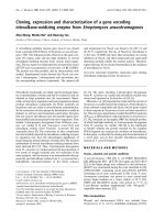
infected donor cells (Figures 5A, B, C). We observed a
significant 2.3-fold and 5.4-fold higher frequency of con-
jugate and MC formation and a 2.75-fold higher fre-
quency of Gag and CD4 polarization to conjugate
interfaces (VS formation - see Figure 5D) in J-ps20
high
vs. J-ps20
inter
populations respectively (Figure 5F). How-
ever, the proportion of conjugates containing VS was
similar, 14.6% vs. 17.3% in J-ps20
inter
vs. J-ps20
high
con-
jugates, in keeping with the notion that the number of
VS formed is determined by the number of conjugates.
Previously, it has been shown postulated that the forma-
tion of multiple virological synapses (termed polysy-
napses-PS) in conjugates of uninfected targets and HIV-
infected donors is an efficient mode of virus dissemina-
tion [12]. We, therefore, enumerated conjugates (target
or donor) containing two or more synapses simulta-
neously (Figure 5D). A marked 28-fold higher frequency
of PS i n co-cultures of J-ps20
high
vs. J-ps20
inter
cell s was
noted (Figure 5F). However, the frequency of remote
contacts (filopodial bridges and nanotubes) formed
between uninfected target and infected donor cells did
not differ between J-ps20
high
vs. J-ps20
inter
cells (Figure
5F). Interestingly, ps20
high
cells were observed to be
more closely apposed to infected donor cells compared
to ps20
inter
cells (Figure 5A vs. 5B). To quantify this
obse rvation, the medial diameter of conjugate interfaces
was measured and found to be significantly larger in
conjugates with ps20
high
targets. J-ps20
high
vs. J-ps20
inter
conjugates had a mean diameter of 7.46 uM (± 0.41) vs.
4.25 uM (± 0.23) respectively (Figure 5G). Together,
these data highlight ps20 to impact the fundamental
biologic process of cell-cell conjugation.
ps20 promotes conjugate and multiple conjugate
formation more effectively than ICAM-1
We assessed the potency of ps20- vs. ICAM-1- mediated
virus transfer and determined the relative importance of
each in T-T conjugate formation, using an si-RNA tar-
geted knock-down strategy in the Jurkat system. Treat-
ment of Jwt-ps20
inter
cells with siR NA against ICAM-1
led to a significant 50% reduction in the levels of
ICAM-1 mRNA, with no significant reduc tion of either
ps20 or GAPDH expression (Figure 6A). However,
siRNA against ps20 led to a significant 60% reduction in
ps20 mRNA, and a concomitant 40% reduction in
ICAM-1 mRNA, with no reduction in GAPDH. This
confirms our previous observations that blocking ps20
can inhibit ICAM-1 expression [23]. Surface ICAM-1
protein expression was reduced by 50% and 45% with
siRNA against ICAM-1 or ps20, respectively (Figure 6B).
Ps20 vs. ICAM-1 knockdown resulted in a 36% vs. 30%
reduction in the levels of virus transfer respectively (Fig-
ure 6C). In addition , ICAM -1 versus ps20 siRNA inhib-
ited single conjugates by 50% vs. 61% respectively
(Figure 6D). ICAM-1 siRNA inhibited multiple conju-
gates by 50% versus a marked 92% by ps20 siRNA (Fig-
ure 6D). Lastly, the size of the conjugate interface was
not affected by ICAM-1 knockdown, whereas ps20
knockdown had a small but consistent effect; a signifi-
cant 1.2-fold reduction in mean conjugate diameter
from 3.601 (± 0.1871) μminNSsiRNAtreatedcontrol
to 2.933 (± 0.2179) μm in ps20 siRNA treated cells was
noted (Figure 6E).
Discussion
Cell-to-cell HIV transfer is a significant mode of virus
spread amongst CD4
+
T cells in-vitro [3-10] and also
likely to be predominant in vivo, since memory CD4
+
T
cells are more likely to become infected while trafficking
through secondary lymphoid tissue, where lymphocyte
velocities decrease allowing for c ell-virus and cell-cell
interactions to take place [10,39,40 ]. Therefore, identify-
ing host factors that regulate this process is of impor-
tance to understanding HIV-1 dissemination in-vivo.
This paper highlights ps20 to be a novel rate-limiting
step in T-T HIV-1 transfer. We demonstrate this by uti-
lising a flow-cytometry and a PCR-based HIV transfer
ass ay in a ps20 transduced Jurkat model system, as well
Alvarez et al. Retrovirology 2011, 8:29
/>Page 7 of 14
Single conjugate ps20
inter
Single conjugate ps20
high
A)
B)
C)
5uM
2uM 2uM
2uM
2uM
2uM 2uM
D) E)
Multiple conjugates
Virological Synapse Remote contacts
Figure 5 ps20
high
CD4+ T cells form a higher frequency of conjugates, multiple conjugates and VS with HIV-1 infected donor cells.1×
10
5
DDAO vital dye labelled ps20
inter
(Figure 5A) and ps20
high
(Figure 5B) target cells were co-cultured with 60% 1 × 10
5
2044-infected donor
cells for 1 hour on Poly-L-lysine coated glass cover slips. Cells were then fixed and stained with a PE anti-Gag (red) and a FITC anti-CD4 (green)
Abs. The frequency of target cells that were involved in a single conjugate was defined as the percentage of dye-labelled target cells apposed
to an HIV-1-infected donor cell. Representative high power fields captured at 63× magnification are shown, which depict single conjugates for
between (A) J-ps20
inter
or (B) J-ps20
high
cells and HIV-infected donor cells. Left panels depict conjugates with no HIV-Gag CD4 polarization to
conjugate interface. Right panels depict conjugates with HIV-Gag and CD4 polarization (yellow) to conjugate interfaces. (C) Picture shows a
representative high power field of dye labelled targets (white arrows) involved in multiple conjugates, defined as a target cell apposed to two or
more HIV-1-infected donor cells. (D) Picture shows a representative high power field of a polysynapse, defined as a cell with two or more
virological synapses (yellow) at conjugate interfaces. (E) Picture shows a representative high power field of remote contacts (RC) (filopodial
bridges or nanotubes) that connect uninfected target cells to HIV-infected targets. (F) Mean frequency of J-ps20
inter
vs. J-ps20
high
cells involved
in single conjugate, multiple conjugates, or which contain virological synapses, polysynapses or are in contact through remote contacts are
shown. A total of 600 random target cells were assessed across quadruplicate experiments. (G) Qualitative analysis of the junction diameter of a
conjugates was measured using LEICA TCS SP2 software, where the diameter was measured across the medial section of a conjugate. Graph
depicts the mean conjugate interface diameter (μM) between J-ps20
inter
vs. J-ps20
high
cells and HIV-1-infected donor cells. A total of at least 30
conjugates per population were measured across quadruplicate experiments. Asterisk denotes statistically significant data as calculated using an
unpaired t-test (*P ≤0.05).
Alvarez et al. Retrovirology 2011, 8:29
/>Page 8 of 14
Number of XY Pairs:46
Spearman r
0.3014
95% confidence interval:0.003234 to 0.5503
P value (two-tailed): 0.0418
10
-6
10
-5
10
-4
10
-3
10
-2
10
-1
10
0
10
-7
10
-6
10
-5
10
-4
10
-3
10
-2
10
-1
(A)
ps20 RCN
ps20 ICAM-1 GAPDH
0
25
50
75
100
125
(B)
siNS
siPs20
siICAM-
1
*
*
*
*
siNS siICAM siPs20
0
25
50
75
100
125
(C)
*
**
siNS siICAM sips20
0
25
50
75
100
125
(D)
*
*
con
j
u
g
ates Multiple con
0
5
10
15
20
25
siNS
siPs20
siICAM-1
*
*
(E)
siNS siICAM-1 siPs20
0
2
4
6
8
10
(F)
*
NS
Figure 6 siRNA-mediated knockdown of ps20 inhibits conjugate and multiple conjugate formation more effectively than siRNA-
mediated knockdown of ICAM-1. (A) ps20 and ICAM-1 mRNA levels were measured in a selection of ps20
high
and
ps20inter/low
cells at 4
different time points. Data show a two-tailed non-parametric Spearman’s r correlation of all data points. CD4 T-cell Jwt ps20
inter
were treated
with either; a non-silencing (NS), ICAM-1 or WFDC1/ps20-silencing siRNA pool for 6 days. (B) After siRNA treatment the expression of ICAM-1,
ps20 and GAPDH mRNA was analyzed by qRT-PCR and relative expression to b-actin was measured. Normalized relative expression was
calculated in reference to siNS control. Data represent the mean of three replicate assays. (C) Surface expression of ICAM-1 in siRNA treated cells
is shown as assessed by standard immunofluorescence. Normalized MFI was calculated in reference to siNS control. Data represent the mean of
three replicate assays (D) 8 × 10
4
NS, ICAM-1, or ps20 siRNA-treated WT Jurkat cells were dye-labelled and co-cultured with donor 40% 2044-
infected donor cells at a T:D ratio of 1:0.2. Mean percentage of Gag
+
target cells after 4-hour co-culture is shown. Normalized % of Gag
+
target
cells was calculated in reference to siNS control. Data represent the mean of three replicate assays. (E) 5 × 10
5
siRNA treated cells were dye-
labelled and co-cultured with 5 × 10
5
60% 2044-infected donor cells. Co-cultures were incubated for 1 hour on Poly-L-lysine coated glass cover
slips, then fixed and stained with a FITC anti-Gag (Green) Ab. Conjugates and MCs were assessed as before in at least 500 random target cells
per population across triplicate experiments. (F) The panel depicts the mean conjugate interface diameter (μM) between siRNA treated Jurkat
cells and HIV-1-infected donor cells. A total of at least 30 conjugates per population were measured across triplicate experiments. Asterisk
denotes statistically significant data as calculated using a paired t-test (*P ≤0.05) in relation to NS siRNA control.
Alvarez et al. Retrovirology 2011, 8:29
/>Page 9 of 14
as in a panel of primary CD4
+
T cell clones. We report
a significant positive correlation between endogenous
ps20 expression levels and T-T virus transfer. Blocking
ps20 activity with siRNA or specific antibody signifi-
cantly inhibited T-T transfer. Conversely, gain of func-
tion studies using ps20-transduced Jurkat CD4
+
T cells
or the exogenous addition of rps20 confirmed ps20 to
promote HIV-1 transfer. We further show inhibition of
virus transfer and spread into ps20
high
cell s by the T-20
fusion inhibitor and RT inhibitors respectively, with dif-
ferences in virus spread between ps20
high
vs. ps20
low/inter
populations reaching upto 5.7-fold in Jurkat cells (Figure
2) and 8.7-fold in primary clones (Figure 3), highlighting
ps20 to be a potentially potent pathway in promoting T-
T virus dissemination.
Divergent data exist with regard to cell-cell transfer
mediated by fusion vs. endocytosis contributing to pro-
ductive HIV infection [6-8,36,37,41]. In studies using
unstimulated CD4
+
T-cells as target populations, virus
transfer through endocytosis [8,36,37] was noted. How-
ever, other studies show T-T virus transfer to be both
co-receptor- and virus fusion- dependent [6,41]. Whilst
the infected donor cell in these divergent studies was
JurkatorMolt4,akeydifferenceappearstobethe
state of target cell activation with evidence of fusion
dependent entry into activated memory CD4
+
T-cells
targets [41]. Our observat ions with activated clonal CD4
T-cells or Jurkat cells are therefore compatible with this
data. Taken together, these findings suggest that the
activation state of the target cell may account for
observed differences in the mode of virus uptake during
cell-cell virus transfer. Indeed, these differences may
account for o ther data show ing ICAM-1 and LFA-1 as
not being critical for HIV transfer to unstimulated ex
vivo CD4 T cells [38], which have been shown to take
up virus via endocytosis [8,36,37]. Thus it is reasonable
to hypothesize that, differences in the molecular deter-
minants and the mechanisms that govern virus transfer
are at least partly dependent on the state of activation
of the target CD4
+
T-cell.
An important step in HIV transfer is the conjugation
between an infected and an uninfected cell, leading to
VS formation, through which virus can be directly trans-
ferred [3-10]. A time-lapse microscopy study highlighted
that up to 80% of T-T conjugates, at some point after
conjugation/contact, form a VS [11]. Consequently, we
examin ed the role of ps20 in the quality and quantity of
T-T conjugate formation. Evidence is provided in sup-
port of ps20 promoting intercellular conjugation and VS
formation. As the overall proportion of conjugates con-
taining VS was similar between ps20
high
and ps20
low
populations, the capacity of ps20 to promote VS forma-
tion may be attributable to the protein enhancing T-cell
conjugation. In addition, we observed a higher frequency
of multiple conjugates and polys ynapses in the presen ce
of ps20. Therefore, the ability of ps20 to promote multi-
ple conjugates and polysynapses may be of critical
importance to virus dissemination in lymphoid and
mucosal tissue by allowing for fewer transient interac-
tions between cells [see [12]]. This notion is further sup-
ported by the observation that the conjugate interface
between infected donor cells and ps20
high
targets was
significantly larger compared to ps20
low
targets. These
characteristics were attributable to ps20, since knocking
down ps20 expression sign ificantly reduced the number
of single conjugates, multiple conjugates, the size of the
conjugate interface and resulting virus transfer.
Molecular determinants of cell-cell conjugate and VS
formation include the actin and microtubule cytoskeletal
networks, cell signalling, tetraspanin and lipid raft
recruitment [6,13-22,42]. In addition to the HIV-1
receptor complex, several adhesion molecules can polar-
ize to, and stabilize these supramolecular synapses
[13,15,17,20,21]. In particular, Jolly et al. showed that
anti-ICAM-1 inhibited VS by 30%, while anti-LFA-1
inhibited VS an d conjugate formation from 15-90%
depending on the blocking Ab used [17]. Previously, we
demonstrated that ps20 enhanced HIV-1 infection
through ICAM-1 modulation [23]. Here we confirm and
extend these observations. Knocking down ps20 by 60%
specifically suppressed ICAM-1 mRNA by 40% and
ICAM-1 surface staining by 45% and both ICAM-1 and
ps20 individually contributed to conjugate formation
and virus transfer. Strikingly, knocking down ps20 inhib-
ited multiple conjugate formation by 90% compared to
50% inhibition by siRN A against ICAM-1. Furthermore,
ICAM-1 did not impact the size of the conjugate inter-
face, whilst ps20 did so, albeit marginally (Figure 6E).
These data suggest that ps20-induced ICAM-1 modula-
tion though important, may not fully account for the
ps20 effect. As ps20 is implicated in regulating extracel-
lular matrix (ECM) components [31,34] and given
recent data that another retrovirus, HTLV-1, can be
captured, and then transmitted through ECM glycopro-
teins’ [43], the identification of other adhesion and ECM
targets regulated by ps20 and th eir role in cell-cell HIV-
1 transfer could enhance understanding of mechanisms
that drive HIV-1 dissemination in-vivo.
The molecular mechanisms by which ps20 regulates
ICAM-1 expression and clustering through putative
binding partners and signalling functions are part of on-
going work in our laboratory. Other work highlights a
fundamental role for ps20 in cell migration and angio-
genesis [31]. Both these processes are recognised to
modulate adhesion molecules [32,33]. As cell-cell adhe-
sion plays a significant role in successful virus infections
in general [44], it could be argued that the potency of
ps20 to promote HIV-1 infection is linked with it’ s
Alvarez et al. Retrovirology 2011, 8:29
/>Page 10 of 14
fundamental role in regulating cell adhesion. The novel
observation that CD4
+
T cells can be segregated into
stable subsets on the basis of ps20 expression coupled
with the observat ion that ps20
high
CD4
+
T cells are
more susceptible to infection than ps20
low
cells, strongly
suggests that ps20
+
CD4
+
T cells may be preferentially
targeted and lost in-vivo. Our contention is that lo cal
concentrations of ps20, in tissue such as the gut, ma y
drive HIV-1 infection a nd CD4
+
T cell loss by increas-
ing adhesion antigen expression on CD4
+
Tcells
through autocrine and paracrine effects, thereby high-
lightin g a novel role for the ancient whey acidic protein,
WFDC1/ps20, in HIV-1 pathogenesis.
Conclusions
This study highlights three novel aspects of T-T HIV
transfer. First, using three approaches to probe T-T HIV
transfer, namely, flow cytometry, PCR and confocal
microscopy, this study highlights ps20 to be a novel
host factor that promotes cell-cell conjugation and viro-
logicalsynapseformationingainandlossoffunction
assay systems. Second, one mechanism by which ps20
promotes intercellular HIV transfer is by regulating sur-
face ICAM-1 expression levels. Importantly, ps20 pro-
moted multiple cell conjugation more efficiently than
ICAM-1 and was identified to promote poly-synapse
formation. Host factors that promote poly-synapse for-
mation may be particularly potent in promoting virus
dissemination in vivo [12] and thereby impact HIV-1
pathogenesis. Thirdly, the observation that primary CD4
T-lymphocyte clones segregate naturally into distinct
subsets based on endogenous ps20 expression and that
ps20 levels correlate with intercellular HIV transfer,
identifies ps20 to a novel marker of CD4 T cells that are
vulnerable to HIV infection. Together, these observa-
tions highlight that ps20 is a novel host factor that
could promote virus d issemination by promoting T-T
cell conjugation.
Methods
CD4
+
T cells
Jurkat CCR5
+
T cells (from National Centre for Biologi-
cal Standards & Contr ols, NIBSC, UK) were maintained
in RPMI 1640 (GIBCO Invitrogen, UK) + 10% Fetal Calf
Serum (FCS) (Helena Bioscience s UK), 20 ug/ml Genta-
mycin (Sigma-Aldrich, UK). A panel of random blood
CD4+ T-cell clones generated by standard limiting dilu-
tion cloning [23,45] was screened for ps20 mRNA, and
cells were identified to be ps20
high
, ps20
inter
, ps20
low
(see
Additional file figure S1A). Additionally, two CD4
+
T
cell clones isolated from Endoscopy sections taken from
non-lesional or lesional portion of the colon of a patient
with Crohns disease (kind gift Dr. Deena Gibbons and
Prof. Adrian H ayday King’ s College London) were
identifie d to be ps20
low
and ps20
high
respectively. Clones
were maintained in RPMI 1640, 10% FCS + 10% Human
AB
+
Serum (First Link, UK), 20 ug/ml Gentamycin. A
typical feeding cycle of CD4
+
T cell clones included
activation with irradiated allogeneic PBMC (1:1 ratio) +
2 μg/ml PHA (Biostat Diagnostic Systems, Germany) +
20 IU/ml IL2 (Prol eukin, Chir on, UK) every 10-14 days.
Cells were split and fed every 3-4 days with fresh 30 IU/
ml IL2.
Stable ps20
high
Jurkat CD4
+
T cells
The WFDC1 gene was digested out from the pHA/
WFDC1 expression plasmid (kind gift D. Rowley, Baylor
University, USA) with EcoRI and cloned into an MMLV
based bi-cistronic retroviral vector, pCxCR encoding red
fluorescent protein (RFP) under the control of a cytome-
galovirus (CMV) promoter (kind gift Greg Towers, Uni-
versity College London). R etroviral particles encoding
WFDC1/RFP (pCps20CR), or RFP alone (pCxCR, empty
vector control) were made by transient transfection of
293T cells with pCxCR, or pCG9CR, along with the
packaging construct pCpg (MMLV Gag/Pol) and an
envelope construct encoding VSV-G (pMD.G) (kind gift
D. Trono, Geneva, Switzerland). Retroviral particles
were harvested 48 hrs after transfection, clarified and
titrated onto 2 × 10
5
CCR5
+
CD4
+
Jurkat cells, three
times over 3 days. Cells were sorted for RFP, expanded
and ps20 expression confirmed by qRT-PCR. The ps20
transduced population is referred to as J-ps20
high
;the
empty vector control as J- ps20
inter
.
Virus Production
The primary HIV-1 X4 strain 2044 was propagated in
PHA activated PBMC [35]. The HIV-1 full-length mole-
cular clones NL4-3 and YU2 (kind gift, M Malim, King’s
College London) were produced by the transient trans-
fection of 293T cells, using Fugene 6 reagent (Roche,
Switzerlan d). Viral stocks were standardised based on
Gag p24 conce ntrations determined by p24 ELISA (NIH
Reagents).
Antibodies
Anti-CD54 RPE (clone 15.2; Serotec, U.K.); anti-ps20
(IG7; kind gift D. Rowley); anti-ICAM-1 Ab (LB-2) (BD
Pharmingen, CA); FITC- or PE- conjugated Gag-p24
antibody (clone KC57; Beckman Coulter, UK) were
used. In addition: anti-CD4 (L120), anti-HIV ENV and
rabbit anti-HIV Gag p24 and p17 were obtained from
NIBSC, UK. Isotype-matched mAbs were from BD Phar-
mingen, CA.
Ps20 ELISA
Affinity purified polyclonal rabbit anti-ps20 antibody
(202-254) specific for residues 206-220 of the ps20
Alvarez et al. Retrovirology 2011, 8:29
/>Page 11 of 14
amino acid sequence was generated through Eurogentec
Ltd, Belgium. Nunc 96-well plates were coated over-
night at 4°C with 1 ug/mL 202-254 diluted in PBS.
Plates were blocked with PBS, 1% BSA for 2H at room
temperature (RT), washed x3 w ith PBS-0.2% T ween-20
and test samples added for 2 hours at RT. Plates were
washed 6 times to remove unbound material. Detection
was with 2 ug/ml IG7 anti-ps20 conjugated to horserad-
ish peroxidase in PBS-1% BSA-0.2% Tween-20 for 2
hours, RT followed by further 6 washes. 150 μLofsub-
strate OPD (Sigma) was added for 30 min. at room tem-
perature and stopped with 50 μL of 4 M sulphur ic acid.
Optical densities we re determined at 492 nm in a Bio-
Rad ELISA plate reader. Ps20 concentrations were deter-
mined in relation to a standard curve using recombinant
ps20 of known concentration generated in drosophila
2
.
Spreading HIV-1 infection
Cells were challenged with virus stocks standardised o n
Gag p24 concentration, and unbound virus was removed
by washing after 24 hour s. Productive infection was
monitored by intracellular staining for HIV-1 G ag p24
using a Fix and Perm kit (AD Serotec, U.K.). Cells were
fixed for 10 minutes at RT in fixation buffer, washed
once with cold PBS, then resuspended in permeabiliza-
tion buffer and a 1/10 final dilution of KC57 RD1 or
FITC added for 25 minutes at RT. Samples were washed
twice with PBS and resuspended in PBS 2%FCS, 2% par-
aformaldehyde and analysed on a FACSCalibur instru-
ment and data analysed using Flow Jo software.
Cell to cell HIV-1 transfer assay
A modified version of an assay described by Sourisseau
et al [10,15] was used. Briefly, WT Jurkat CD4
+
Tcells
(Donor) were infected with HIV-1 strains till cultures
were 10-60% Gag p24+. Targets cells comprising
ps20
high
,ps20
inter
or ps20
low
CD4
+
T cell population
were first labelled with the Cell Trace FarRed DDAO-SE
vital dye (Invitrogen UK). 1 × 10
6
/ml target cells were
incubated in PBS with 10 μM of the DDAO-SE dye for
7 minutes at 37°C, washed twice in PBS 5% FCS and co-
cultured with infected donor cells at varying ratios in a
final volume of 500 μl in a 24-well plate. Infection inhi-
bitors included were 5 μg/ml T-20 (Roche, Hertford-
shire, UK), or 5 μM each of AZT/Lamirudine (AIDS
Repository Reagent Program, MD, USA). Gag transfer
wasmeasured4hourspostco-culturebyenumerating
dye-labelled targets cells that stained positive for HIV-1
Gag p24 by flow cytometry.
siRNA knockdown
Accell siRNA smart pools targeting ps20 (E -013097-
00), ICAM-1 (E-003502-00-0010) or a non-specific tar-
geting control (D-001910-10) were purchased from
Dharmacon, CO, USA. Cell populations were washed 3
times in PBS prior to resuspension at a density of 3 ×
10
5
/ml in Accell siRNA passive uptake media contain-
ing, 3% FCS and 1 μM of specified Accell siRNA
pools. For primary clones, the medium was supple-
mented with 30 IU/ml IL-2. 3 days later cells were
washed and re-incubated at 3 × 10
4
cells/well in a 96-
well plate in a fresh aliquot of comple te passive uptake
media containing 1 μM of specified Accell siRNA pool.
3 days later, cells were washed, and target gene knock-
down efficiency was assessed by qRT-PCR before use
in functional assays.
mRNA measurement
Total RNA was extracted using an RNaeasy kit (Qiagen,
UK), then converted to cDNA (Ambion, Inc., TX, USA).
ps20 was measured using a custom designed Taqman™
assay as described previously [23]. ICAM-1 was mea-
sured using a TaqMan
®
primer and probe set
(Hs99999152_m1). ps20/ICAM-1 expression was mea-
sured relative to HPRT 1(Hs01003268_g1) or b-actin
(Hs99999903_m1) or GAPDH (Hs99999905_m1) (Taq-
Man
®
; Applied Biosystems, UK), according to manufac-
turer’s instructions, on an ABI Prism 7900 HT (Applied
Biosyste ms, CA, USA). Data was analyzed using SDS 2.3
software (Applied biosystems, CA, USA). Relative copy
number (RCN) was calculated by determining the delta
ct values, which was used in the following equation
(RCN) = 2^
(-ΔCt)
.
HIV-1 LTR measurement
DNA was extracted using a DNeasy kit (Qiagen, UK).
Primer sequences for HIV-1 LTR were; L2:
CTGTGGATCTACCACACACAAGGCTAC (forward)
and L3:GCTGCTTATATGTAGCATCTGAGGGC
(reverse) [46] and measured using TaqMan
®
probe
assay relative to b-actin (Applied Biosystems, CA, USA).
Delta Ct was first calculated, then normalized by sub-
tracting the delta ct values generated from 8E5 cells,
which contain one integrated copy of HIV-1. Relative
copy number = 2^
(-ΔΔCt)
.
Confocal microscopy
Far Red DDAO-SE dye-labelled target cells were cul-
tured with HIV-1 infected donor cells along with 5 ug/
ml anti-CD4 and anti-HIV ENV Abs on poly-L-lysine
coated glass cover slips (Sigma, UK). Cells were washed
twice with PBS, fixed for 10 minutes in 4% paraformal-
dehyde, 1% BSA in PBS at RT, washed twice with PBS
1% BSA and permeabilized using 0.2% triton X-100 for
10 minutes at RT. The cells were then blocked and
quenched in 50 mM NH
4
CL, 2% mouse serum, 2% BSA,
0.05% sodium azide. Cells were then stained with an
ant i-Gag Ab, KC57-PE (Beckman Cou lter), and an a nti-
Alvarez et al. Retrovirology 2011, 8:29
/>Page 12 of 14
CD4, L120-FITC (BD, San Jose, CA) at 1/20 total
volume dilutions in block/quench buffer for 60 minutes
at RT. After incubation, cells were washed 3× with PBS
and mounted on Superfrost Plus glass slides (VWR
International, UK), using ultramount aqueous perma-
nent mounting medium (Dako, Denmark). Slides were
allowedtodryovernightat4°Cinthedark.Thenext
day the cells were visualized by scanning laser confocal
microscopy on a Leica DM IR2 (LEICA Microsystems,
Germany) and analyzed using LEICA TCS SP2 confocal
software (LEICA, Microsystems, Germany). Images were
acquired using a 63× oil-immersion objective and pro-
cess ed using Adobe Photoshop CS (Adobe Systems, San
Jose, CA).
Statistical Analysis
Statistical analysis of data was performed using Graph
Pad PRISM Software, San Diego, CA. All p-values
reported are two-tailed. P values of less than or equal to
0.05 were deemed significant.
Additional material
Additional file 1: Figure S1. Jurkat CD4 T cells stably transduced to
express full-length human ps20 are more susceptible to cell-free
HIV-1 infection. A) The mean relative copy number (RCN) of ps20 mRNA
(relative to the HPRT house keeping gene) measured in three biological
replicate samples by qRT-PCR; in the empty vector (EV) transduced
ps20
inter
Jurkat cells, WFDC1/ps20 transduced ps20
high
Jurkat cells, wild-
type Jurkat cells as well as a panel of 10 primary CD4
+
T-cell clones (C1-
C10) is shown. The two horizontal lines are arbitrarily set to define CD4
T-lymphocyte populations as ps20 low (RCN <0.001), ps20 intermediate
(RCN 0.001-0.1) and ps20 high (>0.1RCN). (B) Mean secreted ps20 protein
levels measured in three replicate samples by a ps20 specific ELISA assay
are shown. Positive control was supernatant from the human embryonic
kidney cell line, 293T cells transfected with a WFDC1/ps20 encoding
construct (pBKps20) and supernatant from ps20 mRNA high HeLa cells;
negative control was supernatant from 293T cells transfected with empty
vector alone (pBK). Test samples included 48-hour culture supernatant
from the WFDC1/ps20 transduced population and the empty vector
transduced EV population. (C) 2 × 10
5
J-ps20
inter
or J-ps20
high
cells were
challenged with 2.5 ng/1 × 10
6
cells of the X4-tropic HIV-1 strain, NL4-3.
Productive infectio n was measured by intracellular staining for HIV-1 p24
capsid antigen and the percentage of p24 positive cells determined on
days 3, 5 and 7 post-challenge. (D) J-ps20
inter
or J-ps20
high
cells were first
pre-cultured overnight (16 hrs) in 5 μg/ml of either control mous e IgG
1
or the anti-ps20 Ab, IG7, then challenged with NL4-3 for 24 hours (10 ug
Gag p24 antigen concentration of virus stock/10
5
cells). 24 hours later
equivalent numbers of cells were trypsinized to remove surface bound
virus, washed and cell pellets lysed in PBS with 10% triton-X 100. The
amount of Gag p24 antigen was then measured by ELISA and used to
assess the fold increase in infection over background. The mean and
standard error of triplicate replicate experiments are shown.
Acknowledgements
This work was funded by a PhD studentship funded by the Medical
Research Council, UK & by grants to AV from the Guy’s & St Thomas’ Charity
and the International Consortium for Novel Anti-Virals. The authors thank
Professor M Malim for discussions and critical review of the manuscript; Dr
Nathan Shearer for expert advice on confocal microscopy and critical review
of manuscript; Dr T Tree for CD4 T cell clones from blood and Dr Deena
Gibbons and Professor AH Hayday for CD4
+
T cell clones isolated from gut
tissue. The authors thank Professor Q Sattentau and Dr C Jolly for helpful
discussions, and review of the manuscript.
Authors’ contributions
AV was involved in experimental design, hypothesis generation and
manuscript editing. RAA conducted 80% of the work outlined and wrote the
manuscript. JLR helped with qRT-PCR. GT helped with qDNA-PCR and HIV
transfer assays. SKR helped develop the ps20 ELISA. All authors read and
approved the final manuscript.
Conflict of interests
The authors declare that they have no competing interests.
Received: 18 October 2010 Accepted: 5 May 2011
Published: 5 May 2011
References
1. Sherer NM, Lehmann MJ, Jimenez-Soto LF, Horensavitz C, Pypaert M,
Mothes W: Retroviruses can establish filopodial bridges for efficient cell-
to-cell transmission. Nat Cell Biol 2007, 9:310-315.
2. Sowinski S, Jolly C, Berninghausen O, Purbhoo MA, Chauveau A, Köhler K,
Oddos S, Eissmann P, Brodsky FM, Hopkins C, Onfelt B, Sattentau Q,
Davis DM: Membrane nanotubes physically connect T cells over long
distances presenting a novel route for HIV-1 transmission. Nat Cell Biol
2008, 10:211-219.
3. Jolly C, Sattentau QJ: Retroviral spread by induction of virological
synapses. Traffic 2004, 5:643-650.
4. Sattentau Q: Avoiding the void: cell-to-cell spread of human viruses. Nat
Rev Microbiol 2008, 6:815-826.
5. Dimitrov DS, Willey RL, Sato H, Chang LJ, Blumenthal R, Martin MA:
Quantitation of human immunodeficiency virus type 1infection kinetics.
J Virol 1993, 67:2182-2190.
6. Jolly C, Kashefi K, Hollinshead M, Sattentau QJ: HIV-1 cell-to-cell transfer
across an Env-induced, actin-dependent synapse. J Exp Med 2004,
199:283-293.
7. Carr JM, Hocking H, Li P, Burrell CJ: Rapid and efficient cell-to-cell
transmission of human immunodeficiency virus infection from
monocyte-derived macrophages to peripheral blood lymphocytes.
Virology 1999, 265:319-329.
8. Chen P, Hubner W, Spinelli MA, Chen BK: Predominant mode of human
immunodeficiency virus transfer between T cells is mediated by
sustained Env-dependent neutralization-resistant virological synapses. J
Virol 2007, 81:12582-12595.
9. Sato H, Orenstein J, Dimitrov D, Martin M: Cell-to-cell spread of HIV-1
occurs within minutes and may not involve the participation of virus
particles. Virology 1992, 186:712-724.
10. Sourisseau M, Sol-Foulon N, Porrot F, Blanchet F, Schwartz O: Inefficient
human immunodeficiency virus replication in mobile lymphocytes. J
Virol 2007, 81:1000-1012.
11. Hübner W, McNerney GP, Chen P, Dale BM, Gordon RE, Chuang FY, Li XD,
Asmuth DM, Huser T, Chen BK: Quantitative 3D video microscopy of HIV
transfer across T cell virological synapses. Science 2009, 323:1743-1747.
12. Rudnicka D, Feldmann J, Porrot F, Wietgrefe S, Guadagnini S, Prévost MC,
Estaquier J, Haase AT, Sol-Foulon N, Schwartz O: Simultaneous HIV Cell-to-
Cell Transmission To Multiple Targets Through Polysynapses. J Virol 2009,
83:6234-46.
13. Nobile C, Petit C, Moris A, Skrabal K, Abastado JP, Mammano F, Schwartz O:
Covert human immunodeficiency virus replication in dendritic cells and
in DC-SIGN-expressing cells promotes long-term transmission to
lymphocytes. J Virol 2005, 79:5386-5399.
14. Vasiliver-Shamis G, Cho MW, Hioe CE, Dustin ML: Human
immunodeficiency virus type 1 envelope gp120-induced partial T-cell
receptor signalling creates an F-actin-depleted zone in the virological
synapse. J Virol 2009,
83:11341-11355.
15.
Sol-Foulon N, Sourisseau M, Porrot F, Thoulouze MI, Trouillet C, Nobile C,
Blanchet F, di Bartolo V, Noraz N, Taylor N, Alcover A, Hivroz C, Schwartz O:
ZAP-70 kinase regulates HIV cell-to-cell spread and virological synapse
formation. EMBO J 2007, 26:516-526.
Alvarez et al. Retrovirology 2011, 8:29
/>Page 13 of 14
16. Jolly C, Sattentau QJ: Human immunodeficiency virus type 1 assembly,
budding, and cell-cell spread in T cells take place in tetraspanin-
enriched plasma membrane domains. J Virol 2007, 81:7873-7884.
17. Jolly C, Mitar I, Sattentau QJ: Adhesion molecule interactions facilitate
human immunodeficiency virus type 1-induced virological synapse
formation between T cells. J Virol 2007, 81:13916-13921.
18. Arthos J, Cicala C, Martinelli E, Macleod K, Van Ryk D, Wei D, Xiao Z,
Veenstra TD, Conrad TP, Lempicki RA, McLaughlin S, Pascuccio M, Gopaul R,
McNally J, Cruz CC, Censoplano N, Chung E, Reitano KN, Kottilil S,
Goode DJ, Fauci A: SHIV-1 envelope protein binds to and signals through
integrin alpha4beta7, the gut mucosal homing receptor for peripheral T
cells. Nat Immunol 2008, 9:301-309.
19. Krementsov DN, Weng J, Lambele M, Roy NH, Thali M: Tetraspanins
regulate cell-to-cell transmission of HIV-1. Retrovirology 2009, 6:64.
20. Tsunetsugu-Yokota Y, Yasuda S, Sugimoto A, Yagi T, Azuma M, Yagita H,
Akagawa K, Takemori T: Efficient virus transmission from dendritic cells to
CD4+ T cells in response to antigen depends on close contact through
adhesion molecules. Virology 1997, 239:259-268.
21. McDonald D, Wu L, Bohks SM, KewalRamani VN, Unutmaz D, Hope TJ:
Recruitment of HIV and its receptors to dendritic cell-T cell junctions.
Science 2003, 300:1295-1297.
22. Jolly C, Sattentau QJ: Human immunodeficiency virus type 1 virological
synapse formation in T cells requires lipid raft integrity. J Virol 2005,
79:12088-12094.
23. Alvarez R, Reading J, King DF, Hayes M, Easterbrook P, Farzaneh F, Ressler S,
Yang F, Rowley D, Vyakarnam A: WFDC1/ps20 is a novel innate
immunomodulatory signature protein of human immunodeficiency virus
(HIV)-permissive CD4+ CD45RO+ memory T cells that promotes
infection by upregulating CD54 integrin expression and is elevated in
HIV type 1 infection. J Virol 2008, 82:471-486.
24. Tardif MR, Tremblay MJ: LFA-1 is a key determinant for preferential
infection of memory CD4+ T cells by human immunodeficiency virus
type 1. J Virol 2005, 79:13714-13724.
25. Liao Z, Roos JW, Hildreth JE: Increased infectivity of HIV type 1 particles
bound to cell surface and solid-phase ICAM-1 and VCAM-1 through
acquired adhesion molecules LFA-1 and VLA-4. AIDS Res Hum Retroviruses
2000, 16:355-366.
26. Fortin JF, Cantin R, Lamontagne G, Tremblay M: Host-derived ICAM-1
glycoproteins incorporated on human immunodeficiency virus type 1
are biologically active and enhance viral infectivity. J Virol 1997,
71:3588-3596.
27. Tardif MR, Tremblay MJ: Presence of host ICAM-1 in human
immunodeficiency virus type 1 virions increases productive infection of
CD4+ T lymphocytes by favoring cytosolic delivery of viral material. J
Virol 2003, 77:12299-12309.
28. Bingle CD, Vyakarnam A: Novel innate immune functions of the whey
acidic protein family. Trends Immunol 2008, 29:444-453.
29. Ranganathan S, Simpson KJ, Shaw DC, Nicholas KR: The whey acidic
protein family: a new signature motif and three-dimensional structure
by comparative modeling. J Mol Graph Model
1999, 17:106.
30. Moreau T, Baranger K, Dadé S, Dallet-Choisy S, Guyot N, Zani ML:
Multifaceted roles of human elafin and secretory leukocyte proteinase
inhibitor (SLPI), two serine protease inhibitors of the chelonianin family.
Biochimie 2008, 90:284-295.
31. McAlhany SJ, Ressler SJ, Larsen M, Tuxhorn JA, Yang F, Dang TD,
Rowley DR: Promotion of angiogenesis by ps20 in the differential
reactive stroma prostate cancer xenograft model. Cancer Res 2003,
63:5859-5865.
32. Berrier AL, Yamada KM: Cell-matrix adhesion. J Cell Physiol 2007,
213:565-573.
33. Hynes RO, Lively JC, McCarty JH, Taverna D, Francis SE, Hodivala-Dilke K,
Xiao Q: The diverse roles of integrins and their ligands in angiogenesis.
Cold Spring Harb Symp Quant Biol 2002, 67:143-153.
34. Larsen M, Ressler SJ, Lu B, Gerdes MJ, McBride L, Dang TD, Rowley DR:
Molecular cloning and expression of ps20 growth inhibitor. A novel
WAP-type “four-disulfide core” domain protein expressed in smooth
muscle. J Biol Chem 1998, 273:4574-4584.
35. Ruggiero E, Bona R, Muratori C, Federico M: Virological consequences of
early events following cell-cell contact between human
immunodeficiency virus type 1-infected and uninfected CD4+ cells. J
Virol 2008, 82:7773-7789.
36. Blanco J, Bosch B, Fernández-Figueras MT, Barretina J, Clotet B, Esté JA:
High level of coreceptor-independent HIV transfer induced by contacts
between primary CD4 T cells. J Biol Chem 2004, 279:51305-51314.
37. Bosch B, Grigorov B, Senserrich J, Clotet B, Darlix JL, Muriaux D, Este JA: A
clathrin-dynamin-dependent endocytic pathway for the uptake of HIV-1
by direct T cell-T cell transmission. Antiviral Res 2008, 80:185-193.
38. Puigdomenech I, Massanella M, Cabrera C, Clotet B, Blanco J: On the steps
of cell-to-cell HIV transmission between CD4 T cells. Retrovirology 2009,
6:89.
39. Bousso P, Robey EA: Dynamic behavior of T cells and thymocytes in
lymphoid organs as revealed by two-photon microscopy. Immunity 2004,
21:349-355.
40. Celli S, Garcia Z, Bousso P: CD4 T cells integrate signals delivered during
successive DC encounters in vivo. J Exp Med 2005, 202:1271-1278.
41. Martin N, Welsch S, Jolly C, Briggs JA, Vaux D, Sattentau QJ: Virological
synapse-mediated spread of human immunodeficiency virus type 1
between T cells is sensitive to entry inhibition. J Virol 2010, 84:3516-3527.
42. Vasiliver-Shamis G, Cho MW, Hioe CE, Dustin ML: Human
immunodeficiency virus type 1 envelope gp120-induced partial T-cell
receptor signaling creates an F-actin-depleted zone in the virological
synapse. J Virol 2009, 83:11341-11355.
43. Pais-Correia AM, Sachse M, Guadagnini S, Robbiati V, Lasserre R, Gessain A,
Gout O, Alcover A, Thoulouze MI: Biofilm-like extracellular viral assemblies
mediate HTLV-1 cell-to-cell transmission at virological synapses. Nat Med
2010, 16:83-89.
44. Stewart PL, Nemerow GR: Cell integrins: commonly used receptors for
diverse viral pathogens. Trends Microbiol 2007, 15:500-507.
45. Vyakarnam A, Eyeson J, Teo I, Zuckerman M, Babaahmady K,
Schuitemaker H, Shaunak S, Rostron T, Rowland-Jones S, Simmons G,
Clapham P: Evidence for a post-entry barrier to R5 HIV-1 infection of
CD4 memory T cells. AIDS 2001, 15:1613-1626.
46. Wu Y, Marsh JW: Selective transcription and modulation of resting T cell
activity by preintegrated HIV DNA. Science 2001, 293:1503-1506.
doi:10.1186/1742-4690-8-29
Cite this article as: Alvarez et al.: WFDC1 expression identifies memory
CD4 T- lymphocytes rendered vulnerable to cell-cell HIV-1 transfer by
promoting intercellular adhesive junctions. Retrovirology 2011 8:29.
Submit your next manuscript to BioMed Central
and take full advantage of:
• Convenient online submission
• Thorough peer review
• No space constraints or color figure charges
• Immediate publication on acceptance
• Inclusion in PubMed, CAS, Scopus and Google Scholar
• Research which is freely available for redistribution
Submit your manuscript at
www.biomedcentral.com/submit
Alvarez et al. Retrovirology 2011, 8:29
/>Page 14 of 14