Báo cáo y học: " Phylogenetic analysis consistent with a clinical history of sexual transmission of HIV-1 from a single donor reveals transmission of highly distinct variants" docx
Bạn đang xem bản rút gọn của tài liệu. Xem và tải ngay bản đầy đủ của tài liệu tại đây (1008.47 KB, 14 trang )
RESEARC H Open Access
Phylogenetic analysis consistent with a clinical
history of sexual transmission of HIV-1 from a
single donor reveals transmission of highly
distinct variants
Suzanne English
1
, Aris Katzourakis
2
, David Bonsall
3
, Peter Flanagan
1
, Anna Duda
1
, Sarah Fidler
3
, Jonathan Weber
3
,
Myra McClure
3
, SPARTAC Trial Investigators
1
, Rodney Phillips
1,4,5†
and John Frater
1,4,5*†
Abstract
Background: To combat the pandemic of human immunodeficiency virus 1 (HIV-1), a successful vaccine will need
to cope with the variability of transmissible viruses. Human hosts infected with HIV-1 potentially harbour many viral
variants but very little is known about viruses that are likely to be transmitted, or even if there are viral
characteristics that predict enhanced transmission in vivo. We show for the first time that genetic divergence
consistent with a single transmission event in vivo can represent several years of pre-transmission evolution.
Results: We describe a highly unusual case consistent with a single donor transmitting highly related but distinct
HIV-1 variants to two individuals on the same evening. We confirm that the clustering of viral genetic sequences,
present within each recipient, is consistent with the history of a single donor across the viral env, gag and pol
genes by maximum likelihood and Bayesian Markov Chain Monte Carlo based phylogenetic analyses. Based on an
uncorrelated, lognormal relaxed clock of env gene evolution calibrated with other datasets, the time since the
most recent common ancestor is estimated as 2.86 years prior to transmission (95% confidence interval 1.28 to
4.54 years).
Conclusion: Our results show that an effective design for a preventative vaccine will need to anticipate extensive
HIV-1 diversity within an individual donor as well as diversity at the population level.
Background
A successful HIV-1 vaccine would be designed based
upontheantigenicityoftransmissibleviruses.Atthe
global level, multiple subtypes with evidence of on-going
evolution [1] result in a level of diversity that has
already frustrated all efforts to synthesize a universal
HIV-1 vaccine [2]. Additionally, substantial virus diver-
sity develops within a single host during chronic infec-
tion [3], and it is unclear which viral variants are
transmissible to a new host. Recent efforts have concen-
trated on inferring variant transmissibility by
characterizing the precise genetic and antigenic features
of viruses found during very early stages of infection
[4-9].
Single viral variants are detected in a significant pro-
portion of new HIV-1 infections in vivo,indicatinga
profound genetic bottleneck [6,10]. The degree o f
genetic bottleneck has been associated with the route of
transmission [11-13]. Another factor associated with the
number of infecting variants is the presence of genitour-
inary infections [10]. Together, these data suggest that
differences in the degree of genetic bottleneck are
related to variations in mucosal defence and its integrity.
However, the actual mechanism of this genetic bottle-
neck remains unclear, and studies may be confounded
by variations in both the risk of transmission among
donors and the diversity of transmissible virions within
donors [9]. The highest risk of transmission occurs
* Correspondence:
† Contributed equally
1
Nuffield Department of Clinical Medicine, Peter Medawar Building for
Pathogen Research, Oxford University, South Parks Road, Oxford, OX1 3SY,
UK
Full list of author information is available at the end of the article
English et al . Retrovirology 2011, 8:54
/>© 2011 English et al; licensee BioMed Central Ltd. This is an Open Access article dist ributed under th e terms of the Creative Commons
Attribution License ( which permits unrestricted use, distributio n, and repro duction in
any medium, provided the original work is properly cited.
during primary infection when the population size of
infectious virus peaks [14]. However, viral diversity
within the acutely-infected donor is limited, potentially
making transmitted viruses indistinguishable in the reci-
pient [4-6,11,15].
Furthermore, genetic analysis has also indicated that
mucosal defence and integrity are not the only explana-
tions for the apparent genetic bottleneck. Demographic
models have been developed that avoid unsupported
prior assumptions about the degree of genetic bottle-
neck [16]. Viral variability was compared [9] in gag and
env genes after transmission in mother-to-child trans-
mission cases and in men who have sex with men
(MSM ). Viral variability after transmi ssion was not con-
sistently associated with the route of transmission [9]. In
addition , a severe genetic bottleneck may be a sufficient,
but not a necessary, condition for random transmission
of genetic variability [9].
If transmission of viral variability is not random, then
transmission may occur by natural selection [17,18].
However, transmissibility has not yet been associated
with specific viral characteristi cs. Most ne w, sexually-
transmitted HIV-1 infections are CCR5-tropic [4,19],
but this may reflect biased representation of these var-
iants in genital fluids [20,21]. In eight cases of hetero-
sexual transmission of subtype C [22], transmitted
variants tended to have few er potential N-linked glyco-
sylation sites (PNLGSs) and shorter hyp ervariable loops
than the average variant in the donor host. In addition,
recipient env-pseudotyped virus was more susceptible to
neutralization by donor serum than donor env-pseudo-
typed virus [22]. A study of 35 subtype A cases from
Kenya, and 13 subtype B cases from the USA [23] found
that recently-infected persons ha d viruses with shorter,
less-glycosylated V1V2 l oops compared with a database
of viruses [23]. However, studies of subtype B have not
shown a consistent decrease in hypervariable loop length
or the number of PNLGSs [24,25]. Therefore, there is
no firm evidence that natural selection determines
transmission of viral variants.
Animal models of HIV infection that use the closely-
related simian immunodeficiency virus (SIV) have also
demonstrated that many differe nt variants circu lating
within the host are transmissible. A low-dose, intrarectal
inoculum of SIV was given to 18 rhesus macaques [26]
to mimic physiological concentrations. Although
between one and five variants initiated new infections,
the viruses transmitted to all maca ques collectively
reflected the diversity within the inoculum [26]. Another
study [27] demonstrated a stochastic pattern of V1V2
variant transmission from an inoculum. Therefore, a
broad range of viruses circulati ng in a single donor may
be potentially transmissible at any one time, consistent
with the hypoth esis that transmission of viral variants is
a random process.
To demonstrate that this lack of predictability is also
true for HIV-1 transmission in humans, w e present an
unusual case consistent with a clinical history of one
male having transmitted significantly divergent HIV-1
variants to two recipients on the same evening. We
show that, as with macaques, diversity in early infection
is limited and compatible with transmission of a single
variant to each recipient, but also that a single donor
can transmit two very different HIV-1 strains contem-
poraneously. Furthermore, we do not find any evidence
that this between-host genetic divergence is evidence of
selection pressure from either humoral or cellular
immunity during or since transmission. Finally, if trans-
mission is a random process, we hypothesize that a pro-
tective vaccine would need to cover the breadth of
transmissible variation within individual donors as well
as population-wide diversity.
Results and Discussion
Case history of a single, third party exposure and recent
seroconversion
Two adult males, P1 and P2, reported a single sexual
encounter each with the same third-party that occurred
on day 0 (Figure 1). P1 and P2 reported subsequent
exposure only to each other prior to enrolment in the
Sho rt Pulse AntiRetroviral Therapy at HIV seroConver-
sion (SPARTAC) trial. Despite repeated efforts, the
third-party donor could not be traced. On day 6 post-
exposure, P1 presented to his primary care physician
with symptoms compatible with HIV seroconversion.
On day 25, P1 tested positive for HIV-1 by ELISA with
an incident result on a detuned ELISA, suggestive of
recent infection [28,29]. P2 had a positive HIV-1 PCR
and negative HIV-1 ELISA on day 22, and on day 35
was p24 positive, but negative by Murex ELISA (R&D
Systems, UK) [30]. The M urex ELISA was repeated on
day 56 and had become clearly positive. Although, the
Murex ELISA was p ositive in P1 earlier than in P2, the
result was consistent with reported between-host varia-
bility in both the duration of the pre-viraemic phase and
the timing of the appearance of markers of seroconver-
sion [30,31]. Therefore, clinical and laboratory evidence
supported recent seroconversion in P1 and P2.
P1 and P2 were sampled on the same day when they
enrolled in the SPARTAC trial, 63 days post-exposure.
Both participants were randomized to receive no ther-
apy. Plasma for sequencing was re-sampled on the same
date from both participants, on day 235 post-exposure.
P1 reported exposure to a fourth party afte r day 63 and
before day 235. Evidence of HIV-1 super-infection in P1
was seen on plasma collected at day 235 (data not
English et al . Retrovirology 2011, 8:54
/>Page 2 of 14
shown). On day 245, P1 was diagnosed with acute hepa-
titisCvirus(HCV)infection(Figure1)havingbeen
negative for HCV by PCR and antibody on day 29. He
commenced treatment with ribavirin and interferon
after day 245. Therefore, all time-points after day 63
were excluded from further phylogenetic analysis.
The CD4+ count and plasma viral load values for P1
and P2 are shown in Figure 1. Despite the same expo-
sure, P1 and P2 followed different clinical courses. P1
maintained a CD4+ T cell count great er than 350 cells/
mm
3
during the first 310 days of untreated infection
compared with P2, who had only two CD4+ readings
greater than 350 ce lls/mm
3
over the first 249 days of
infection. The plasma viral load for P1 was consistently
lower than P2 after day 96, with the exception of a sec-
ondpeakreadinginP1takenonday249,afterthe
detection of HIV-1 super-infection a nd acute HCV
infection. Therefore, P2 appeared to progress more
rapidly than P1.
Further clinical laboratory evidence was consistent
with the history of a single donor because the time
window for one participant to have infected the other
was short. Participants P1 and P2 were both positive
for p31 antigen on Western Blot on day 63. Therefore,
the minimum estimated time since the onset of detect-
able viraemia (> 50 copies/ml) of approximately 47.4
days [30,31]. Thus, the estimated maximum pre-virae-
mic phase for either participant was 15 to 16 days.
Since, the estimated pre-viraemic phase for HIV-1 lasts
between 7 and 25 days [30-33], one participant could
have infected the other only between day 7 and day 9
post-exposure to the third party. However, peak viral
load in acutely infected subjects is reached 7 or more
days after the onset of detecta ble viraemia [6,12,34]
and the infectiousness of a donor MSM is low if his
viral load is 400 copies/ml or less [35]. Therefore,
while the laboratory evidence did not exclude this
alternative scenario, it was unlikely that one participant
infected the other.
Sequences for phylogenetic analysis obtained from
multiple viral genes
If P1 and P2 had indeed been infected by the same
third person on the same night, we expected that viral
sequences sampled from one recipient would be highly
similar, or even identical, to sequences sampled from
the other recipient. We sampled fragments of three dif-
ferent HIV-1 genes, 63 days post-exposure (Figure 2).
The gene fragments were located within the env, gag
and pol genes. We sampled an env fragment from the
start of the gp160 coding region to the end of the gp120
coding region (HXB2 nucleotide position 6225 to 7757)
by single genome amplification ( SGA)[4-6,12,13,36].
After 5% gap-stripping with GapStreeze, the env gene
fragment alignment was 1305 base pairs in length. The
more conserved gag p24 to p6 (HXB2 1471 to 1976)
and pol Reverse Transcriptase (RT, HXB2 2643 to 3428)
gene fragments were sampled by bacterial cloning [37].
We included reference sequences from individu als in
the same geographical area and demographic risk
group, drawn from the SPARTAC trial and the St
Mary’s Hospital Acute Infection Cohort [38], as well as
the LANL UK reference database. Trees were rooted
with outlier sequences from different HIV-1 subtypes
and non-M groups in the LANL database. Sequences
from both participants clustered with subtype B refer-
ence sequences in phylogenetic analyses of all three
genes. GenBank accession numbers for sequences from
the SPARTAC trial UK cohort and the St Mary’s Hospi-
tal Acute Infection Cohort in this study are FJ645274 to
FJ5645360, JF440652 to JF440693, JF499738 to
JF499786, JF506093 to JF506179, and JF692885 to
JF693023.
C
D4+
C
ount
f
or P1 and P2
0 100 200 300 400 500 600 700 800 900 1000
0
100
200
300
400
500
600
700
P1 CD4+
P2 CD4+
P2 commenced
ART on day 249
P1 commenced
ART on day 930
P1 diagnosed with acute
HCV on day 245
Days Post-Exposure
Log Viral Load for P1 and P2
0 100 200 300 400 500 600 700 800 900 1000
0
1
2
3
4
5
6
7
8
P1 Viral Load
P2 Viral Load
P1 commenced
ART on day 930
P2 commenced
ART on day 249
P1 diagnosed with acute
HCV on day 245
Da
y
s Post-Exposure
CD4+ Count (mm
-3
)
Viral Load (copies/ml)
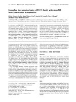
Figure 1 Clinical data for P1 compared with that of P2.Thea.
CD4+ counts (/mm
3
) and b. log viral loads (copies/ml) for P1 (blue)
and P2 (red) are shown. P1 and P2 were exposed to the same third
party on day 0. P1 remained off therapy for 930 days post-exposure
whilst P2 progressed more rapidly and commenced HAART 249
days post-exposure. Plasma for baseline sequencing was collected
on day 63 but the CD4+ count or VL were not recorded. At day
245, P1 was diagnosed with acute HCV infection and had evidence
of super-infection in plasma collected at day 235, having been
exposed to a fourth person after day 63.
English et al . Retrovirology 2011, 8:54
/>Page 3 of 14
Between-host phylogenetic analysis supports the clinical
history of a single donor
By both maximum likelihood (ML) and Bayesian
MCMC based analyses, sequences from P1 and P2 were
highly related and clustered to the exclusion of all other
sequences, consistent with a common donor (Figure 2,
Additiona l Files 1 and 2). We demonstrated the statisti-
cal support for the robustness of the cluster by both
methods ( Figure 2 - ML bootstrap values for three
genes were: env 100%, gag 99.9% and pol 99 .3%, and
Bayesian MCMC based posterior probabilities were:
100% for env, gag and pol). We could not use phyloge-
net ic inference to exclude the possibil ity that one part i-
cipant infected the other, s ince such techniques cannot
prove the direction of transmission in a forensic sense
[39]. For example, we could not exclude the possibility
that two strains were transmitted to one par ticipant
and that an initially infectious strain was out-competed
to extinction prior to day 63. However, results from
other studies sugge sted this was unlikely [5,6,13,40,41].
Therefore, phylogenetic analyses were consistent with
the clinical history that a single, third party contem-
poraneously transmitted the divergent strains that
infected P1 and P2.
Significant between-host divergence observed in
transmitted HIV-1 env and pol genes
We measured the inter-host distance for stem branches,
which are the internal branches separating the within-
patient sequences. For the gag gene fragment, which we
f.
e.
b.
a.
c.
d.
0.05
0.05
0
.
05
0.05
0.05
0.05
Figure 2 Trees generated for phylogenetic cluster analysis. Phylogenetics cluster analysis was carried out using day 63 viral sequences from
P1 and P2. Zoomed-in images of trees are shown in Figure 2 for the env fragment in a. and b., the gag fragment in c. and d., and the pol
fragment in e. and f Results from two different methods of cluster analysis are shown for each fragment: ML (PhyML) trees in a., c., and e., and
Bayesian MCMC based consensus trees in b., d., and f Terminal nodes represent sequences sampled from P1 (blue circles) or P2 (red circles), as
well as reference sequences. Env sequences for P1 and P2 were sampled by SGA and represent gap-stripped alignments 1305 nucleotides in
length. Gag and pol fragments were sampled by bacterial cloning. The full tree images can be viewed in Additional Figures 1 and 2. All scale
bars show 0.05, equivalent to 5% divergence. ML bootstrap values or Bayesian MCMC based posterior probabilities for the clustering of P1 and
P2 are given as percentages next to the common ancestor node.
English et al . Retrovirology 2011, 8:54
/>Page 4 of 14
expected to be the most conserved fragment, the inter-
host distance was 0.54% by ML analysis (Figure 2c). The
inter-host distance for the env fragment, which we
expected to be the least conserved of the three, was
3.81%(Figure 2a). For the pol fragment, the inter-host
distance was 1.93% (Figure 2e). The inter-host distance
for env contrasts with the smaller mean distance within
each participant. For env, the mean within-patient dis-
tance was 0.54% by ML analysis in both participants
across the gap-stripped 1305 nucleotide alignment, con-
sistent with the history of recent infection (Figure 2a).
In addition, sequence analysis of day 235 plasma also
failed to detect env or pol sequences from P1 in P2 and
vice versa (data not shown). Therefore, despite sharing
highly similar gag genes, consistent with the clinical his-
tory of a common origin, P1 and P2 appeared to be
infected with remarkably different env variants and, to a
lesser extent, pol variants.
Current implementations of ML and Bayesian tree
analysis do not model gaps or non-aligned regions infor-
matively [42]. As phylogenetic analysis of the env region
meant removing gaps and non-aligned portions, we
compared full-fragment, non-stripped env sequences
from P1 and P2 with the baseline consensus sequence
for P1 in a Highlighter plot (Figure 3). There was
sequence homogeneity within both P1 and P2, compati-
ble with a single strain initiating a recent infection for
each. However, there were multiple sites of variation
when P1 was compared with P2. Secondly, we quantified
the percentage phylogenetic signal-to-noise (STN)[43] in
env.Wecomparedourfullenv fragment with gaps to
the same fragment with 5% gap-strip ping. The percen-
tage STN between P1 and P2 was 70.7% to 24.3% in the
unstripped env fragment and 62.0% to 30.7% for the
stripped env. Nevertheless, the percentage STN in the
stripped alignment between hosts was greater than in
previous studies of multiple-variant transmissions in this
genomic region [6,12]. Our analyses indicated that there
was a small loss of between-host phylogenetic s ignal in
env by stripping gaps or poorly aligned regions. How-
ever, stripped env fragment alignments contained a
higher percentage STN than either the shorter gag align-
ment (49.4% to 50.5%) or shorter pol alignment (4.2% to
35.5%). The gag and pol fragment alignments did not
require stripping. Noise ≥ 30% was consistent with a
phylogenetic cluster [44,45], but we needed to quantify
between-ho st evolution prior to transmission by another
method.
Env divergence quantified by estimating the tMRCA
To quantify pre-transmission evolution, we estimated
the time since divergence of the two env variants infect-
ing P1 and P2 by calibrating the sequence evolution rate
for the env C2V5 region of gp120 against another
dataset and by measuring the degree of within-host
diversification since transmission [3,15]. Using Bayesian
MCMC based inference, we estimated the inter-host dis-
tance as the time to the most recent common ancestor
(tMRCA) which was 2.82 years (95% confidence interval:
1.28 to 4.54 years) of viral evolution (Figure 4). We
repeated this analysis with different priors (Additional
File 3). All of these results were consistent, and the
common ancestor of the HIV-1 env genes infecting P1
and P2 was estimated to have existed at least 1.14 years
prior to transmission, either in a chronically infected
donor or in a recent previous host. These estimates
were again consistent with the clinical history of a sin-
gle, third party having infected both P1 and P2, and that
highly divergent sequences could be transmitted by a
single donor within a very short period of time.
Potential antigenic variation in the gp120 proteins
of transmitted viruses
However, demonstrating a high level of divergence did
not answer whether each patient received divergent var-
iants at random or whether there was selection at
Master - P1 gp120 day 63 consensus
P1 gp120 day 63 consensus
P1 gp120 day 63 SGA 1
P1 gp120 day 63 SGA 2
P1 gp120 day 63 SGA 3
P1 gp120 day 63 SGA 4
P1 gp120 day 63 SGA 5
P1 gp120 day 63 SGA 6
P1 gp120 day 63 SGA 7
P1 gp120 day 63 SGA 8
P1 gp120 day 63 SGA 9
P1 gp120 day 63 SGA 10
P1 gp120 day 63 SGA 11
P1 gp120 day 63 SGA 12
P1 gp120 day 63 SGA 13
P1 gp120 day 63 SGA 14
P1 gp120 day 63 SGA 15
P1 gp120 day 63 SGA 16
P1 gp120 day 63 SGA 17
P1 gp120 day 63 SGA 18
P1 gp120 day 63 SGA 19
P1 gp120 day 63 SGA 20
P1 gp120 day 63 SGA 21
P2 gp120 day 63 SGA 1
P2 gp120 day 63 SGA 2
P2 gp120 day 63 SGA 3
P2 gp120 day 63 SGA 4
P2 gp120 day 63 SGA 5
P2 gp120 day 63 SGA 6
P2 gp120 day 63 SGA 7
P2 gp120 day 63 SGA 8
P2 gp120 day 63 SGA 9
P2 gp120 day 63 SGA 10
P2 gp120 day 63 SGA 11
P2 gp120 day 63 SGA 12
P2 gp120 day 63 SGA 13
P2 gp120 day 63 SGA 14
P2 gp120 day 63 SGA 15
P2 gp120 day 63 SGA 16
P2 gp120 day 63 SGA 17
P2 gp120 day 63 SGA 18
P2 gp120 day 63 SGA 19
Figure 3 Highlighter plot of env gp120 nucleotide sequences.
Full-length env gp120 sequences from day 63 were sampled by
SGA. The Highlighter plot shows gaps in grey and nucleotide
substitutions (A = green, T = red, G = orange, C = light blue),
revealing difficult-to-align regions. The master sequence against
which all other sequences are compared is the majority-rule P1
consensus sequence at day 63, shown as the top sequence.
English et al . Retrovirology 2011, 8:54
/>Page 5 of 14
transmission. Transmission of divergent env gp120 var-
iants could be due to hard selection for differences in
antigenicity in each recipient. Hard selection involves
selective mortality of variants [46]. In rhesus macaques,
SIV envelope pro teins appear be under hard selection at
transmission due t o neutralizing antibodies [47].
Attempts have been made to infer the antigenicity of
HIV-1 envelope proteins to neutralizing antibodies from
the number of potential N-linked glycosylation sites
(PNLGSs) in gp120 [22,48]. Therefore, we hypothesized
that differences in the number of PNLGSs in gp120
would indicate potential between-host differences in
viral antigenicity.
We compared PNLGSs within inferred amino acid
sequences for gp120 from P1 and P2 using N-Glycosite
(Figure 5). P1 had a mean of 24 PNLGSs (range 23 to
25). P2 had a mean of 29 PNLGSs (range 28 to 29).
Firstly, we looked for p ositions where P1 and P2 were
identical. P1 and P2 shared PNLGSs in 100% of
sequences at 17 positions. To demonstrate that this
degree of identity was consistent with a phylogenetic
cluster, we compared these sequences with 242 unre-
lated sequences. We studied 87 full-length, inferred
amino acid sequences for gp120 sampled from other
SPARTAC participants at trial baseline by population
sequencing, as well a 155 subtype B sequences from the
LANL database sampled during acute infection. The
combined SPARTAC/LANL reference sequences had
100% PNLGS predictions at only one site, located in C1.
Greater than 90% of the reference sequences had a
PNLGS s at only seven positions. We concluded that the
degree of similarity between P1 and P2 was consistent
with a phylogenetic cluster due to transmission from a
single donor.
We then look ed at t he positions that were not 100%
identical, to see if there was any evidence of potential
hard selection in each recipient during transmission. In
particular, we focussed on the V1V4 region that is
implicated in susceptibility to neutralizing antibodies.
Previous studies of this region have suggested that fewer
PNLGSs in this region increases the susceptibility of
highly related strains to neutralizing antibody
[22,24,25,49]. We found a higher mean number of
PNLGSs across V1V4 in P2 (24 sites, range 23-25) than
P1 (19 sites, range 18-20; p < 0.0001, unpaired T-test).
Thesedataindicatedthattherecouldbeadifferencein
susceptibility to neutralizing antibodies between these
two strains, consistent with a non-random model o f
transmission.
No autologous or cross-neutralization observed despite
potential antigenic variation
We hypothesized that differences at PNLGSs might
equate to differences in neutralization that would
explain the transmission of divergent env variants
[22,24,25,49 ]. Therefore, we investigated whether the
viral isolates from P1 and P2 had different neutralization
profiles. Viruses pseudotyped with full-length day 63 env
sequences from P 1 and P2 were t ested against
Figure 4 Relaxed-clock tree for env. Between-h ost divergen ce, in
terms of pre-transmission evolution, was quantified as the estimated
tMRCA using a Bayesian MCMC based approach. Env C2V5 fragment
sequences from P1 and P2, sampled at day 63 by SGA, were
calibrated against within-host divergence since the estimated time
since transmission as well as the mean rate of substitution from the
reference dataset.
Figure 5 Comparison of PNLGSs in inferred env gp120 amino
acid sequences. Full-length gp120 amino acid sequences, inferred
from day 63 SGA nucleotide sequences, are shown. The proportion
of P1 sequences with PNLGS at a particular position are shown as a
‘positive’ blue bar and the proportion of P2 sequences with a
PNLGS is shown as a ‘negative’ red bar. Positions where 100% of
sequences have and PNLGS in both P1 and P2 are indicated by
small stars.
English et al . Retrovirology 2011, 8:54
/>Page 6 of 14
autologo us or heterologous serum from each participant
sampled at day 186 post-exposure. However, the env
pseudotypes for both P1 and P2 were only po orly neu-
tralized or cross-neutralized (half maximal inhibitory
concentration, IC
50
,ofserum≤ 1:20, Additional File 4).
Therefore, it seemed unlikely that a humoral response
was responsible for the detection of different env var-
iants in P1 and P2, consistent with transmission being a
random process.
However, envelope proteins are not only potentially
under immune selection at transmission but also
might be selected for an increased ability to enter
cells. We used the data from our neutralization assay
to estimate the infectivity of the env pseudotyped
viruses in vitro. Pseudoviruses derived from P1
sequences were approximately 2.5 times (P < 0.05)
more infectious in vitro than pseudoviruses from P2,
after normalization to reverse transcriptase levels
(Additional File 5). We noted between-host diversity
in C2C4, including differences in glycosylation. C2C4
encodes discontinuous re gions involved in CD4 and
co-receptor binding [50-52]. Inferred gp120 protein
sequences were analysed with several algorithms that
were evaluated by Low and colleagues [53], to detect
differences in predicted co-receptor usage and mini-
mize the possibility of missing CXCR4/CCR5 dual-use
variants. However, these algorithms predicted that all
sampled viruses from P1 and P2 would use CCR5.
Our experiment was not specifically set up to test
infectivity so all these results must be interpreted with
caution. In addition, potential differences in infectivity
do not explain why both viruses were able to cause
productive infection in different individuals. Therefore,
we found no evidence to reject a random model of
transmission.
HLA Class 1 restricted responses and potential selection
pressure around transmission
We also investigated HIV-1 specific cellular immune
responses, to exclude another potential source of hard
selection in each participant that might influence our
results. Clinical progression and viral load have been
associated with host HLA Class I type in chronic infec-
tion [54-56]. HLA Class I restricts the ability of host
cytotoxic T lymphocytes (CTLs) to recognize and
destroy infected cells. Furthermore, sequencing studies
have detected evidence consistent with escape from
CTL responses within weeks of HIV-1 infection [57].
TherolethatCTLsplayinpreventingestablishedviral
infection in humans remains unclear. However, vaccina-
tion of rhesus macaques to produce detectable CTL
responses is associated with partial protection from
infection [58], and HIV-1 specific CTL responses have
been detected in persons who remain PCR/ELISA
negative despite high-risk exposure [59-61]. Therefore,
we hypothesized that CTL responses during and after
transmission were a potential source of hard selection in
P1 and P2.
Firstly, we compared the Class I HLA type of P1 a nd
P2 with the clinical data to see if there was evidence of
selection. P1 possessed HLA-A*0201, A*2402, HLA-
B*1402, B*3543, Cw*0102, Cw*0802; P2 possessed HLA-
A*0101, A*2901, B*0801, B*5001, Cw*06 02, Cw*0701.
Neither participant possessed HLA types that are
strongly associated with protection from progression in
chronic infection [62,63]. However, P2, who progressed
quickest, possessed the HLA-A*0101 B*0801 haplotype
that is associated with more rapid progression [64].
Therefore, we hypothesize that host factors contribute
to the different clinical out come in t hese participants
and that the viruses had been under different selection
pressures since transmission.
Detectable CTL responses do not explain between-host
divergence in env
We investigated whether different CTL responses could
have influenced detection of divergent variants in our
study. Phylogenetic analysis assumes neutral evolution
rather than natural selection [44]. Therefore, we com-
pared viral sequence data and g-interferon ELISpot data
from each participant to see if cytotoxic T lymphocyte
responses since transmission may have accounted for
observed between-host divergence in env [65,66 ].
Sequence data were available for the two env gp120
optimal peptides against which P2 had a significant
response: TVYYGVPVWK (HXB2 gp160 30-46) and
SFEPIPIHY (HXB2 gp160 202-221). The inferred amino
acid sequences for P1 were identical to the wild-type
peptides at t hese epitopes: TVYYGVPVWR and
SFEPIPIHY. P2 was also infected with wild-type
TVYYGVPVWR, as well as both wild-type and mutant
SFEPIPIHK sequences. Therefore, between-host genetic
differences in env could not be attributed to detectable,
env-directed CTL responses, and our data were still
consistent with transmission of env variants being a ran-
dom process.
Conclusions
We have quantified for the first time significant,
between-host genetic divergence in HIV-1 variants that
are likely to have been transmitted by a single donor to
two recipients on the same night. Furthermore, these
data indicate that currently it is not possible to predict
which of the many HIV-1 variants circulating at the
time of transmission will successfully seed a new infec-
tion. If transmission is a random process, then this
represents a major hurdle that any HIV-1 vaccine design
will need to overcome.
English et al . Retrovirology 2011, 8:54
/>Page 7 of 14
Methods
Participants
360 participants, 151 of whom were from the UK or Ire-
land, were recruited to the Short Pulse AntiRetroviral
Therapy at HIV seroConversion (SPARTAC) trial
(ISRCTN number 76742797; EudraCT number 2004-
000446-20). Two male individuals from the UK cohort,
P1 and P2, were identified on clinical history as having
epidemiologically-linked infections: they were partners
and had shared a sexual encounter with a single, third
male on the same night. P1 and P2 were enrolled in the
trial on the same day and followed up at the Jefferiss
Trust Clinic, St. Mary’s Hospital, Paddington, London,
UK. They were both randomized to receive no therapy.
Ethics Statement
This study has been approved by the Multicentre
Research Ethics Committee (MREC). All participants
provided written informed consent before participating
in this study.
HLA typing
Participant HLA type was determined to the oligo-allelic
level using Dynal RELITM Reverse Sequence-Specific Oli-
gonucleotide kits for the HLA-A, -B and -C loci (Dynal
Biotech). To obtain four-digit typing, Dynal Biotech
Sequence-Specific priming kits were used, in conjunction
with the Sequence-Specific Oligonucleotide type.
Separation of PBMCs and plasma
Peripheral blood mononucleocyte (PBMC) and plasma
samples wer e separated fro m fresh EDTA blood by
Ficoll/Hypaque density gradient centrifugation. For
PBMC collection, blood was diluted with R10 solution:
RPMI 1640 (Sigma UK) with 10% fetal calf serum (FCS;
Sigma, UK), 50 units/ml penicillin/streptomycin mix
and 2 μM L-glutamine. The mixture w as then layered
over Lymphoprep separation medium (Gibco, UK). Sam-
ples were centrifuged at 100 × g at room temperature.
The resultant layer of PBMC was removed and washed.
1 ml aliquots containing 5 × 10
6
cells were stored in
cryotubes in liquid nitrogen at -180±C. For plasma col-
lection, blood samples were prepared as above with dilu-
tion with R10, and the resulting plasma was collected in
1 ml aliquots and stored at -80±C.
Viral RNA extraction
1 ml aliquots of frozen plasma were used for each
ext ract ion. The plasma was centrifuged at 1600 × g and
4±C for 1 hour to pellet the virus. Excess plasma was
removed and the pellet was resuspended in 140 μlof
remaining plasma. RNA was then extracted wit h the
QIAamp Viral RNA Minikit (Qiagen, UK) according to
the manufacturer’s instructions.
Reverse transcription and polymerase chain reaction
(PCR)
For env, viral RNA was reverse transcribed using the
SuperScript III Kit (Invitrogen, UK) to produce cDNA.
15 μl of viral RNA was added to 1.5 μldH
2
O, 1.5 μl pri-
mer OFM19 [6] (concentration 20 μM) and 1.5 μl
dNTPs (concentration 10 mM). The mix was heated to
65°C for 5 min followed by 4°C for 1 mins to anneal the
primers to the RNA. The reverse transcription (RT)
reaction mix ( 5xBuffer: 6 μl, DTT: 1.5 μl; RNaseOUT
1.5 μl; SuperScript III 1.5 μl) was then added to make a
final volume of 29 μl. The reaction mix was heated to
50°C for 60 min, followed by 55°C for 60 min and finally
75°C for 10 minutes. For gag and pol, viral RNA was
reverse transcribed using the Reverse-iT 1
st
Strand
Synthesis Kit (Abgene, UK). 18 μlofviralRNAwas
added to 1.5 μl primer (random decamers and oligodT
supplied with the kit, concentration 20 μM). The mix
was heated to 75°C for 5 min followed by 4°C for 2 min
to anneal the primers to the RNA. The RT reaction mix
(5×Buffer: 6 μl; dNTPs: 3 μl concentration 10 mM;
RTase Blend 1.5 μl) was then added to make a final
volume of 30 μl. The reaction mixture was heated to 42°
C for 60 min followed by 75°C for 10 min. The HIV gag
and pol genes were amplified by separate PCR reactions
as described in detail elsewhere [67]. The HIV env genes
were amplified by PCR using a protocol for single gen-
ome amplification as described in detail elsewhere [5,6].
Single genome amplification
Single genome amplification (SGA) of env was carried as
described elsewhere [5,6]. A 30% cut-off for positive
wells was used [5,6,36].
Bacterial cloning
Bacterial cloning was carried out for gag and pol using
the TOPO TA “One Shot” Cloning Kit for Sequencing
(Invitrogen, UK). Purified PCR products were ligated
into the pCR4-TOPO vector. Escherichia coli were
mixe d on ice with the ligation mix and then transfected
by heat shock at 42°C for 30 s. Cells were immediately
removed to ice and then added to SOC medium (Invi-
trogen, UK) and placed on a shaking incubator at 37°C
and < 1 × g for 1 hour. Cells were then spread on plates
of 1× lysogeny broth (LB) agar (Sigma, UK) containing
0.1 μ g/ml ampicillin (Sigma, UK) and incubated over-
night at 37°C. Negative controls were included. Colonies
were then selected and added to individual wells con-
taining 2× LB medium (Sigma, UK) with 0.05 μ g/ml
kanamycin (Sigma, UK). The wells were incubated on a
shaking incubator overnight at 37°C and < 1 × g. Bac-
teria were lysed and minipreps of clonal plasmid DNA
(pDNA) were prepared using t he Montage Miniprep
96
Kit (Millipore, US).
English et al . Retrovirology 2011, 8:54
/>Page 8 of 14
Sequencing
Sequencing of population PCR, SGA and bacterial clon-
ing DNA products was performed using BigDye technol-
ogy in a 96-well plate. For population PCR and SGA
products, 3 μl DNA was added to a mix containing 0.8
μl BigDye Terminator (Applied Biosystems, UK), 1.5 μl
5× sequencing buffer (Applied Biosystems, UK), 2 μlof
primer (3.3 μM) and 2.7 μldH
2
O. For b acteria- cloned
pDNA, 4 μl of miniprep was added to a mix containing
1 μl BigDye Terminator, 1.5 μl5×sequencingbuffer,1
μlofprimer(3.3μM) and 3.5 μldH
2
O. The following
cycling conditions were used: 96°C for 30 s, then 30
cycles of 96°C for 30 s, 50°C for 15 s and 60°C for 4
min. DNA for sequencing was precipitated on ice with 2
μl3Msodiumacetate,10μldH
2
O, 50 μl ice-cold 100%
ethanol for 5 min at -20°C, centrifuged at 600 × g for 80
min at 4°C, washed twice with ice-cold 70% ethanol and
run on an ABI 3700 sequencer.
Sequence alignment
All sequences were manually edited using Sequencher
v4.8 (Gene Codes Corporation, US) and manually
aligned using Se-Al v2.0a11 [68,69]. For env alignment,
sequences were first aligned with MUSCLE v3.7 [70] fol-
lowed by manual alignment. Sequences containing stop
codons or frameshifts were deleted prior to subsequent
analysis. Where appropriate, reference sequences were
obtained from the Los Alamos National Laboratory
(LANL) HIV sequence database [71]. For env,which
contains many gaps and poorly aligned regions, gap
stripping was undertaken first with GapStreeze set to
5% [72]. In GapStreeze, the user sets a gap tolerance
between 0% and 100%. A value of 5% will cause all col-
umns in the alignment to be deleted if more than 5% of
sequences contain a gap at that position. Sequences
were manually edited in Se-Al v2.0a11 before and after
gap-stripping.
Between-host phylogenetic analysis
Phylogenetic analysis of viral sequences sampled from
P1 and P2 w as carried out by severa l methods across
the env, gag and pol gene-fragments. Prior to gap-strip-
ping with GapStreeze, a likelihood mapping [45] analysis
was run to ensure phylogenetic signal within env was
significant. Likelihood mapping was implemented in
Tree-Puzzle v5.3.rc7 [73] and the env fragment was
screened from the beginning of the c oding start region
to the end of gp120 (HXB2 nucleotide position 6225 to
7757). Additionally, full nucleotide sequences for the
fragme nts from all three genes we re visually screened in
Highlighter [74], and the inferred protein sequence were
screened visually using Jalview v2.6 [75,76]. Phylogenetic
trees were initially constructed using the maximum like-
lihood (ML) method with PhyML v3.0 software [77],
and visualized i n FigTree v1.3.1 [78]. We chose the sub-
stitution model that gave the highest likelihood with
PAUP*v4.0 [79]: the generalized time reversible (GTR)
model incorporating estimates of the proportion of
invariant sites (I), and the shape parameter of a gamma
distribution [80]. ML branch support values were
obtained by non-parametric bootstrapping using PhyML
v3.0 (1000 replicates). Finally, phylogenetic analysis
using a Bayesian MCMC based method was implemen-
ted in Mr Bayes v3.1.2 [81,82]. An unconstrained branch
length (exponential) prior was used to avoid enforcing a
molecular clock [44]. MrBayes v3.1.2 was run in dupli-
cate for at least 50,000,000 steps for env and pol,sam-
pling trees every 1,000 steps. MrBayes v3.1.2 was run in
duplicate for at least 100,000,00 0 steps for gag, sampling
every 10,000 steps. Convergence was assessed with Tra-
cer v1.5 [83] w ith all parameter estimates having effec-
tive sample sizes (ESSs) of > 300, because a high ESS
reflects a low degree of correlation among samples [44].
The consensus tree for each gene, with posterior prob-
abilities for branch support, was generated and visua-
lized in FigTree v1.3.1.
Inferring the tMRCA using a relaxed molecular clock
To determine the time to the most recent common
ancestor (tMRCA) of the sequences isolated from the
two participants, we used a Bayesian MCMC based
approach. We tested our assumption that all of the
observed evolution in env within the viral sequence sets
from each participant had occurr ed wit hin each host by
demonstrating a star-like intra-host phylogeny, and con-
firming that intra-host divergence by ML was consistent
with that predicted for early, monophyletic infection
against other datasets [3,5,6,12,15]. We used a normal
tMRCA prior for the s equences within each participant,
calibrated to a mean of 63 days since exposure (standard
deviation 1 day). We ran BEAST v1.5.4 [84] for at least
100,000,000 steps, sampling every 10,000 steps, and
employing an uncorr elated lognormal relaxed clock to
allow for rate variation among branches [15,44,85-87].
Rate variation may occur if the two variants evolved at
different rates, before or after transmission [15,44]. The
substitution model was the GTR model. The underlying
demographic model was the Bayesian skyline plot with
10 steps, and was used as a fle xible prior on the distri-
bution of the inter-node intervals on the sampled phylo-
genetic topologies [15,44,85-87]. Convergence was
assessed with Tracer v1.5, and all parameter estimates
had ESSs of > 300 [15,44,85-87]. Convergence was not
achieved when us ing estimated transmission time as the
only prior; the ESSs for the prior and posterior probabil-
ities remained < 100 after 300,000,000 steps
[15,44,85-87]. To d eal with this issue, a posterior mean
rate of substitution prior was estimated from the
English et al . Retrovirology 2011, 8:54
/>Page 9 of 14
posterior mean rate of another dataset, for a fragment of
the env C2V5 region [15]. This mean rate prior was nor-
mally distributed, with a mean of 8.18 × 10
-3
substitu-
tions per site per year (standard deviation of 1.15×10
-3
substitutions per site per year) [15]. The hypervariable
region s were cut to be consistent with the original data-
set after consultation with the authors [88]. To achieve
convergence, our relaxed-clock analysis also required
the full-length C2V5 fragment, rather than the part-frag-
ment used in the reference dataset that was missing the
5’ end of the C2 region [15].
To determine the sensitivity of o ur results to the
choice of prior, we also analysed the data under a strict
molecular clock, calibrating the time of transmission to
thesamepriorasundertherelaxed molecular clock,
but not enforcing a strong prior on the rate [15,44]. We
performed this analysis for C2V5 and our entire 1305
stripped env fragment. The mean rate prior from the
reference dataset was necessary for the, relaxed clock
analysis to converge, but our tMRCA estimate was
robust to this choice of prior, as the most important
prior for the tMRCA estimate was the time of transmis-
sion. Although calibration to the time since transmission
may lead to an overestimate of the posterior substitution
rate estimate [15], other studies have found that this
effect is sma ll for monophyletic infections [6,12]. Both
strict-clock and relaxed-clock analyses using the gag and
pol fragments failed to achieve convergence after
300,000,000 steps, and no r eference datasets were avail-
able for calibration of evolution in these fragments.
Potential N-linked glycosylation site analysis
We compared potential N-linked glycosylation sites
(PNLGSs) between inferred amino acid sequences for
the SGA samples env in from P1 and P2, using N-Gly-
cosite [89].
Neutralization and infectivity assays
HIV env genes were amplified from reverse transcribed
viral RNA, restriction-clonedinpcDNA3.1(Invitrogen,
UK) and co-transfected into 293T cells with an env defi-
cient backbone, nl4.3Δenv (Dr M. Pizzato, University of
Geneva). Virus-containing supernatants were harvested,
assayed for reverse transcript ase activity [90], a nd
titrated onto the HIV permissible cell-line, TZM-BL
(also known as JC53-BL) using previously described
techniques [91] with the following modifications: cell
monolayers were fixed with 0.2% gluteraldehyde, stained
with an X-gal substrate and air dried. Infected cells were
counted with an AID v2.9 EliSpot plate-counter (AID
GmbH, Germany). To test serum-mediated neutralizing
responses, 400 focus forming units (FFUs) of titrated-
pseudovirus were incubated with serial dilutions of heat
inactivated autologous sera from participants.
Neutralization was calculated as the percentage-reduc-
tion of FFUs compared to virus-only controls.
IFN-g ELISpot assay
100 μl of 0.5 μ g/ml mouse anti-human IFN-g monoclo-
nal antibody solution (Mabtech, Sweden) was added to
each well on an ELISpot plate (Millipore, US). Frozen
PBMCs were rapidly defrosted and then pipetted into 10
ml of a solution containing RPMI 1640 and pig skin
gelatine (PSG) with added DNAse (Sigma, UK). The
solution was centrifuged at 300 × g for 5 min. The
PBM Cs were resuspended in 20 ml of R10 soluti on and
incubated overnight at 37±C. Cells were then counted
and resuspended in a volume of R10 solution to give a
final concentration of 5 × 10
5
cells per 100 μl. The ELI-
Spot plate was washed three times with 200 μlperwell
of phosphate buffered solution (PBS; Gibco, US) con-
taining 1% FCS. Peptides were added to the appro-
priated wells, with a final concentration of each peptide
being 10 μM. We used overlapping 15 mer peptides
covering HIV-1 proteins gag p17 and gag p24 as well as
optimal epitopes covering gag, pol, nef and env proteins.
100 μl of PBMC suspension was then added to each
well. Duplicate negative controls were prepared, con-
taining R10. Duplicate positive controls were prepared,
containing 5 μ g/ml PHA-P (Sigma, UK). The p late was
incubated for 16 hours at 37±C. The PBMCs were then
discarded and the plate was then washed seven times
with PBS. 100 μlof0.5μ g/ml biotinylated anti-human
IFN-g monoclonal antibody (Mabtech, Sweden) was
added to each well. The plate was incubated for 90 min
at room temperature. The antibody was then discarded
and the plate washed seven times with PBS. 100 μlof
0.5 μ g/ml streptavidin-conjugated alkaline phosphatase
(ALP; Mabtech, Sweden) was added. The plate was incu-
bated at room temperature for 40 min. The streptavidin-
ALP was then discarded and the plate washed seven
times with PBS. 100 μl of substrate solution from the
ALP conjugate substrate kit (Bio-Rad, US) was added to
each well. The plate was incubated at room temperature
for 10 min, or until a colour change was noted in the
positive control well. The plate was then washed with
ordinary tap water and dried. Spots were counted on
the AID version 2.9 EliSpot plate-reader. The normal-
ized magnitude of the response (NMOR) was calculated
as follows [92]:
NMOR = M
exp
− (
¯
x
neg
+3× SD
neg
) − 50
Where M
exp
is the numb er of spots in the experimen-
tal well,
¯
x
neg
is the mean number of spots in the nega-
tive control wells, and SD
neg
is the standard deviation of
the negative c ontrol wells. NMOR is always a positive
integer and all negative values are set to 0.
English et al . Retrovirology 2011, 8:54
/>Page 10 of 14
Additional material
Additional file 1: Images of the entire ML (PhyML) trees for a. env,
b. gag and c. pol. Terminal nodes representing day 63 sequences
sampled from P1 (blue circles) and P2 (red circles), as well as reference
sequences are shown. Env sequences for P1 and P2 were sampled by
SGA and represent gap-stripped alignments of full-length gp120 . Gag
and pol fragment sequences were sampled by bacterial cloning.
Additional file 2: Images of the entire Bayesian MCMC based
consensus trees for a. env,b.gag and c. pol. Terminal nodes
representing day 63 sequences sampled from P1 (blue circles) and P2
(red circles), as well as reference sequences are shown. Env sequences for
P1 and P2 were sampled by SGA and represent gap-stripped alignments
of full-length gp120. Gag and pol fragment sequences were sampled by
bacterial cloning.
Additional file 3: Robustness analysis for Bayesian MCMC based
approach for a. env C2V5 and b. the entire env fragment. The tMRCA
estimation analysis was repeated for env SGA sequences using a strict
molecular clock. The estimated time since transmission was the only
prior.
Additional file 4: Neutralization assay results. Neutralization assay
results are shown for day 186 post-exposure sera from P1 and P2 against
pseudoviruses typed with day 63 P1 and P2 envelopes. Results for two
clones from each participant are shown for both autologous and cross-
neutralization assays at two serum dilutions, 1:20 and 1:60.
Additional file 5: Results of the infectivity assays. Infectivity assays
were used to titre pseudoviruses prior to infection for the neutralization
assay. Fold virus dilutions are shown in the legend. The results for the
two clones used in the assay shown in Additional File 4 are shown but
these results were consistent for the nine clones screened for each
participant. Infectivity is corrected against viral reverse transcriptase
expression.
Acknowledgements
The authors would like to thank the participants and staff of the St. Mary’s
Hospital Jefferiss Wing clinic and the participants and staff involved in the
SPARTAC trial. The authors would like to thank Mr David English for
computing assistance in the rapid processing of MrBayes analyses. The
authors would also like to thank the anonymous reviewers of this
manuscript for their comments and suggestions. JF is supported by the
Medical Research Council. JW and RP are supported by the Wellcome Trust
(UK). REP is a NIHR Senior Investigator. SE is supported by the
Commonwealth Scholarship Commission. AK is supported by the Royal
Society.
SPARTAC Investigators. Trial Steering Committee A Breckenridge (Chair),
C Conlon, D Cooper, F Conradie, J Kaldor, M Schechter, P Claydon, P
Kaleebu, G Ramjee, F Ssali, G Tambussi, J Weber. Trial Physician Sarah
Fidler. Trial Statistician Abdel Babiker. Data and Safety Monitoring
Committee A McLaren (in memoriam), V Beral, G Chene, J Hakim. Central
Virology Laboratories and Repositories Jefferiss Trust Laboratories,
Imperial College, London, UK (M McClure, D Muir, I Blain, A Helander, O
Erlwien, S Kaye). Clinical Endpoint Review Committee N Paton, S Fidler.
Co-ordinating Trial Centres Australia: National Centre in HIV Epidemiology
and Clinical Research, University of New South Wales, Sydney (P Grey, D
Cooper, T Kelleher, M Law). UK and Ireland: MRC Clinical Trials Unit, London
(A Babiker, K Porter, P Kelleher, K Boyd, D Johnson, D Nock) Investigators
and Staff at Participating Sites Australia: St Vincent’s Hospital, Sydney (D
Cooper), Carlton Clinic, Melbourne (J Anderson), 407 Doctors, Sydney, (R
McFarlane), Prahran Market Clinic, Melbourne (N Roth), Taylor Square Private
Clinic, Sydney (R Finlayson), The Centre Clinic, Melbourne (B Kiem Tee),
Sexual Health Centre, Melbourne (T Read), AIDS Medical Unit, Brisbane (M
Kelly), Centre for Immunology, Sydney (P Cunningham). Brazil: Projeto Praça
Onze, Hospital Escola São Francisco de Assis, Universidade federal do Rio de
Janeiro,
Rio de Janeiro. (M Schechter, R Zajdenverg, M Merçon). Italy:
Ospedale San Raffaele, Milan (G Tambussi, C Tassan Din, C Ronchetti, G Travi,
V Rusconi, G de Bartolo), Ospedale Lazzaro Spallanzani, Roma (G D’Offizi, C
Vlassi, A Corpolongo). South Africa:Capetown: Desmond Tutu HIV Centre,
Institute of Infectious Diseases, Capetown (R Wood, J Pitt, L-G Bekker, J
Aploon, L Fielder, N Killa, T Buhler) Johannesburg: Reproductive Health and
HIV Research Unit, Bara Clinic, Chris Hani Baragwanath Hospital,
Johannesburg (H Rees, J Moyes, S Walaza, K Moitse), Contract Laboratory
Services, Johannesburg Hospital, Johannesburg (W Stevens, C Wallis, C
Ingram, M Majam) Kwazulu-Natal: HIV Prevention Unit, Medical Research
Council, Durban (G Ramjee, D Singh, T Mtambo, S Gappoo, H Somaroo, J
Moodley, M Mills, A Premrajh, N Nozulu, K Naidoo). Uganda: MRC/Uganda
Virus Research Institute, Entebbe (H Grosskurth, A Kamali, P Kaleebu, J
Mugisha, U Bahemuka, F Lyagoba, P Tabuga). Spain: Hospital Clinic-IDIBAPS.
Univ. of Barcelona. Barcelona (J M Miro, M López-Dieguez, F. Agüero, JA
Arnaiz, T. Pumarola, M. Plana, M. Tuset, MC Ligero, C. Gil, T. Gallart, JM Gatell)
UK and Ireland: Royal Sussex County Hospital, Brighton (M Fisher, L Heald, N
Perry, D Pao, D Maitland), St James’s Hospital, Dublin (F Mulcahy, G
Courtney, D Reidy), Regional Infectious Diseases Unit, Western General
Hospital and Genitourinary Dept, Royal Infirmary of Edinburgh, Edinburgh (C
Leen, G Scott, L Ellis, S Morris, P Simmonds, T Shaw), Chelsea and
Westminster Hospital, London (B Gazzard, D Hawkins, C Higgs, C Mahuma),
Homerton Hospital, London (J Anderson, L Muromba), Mortimer Market
Centre, London (I Williams, J Turner, D Mullan, D Aldam), North Middlesex
Hospital (J Ainsworth, A Waters), Royal Free Hospital (M Johnson, S Kinloch,
A Carroll, P Byrne, Z Cuthbertson), St Bartholomew’s Hospital, London (C
Orkin, J Hand, C De Souza), St Mary’s Hospital, London (J Weber, S Fidler, E
Thomson, J Fox, K Legg, S Mullaney, A Winston, N Poulter, S Wilson) Trial
Secretariat D Winogron, S Keeling.
Author details
1
Nuffield Department of Clinical Medicine, Peter Medawar Building for
Pathogen Research, Oxford University, South Parks Road, Oxford, OX1 3SY,
UK.
2
Department of Zoology, Oxford University, South Parks Road, Oxford,
OX1 3PS, UK.
3
Division of Medicine, Wright Fleming Institute, Imperial
College, St. Mary’s Hospital, Norfolk Place, Paddington, London W2 1PG, UK.
4
The James Martin 21st Century School, Peter Medawar Building for
Pathogen Research, South Parks Road, Oxford, OX1 3SY, UK.
5
Oxford NIHR
Biomedical Research Centre, Oxford, UK.
Authors’ contributions
RP and JF conceived the study; JF, SE and AK designed the study; SE, DB, PF
and AD performed the experiments; SE, AK and DB analysed the data; SE
and JF wrote the paper; RP, MM, JW, SF and STSC contributed participant
information and samples; all authors were involved in drafting this paper; all
authors have read and approved the final manuscript.
Competing interests
The authors declare that they have no competing interests.
Received: 15 January 2011 Accepted: 7 July 2011 Published: 7 July 2011
References
1. Kawashima Y, Pfafferott K, Frater J, Matthews P, Payne R, Addo M,
Gatanaga H, Fujiwara M, Hachiya A, Koizumi H, et al: Adaptation of HIV-1
to human leukocyte antigen class I. Nature 2009, 458:641-645.
2. Gaschen B, Taylor J, Yusim K, Foley B, Gao F, Lang D, Novitsky V, Haynes B,
Hahn BH, Bhattacharya T, Korber B: Diversity considerations in HIV-1
vaccine selection. Science 2002, 296:2354-2360.
3. Shankarappa R, Margolick JB, Gange SJ, Rodrigo AG, Upchurch D,
Farzadegan H, Gupta P, Rinaldo CR, Learn GH, He X, et al: Consistent viral
evolutionary changes associated with the progression of human
immunodeficiency virus type 1 infection. J Virol 1999, 73:10489-10502.
4. Salazar-Gonzalez JF, Salazar MG, Keele BF, Learn GH, Giorgi EE, Li H,
Decker JM, Wang S, Baalwa J, Kraus M H, et al: Genetic identity,
biological phenotype, and evolutionary pathways o f transmitted/
founder vir uses in acute and early HIV-1 infec tion. JExpMed2009,
206:1273-1289.
5. Salazar-Gonzalez JF, Bailes E, Pham KT, Salazar MG, Guffey MB, Keele BF,
Derdeyn CA, Farmer P, Hunter E, Allen S, et al: Deciphering human
immunodeficiency virus type 1 transmission and early envelope
diversification by single-genome amplification and sequencing. J Virol
2008, 82:3952-3970.
6. Keele BF, Giorgi EE, Salazar-Gonzalez JF, Decker JM, Pham KT, Salazar MG,
Sun C, Grayson T, Wang S, Li H, et al: Identification and characterization of
English et al . Retrovirology 2011, 8:54
/>Page 11 of 14
transmitted and early founder virus envelopes in primary HIV-1
infection. Proc Natl Acad Sci USA 2008, 105:7552-7557.
7. Derdeyn CA, Hunter E: Viral characteristics of transmitted HIV. Curr Opin
HIV AIDS 2008, 3:16-21.
8. The Global HIV/AIDS Vaccine Enterprise: Scientific Strategic Plan. PLoS
Med 2005, 2:111-121.
9. Edwards CT, Holmes EC, Wilson DJ, Viscidi RP, Abrams EJ, Phillips RE,
Drummond AJ: Population genetic estimation of the loss of genetic
diversity durin g horizontal transmission of HIV-1. BMC Evol Biol 2006,
6:28.
10. Haaland RE, Hawkins PA, Salazar-Gonzalez J, Johnson A, Tichacek A, Karita E,
Manigart O, Mulenga J, Keele BF, Shaw GM, et al: Inflammatory genital
infections mitigate a severe genetic bottleneck in heterosexual
transmission of subtype A and C HIV-1. PLoS Pathog 2009, 5:e1000274.
11. Abrahams MR, Anderson JA, Giorgi EE, Seoighe C, Mlisana K, Ping LH,
Athreya GS, Treurnicht FK, Keele BF, Wood N, et al: Quantitating the
multiplicity of infection with human immunodeficiency virus type 1
subtype C reveals a non-poisson distribution of transmitted variants. J
Virol 2009, 83:3556-3567.
12. Li H, Bar KJ, Wang S, Decker JM, Chen Y, Sun C, Salazar-Gonzalez JF,
Salazar MG, Learn GH, Morgan CJ, et al: High Multiplicity Infection by HIV-
1 in Men Who Have Sex with Men. PLoS Pathog 2010, 6:e1000890.
13. Bar KJ, Li H, Chamberland A, Tremblay C, Routy JP, Grayson T, Sun C,
Wang S, Learn GH, Morgan CJ, et al: Wide variation in the multiplicity of
HIV-1 infection among injection drug users. J Virol 2010, 84:6241-6247.
14. Hollingsworth TD, Anderson RM, Fraser C: HIV-1 transmission, by stage of
infection. J Infect Dis 2008, 198:687-693.
15. Lemey P, Rambaut A, Pybus OG: HIV evolutionary dynamics within and
among hosts. AIDS Rev 2006, 8:125-140.
16. Drummond AJ, Rambaut A, Shapiro B, Pybus OG: Bayesian coalescent
inference of past population dynamics from molecular sequences. Mol
Biol Evol 2005, 22:1185-1192.
17. Zhang LQ, MacKenzie P, Cleland A, Holmes EC, Brown AJ, Simmonds P:
Selection for specific sequences in the external envelope protein of
human immunodeficiency virus type 1 upon primary infection. J Virol
1993, 67:3345-3356.
18. Zhu T, Mo H, Wang N, Nam DS, Cao Y, Koup RA, Ho DD: Genotypic and
phenotypic characterization of HIV-1 patients with primary infection.
Science 1993, 261:1179-1181.
19.
Butler DM, Delport W, Kosakovsky Pond SL, Lakdawala MK, Cheng PM,
Little SJ, Richman DD, Smith DM: The origins of sexually transmitted HIV
among men who have sex with men. Sci Transl Med 2:18re11.
20. Bull M, Learn G, Genowati I, McKernan J, Hitti J, Lockhart D, Tapia K, Holte S,
Dragavon J, Coombs R, et al: Compartmentalization of HIV-1 within the
female genital tract is due to monotypic and low-diversity variants not
distinct viral populations. PLoS One 2009, 4:e7122.
21. Diem K, Nickle DC, Motoshige A, Fox A, Ross S, Mullins JI, Corey L,
Coombs RW, Krieger JN: Male genital tract compartmentalization of
human immunodeficiency virus type 1 (HIV). AIDS Res Hum Retroviruses
2008, 24:561-571.
22. Derdeyn CA, Decker JM, Bibollet-Ruche F, Mokili JL, Muldoon M,
Denham SA, Heil ML, Kasolo F, Musonda R, Hahn BH, et al: Envelope-
constrained neutralization-sensitive HIV-1 after heterosexual
transmission. Science 2004, 303:2019-2022.
23. Chohan B, Lang D, Sagar M, Korber B, Lavreys L, Richardson B, Overbaugh J:
Selection for human immunodeficiency virus type 1 envelope
glycosylation variants with shorter V1-V2 loop sequences occurs during
transmission of certain genetic subtypes and may impact viral RNA
levels. J Virol 2005, 79:6528-6531.
24. Liu Y, Curlin ME, Diem K, Zhao H, Ghosh AK, Zhu H, Woodward AS,
Maenza J, Stevens CE, Stekler J, et al: Env length and N-linked
glycosylation following transmission of human immunodeficiency virus
Type 1 subtype B viruses. Virology 2008, 374:229-233.
25. Frost SD, Liu Y, Pond SL, Chappey C, Wrin T, Petropoulos CJ, Little SJ,
Richman DD: Characterization of human immunodeficiency virus type 1
(HIV-1) envelope variation and neutralizing antibody responses during
transmission of HIV-1 subtype B. J Virol 2005, 79:6523-6527.
26. Keele BF, Li H, Learn GH, Hraber P, Giorgi EE, Grayson T, Sun C, Chen Y,
Yeh WW, Letvin NL, et al: Low-dose rectal inoculation of rhesus
macaques by SIVsmE660 or SIVmac251 recapitulates human mucosal
infection by HIV-1. J Exp Med 2009, 206:1117-1134.
27. Rybarczyk BJ, Montefiori D, Johnson PR, West A, Johnston RE, Swanstrom R:
Correlation between env V1/V2 region diversification and neutralizing
antibodies during primary infection by simian immunodeficiency virus
sm in rhesus macaques. J Virol 2004, 78:3561-3571.
28. Highleyman L: Detuned assay used to track recent infections. BETA 1999,
12:6-78.
29. Kothe D, Byers RH, Caudill SP, Satten GA, Janssen RS, Hannon WH, Mei JV:
Performance characteristics of a new less sensitive HIV-1 enzyme
immunoassay for use in estimating HIV seroincidence. J Acquir Immune
Defic Syndr 2003, 33:625-634.
30. Fiebig EW, Wright DJ, Rawal BD, Garrett PE, Schumacher RT, Peddada L,
Heldebrant C, Smith R, Conrad A, Kleinman SH, Busch MP: Dynamics of HIV
viremia and antibody seroconversion in plasma donors: implications for
diagnosis and staging of primary HIV infection. Aids 2003,
17:1871-1879.
31.
Fiebig EW, Heldebrant CM, Smith RI, Conrad AJ, Delwart EL, Busch MP:
Intermittent low-level viremia in very early primary HIV-1 infection. J
Acquir Immune Defic Syndr 2005, 39:133-137.
32. Little SJ, McLean AR, Spina CA, Richman DD, Havlir DV: Viral dynamics of
acute HIV-1 infection. J Exp Med 1999, 190:841-850.
33. Clark SJ, Saag MS, Decker WD, Campbell-Hill S, Roberson JL, Veldkamp PJ,
Kappes JC, Hahn BH, Shaw GM: High titers of cytopathic virus in plasma
of patients with symptomatic primary HIV-1 infection. N Engl J Med 1991,
324:954-960.
34. Stafford MA, Corey L, Cao Y, Daar ES, Ho DD, Perelson AS: Modeling
plasma virus concentration during primary HIV infection. J Theor Biol
2000, 203:285-301.
35. Wilson DP, Law MG, Grulich AE, Cooper DA, Kaldor JM: Relation between
HIV viral load and infectiousness: a model-based analysis. Lancet 2008,
372:314-320.
36. Palmer S, Kearney M, Maldarelli F, Halvas EK, Bixby CJ, Bazmi H, Rock D,
Falloon J, Davey RT, Dewar RL, et al: Multiple, linked human
immunodeficiency virus type 1 drug resistance mutations in treatment-
experienced patients are missed by standard genotype analysis. J Clin
Microbiol 2005, 43:406-413.
37. Jordan MR, Kearney M, Palmer S, Shao W, Maldarelli F, Coakley EP,
Chappey C, Wanke C, Coffin JM: Comparison of standard PCR/cloning to
single genome sequencing for analysis of HIV-1 populations. J Virol
Methods 2010, 168:114-120.
38. Fidler S, Oxenius A, Brady M, Clarke J, Cropley I, Babiker A, Zhang HT,
Price D, Phillips R, Weber J: Virological and immunological effects of
short-course antiretroviral therapy in primary HIV infection. Aids 2002,
16:2049-2054.
39. Pillay D, Rambaut A, Geretti AM, Brown AJ: HIV phylogenetics. BMJ 2007,
335:460-461.
40. Liu J, Keele BF, Li H, Keating S, Norris PJ, Carville A, Mansfield KG,
Tomaras GD, Haynes BF, Kolodkin-Gal D, et al: Low-dose mucosal simian
immunodeficiency virus infection restricts early replication kinetics and
transmitted virus variants in rhesus monkeys. J Virol 2010,
84:10406-10412.
41. Liu W, Worobey M, Li Y, Keele BF, Bibollet-Ruche F, Guo Y, Goepfert PA,
Santiago ML, Ndjango JB, Neel C, et al: Molecular ecology and natural
history of simian foamy virus infection in wild-living chimpanzees. PLoS
Pathog 2008, 4:e1000097.
42. Salemi M, Burkhardt BR, Gray RR, Ghaffari G, Sleasman JW, Goodenow MM:
Phylodynamics of HIV-1 in lymphoid and non-lymphoid tissues reveals a
central role for the thymus in emergence of CXCR4-using quasispecies.
PLoS One 2007, 2:e950.
43. Hillis DM, Huelsenbeck JP: Signal, noise, and reliability in molecular
phylogenetic analyses. J
Hered 1992, 83:189-195.
44. Salemi M, Vandamme A-M, Lemey P: The phylogenetic handbook: a practical
approach to phylogenetic analysis and hypothesis testing. 2 edition.
Cambridge, UK; New York: Cambridge University Press; 2009.
45. Strimmer K, von Haeseler A: Likelihood-mapping: a simple method to
visualize phylogenetic content of a sequence alignment. Proc Natl Acad
Sci USA 1997, 94:6815-6819.
46. Hurst LD: Fundamental concepts in genetics: genetics and the
understanding of selection. Nat Rev Genet 2009, 10:83-93.
47. Mascola JR, Stiegler G, VanCott TC, Katinger H, Carpenter CB, Hanson CE,
Beary H, Hayes D, Frankel SS, Birx DL, Lewis MG: Protection of macaques
against vaginal transmission of a pathogenic HIV-1/SIV chimeric virus by
passive infusion of neutralizing antibodies. Nat Med 2000, 6:207-210.
English et al . Retrovirology 2011, 8:54
/>Page 12 of 14
48. Simm onds P, Bal fe P, Ludlam CA, Bishop JO, Brown AJ: Analysis of
sequence diversity in hypervariable regions of the external
glycoprotein of human immuno deficiency virus type 1. JVirol1990,
64:5840-5850.
49. Zhang M, Gaschen B, Blay W, Foley B, Haigwood N, Kuiken C, Korber B:
Tracking global patterns of N-linked glycosylation site variation in highly
variable viral glycoproteins: HIV, SIV, and HCV envelopes and influenza
hemagglutinin. Glycobiology 2004, 14:1229-1246.
50. Valenzuela A, Blanco J, Krust B, Franco R, Hovanessian AG: Neutralizing
antibodies against the V3 loop of human immunodeficiency virus type 1
gp120 block the CD4-dependent and -independent binding of virus to
cells. J Virol 1997, 71:8289-8298.
51. Rizzuto CD, Wyatt R, Hernandez-Ramos N, Sun Y, Kwong PD,
Hendrickson WA, Sodroski J: A conserved HIV gp120 glycoprotein
structure involved in chemokine receptor binding. Science 1998,
280:1949-1953.
52. Thali M, Olshevsky U, Furman C, Gabuzda D, Posner M, Sodroski J:
Characterization of a discontinuous human immunodeficiency virus type
1 gp120 epitope recognized by a broadly reactive neutralizing human
monoclonal antibody. J Virol 1991, 65:6188-6193.
53. Low AJ, Dong W, Chan D, Sing T, Swanstrom R, Jensen M, Pillai S, Good B,
Harrigan PR: Current V3 genotyping algorithms are inadequate for
predicting X4 co-receptor usage in clinical isolates. AIDS 2007, 21:F17-24.
54. O’Brien SJ, Gao X, Carrington M: HLA and AIDS: a cautionary tale. Trends
Mol Med 2001, 7:379-381.
55. Carrington M, Nelson GW, Martin MP, Kissner T, Vlahov D, Goedert JJ,
Kaslow R, Buchbinder S, Hoots K, O’Brien SJ: HLA and HIV-1:
heterozygote advantage and B*35-Cw*04 disadvantage. Science 1999,
283:1748-1752.
56. Brumme ZL, Tao I, Szeto S, Brumme CJ, Carlson JM, Chan D, Kadie C,
Frahm N, Brander C, Walker B, et al: Human leukocyte antigen-specific
polymorphisms in HIV-1 Gag and their association with viral load in
chronic untreated infection. AIDS 2008, 22:1277-1286.
57. Fischer W, Ganusov VV, Giorgi EE, Hraber PT, Keele BF, Leitner T, Han CS,
Gleasner CD, Green L, Lo CC, et al: Transmission of single HIV-1 genomes
and dynamics of early immune escape revealed by ultra-deep
sequencing. PLoS One 2010, 5:e12303.
58. Hansen SG, Vieville C, Whizin N, Coyne-Johnson L, Siess DC,
Drummond DD, Legasse AW, Axthelm MK, Oswald K, Trubey CM, et al:
Effector memory T cell responses are associated with protection of
rhesus monkeys from mucosal simian immunodeficiency virus
challenge. Nat Med 2009, 15:293-299.
59. Langlade-Demoyen P, Ngo-Giang-Huong N, Ferchal F, Oksenhendler E:
Human immunodeficiency virus (HIV) nef-specific cytotoxic T
lymphocytes in noninfected heterosexual contact of HIV-infected
patients. J Clin Invest 1994, 93:1293-1297.
60. Clerici M, Giorgi JV, Chou CC, Gudeman VK, Zack JA, Gupta P, Ho HN,
Nishanian PG, Berzofsky JA, Shearer GM: Cell-mediated
immune response
to human immunodeficiency virus (HIV) type 1 in seronegative
homosexual men with recent sexual exposure to HIV-1. J Infect Dis 1992,
165:1012-1019.
61. Rowland-Jones S, Sutton J, Ariyoshi K, Dong T, Gotch F, McAdam S,
Whitby D, Sabally S, Gallimore A, Corrah T, et al: HIV-specific cytotoxic T-
cells in HIV-exposed but uninfected Gambian women. Nat Med 1995,
1:59-64.
62. Gao X, Nelson GW, Karacki P, Martin MP, Phair J, Kaslow R, Goedert JJ,
Buchbinder S, Hoots K, Vlahov D, et al: Effect of a single amino acid
change in MHC class I molecules on the rate of progression to AIDS. N
Engl J Med 2001, 344:1668-1675.
63. Kiepiela P, Leslie AJ, Honeyborne I, Ramduth D, Thobakgale C, Chetty S,
Rathnavalu P, Moore C, Pfafferott KJ, Hilton L, et al: Dominant influence of
HLA-B in mediating the potential co-evolution of HIV and HLA. Nature
2004, 432:769-775.
64. Kaslow RA, Duquesnoy R, VanRaden M, Kingsley L, Marrari M, Friedman H,
Su S, Saah AJ, Detels R, Phair J, et al: A1, Cw7, B8, DR3 HLA antigen
combination associated with rapid decline of T-helper lymphocytes in
HIV-1 infection. A report from the Multicenter AIDS Cohort Study. Lancet
1990, 335:927-930.
65. Frater AJ, Edwards CT, McCarthy N, Fox J, Brown H, Milicic A, Mackie N,
Pillay T, Drijfhout JW, Dustan S, et al: Passive sexual transmission of
human immunodeficiency virus type 1 variants and adaptation in new
hosts. J Virol 2006, 80:7226-7234.
66. Streeck H, Jolin JS, Qi Y, Yassine-Diab B, Johnson RC, Kwon DS, Addo MM,
Brumme C, Routy JP, Little S, et al: Human immunodeficiency virus type
1-specific CD8+ T-cell responses during primary infection are major
determinants of the viral set point and loss of CD4+ T cells. J Virol 2009,
83:7641-7648.
67. Frater AJ, Brown H, Oxenius A, Gunthard HF, Hirschel B, Robinson N,
Leslie AJ, Payne R, Crawford H, Prendergast A, et al: Effective T-cell
responses select human immunodeficiency virus mutants and slow
disease progression. J Virol 2007, 81:6742-6751.
68. Rambaut A: Se-Al. Sequence Alignment Editor v2.0a11. Book Se-Al
Sequence Alignment Editor v2.0a11 (Editor ed.^eds.) City; 2002.
69. Helseth E, Kowalski M, Gabuzda D, Olshevsky U, Haseltine W, Sodroski J:
Rapid complementation assays measuring replicative potential of
human immunodeficiency virus type 1 envelope glycoprotein mutants. J
Virol 1990, 64:2416-2420.
70. Wyatt R, Sullivan N, Thali M, Repke H, Ho D, Robinson J, Posner M,
Sodroski J: Functional and immunologic characterization of human
immunodeficiency virus type 1 envelope glycoproteins containing
deletions of the major variable regions. J Virol 1993, 67:4557-4565.
71. Truckenmiller ME, Kulaga H, Coggiano M, Wyatt R, Snyder SH,
Sweetnam PM: Human
cortical neuronal cell line: a model for HIV-1
infection in an immature neuronal system. AIDS Res Hum Retroviruses
1993, 9:445-453.
72. Williams MF, Eisele DW, Wyatt SH: Neck needle foreign bodies in
intravenous drug abusers. Laryngoscope 1993, 103:59-63.
73. Schmidt HA, Strimmer K, Vingron M, von Haeseler A: TREE-PUZZLE:
maximum likelihood phylogenetic analysis using quartets and parallel
computing. Bioinformatics 2002, 18:502-504.
74. Jansson M, Popovic M, Karlsson A, Cocchi F, Rossi P, Albert J, Wigzell H:
Sensitivity to inhibition by beta-chemokines correlates with biological
phenotypes of primary HIV-1 isolates. Proc Natl Acad Sci USA 1996,
93:15382-15387.
75. Waterhouse AM, Procter JB, Martin DM, Clamp M, Barton GJ: Jalview
Version 2–a multiple sequence alignment editor and analysis
workbench. Bioinformatics 2009, 25:1189-1191.
76. Clamp M, Cuff J, Searle SM, Barton GJ: The Jalview Java alignment editor.
Bioinformatics 2004, 20:426-427.
77. Guindon S, Gascuel O: A simple, fast, and accurate algorithm to estimate
large phylogenies by maximum likelihood. Syst Biol 2003, 52:696-704.
78. FigTree v1.3.1. [ />79. Valenzuela A, Blanco J, Callebaut C, Jacotot E, Lluis C, Hovanessian AG,
Franco R: Adenosine deaminase binding to human CD26 is inhibited by
HIV-1 envelope glycoprotein gp120 and viral particles. J Immunol 1997,
158:3721-3729.
80. Büchen-Osmond CE: 00.061.1.06.001. Human immunodeficiency virus 1.
ICTVdB - The Universal Virus Database, version 3 2004.
81. Huelsenbeck JP, Ronquist F: MRBAYES: Bayesian inference of phylogenetic
trees. Bioinformatics 2001, 17:754-755.
82. Ronquist F, Huelsenbeck JP: MrBayes 3: Bayesian phylogenetic inference
under mixed models. Bioinformatics 2003, 19:1572-1574.
83. Tracer v1.5. [ />84. Drummond AJ, Rambaut A: BEAST: Bayesian evolutionary analysis by
sampling trees. BMC Evol Biol 2007, 7:214.
85. Drummond AJ, Ho SY, Phillips MJ, Rambaut A: Relaxed phylogenetics and
dating with confidence. PLoS Biol 2006, 4:e88.
86. Lewis F, Hughes GJ, Rambaut A, Pozniak A, Leigh Brown AJ: Episodic
sexual transmission of HIV revealed by molecular phylodynamics. PLoS
Med 2008, 5:e50.
87.
Drummond A, Pybus OG, Rambaut A: Inference of viral evolutionary rates
from molecular sequences. Adv Parasitol 2003, 54:331-358.
88. Lemey P: Personal Communication April 9, 2011.
89. Seminari E, Cocchi L, Antoniazzi E, Giacchino R, Maserati R: Clinical
significance of diplopia in HIV infection. Assessment of a personal
caseload and review of the literature. Minerva Med 1996, 87:515-523.
English et al . Retrovirology 2011, 8:54
/>Page 13 of 14
90. Pizzato M, Erlwein O, Bonsall D, Kaye S, Muir D, McClure MO: A one-step
SYBR Green I-based product-enhanced reverse transcriptase assay for
the quantitation of retroviruses in cell culture supernatants. J Virol
Methods 2009, 156:1-7.
91. Montefiori DC: Evaluating neutralizing antibodies against HIV, SIV, and
SHIV in luciferase reporter gene assays. Curr Protoc Immunol 2005,
Chapter 12; Unit 12.11.
92. Duda A, Lee-Turner L, Fox J, Robinson N, Dustan S, Kaye S, Fryer H,
Carrington M, McClure M, McLean AR, et al: HLA-associated clinical
progression correlates with epitope reversion rates in early human
immunodeficiency virus infection. J Virol 2009, 83:1228-1239.
doi:10.1186/1742-4690-8-54
Cite this article as: English et al.: Phylogenetic analysis consistent with a
clinical history of sexual transmission of HIV-1 from a single donor
reveals transmission of highly distinct variants. Retrovirology 2011 8:54.
Submit your next manuscript to BioMed Central
and take full advantage of:
• Convenient online submission
• Thorough peer review
• No space constraints or color figure charges
• Immediate publication on acceptance
• Inclusion in PubMed, CAS, Scopus and Google Scholar
• Research which is freely available for redistribution
Submit your manuscript at
www.biomedcentral.com/submit
English et al . Retrovirology 2011, 8:54
/>Page 14 of 14