Báo cáo y học: "Distinct roles of CD4+ T cell subpopulations in retroviral immunity: lessons from the Friend virus mouse model" pptx
Bạn đang xem bản rút gọn của tài liệu. Xem và tải ngay bản đầy đủ của tài liệu tại đây (484.11 KB, 12 trang )
Nair et al. Retrovirology 2011, 8:76
/>
REVIEW
Open Access
Distinct roles of CD4+ T cell subpopulations in
retroviral immunity: lessons from the Friend virus
mouse model
Savita Nair1,4, Wibke Bayer1, Mickaël JY Ploquin3, George Kassiotis3, Kim J Hasenkrug2† and Ulf Dittmer1,2*†
Abstract
It is well established that CD4+ T cells play an important role in immunity to infections with retroviruses such as
HIV. However, in recent years CD4+ T cells have been subdivided into several distinct populations that are
differentially regulated and perform widely varying functions. Thus, it is important to delineate the separate roles of
these subsets, which range from direct antiviral activities to potent immunosuppression. In this review, we discuss
contributions from the major CD4+ T cell subpopulations to retroviral immunity. Fundamental concepts obtained
from studies on numerous viral infections are presented along with a more detailed analysis of studies on murine
Friend virus. The relevance of these studies to HIV immunology and immunotherapy is reviewed.
Introduction
CD4+ T lymphocytes are a specialized subpopulation of T
cells that recognize antigenic peptides in the context of
MHC class II molecules. Historically, CD4+ T cells have
been regarded as ‘helper’ T (Th) cells, since CD4+ T-cell
help is required for both the induction of neutralizing
antibodies by mature B cells and for the maintenance of
effective cytotoxic T cell (CTL) responses. In the mid1980s functional attributes were discovered that allowed
CD4+ T cells to be subdivided into dichotomous subpopulations of Th1 and Th2 cells [1].
Th1 cells are defined by their property to produce IFNg,
TNFa and IL-2 cytokines, and play critical roles in antitumor immunity [2] and immune responses to many virus
infections including lymphocytic choriomeningitis virus
(LCMV) [3], influenza virus [4], vesicular stomatitis virus
(VSV) [5], polio virus [6], and murine g herpes virus [7].
Besides helper functions, Th1 cells also have important
effector functions. For example, in addition to their immunoregulatory activities, both IFNg and TNFa cytokines
mediate direct anti-viral activities as observed in murine
infections of LCMV [8], herpes simplex virus (HSV) [9],
vaccinia virus [10], measles virus (MV) [11] and Friend
* Correspondence:
† Contributed equally
1
Institute for Virology, University Clinics Essen, University of Duisburg-Essen,
Hufelandstrasse 55, 45122 Essen, Germany
Full list of author information is available at the end of the article
virus (FV) [12]. Th1 cells may also have cytotoxic potential
as observed in a number of viral infections, including dengue virus [13], HSV [14], hepatitis B virus (HBV) [15], MV
[16], human herpesvirus 6 [17], HIV [18] and Epstein-Barr
virus (EBV) [19].
By contrast, Th2 cells secrete IL-4, IL-5, IL-9, IL-13 and
IL-25 when activated in response to bacterial, helminth or
parasitic pathogens such as Clostridium tetani, Staphylococcus aureus, Streptococcus pneumonia, Pneumocystis
carinii, Schistosoma mansoni, and Trichinella spiralis [20].
Th2 cells provide help for B cells to produce IgM, IgA,
IgE, and IgG isotype antibodies, which form the effector
molecules of the humoral immune response [21].
The Th1/Th2 paradigm introduced by Mossman and
Coffman has been expanded by identification of other
CD4+ T cell sub-populations. IL-17 secreting cells designated as Th17 cells [22,23] are important for resistance to
extracellular bacteria and fungi, but may also contribute to
allergic responses [24] and autoimmune pathogenesis in
diseases such as multiple sclerosis, rheumatoid arthritis,
psoriasis and inflammatory bowel disease [25]. Yet another
sub-population of CD4+ T cells is the follicular helper T
(Tfh) cell. Upon antigenic stimulation, Tfh produce IL-21
and home to B cell follicles where they are essential for
the differentiation of B cells into germinal center B cells
and antibody secreting plasma cells [26,27].
Finally, there is a unique subset of CD4+ T cells called
regulatory T cell (Tregs) subset that negatively regulates
© 2011 Nair et al; licensee BioMed Central Ltd. This is an Open Access article distributed under the terms of the Creative Commons
Attribution License ( which permits unrestricted use, distribution, and reproduction in
any medium, provided the original work is properly cited.
Nair et al. Retrovirology 2011, 8:76
/>
the immune system and serves to prevent autoimmunity
and immunopathology [28]. During many different types
of infection natural and/or induced Tregs expand to
control the pathogen-specific effector T cell response.
Evidence indicates that this negative control mechanism
is important in limiting T-cell-mediated collateral
damage that may occur during immune responses
against microbial pathogens. Along these lines, Tregs
inhibit the development of immunopathogenesis in
Hepatitis C virus (HCV) infections [29], HSV infections
[30,31], and FV infections [32]. On the other hand,
Treg-mediated suppression of immune responses may
delay pathogen clearance as observed in chronic HCV
[33-35], HIV [36], EBV [37], HSV [38], and FV [39]
infections. In the same context, Tregs also inhibit antitumor immune responses and restoration of anti-tumor
immunity requires attenuation of Treg functions [40].
The general importance of CD4 + T cells in human
health and immunity was dramatically displayed early in
the AIDS epidemic as patients presenting with reduced
CD4+ T cell counts developed opportunistic infections.
CD4 + T cells, the main targets for HIV infection, are
rapidly depleted during HIV infection [41,42], eventually
leading to the acquired immunodeficiency syndrome
known as AIDS. Loss of antiviral IFNg production by
CD4+ T cells, as well as loss of direct cytotoxic activity
against infected cells [43-45], contribute to immunodeficiency, but more important may be the loss of CD4 +
T cell helper activity. CD4+ T cell help is necessary for
long-term CD8+ T cell memory and the development of
high-avidity antibody responses, both of which are deficient in HIV infections [46-48]. Another major factor contributing to HIV-induced immunodeficiency is immune
system hyperactivation, which appears to be the result of
HIV-induced pathology in the gut-associated lymphoid tissue [49,50]. Damage to the gastrointestinal tract early in
HIV infection allows immunostimulatory microbial products such as lipopolysaccharide to translocate into the
bloodstream [51]. The resulting non-specific activation of
immune cells can cause activation-induced cell death and
contribute to HIV-associated CD4+ T cell depletion. This
dysregulation of the immune response not only reduces
the ability to mount pathogen-specific responses, but can
cause immunopathogenic effects. Dysregulation is further
exacerbated by the loss of CD4+ Tregs, which would normally dampen immunopathogenic responses [52,53].
The reported loss of CD4+ Tregs from the peripheral
blood in HIV patients [54], is associated with an accumulation of these same cells in infected lymphatic tissues,
suggesting that Tregs either redistribute to infected tissues,
proliferate there, or both [36,55]. Tregs at the sites of
infection are associated with dysfunctional CD8+ T cells
and can inhibit both HIV-specific CD4+ and CD8+ T cell
Page 2 of 12
responses in vitro [36,54,56]. Interestingly, HIV-infected
patients who exert control over virus loads have lower
Treg responses [52], suggesting that Tregs indeed contribute to effector T cell dysfunction and inability to clear
the infection.
Acute HIV-1 infection is usually characterized by mild
flu-like symptoms and hence, only few patients are diagnosed with acute HIV infection. Thus, there is limited
opportunity to study the early immunological responses
to HIV infection. Another limitation in HIV research is
the lack of a tractable small animal model susceptible to
HIV infection. The most widely used model is the infection of macaques with simian immunodeficiency virus
(SIV), which is closely related to HIV, and an enormous
amount of knowledge has been gained from studies in
this model. However, there are limitations in the studies
that can be done in non-human primates as compared to
a mouse model. For example, there are no colonies of
congenic, transgenic, or targeted gene knockout macaques available for study. Since there is no perfect solution
for scientists to study HIV infections, the approach has
been to gain information from studies in humans, nonhuman primates, and also mouse models, which are useful for elucidating fundamental concepts in retroviral
immunology that may have relevance to HIV infections
in humans.
A mouse virus that has been particularly informative is
the Friend retrovirus, which has provided information
regarding basic mechanisms of immunological control and
escape in both acute and persistent retroviral infections.
Studies of mice infected with FV have revealed a complex
balance of immune responses induced by at least two subsets of CD4+ T cells with opposing effects. On one hand,
CD4+ Tfh and Th1 cells coordinate B cell and CD8+ T cell
immune responses, and additionally induce direct antiviral effects fortifying the immunological control of FV
[57-59]. On the other hand, CD4+ Tregs down-regulate
the immune responses of CD4+ Th cells [32,58] and CD8+
CTLs [39,60-62] thus, prolonging the recovery from acute
FV infection, and allowing the establishment of a chronic
infection. The interplay of different subsets of CD4+ T
cells in FV infection and the relevance to HIV infection in
humans will be discussed in this review.
Friend retrovirus infection of mice
FV was isolated from leukemic mice by Charlotte Friend
[63] and has since been used for identifying genes that
control susceptibility to viral infection and virus-induced
cancer [64]. FV is a retroviral complex comprising
Friend murine leukemia virus (F-MuLV), a replication
competent helper virus that is nonpathogenic in adult
mice, and spleen focus-forming virus (SFFV), a replication-defective virus responsible for pathogenesis [65].
Nair et al. Retrovirology 2011, 8:76
/>
SFFV cannot produce its own particles; so it spreads by
being packaged in F-MuLV-encoded particles produced
in cells co-infected by both viruses. FV infection induces
lethal erythroleukemia in susceptible strains of mice
[65]. Recovery from FV-induced disease partly depends
upon genes mapped to the MHC (H-2) region on chromosome 17 of the mouse. Resistance of adult mice
against FV-induced disease is determined by the presence of the ‘b’ alleles at the H-2D and H-2A regions,
important for the induction of rapid and strong FV-specific CTL and CD4+ T-cell responses, respectively [66].
Mice that are resistant to FV-induced disease are homozygous for the ‘b’ allele at the H-2A region and display a
higher magnitude of CD4+ T-cell responses than FVsusceptible mice that have none or only one ‘b’ allele in
the H-2A region [58]. However, despite protection from
FV-induced leukemia, resistant mice are unable to clear
the virus completely and remain persistently infected for
life [64,67].
In the recent past it was discovered that mouse-passaged
FV stocks also contained lactate dehydrogenase-elevating
virus (LDV), an endemic mouse virus. LDV interferes with
anti-FV immune responses compromising early recovery
from FV infection [68,69]. Subsequently, much of the data
generated with virus stocks containing LDV have been
repeated with FV/LDV- stocks, and in this review we discuss results from experiments performed with both FV/
LDV+ and FV/LDV- virus stocks.
The role of CD4+ T cells in FV infection and
vaccination
Specificity of CD4+ T cells in FV infection
CD4 + T cells are indispensable for natural immunity
against FV since the absence of CD4+ T cells during the
acute or chronic phase of FV infection causes loss of control over FV replication in resistant mice [57-59,70]. CD4
+
T cells mediate immunity during FV as well as FV/LDV
+ co-infection, as comparable results are obtained in
CD4-depletion experiments using FV alone or FV/LDV+
infected mice [58,70]. Use of congenic recombinant mice
allowed the identification of two CD4+ T cell epitopes of
the F-MuLV gp70 Env molecule that stimulate CD4+ T
cell responses in FV infected mice. One of the epitopes
lies in the N-terminal region of F-MuLV env 122-141
(DEPLTSLTPRCNTAWNRLKL) and is presented in the
context of H-2 IAb molecules while the second epitope is
in the C-terminal region of F-MuLV env462-479 (HPPSYVYSQFEKSYRHKR) and is presented in the context of
H-2 IEb/d molecules [71-73]. In addition, an Ab/k or Eb/k
restricted CD4+ T cell epitope in the p15 (MA) region of
the F-MuLV gag 83-97 (IVTWEAIAVDPPPWV) protein
[74] is associated with the induction of effective CD4+T
cell immune responses against FV challenge.
Page 3 of 12
Role of CD4+ T cells in vaccine-induced protection
against FV
Protection from FV infection can be elicited by several
different types of vaccines including killed and attenuated
viruses, viral proteins, peptides, and recombinant vaccinia
or adenovirus vectors expressing FV genes. Vaccination
with recombinant vaccinia viruses using different combinations of FV protein fragments identified protective epitopes in the F-MuLV Gag and Env proteins, although
vaccination with F-MuLV Env vectors protects better
against infection than vaccination with a gag vector alone
[75,76]. These studies were done in congenic mice to
eliminate host genes as variables affecting protection.
Adenovirus vectors expressing F-MuLV Env and Gag
also induce varying degrees of protection against FV,
which can be significantly improved by adding vectors
that not only expresses F-MuLV proteins but also displayed F-MuLV gp70 on the viral surface [77]. In these
experiments, protection correlated with an enhanced
neutralizing antibody and FV-specific CD4 + T cell
response after virus challenge. Immunization with synthetic peptide vaccines containing the CD4+ T cell epitopes env 121-141 or env 462-479 from the gp70 Env
glycoprotein of F-MuLV induces protection in most of
the vaccinated mice [78]. Surprisingly, it was suggested
that the protective effect of the CD4 epitope vaccine was
dependent on NK cells, as NK cell depletion after vaccination abolished the effect of peptide immunization [79].
Studies using congenic and congenic recombinant mice
have demonstrated that the MHC background of the
mice used for immunization plays an important role in
determining the efficacy of vaccines [64,80]. As expected,
only mice expressing MHC class II alleles such as H-2Ab,
which can present the immunodominant CD4 + T cell
epitopes are protected when immunized with vaccinia
virus recombinants expressing F-MuLV Env protein
[71,81]. Of note, recovery of immunized mice from challenge with pathogenic FV requires induction of neutralizing antibodies (IgG) and virus-specific T cell responses
[75,81]. The requirement for complex immune responses
in inducing protection against FV was confirmed using a
live attenuated FV vaccine. Nonpathogenic F-MuLV,
which replicates poorly in adult mice, was used as attenuated vaccine. Further attenuation of the virus was
achieved by crossing the Fv-1 genetic resistance barrier
in mice [82]. Adoptive transfer experiments between congenic mice illustrated that the sterilizing immunity
induced by this vaccine depends on virus-specific CD4+
and CD8 + T cell as well as on B cell responses [83].
Whereas the CD8+ T cells and antibodies have some protective activity on their own, vaccine-primed CD4 +
T cells alone did not induce protection [84], suggesting
that their role in protection against FV is mainly to
Nair et al. Retrovirology 2011, 8:76
/>
Page 4 of 12
provide help for effector B and T cell responses. However, high numbers of FV-specific CD4+ T cells mediate
direct antiviral effects even in the absence of effector
CD8+ T or B cells [59].
Helper functions of CD4+ T cells in FV infection
Antibodies are critical for most effective antiviral immune
responses and utilize a number of different mechanisms to
mediate protection. These include blockade of receptorbinding proteins on viruses, lysis of virally infected cells,
and lysis of the viruses themselves [85-88]. Passive immunization studies demonstrated that antibodies alone, at
concentrations inducible by vaccines, reduce virus loads in
FV infected mice but cannot completely prevent infection
[83,89]. At these physiological concentrations of antibody,
the mice also require T cell-mediated immune responses
for protection. The development of effective antibody
responses against most viruses, including FV requires help
from CD4+ T cells [58,90], and recent evidence indicates
that a specialized subset called Tfh cells is essential for B
cell help (Figure 1).
The differentiation of Tfh is controlled by expression of
the B cell lymphoma 6 (Bcl-6) gene [91-93], and Tfh
express several distinct molecules involved in B cell help
including CXCR5, PD-1, ICOS, CD40L and OX40. Recent
analysis of the differentiation of virus-specific CD4+ T
cells during FV infection revealed a prominent Tfh profile
[94]. At the peak of the response, up to 40% of the virusspecific CD4+ T cells in the spleen were defined as Tfh by
expression of a combination of surface markers (CXCR5,
PD-1 and ICOS), transcription factors (Bcl-6) and by their
cytokine profile (IL-21). In contrast, little differentiation of
virus-specific CD4+ T cells towards Th2, Treg, or Th17
subsets was observed. These studies were made possible
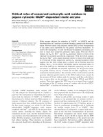
Acute phase of Friend Virus infection
(0-4 weeks post infection)
FV
CD4
Teffector
FV-induced
splenomegaly
IFN-γ
CD4
Thelper or Tfh
FV-infected
cells
CD4
Tregulatory
help
B
cells
Antibodies
IFN-γ, Perforin,
Granzymes
Maintenance &
Survival
CD8
Teffector
Figure 1 Distinct populations of CD4+ T cells regulate the virus-specific immune response during acute Friend Retrovirus infection.
CD4+ helper T cells and follicular helper T cells augment virus-specific cytotoxic T cell and antibody responses. In addition, a subpopulation of
effector CD4+ T cells directly inhibits virus replication. However, at the same time natural regulatory T cells expand and start to suppress effector
T cell responses, which interferes with control of virus replication. (Arrows indicate enhancement of responses, whereas blocked lines indicate
inhibition).
Nair et al. Retrovirology 2011, 8:76
/>
with the use of mice carrying a transgenic T cell receptor
chain specific for FV. The strong Tfh differentiation of
FV-specific CD4+ T cells can be a result of the specific
cytokine environment that this infection creates, as it is
likely to be the case for Th1 differentiation. Also, the efficient infection of B cells by FV [68,95], which then present
FV antigens to specific CD4+ T cells, may contribute to
enhance Tfh differentiation [96].
Although Tfh differentiation probably requires highavidity TCR interactions with antigen-presenting cells
following peptide immunization [97], no such requirement is observed during acute FV infection [94]. This
finding indicates that levels and/or persistence of antigen
presentation during viral infections may exceed those
achieved by peptide immunization, and therefore the
requirement for high-avidity TCR signaling is bypassed.
In HIV infections, the relative control of viremia is associated with the presence of IL-21-producing CD4 +
T cells [98]. Interestingly, evidence suggests that IL-21producing CD4+ T cells may be critical for the maintenance of CD8 + T cell responses during chronic virus
infections [99-101], although it remains to be determined
whether in all these cases IL-21 is produced by Tfh cells
or another T cell subset.
It is known that CD4+ T cells are generally important
for the clonal expansion, development of effector function, and the generation of long-term memory CD8 +
T cells [102]. The requirement of CD4+ T cell-help for
primary CD8+ T cell responses is determined by the nature of the infectious agent and the inflammatory milieu
formed by the pathogen [103-105]. Although T cell help
may be dispensable in the priming phase of the CD8 +
T cell response, it is essential in the generation and maintenance of long-lived memory CD8 + T cells [106-109],
and the function of CD8+ T cells during chronic infection
[110]. During the first two weeks of acute FV infection
the priming and expansion of CD8+ T cells occurs independently of CD4+ T-cell help [68]. In contrast, CD4 +
T cells are required for the maintenance of effector and
memory FV-specific CD8 + T cells during the recovery
phase of FV infection [58] (Figure 1). The situation is
slightly different in HIV-1 infections where the development of effector CD8+ T cell responses is compromised
in the absence of help from CD4+ T cells [47]. As mentioned above, there appears to be a role for CD4+ T cellproduced IL-21 in the development of HIV-specific
CD8 + T cell responses [98], and IL-21 has also been
shown to be an important cytokine in the maintenance of
CD8+ T cell functionality during chronic viral infections
[99-101].
Direct anti-viral functions of CD4+ T cells against FV
In addition to classical helper functions, CD4+ T cells
possess direct effector functions important in controlling
Page 5 of 12
infectious agents. As demonstrated in vitro, IFNg
secreted by CD4+ Th1 cells during FV infection is a key
component involved in the direct anti-viral effects of
CD4+ T cells [12]. Studies in genetic knockout mice and
mice depleted of IFNg-producing CD4+ T cells suggest
an especially important role in the long-term control of
persistent FV infection [12,57,111,112] (Figure 2). FVspecific CD4+ T cells from CD4+ TCRb-transgenic mice
with a TCRb chain specific for the F-MuLV env122-141
epitope rapidly expand in an antigen-dependent manner
when adoptively transferred into acutely infected mice.
The cells differentiate into Th1-type effector CD4 +
T cells that produce IFNg [58,59] (Figure 1). Adoptive
transfers of FV-specific CD4+ T cells into FV-infected
mice that are either lymphocyte-deficient or depleted,
protect from acute disease even in the absence of cytotoxic T cell or antibody responses [59]. These results
indicate potent and direct anti-viral effects by CD4+
T cells. Protection is not solely based on IFNg production, since protection against acute disease is also seen
in IFNg receptor deficient mice [59]. However, FV-specific CD4 + T cells only protect immunodeficient mice
against acute disease, and all animals eventually succumb to the infection in the absence of CD8 + T cells
and B cells [59]. In HIV infection too, anti-viral effector
responses in HIV-1-infected long-term non-progressors
are associated with increased levels of IFNg, the chemokine RANTES, and the macrophage inflammatory proteins
MIP-1a and MIP-1b that are produced by virus-specific
CD4 + T cells [113]. The rare individuals who display
immunological control over HIV not only possess effective
CD8 + CTL [114,115], but also contain multiple CD4 +
T cell clones with the characteristics of highly efficient
effector cells that have high-avidity to HIV gag peptides
and produce IFNg [116]. A most interesting and poorly
understood aspect of HIV controllers is that they can
maintain cell-mediated immune responses over long periods of chronic infection, a situation where most cellmediated responses become exhausted and ineffective.
In addition to providing help and secreting antiviral
factors, it has also been shown that CD4 + T cells can
develop the capacity to lyse infected cells. Although most
data come from cell lines and CD4+ T cell clones, it has
been shown that CD4+ T cells specific for LCMV [117],
influenza [118] have cytotoxic activity in vivo. Furthermore, cytotoxic CD4+ T cells from the peripheral blood
of individuals infected with HIV-1, influenza, EBV or
CMV display cytotoxic activity directly ex vivo [119-124].
One obvious limitation on CD4+ T cell-mediated cytotoxic activity is that cognate antigen is only recognized
on target cells that express MHC class II molecules.
Direct antiviral activity by CD4+ T cells seems to be critical during chronic FV infection while the presence of
virus-specific CD8 + T cells and virus-neutralizing
Nair et al. Retrovirology 2011, 8:76
/>
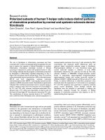
FV
Page 6 of 12
Chronic phase of Friend Virus infection
( > 6 weeks post infection)
CD4
Tregulatory
CD8
Teffector
FV-induced
splenomegaly
mild FV-induced
splenomegaly
FV-infected cells
CD4
Teffector
Figure 2 Distinct roles for CD4+ T cell subpopulations in chronic Friend Retrovirus infection. During chronic infection, effector CD8+ T cell
responses are suppressed by regulatory T cells but a subpopulation of effector CD4+ T cells prevents virus reactivation. (Blocked lines indicate
inhibition of immune responses or virus replication; the dotted line indicates that this response is suppressed during chronic infection).
antibodies have no correlation with chronic virus control
(Figure 2). FV replicates mainly in nucleated erythroid
precursors which are MHC class II-negative, and cytolysis of these cells is only observed as a by-stander effect in
the presence of APCs [12]. However, MHC class II-positive B cells are the main reservoirs of persistent FV [57]
and are susceptible to CD4+ T cell-mediated cytolysis.
The mechanisms underlying CD4+ T-cell mediated killing during acute and persistent FV infection are not fully
understood. Perforin, granzyme A, and granzyme B are
effector molecules of the granule exocytosis pathway that
are mainly produced by CD8+ T cells and control acute
FV infection. However, these molecules are not essential
during the chronic phase while Fas-FasL interaction, a
cytotoxic pathway that CD4+ T cells can use [125,126], is
mandatory for effective control of FV replication during
persistent infection [127].
CD4+ regulatory T cells in FV infection
Pioneering work using the FV model established that
mice persistently infected with FV display elevated levels
of activated CD4+CD25+ natural Tregs with potent inhibitory activity including the suppression of CD8+ T cellmediated killing of FV-induced tumors [60]. Later studies
showed that FV-induced Tregs rapidly suppressed the
function of TCR transgenic, FV-specific CD8 + T cells
adoptively transferred into chronically infected mice [61]
(Figure 2). Kinetic studies indicated that deterioration in
the ability of effector CD8+ T cells to produce cytotoxic
molecules and cytokines begins at 2 weeks post infection
Nair et al. Retrovirology 2011, 8:76
/>
(wpi), the same time point when CD4+ Treg expansion is
peaking [62] (Figure 1). The kinetic properties of FVmediated Treg expansion and CD8+ T-cell dysfunction
are not changed by LDV co-infection, and the expansion
of Treg occurs during FV infection but not LDV infection
[62,128]. To investigate the correlation between dysfunction of effector T cells and expansion of CD4+ Tregs, the
DEREG mouse was employed. These mice express a
Diptheria Toxin (DT) receptor/GFP fusion gene under
the control of the Foxp3 promoter, which is a transcription factor critical for the development and function of
CD4+ Tregs [129]. Foxp3 expressing cells can be experimentally depleted by treatment with DT. When FV
infected DEREG mice receive DT, it leads to specific
deletion of CD4+Foxp3+ Tregs. During acute FV infection, Treg depletion results in strongly augmented peak
CD8+ T cell responses, including a rise in the frequency
of FV-specific effector CD8 + T cells, dramatically
enhanced expression and degranulation of cytotoxic
molecules, and increased in vivo CTL-mediated lysis of
infected target cells [128]. Most importantly, this increase
in CD8+ T cell activity results in a significant reduction
in virus loads. During chronic FV infection, ablation of
Tregs induces proliferation of FV-specific CD8+ T cells
as well as reactivation of the residual but functionally
exhausted CD8+ T cells [39]. Importantly, the reactivation of the suppressed CD8 + T cell response in Tregdepleted mice results in reduced viral set points during
chronic retroviral infection.
CD4+ natural Tregs also influence the outcome of CD4+
effector T cell responses during acute FV infection. FVspecific CD4+ T cells display anti-viral effector functions
until 2 wpi, but thereafter their ability to produce IFNg is
reduced [67] (Figure 1). These FV-specific CD4+ T cells
with reduced IFNg expression at 3 wpi regain their ability
to produce IFNg following depletion of CD4 + Foxp3 +
Tregs in infected DEREG mice [67]. Thus, it is evident
that CD4+ Tregs negatively influence effector functions of
CD4+ T cells during acute FV infection thereby impairing
initial control over viral replication [67,129]. These findings are supported by the work of Antunes et al., who
showed that bone-marrow pathology observed in FVinfected lymphopenic mice, which is mediated by FV-specific CD4+ T cells is inhibited by Tregs [32].
Expansion of CD4+ Tregs during FV infection is highly
compartmentalized with CD4+ Tregs expanding in organs
with high viral replication and associated inflammation.
Interestingly, depletion experiments showed that the presence of CD8+ T cells supports the expansion of CD4+
Tregs in lymphatic tissues such as spleen and bone marrow [128]. Likewise, in HIV infection, CD4+ Tregs expand
predominantly in lymph nodes where the virus replicates
most efficiently and virus-specific CD8+ T cells accumulate. Thus, Treg numbers in lymph nodes correlate very
Page 7 of 12
well with disease progression of HIV infected individuals
[36,55,130]. Moreover, increased expression of aEb7 integrin (CD103) on CD4 + Foxp3 + Tregs suggests a role of
integrins in compartmentalization of Tregs in FV infection
[62]. Hence, it becomes imperative to investigate local
Treg responses before drawing conclusions on the role of
Tregs in retroviral infections.
In addition to CD8+ T cells, dendritic cells (DC) also
seem to be involved in the expansion of Tregs. We have
previously shown that FV-infected DCs do not fully
mature and specifically expand Foxp3 + Tregs in vitro
[131]. This has also been described for HIV-infected DCs
[132] suggesting a possible mechanism that retroviruses
may use to increase numbers of Tregs at sites of infection.
If DCs are involved in Treg expansion, one might presume
that they present viral antigens to Tregs that then proliferate in an antigen-specific manner. However, FV-specific
induced Tregs are undetectable in FV-infected mice either
by using class II tetramers or after adoptive transfer of FVspecific CD4+ T cells from TCR transgenic mice [128].
This finding is in agreement with the fact that CD4+ Tcell mediated bone-marrow pathology observed in
FV-infected lymphopenic hosts is impeded by immunosuppressive natural Tregs that are not specific for FV [32].
Recent findings from LCMV studies may explain these
seemingly contradictory results. Tregs expanding after an
LCMV clone 13 infection are not LCMV-specific, but at
least a fraction of them expand in response to an endogenous retroviral superantigen (Sag) [133]. The chronic
LCMV infection upregulates expression of the Sag in DCs,
which then induce proliferation of Tregs with certain
T cell receptors that can bind Sag. There is experimental
evidence that the percentage of Tregs with the same T cell
receptor (Vb5) increases during FV infection (own unpublished results), so a similar mechanism may also be
involved in the expansion of Tregs after FV infection.
Knowledge about the mechanisms underlying Tregmediated immunosupression during FV infection is limited. Studies using transgenic mice have demonstrated
that CD4+CD25+ T cells isolated from mice chronically
infected with FV suppress IFNg and granzyme B production by activated CD8+ T cells. Suppression occurs in a
direct cell-to-cell contact dependent manner independent
of the presence of APCs [134]. CD4+ Tregs may do so via
the expression of connexins, which are gap-junction proteins that have been found to be critical for the transfer of
the potent inhibitory second messenger cyclic AMP
(cAMP) into effector T cells [135,136]. In contrast, soluble
factors such as IL-10 and transforming growth factor
(TGF)-b secreted by CD4 + Tregs do not contribute
towards Treg-mediated immunosuppression in in vitro
and in vivo experiments [61,134]. Furthermore, FVinduced Tregs do not secrete granzymes, ruling out granzyme-dependent Treg-mediated apoptosis of effector
Nair et al. Retrovirology 2011, 8:76
/>
T cells [62]. The mechanism of suppression by Tregs in
FV infected mice is still under investigation. In HIV infections, immunosuppressive IL-10 production by CD4+ T
cells has been associated with disease progression, but it is
unclear whether these CD4+ T cells were Tregs [137]. It
has very recently been shown that Tregs control HIV
replication in activated T cells via a contact-dependent
mechanism involving cAMP [138].
Given the well-established role of Tregs in pathogen
persistence, it is now of great interest to develop therapeutic approaches to manipulate this immunosuppressive subset of cells. Treg functions are reversed by blocking
glucocorticoid-induced tumour necrosis factor receptor
(GITR), a member of the TNF receptor superfamily. GITR
is also a phenotypic marker of CD4+Foxp3+ Tregs and it is
highly expressed on Tregs during FV infection [62]. Blockade by antibodies leads to heightened production of IFNg
and TNFa by CD8+ T cells [61]. Antibody-mediated signaling through CD137 (4-1BB), a co-stimulatory molecule
also from the TNF receptor superfamily, renders CD8+ T
cells resistant to suppression by Tregs. Thus, anti-CD137
antibody therapy promotes virus-specific CD8+ T cell proliferation and development of effector functions to exert
control over chronic FV infections [139]. In vitro experiments with CD8+ T cells from HIV-infected patients also
show restored functional properties following treatment
with anti-CD137 antibodies [140].
In mice, an alternative therapeutic approach is the
depletion of Tregs, such as is done experimentally in the
DEREG mouse experiments [39,58,128]. Depletion of
Tregs leads to concerns that autoimmunity or other
immunopathology might be induced, but transient depletion of Tregs in the DEREG mice is not associated with
detectable immunopathology even during an ongoing antiretroviral immune response [128]. Such a therapeutic
approach may be a possible treatment in HIV infected
humans using an IL-2-toxin fusion protein (ONTAK)
[141] that kills CD4+CD25+ Tregs by binding to the IL-2
receptor via their expression of CD25. Treatment of cancer patients with ONTAK did not induce serious clinical
side effects. Jiang and co-workers performed an interesting
experiment to show that IL-2-toxin fusion proteinmediated depletion of CD4 + CD25 + Tregs in HIV-1
infected humanized mice resulted in a significant reduction of viral loads during acute HIV infection [142]. However, it is not known whether the reduction of viral loads
is mediated by an enhanced immune response.
In addition to HIV, Treg-mediated dysfunction of
effector T cells is a matter of concern in other chronic
virus infections such as HCV, HBV, and EBV [143].
Therefore, therapeutic manipulation of Tregs in vivo
with respect to enhancing virus-specific immunity and
balancing immunopathology could have widespread clinical applications.
Page 8 of 12
Conclusion
It has been known for some time that CD4+ T cells play a
critical role in retroviral immunity, but only recently has
the complexity of this subpopulation begun to be realized. Several distinct functions ascribed to subpopulations of CD4+ T cell have now been defined in mouse
retrovirus models. Type 1 helper CD4 + T cells were
important for the maintenance and survival of effector
CD8+ T cells, and follicular helper T cells critically supported antibody responses. CD4+ T cells with direct antiviral activity were also described, mainly during chronic
retroviral infection, but which may be active during acute
infections as well. Concurrent with the kinetics of the
antiviral CD4 + T cell response during acute retroviral
infection were the expansion and activation of a subpopulation of natural regulatory T cells at sites of infection.
The natural regulatory T cells suppressed effector T cell
responses, which interfered with immune control of virus
replication and contributed to viral chronicity. Similar
findings have also been made in HIV infected humans
and the therapeutic manipulation of regulatory T cells in
vivo with respect to enhancing retrovirus-specific immunity is a new frontier of high interest in the treatment of
viral infections.
Acknowledgements and funding
This work was supported by the Division of Intramural Research at the
National Institute of Allergy and Infectious Diseases, NIH.
Author details
1
Institute for Virology, University Clinics Essen, University of Duisburg-Essen,
Hufelandstrasse 55, 45122 Essen, Germany. 2Laboratory of Persistent Viral
Diseases, Rocky Mountain Laboratories, NIAID, NIH, Hamilton, MT 59840, USA.
3
Division of Immunoregulation, MRC National Institute for Medical Research,
The Ridgeway, London NW7 1AA, UK. 4Immune Cell Development and Host
Defense Program, Fox Chase Cancer Center, 333 Cottman Avenue,
Philadelphia, PA 19111, USA.
Authors’ contributions
SN, UD and KJH were responsible for drafting and revising the manuscript
as well as organizing the content. WB, MJ-YP, and GK contributed
significantly in drafting the manuscript and revising it critically. All authors
read and approved the final manuscript.
Competing interests
The authors declare that they have no competing interests.
Received: 24 May 2011 Accepted: 26 September 2011
Published: 26 September 2011
References
1. Mosmann TR, Cherwinski H, Bond MW, Giedlin MA, Coffman RL: Two types
of murine helper T cell clone. I. Definition according to profiles of
lymphokine activities and secreted proteins. J Immunol 1986,
136:2348-2357.
2. Kennedy R, Celis E: Multiple roles for CD4+ T cells in anti-tumor immune
responses. Immunol Rev 2008, 222:129-144.
3. Fayolle C, Deriaud E, Leclerc C: In vivo induction of cytotoxic T cell
response by a free synthetic peptide requires CD4+ T cell help.
JImmunol 1991, 147:4069-4073.
Nair et al. Retrovirology 2011, 8:76
/>
4.
5.
6.
7.
8.
9.
10.
11.
12.
13.
14.
15.
16.
17.
18.
19.
20.
21.
22.
23.
24.
Graham MB, Braciale VL, Braciale TJ: Influenza virus-specific CD4+ T helper
type 2 T lymphocytes do not promote recovery from experimental virus
infection. J Exp Med 1994, 180:1273-1282.
Maloy KJ, Burkhart C, Freer G, Rulicke T, Pircher H, Kono DH,
Theofilopoulos AN, Ludewig B, Hoffmann-Rohrer U, Zinkernagel RM,
Hengartner H: Qualitative and quantitative requirements for CD4+ T cellmediated antiviral protection. J Immunol 1999, 162:2867-2874.
Mahon BP, Katrak K, Nomoto A, Macadam AJ, Minor PD, Mills KH:
Poliovirus-specific CD4+ Th1 clones with both cytotoxic and helper
activity mediate protective humoral immunity against a lethal poliovirus
infection in transgenic mice expressing the human poliovirus receptor. J
Exp Med 1995, 181:1285-1292.
Cardin RD, Brooks JW, Sarawar SR, Doherty PC: Progressive loss of CD8+ T
cell-mediated control of a gamma-herpesvirus in the absence of CD4+ T
cells. J Exp Med 1996, 184:863-871.
Varga SM, Welsh RM: Stability of virus-specific CD4+ T cell frequencies
from acute infection into long term memory. J Immunol 1998,
161:367-374.
Manickan E, Rouse RJ, Yu Z, Wire WS, Rouse BT: Genetic immunization
against herpes simplex virus. Protection is mediated by CD4+ T
lymphocytes. J Immunol 1995, 155:259-265.
Maloy KJ, Burkhart C, Junt TM, Odermatt B, Oxenius A, Piali L,
Zinkernagel RM, Hengartner H: CD4(+) T cell subsets during virus
infection. Protective capacity depends on effector cytokine secretion
and on migratory capability. J Exp Med 2000, 191:2159-2170.
Reich A, Erlwein O, Niewiesk S, ter Meulen V, Liebert UG: CD4+ T cells
control measles virus infection of the central nervous system.
Immunology 1992, 76:185-191.
Iwashiro M, Peterson K, Messer RJ, Stromnes IM, Hasenkrug KJ: CD4(+) T
cells and gamma interferon in the long-term control of persistent friend
retrovirus infection. J Virol 2001, 75:52-60.
Gagnon SJ, Ennis FA, Rothman AL: Bystander target cell lysis and cytokine
production by dengue virus-specific human CD4(+) cytotoxic Tlymphocyte clones. J Virol 1999, 73:3623-3629.
Yasukawa M, Inatsuki A, Kobayashi Y: Differential in vitro activation of CD4
+CD8- and CD8+CD4- herpes simplex virus-specific human cytotoxic T
cells. J Immunol 1989, 143:2051-2057.
Barnaba V, Franco A, Paroli M, Benvenuto R, De Petrillo G, Burgio VL,
Santilio I, Balsano C, Bonavita MS, Cappelli G, et al: Selective expansion of
cytotoxic T lymphocytes with a CD4+CD56+ surface phenotype and a T
helper type 1 profile of cytokine secretion in the liver of patients
chronically infected with Hepatitis B virus. J Immunol 1994,
152:3074-3087.
Jacobson S, Richert JR, Biddison WE, Satinsky A, Hartzman RJ, McFarland HF:
Measles virus-specific T4+ human cytotoxic T cell clones are restricted
by class II HLA antigens. J Immunol 1984, 133:754-757.
Yakushijin Y, Yasukawa M, Kobayashi Y: Establishment and functional
characterization of human herpesvirus 6-specific CD4+ human T-cell
clones. J Virol 1992, 66:2773-2779.
Orentas RJ, Hildreth JE, Obah E, Polydefkis M, Smith GE, Clements ML,
Siliciano RF: Induction of CD4+ human cytolytic T cells specific for HIVinfected cells by a gp160 subunit vaccine. Science 1990, 248:1234-1237.
Bickham K, Munz C, Tsang ML, Larsson M, Fonteneau JF, Bhardwaj N,
Steinman R: EBNA1-specific CD4+ T cells in healthy carriers of EpsteinBarr virus are primarily Th1 in function. J Clin Invest 2001, 107:121-130.
Zhu J, Paul WE: CD4 T cells: fates, functions, and faults. Blood 2008,
112:1557-1569.
McHeyzer-Williams LJ, McHeyzer-Williams MG: Antigen-specific memory B
cell development. Annu Rev Immunol 2005, 23:487-513.
Harrington LE, Hatton RD, Mangan PR, Turner H, Murphy TL, Murphy KM,
Weaver CT: Interleukin 17-producing CD4+ effector T cells develop via a
lineage distinct from the T helper type 1 and 2 lineages. Nat Immunol
2005, 6:1123-1132.
Park H, Li Z, Yang XO, Chang SH, Nurieva R, Wang YH, Wang Y, Hood L,
Zhu Z, Tian Q, Dong C: A distinct lineage of CD4 T cells regulates tissue
inflammation by producing interleukin 17. Nat Immunol 2005,
6:1133-1141.
Yen D, Cheung J, Scheerens H, Poulet F, McClanahan T, McKenzie B,
Kleinschek MA, Owyang A, Mattson J, Blumenschein W, et al: IL-23 is
essential for T cell-mediated colitis and promotes inflammation via IL-17
and IL-6. J Clin Invest 2006, 116:1310-1316.
Page 9 of 12
25. Langrish CL, Chen Y, Blumenschein WM, Mattson J, Basham B, Sedgwick JD,
McClanahan T, Kastelein RA, Cua DJ: IL-23 drives a pathogenic T cell
population that induces autoimmune inflammation. J Exp Med 2005,
201:233-240.
26. Breitfeld D, Ohl L, Kremmer E, Ellwart J, Sallusto F, Lipp M, Forster R:
Follicular B helper T cells express CXC chemokine receptor 5, localize to
B cell follicles, and support immunoglobulin production. J Exp Med 2000,
192:1545-1552.
27. Crotty S: Follicular Helper CD4 T Cells (T(FH)). Annu Rev Immunol 2011.
28. Wing K, Sakaguchi S: Regulatory T cells exert checks and balances on self
tolerance and autoimmunity. Nat Immunol 2010, 11:7-13.
29. Boyer O, Saadoun D, Abriol J, Dodille M, Piette JC, Cacoub P, Klatzmann D:
CD4+CD25+ regulatory T-cell deficiency in patients with hepatitis Cmixed cryoglobulinemia vasculitis. Blood 2004, 103:3428-3430.
30. Suvas S, Azkur AK, Kim BS, Kumaraguru U, Rouse BT: CD4+CD25+
regulatory T cells control the severity of viral immunoinflammatory
lesions. J Immunol 2004, 172:4123-4132.
31. Lund JM, Hsing L, Pham TT, Rudensky AY: Coordination of early protective
immunity to viral infection by regulatory T cells. Science 2008,
320:1220-1224.
32. Antunes I, Tolaini M, Kissenpfennig A, Iwashiro M, Kuribayashi K, Malissen B,
Hasenkrug K, Kassiotis G: Retrovirus-specificity of regulatory T cells is
neither present nor required in preventing retrovirus-induced bone
marrow immune pathology. Immunity 2008, 29:782-794.
33. Brady MT, MacDonald AJ, Rowan AG, Mills KH: Hepatitis C virus nonstructural protein 4 suppresses Th1 responses by stimulating IL-10
production from monocytes. Eur J Immunol 2003, 33:3448-3457.
34. Boettler T, Spangenberg HC, Neumann-Haefelin C, Panther E, Urbani S,
Ferrari C, Blum HE, von Weizsacker F, Thimme R: T cells with a CD4+CD25
+ regulatory phenotype suppress in vitro proliferation of virus-specific
CD8+ T cells during chronic hepatitis C virus infection. J Virol 2005,
79:7860-7867.
35. Rushbrook SM, Ward SM, Unitt E, Vowler SL, Lucas M, Klenerman P,
Alexander GJ: Regulatory T cells suppress in vitro proliferation of virusspecific CD8+ T cells during persistent hepatitis C virus infection. J Virol
2005, 79:7852-7859.
36. Andersson J, Boasso A, Nilsson J, Zhang R, Shire NJ, Lindback S,
Shearer GM, Chougnet CA: The prevalence of regulatory T cells in
lymphoid tissue is correlated with viral load in HIV-infected patients. J
Immunol 2005, 174:3143-3147.
37. Marshall NA, Vickers MA, Barker RN: Regulatory T cells secreting IL-10
dominate the immune response to EBV latent membrane protein 1. J
Immunol 2003, 170:6183-6189.
38. Suvas S, Kumaraguru U, Pack CD, Lee S, Rouse BT: CD4+CD25+ T cells
regulate virus-specific primary and memory CD8+ T cell responses. J Exp
Med 2003, 198:889-901.
39. Dietze KK, Zelinskyy G, Gibbert K, Schimmer S, Francois S, Myers L,
Sparwasser T, Hasenkrug KJ, Dittmer U: Transient depletion of regulatory T
cells in transgenic mice reactivates virus-specific CD8+ T cells and
reduces chronic retroviral set points. Proc Natl Acad Sci USA 2011,
108:2420-2425.
40. Nishikawa H, Sakaguchi S: Regulatory T cells in tumor immunity. Int J
Cancer 2010, 127:759-767.
41. Doitsh G, Cavrois M, Lassen KG, Zepeda O, Yang Z, Santiago ML,
Hebbeler AM, Greene WC: Abortive HIV infection mediates CD4 T cell
depletion and inflammation in human lymphoid tissue. Cell 2010,
143:789-801.
42. Grossman Z, Meier-Schellersheim M, Sousa AE, Victorino RM, Paul WE: CD4+
T-cell depletion in HIV infection: are we closer to understanding the
cause? Nat Med 2002, 8:319-323.
43. Norris PJ, Sumaroka M, Brander C, Moffett HF, Boswell SL, Nguyen T,
Sykulev Y, Walker BD, Rosenberg ES: Multiple effector functions mediated
by human immunodeficiency virus-specific CD4(+) T-cell clones. J Virol
2001, 75:9771-9779.
44. Sethi KK, Naher H, Stroehmann I: Phenotypic heterogeneity of
cerebrospinal fluid-derived HIV-specific and HLA-restricted cytotoxic Tcell clones. Nature 1988, 335:178-181.
45. Littaua RA, Oldstone MB, Takeda A, Ennis FA: A CD4+ cytotoxic Tlymphocyte clone to a conserved epitope on human immunodeficiency
virus type 1 p24: cytotoxic activity and secretion of interleukin-2 and
interleukin-6. J Virol 1992, 66:608-611.
Nair et al. Retrovirology 2011, 8:76
/>
46. Altfeld M, Rosenberg ES: The role of CD4(+) T helper cells in the cytotoxic
T lymphocyte response to HIV-1. Curr Opin Immunol 2000, 12:375-380.
47. Spiegel HM, Ogg GS, DeFalcon E, Sheehy ME, Monard S, Haslett PA,
Gillespie G, Donahoe SM, Pollack H, Borkowsky W, et al: Human
immunodeficiency virus type 1- and cytomegalovirus-specific cytotoxic T
lymphocytes can persist at high frequency for prolonged periods in the
absence of circulating peripheral CD4(+) T cells. J Virol 2000,
74:1018-1022.
48. Poignard P, Sabbe R, Picchio GR, Wang M, Gulizia RJ, Katinger H, Parren PW,
Mosier DE, Burton DR: Neutralizing antibodies have limited effects on the
control of established HIV-1 infection in vivo. Immunity 1999, 10:431-438.
49. Veazey RS, DeMaria M, Chalifoux LV, Shvetz DE, Pauley DR, Knight HL,
Rosenzweig M, Johnson RP, Desrosiers RC, Lackner AA: Gastrointestinal
tract as a major site of CD4+ T cell depletion and viral replication in SIV
infection. Science 1998, 280:427-431.
50. Guadalupe M, Reay E, Sankaran S, Prindiville T, Flamm J, McNeil A,
Dandekar S: Severe CD4+ T-cell depletion in gut lymphoid tissue during
primary human immunodeficiency virus type 1 infection and substantial
delay in restoration following highly active antiretroviral therapy. J Virol
2003, 77:11708-11717.
51. Brenchley JM, Price DA, Schacker TW, Asher TE, Silvestri G, Rao S, Kazzaz Z,
Bornstein E, Lambotte O, Altmann D, et al: Microbial translocation is a
cause of systemic immune activation in chronic HIV infection. Nat Med
2006, 12:1365-1371.
52. Hunt PW, Landay AL, Sinclair E, Martinson JA, Hatano H, Emu B, Norris PJ,
Busch MP, Martin JN, Brooks C, et al: A low T regulatory cell response may
contribute to both viral control and generalized immune activation in
HIV controllers. PLoS One 2011, 6:e15924.
53. Marques R, Williams A, Eksmond U, Wullaert A, Killeen N, Pasparakis M,
Kioussis D, Kassiotis G: Generalized immune activation as a direct result
of activated CD4+ T cell killing. J Biol 2009, 8:93.
54. Kinter AL, Hennessey M, Bell A, Kern S, Lin Y, Daucher M, Planta M,
McGlaughlin M, Jackson R, Ziegler SF, Fauci AS: CD25(+)CD4(+) regulatory
T cells from the peripheral blood of asymptomatic HIV-infected
individuals regulate CD4(+) and CD8(+) HIV-specific T cell immune
responses in vitro and are associated with favorable clinical markers of
disease status. J Exp Med 2004, 200:331-343.
55. Nilsson J, Boasso A, Velilla PA, Zhang R, Vaccari M, Franchini G, Shearer GM,
Andersson J, Chougnet C: HIV-1-driven regulatory T-cell accumulation in
lymphoid tissues is associated with disease progression in HIV/AIDS.
Blood 2006, 108:3808-3817.
56. Aandahl EM, Michaelsson J, Moretto WJ, Hecht FM, Nixon DF: Human CD4
+ CD25+ regulatory T cells control T-cell responses to human
immunodeficiency virus and cytomegalovirus antigens. J Virol 2004,
78:2454-2459.
57. Hasenkrug KJ, Brooks DM, Dittmer U: Critical role for CD4(+) T cells in
controlling retrovirus replication and spread in persistently infected
mice. J Virol 1998, 72:6559-6564.
58. Nair SR, Zelinskyy G, Schimmer S, Gerlach N, Kassiotis G, Dittmer U:
Mechanisms of control of acute Friend virus infection by CD4+ T helper
cells and their functional impairment by regulatory T cells. J Gen Virol
2010, 91:440-451.
59. Pike R, Filby A, Ploquin MJ, Eksmond U, Marques R, Antunes I, Hasenkrug K,
Kassiotis G: Race between retroviral spread and CD4+ T-cell response
determines the outcome of acute Friend virus infection. J Virol 2009,
83:11211-11222.
60. Iwashiro M, Messer RJ, Peterson KE, Stromnes IM, Sugie T, Hasenkrug KJ:
Immunosuppression by CD4+ regulatory T cells induced by chronic
retroviral infection. Proc Natl Acad Sci USA 2001, 98:9226-9230.
61. Dittmer U, He H, Messer RJ, Schimmer S, Olbrich AR, Ohlen C,
Greenberg PD, Stromnes IM, Iwashiro M, Sakaguchi S, et al: Functional
impairment of CD8(+) T cells by regulatory T cells during persistent
retroviral infection. Immunity 2004, 20:293-303.
62. Zelinskyy G, Kraft AR, Schimmer S, Arndt T, Dittmer U: Kinetics of CD8+
effector T cell responses and induced CD4+ regulatory T cell responses
during Friend retrovirus infection. Eur J Immunol 2006, 36:2658-2670.
63. Friend C: Cell-free transmission in adult Swiss mice of a disease having
the character of a leukemia. J Exp Med 1957, 105:307-318.
64. Chesebro B, Miyazawa M, Britt WJ: Host genetic control of spontaneous
and induced immunity to Friend murine retrovirus infection. Annu Rev
Immunol 1990, 8:477-499.
Page 10 of 12
65. Kabat D: Molecular biology of Friend viral erythroleukemia. Curr Top
Microbiol Immunol 1989, 148:1-42.
66. Hasenkrug KJ, Chesebro B: Immunity to retroviral infection: the Friend
virus model. Proc Natl Acad Sci USA 1997, 94:7811-7816.
67. Hasenkrug KJ: Lymphocyte deficiencies increase susceptibility to friend
virus-induced erythroleukemia in Fv-2 genetically resistant mice. J Virol
1999, 73:6468-6473.
68. Robertson SJ, Ammann CG, Messer RJ, Carmody AB, Myers L, Dittmer U,
Nair S, Gerlach N, Evans LH, Cafruny WA, Hasenkrug KJ: Suppression of
acute anti-friend virus CD8+ T-cell responses by coinfection with lactate
dehydrogenase-elevating virus. J Virol 2008, 82:408-418.
69. Marques R, Antunes I, Eksmond U, Stoye J, Hasenkrug K, Kassiotis G: B
lymphocyte activation by coinfection prevents immune control of friend
virus infection. J Immunol 2008, 181:3432-3440.
70. Robertson MN, Spangrude GJ, Hasenkrug K, Perry L, Nishio J, Wehrly K,
Chesebro B: Role and specificity of T-cell subsets in spontaneous
recovery from Friend virus-induced leukemia in mice. J Virol 1992,
66:3271-3277.
71. Miyazawa M, Nishio J, Chesebro B: Genetic control of T cell
responsiveness to the Friend murine leukemia virus envelope antigen.
Identification of class II loci of the H-2 as immune response genes. J Exp
Med 1988, 168:1587-1605.
72. Iwashiro M, Kondo T, Shimizu T, Yamagishi H, Takahashi K, Matsubayashi Y,
Masuda T, Otaka A, Fujii N, Ishimoto A, et al: Multiplicity of virus-encoded
helper T-cell epitopes expressed on FBL-3 tumor cells. J Virol 1993,
67:4533-4542.
73. Shimizu T, Uenishi H, Teramura Y, Iwashiro M, Kuribayashi K, Tamamura H,
Fujii N, Yamagishi H: Fine structure of a virus-encoded helper T-cell
epitope expressed on FBL-3 tumor cells. J Virol 1994, 68:7704-7708.
74. Sugahara D, Tsuji-Kawahara S, Miyazawa M: Identification of a protective
CD4+ T-cell epitope in p15gag of Friend murine leukemia virus and role
of the MA protein targeting the plasma membrane in immunogenicity. J
Virol 2004, 78:6322-6334.
75. Earl PL, Moss B, Morrison RP, Wehrly K, Nishio J, Chesebro B: T-lymphocyte
priming and protection against Friend leukemia by vaccinia-retrovirus
env gene recombinant. Science 1986, 234:728-731.
76. Miyazawa M, Nishio J, Chesebro B: Protection against Friend retrovirusinduced leukemia by recombinant vaccinia viruses expressing the gag
gene. J Virol 1992, 66:4497-4507.
77. Bayer W, Tenbusch M, Lietz R, Johrden L, Schimmer S, Uberla K, Dittmer U,
Wildner O: Vaccination with an adenoviral vector that encodes and
displays a retroviral antigen induces improved neutralizing antibody and
CD4+ T-cell responses and confers enhanced protection. J Virol 2010,
84:1967-1976.
78. Miyazawa M, Fujisawa R, Ishihara C, Takei YA, Shimizu T, Uenishi H,
Yamagishi H, Kuribayashi K: Immunization with a single T helper cell
epitope abrogates Friend virus-induced early erythroid proliferation and
prevents late leukemia development. J Immunol 1995, 155:748-758.
79. Iwanami N, Niwa A, Yasutomi Y, Tabata N, Miyazawa M: Role of natural
killer cells in resistance against friend retrovirus-induced leukemia. J Virol
2001, 75:3152-3163.
80. Chesebro B: MHC recombinant mice: genetic tools for study of resistance
to viral disease. Int Rev Immunol 1988, 3:393-396.
81. Morrison RP, Earl PL, Nishio J, Lodmell DL, Moss B, Chesebro B: Different H2 subregions influence immunization against retrovirus and
immunosuppression. Nature 1987, 329:729-732.
82. Dittmer U, Brooks DM, Hasenkrug KJ: Characterization of a live-attenuated
retroviral vaccine demonstrates protection via immune mechanisms. J
Virol 1998, 72:6554-6558.
83. Dittmer U, Brooks DM, Hasenkrug KJ: Requirement for multiple
lymphocyte subsets in protection by a live attenuated vaccine against
retroviral infection. Nat Med 1999, 5:189-193.
84. Dittmer U, Hasenkrug KJ: Different immunological requirements for
protection against acute versus persistent Friend retrovirus infections.
Virology 2000, 272:177-182.
85. Chesebro B, Wehrly K: Identification of a non-H-2 gene (Rfv-3) influencing
recovery from viremia and leukemia induced by Friend virus complex.
Proc Natl Acad Sci USA 1979, 76:425-429.
86. Chesebro B, Wehrly K: Studies on the role of the host immune response
in recovery from Friend virus leukemia. I. Antiviral and antileukemia cell
antibodies. J Exp Med 1976, 143:73-84.
Nair et al. Retrovirology 2011, 8:76
/>
87. Doig D, Chesebro B: Anti-Friend virus antibody is associated with
recovery from viremia and loss of viral leukemia cell-surface antigens in
leukemic mice. Identification of Rfv-3 as a gene locus influencing
antibody production. J Exp Med 1979, 150:10-19.
88. Zwick MB, Burton DR: HIV-1 neutralization: mechanisms and relevance to
vaccine design. Curr HIV Res 2007, 5:608-624.
89. Hasenkrug KJ, Brooks DM, Chesebro B: Passive immunotherapy for
retroviral disease: influence of major histocompatibility complex type
and T-cell responsiveness. Proc Natl Acad Sci USA 1995, 92:10492-10495.
90. Super HJ, Brooks D, Hasenkrug K, Chesebro B: Requirement for CD4(+) T
cells in the Friend murine retrovirus neutralizing antibody response:
evidence for functional T cells in genetic low-recovery mice. J Virol 1998,
72:9400-9403.
91. Johnston RJ, Poholek AC, DiToro D, Yusuf I, Eto D, Barnett B, Dent AL,
Craft J, Crotty S: Bcl6 and Blimp-1 are reciprocal and antagonistic
regulators of T follicular helper cell differentiation. Science 2009,
325:1006-1010.
92. Nurieva RI, Chung Y, Martinez GJ, Yang XO, Tanaka S, Matskevitch TD,
Wang YH, Dong C: Bcl6 mediates the development of T follicular helper
cells. Science 2009, 325:1001-1005.
93. Yu D, Rao S, Tsai LM, Lee SK, He Y, Sutcliffe EL, Srivastava M, Linterman M,
Zheng L, Simpson N, et al: The transcriptional repressor Bcl-6 directs T
follicular helper cell lineage commitment. Immunity 2009, 31:457-468.
94. Ploquin MJ, Eksmond U, Kassiotis G: B cells and TCR avidity determine
distinct functions of CD4+ T cells in retroviral infection. J Immunol. 2011
Sep 15;187(6):3321-30. 2011 Sep 15, 187(6):3321-3330.
95. Bila C, Oberhauser V, Ammann CG, Ejaz A, Huber G, Schimmer S, Messer R,
Pekna M, von Laer D, Dittmer U, et al: Complement opsonization
enhances friend virus infection of B cells and thereby amplifies the
virus-specific CD8+ T cell response. J Virol 2011, 85:1151-1155.
96. Deenick EK, Chan A, Ma CS, Gatto D, Schwartzberg PL, Brink R, Tangye SG:
Follicular helper T cell differentiation requires continuous antigen
presentation that is independent of unique B cell signaling. Immunity
2010, 33:241-253.
97. Fazilleau N, McHeyzer-Williams LJ, Rosen H, McHeyzer-Williams MG: The
function of follicular helper T cells is regulated by the strength of T cell
antigen receptor binding. Nat Immunol 2009, 10:375-384.
98. Yue FY, Lo C, Sakhdari A, Lee EY, Kovacs CM, Benko E, Liu J, Song H,
Jones RB, Sheth P, et al: HIV-specific IL-21 producing CD4+ T cells are
induced in acute and chronic progressive HIV infection and are
associated with relative viral control. J Immunol 2010, 185:498-506.
99. Frohlich A, Kisielow J, Schmitz I, Freigang S, Shamshiev AT, Weber J,
Marsland BJ, Oxenius A, Kopf M: IL-21R on T cells is critical for sustained
functionality and control of chronic viral infection. Science 2009,
324:1576-1580.
100. Elsaesser H, Sauer K, Brooks DG: IL-21 is required to control chronic viral
infection. Science 2009, 324:1569-1572.
101. Yi JS, Du M, Zajac AJ: A vital role for interleukin-21 in the control of a
chronic viral infection. Science 2009, 324:1572-1576.
102. Bevan MJ: Helping the CD8(+) T-cell response. Nat Rev Immunol 2004,
4:595-602.
103. Jennings SR, Bonneau RH, Smith PM, Wolcott RM, Chervenak R: CD4positive T lymphocytes are required for the generation of the primary
but not the secondary CD8-positive cytolytic T lymphocyte response
to herpes simplex virus in C57BL/6 mice. Cell Immunol 1991,
133:234-252.
104. Riberdy JM, Christensen JP, Branum K, Doherty PC: Diminished primary
and secondary influenza virus-specific CD8(+) T-cell responses in CD4depleted Ig(-/-) mice. J Virol 2000, 74:9762-9765.
105. Smith CM, Wilson NS, Waithman J, Villadangos JA, Carbone FR, Heath WR,
Belz GT: Cognate CD4(+) T cell licensing of dendritic cells in CD8(+) T
cell immunity. Nat Immunol 2004, 5:1143-1148.
106. Frank GM, Lepisto AJ, Freeman ML, Sheridan BS, Cherpes TL, Hendricks RL:
Early CD4(+) T cell help prevents partial CD8(+) T cell exhaustion and
promotes maintenance of Herpes Simplex Virus 1 latency. J Immunol
2010, 184:277-286.
107. Shedlock DJ, Shen H: Requirement for CD4 T cell help in generating
functional CD8 T cell memory. Science 2003, 300:337-339.
108. Novy P, Quigley M, Huang X, Yang Y: CD4 T cells are required for CD8 T
cell survival during both primary and memory recall responses. J
Immunol 2007, 179:8243-8251.
Page 11 of 12
109. Janssen EM, Lemmens EE, Wolfe T, Christen U, von Herrath MG,
Schoenberger SP: CD4+ T cells are required for secondary expansion and
memory in CD8+ T lymphocytes. Nature 2003, 421:852-856.
110. Matloubian M, Concepcion RJ, Ahmed R: CD4+ T cells are required to
sustain CD8+ cytotoxic T-cell responses during chronic viral infection. J
Virol 1994, 68:8056-8063.
111. Stromnes IM, Dittmer U, Schumacher TN, Schepers K, Messer RJ, Evans LH,
Peterson KE, Race B, Hasenkrug KJ: Temporal effects of gamma interferon
deficiency on the course of Friend retrovirus infection in mice. J Virol
2002, 76:2225-2232.
112. Dittmer U, Peterson KE, Messer R, Stromnes IM, Race B, Hasenkrug KJ: Role
of interleukin-4 (IL-4), IL-12, and gamma interferon in primary and
vaccine-primed immune responses to Friend retrovirus infection. J Virol
2001, 75:654-660.
113. Rosenberg ES, Billingsley JM, Caliendo AM, Boswell SL, Sax PE, Kalams SA,
Walker BD: Vigorous HIV-1-specific CD4+ T cell responses associated with
control of viremia. Science 1997, 278:1447-1450.
114. Saez-Cirion A, Lacabaratz C, Lambotte O, Versmisse P, Urrutia A, Boufassa F,
Barre-Sinoussi F, Delfraissy JF, Sinet M, Pancino G, Venet A: HIV controllers
exhibit potent CD8 T cell capacity to suppress HIV infection ex vivo and
peculiar cytotoxic T lymphocyte activation phenotype. Proc Natl Acad Sci
USA 2007, 104:6776-6781.
115. Migueles SA, Osborne CM, Royce C, Compton AA, Joshi RP, Weeks KA,
Rood JE, Berkley AM, Sacha JB, Cogliano-Shutta NA, et al: Lytic granule
loading of CD8+ T cells is required for HIV-infected cell elimination
associated with immune control. Immunity 2008, 29:1009-1021.
116. Vingert B, Perez-Patrigeon S, Jeannin P, Lambotte O, Boufassa F, Lemaitre F,
Kwok WW, Theodorou I, Delfraissy JF, Theze J, Chakrabarti LA: HIV
controller CD4+ T cells respond to minimal amounts of Gag antigen due
to high TCR avidity. PLoS Pathog 2010, 6:e1000780.
117. Jellison ER, Kim SK, Welsh RM: Cutting edge: MHC class II-restricted killing
in vivo during viral infection. J Immunol 2005, 174:614-618.
118. Brown DM, Dilzer AM, Meents DL, Swain SL: CD4 T cell-mediated
protection from lethal influenza: perforin and antibody-mediated
mechanisms give a one-two punch. J Immunol 2006, 177:2888-2898.
119. Appay V, Zaunders JJ, Papagno L, Sutton J, Jaramillo A, Waters A,
Easterbrook P, Grey P, Smith D, McMichael AJ, et al: Characterization of
CD4(+) CTLs ex vivo. J Immunol 2002, 168:5954-5958.
120. Lotti B, Wendland T, Furrer H, Yawalkar N, von Greyerz S, Schnyder K,
Brandes M, Vernazza P, Wagner R, Nguyen T, et al: Cytotoxic HIV-1
p55gag-specific CD4+ T cells produce HIV-inhibitory cytokines and
chemokines. J Clin Immunol 2002, 22:253-262.
121. Norris PJ, Moffett HF, Yang OO, Kaufmann DE, Clark MJ, Addo MM,
Rosenberg ES: Beyond help: direct effector functions of human
immunodeficiency virus type 1-specific CD4(+) T cells. J Virol 2004,
78:8844-8851.
122. Hammond SA, Bollinger RC, Stanhope PE, Quinn TC, Schwartz D,
Clements ML, Siliciano RF: Comparative clonal analysis of human
immunodeficiency virus type 1 (HIV-1)-specific CD4+ and CD8+ cytolytic
T lymphocytes isolated from seronegative humans immunized with
candidate HIV-1 vaccines. J Exp Med 1992, 176:1531-1542.
123. Zaunders JJ, Dyer WB, Wang B, Munier ML, Miranda-Saksena M, Newton R,
Moore J, Mackay CR, Cooper DA, Saksena NK, Kelleher AD: Identification of
circulating antigen-specific CD4+ T lymphocytes with a CCR5+, cytotoxic
phenotype in an HIV-1 long-term nonprogressor and in CMV infection.
Blood 2004, 103:2238-2247.
124. Jameson J, Cruz J, Terajima M, Ennis FA: Human CD8+ and CD4+ T
lymphocyte memory to influenza A viruses of swine and avian species. J
Immunol 1999, 162:7578-7583.
125. Appay V: The physiological role of cytotoxic CD4(+) T-cells: the holy
grail? Clin Exp Immunol 2004, 138:10-13.
126. Stalder T, Hahn S, Erb P: Fas antigen is the major target molecule for CD4
+ T cell-mediated cytotoxicity. J Immunol 1994, 152:1127-1133.
127. Zelinskyy G, Balkow S, Schimmer S, Schepers K, Simon MM, Dittmer U:
Independent roles of perforin, granzymes, and Fas in the control of
Friend retrovirus infection. Virology 2004, 330:365-374.
128. Zelinskyy G, Dietze KK, Husecken YP, Schimmer S, Nair S, Werner T,
Gibbert K, Kershaw O, Gruber AD, Sparwasser T, Dittmer U: The regulatory
T-cell response during acute retroviral infection is locally defined and
controls the magnitude and duration of the virus-specific cytotoxic Tcell response. Blood 2009, 114:3199-3207.
Nair et al. Retrovirology 2011, 8:76
/>
Page 12 of 12
129. Lahl K, Loddenkemper C, Drouin C, Freyer J, Arnason J, Eberl G, Hamann A,
Wagner H, Huehn J, Sparwasser T: Selective depletion of Foxp3+
regulatory T cells induces a scurfy-like disease. J Exp Med 2007, 204:57-63.
130. Kinter A, McNally J, Riggin L, Jackson R, Roby G, Fauci AS: Suppression of
HIV-specific T cell activity by lymph node CD25+ regulatory T cells from
HIV-infected individuals. Proc Natl Acad Sci USA 2007, 104:3390-3395.
131. Balkow S, Krux F, Loser K, Becker JU, Grabbe S, Dittmer U: Friend retrovirus
infection of myeloid dendritic cells impairs maturation, prolongs contact
to naive T cells, and favors expansion of regulatory T cells. Blood 2007,
110:3949-3958.
132. Che KF, Sabado RL, Shankar EM, Tjomsland V, Messmer D, Bhardwaj N,
Lifson JD, Larsson M: HIV-1 impairs in vitro priming of naive T cells and
gives rise to contact-dependent suppressor T cells. Eur J Immunol 2010,
40:2248-2258.
133. Punkosdy GA, Blain M, Glass DD, Lozano MM, O’Mara L, Dudley JP,
Ahmed R, Shevach EM: Regulatory T-cell expansion during chronic viral
infection is dependent on endogenous retroviral superantigens. Proc
Natl Acad Sci USA 108:3677-3682.
134. Robertson SJ, Messer RJ, Carmody AB, Hasenkrug KJ: In vitro suppression
of CD8+ T cell function by Friend virus-induced regulatory T cells. J
Immunol 2006, 176:3342-3349.
135. Bopp T, Becker C, Klein M, Klein-Hessling S, Palmetshofer A, Serfling E,
Heib V, Becker M, Kubach J, Schmitt S, et al: Cyclic adenosine
monophosphate is a key component of regulatory T cell-mediated
suppression. J Exp Med 2007, 204:1303-1310.
136. Bopp T, Dehzad N, Reuter S, Klein M, Ullrich N, Stassen M, Schild H, Buhl R,
Schmitt E, Taube C: Inhibition of cAMP degradation improves regulatory
T cell-mediated suppression. J Immunol 2009, 182:4017-4024.
137. Ostrowski MA, Gu JX, Kovacs C, Freedman J, Luscher MA, MacDonald KS:
Quantitative and qualitative assessment of human immunodeficiency
virus type 1 (HIV-1)-specific CD4+ T cell immunity to gag in HIV-1infected individuals with differential disease progression: reciprocal
interferon-gamma and interleukin-10 responses. J Infect Dis 2001,
184:1268-1278.
138. Moreno-Fernandez ME, Rueda CM, Rusie LK, Chougnet CA: Regulatory T
cells control HIV replication in activated T cells through a cAMPdependent mechanism. Blood 2011, 117:5372-5380.
139. Robertson SJ, Messer RJ, Carmody AB, Mittler RS, Burlak C, Hasenkrug KJ:
CD137 costimulation of CD8+ T cells confers resistance to suppression
by virus-induced regulatory T cells. J Immunol 2008, 180:5267-5274.
140. Wang C, Wen T, Routy JP, Bernard NF, Sekaly RP, Watts TH: 4-1BBL induces
TNF receptor-associated factor 1-dependent Bim modulation in human
T cells and is a critical component in the costimulation-dependent
rescue of functionally impaired HIV-specific CD8 T cells. J Immunol 2007,
179:8252-8263.
141. Dannull J, Su Z, Rizzieri D, Yang BK, Coleman D, Yancey D, Zhang A,
Dahm P, Chao N, Gilboa E, Vieweg J: Enhancement of vaccine-mediated
antitumor immunity in cancer patients after depletion of regulatory T
cells. J Clin Invest 2005, 115:3623-3633.
142. Jiang Q, Zhang L, Wang R, Jeffrey J, Washburn ML, Brouwer D, Barbour S,
Kovalev GI, Unutmaz D, Su L: FoxP3+CD4+ regulatory T cells play an
important role in acute HIV-1 infection in humanized Rag2-/-gammaC-/mice in vivo. Blood 2008, 112:2858-2868.
143. Li S, Gowans EJ, Chougnet C, Plebanski M, Dittmer U: Natural regulatory T
cells and persistent viral infection. J Virol 2008, 82:21-30.
doi:10.1186/1742-4690-8-76
Cite this article as: Nair et al.: Distinct roles of CD4+ T cell
subpopulations in retroviral immunity: lessons from the Friend virus
mouse model. Retrovirology 2011 8:76.
Submit your next manuscript to BioMed Central
and take full advantage of:
• Convenient online submission
• Thorough peer review
• No space constraints or color figure charges
• Immediate publication on acceptance
• Inclusion in PubMed, CAS, Scopus and Google Scholar
• Research which is freely available for redistribution
Submit your manuscript at
www.biomedcentral.com/submit