Báo cáo khoa học: "Alternative protocol to initiate high-frequency oscillatory ventilation: an experimental study" pot
Bạn đang xem bản rút gọn của tài liệu. Xem và tải ngay bản đầy đủ của tài liệu tại đây (321.69 KB, 9 trang )
Open Access
Available online />Page 1 of 9
(page number not for citation purposes)
Vol 10 No 5
Research
Alternative protocol to initiate high-frequency oscillatory
ventilation: an experimental study
Jens Karmrodt, Matthias David, Shying Yuan and Klaus Markstaller
Department of Anesthesiology, Johannes Gutenberg-University, Langenbeckstraße 1, D-55101 Mainz, Germany
Corresponding author: Jens Karmrodt,
Received: 15 May 2006 Revisions requested: 13 Jun 2006 Revisions received: 4 Sep 2006 Accepted: 25 Sep 2006 Published: 25 Sep 2006
Critical Care 2006, 10:R138 (doi:10.1186/cc5052)
This article is online at: />© 2006 Karmrodt et al.; licensee BioMed Central Ltd.
This is an open access article distributed under the terms of the Creative Commons Attribution License ( />),
which permits unrestricted use, distribution, and reproduction in any medium, provided the original work is properly cited.
Abstract
Introduction The objective was to study the effects of a novel
lung volume optimization procedure (LVOP) using high-
frequency oscillatory ventilation (HFOV) upon gas exchange, the
transpulmonary pressure (TPP), and hemodynamics in a porcine
model of surfactant depletion.
Methods With institutional review board approval, the
hemodynamics, blood gas analysis, TPP, and pulmonary shunt
fraction were obtained in six anesthetized pigs before and after
saline lung lavage. Measurements were acquired during
pressure-controlled ventilation (PCV) prior to and after lung
damage, and during a LVOP with HFOV. The LVOP comprised
a recruitment maneuver with a continuous distending pressure
(CDP) of 45 mbar for 2.5 minutes, and a stepwise decrease of
the CDP (5 mbar every 5 minute) from 45 to 20 mbar. The TPP
level was identified during the decrease in CDP, which assured
a change of the P
a
O
2
/F
I
O
2
ratio < 25% compared with
maximum lung recruitment at CDP of 45 mbar (CDP45). Data
are presented as the median (25th–75th percentile);
differences between measurements are determined by
Friedman repeated-measures analysis on ranks and multiple
comparisons (Tukey's test). The level of significance was set at
P < 0.05.
Results The PaO
2
/FiO
2
ratio increased from 99.1 (56.2–128)
Torr at PCV post-lavage to 621 (619.4–660.3) Torr at CDP45
(CDP45) (P < 0.031). The pulmonary shunt fraction decreased
from 51.8% (49–55%) at PCV post-lavage to 1.03% (0.4–3%)
at CDP45 (P < 0.05). The cardiac output and stroke volume
decreased at CDP45 (P < 0.05) compared with PCV, whereas
the heart rate, mean arterial pressure, and intrathoracic blood
volume remained unchanged. A TPP of 25.5 (17–32) mbar was
required to preserve a difference in P
a
O
2
/F
I
O
2
ratio < 25%
related to CDP45; this TPP was achieved at a CDP of 35 (25–
40) mbar.
Conclusion This HFOV protocol is easy to perform, and allows
a fast determination of an adequate TPP level that preserves
oxygenation. Systemic hemodynamics, as a measure of safety,
showed no relevant deterioration throughout the procedure.
Introduction
Current ventilatory strategies for 'lung-protective' ventilation in
acute respiratory distress syndrome (ARDS) include low tidal
volumes to avoid alveolar overdistension, adequate end-expir-
atory lung volume by positive end-expiratory pressure to pre-
vent end-expiratory alveolar collapse, and inspiratory pressure
limitation to minimize further stress and strain to the lung
fibrous skeleton [1]. The excessive and nonphysiological strain
to lung structures is caused by high transpulmonary pressures
(TPP), which in turn depend on the respiratory system
elastance [2]. High-frequency oscillatory ventilation (HFOV)
offers several advantages over conventional ventilation. Oscil-
lations in HFOV are superimposed on a constant fresh gas
flow and induce active inspiratory and expiratory gas move-
ment, resulting in high constant mean airway pressures at low
tidal volumes. Atelectatic lung regions are reopened by the
continuous distending airway pressure (CDP), and the super-
imposed small oscillations provide alveolar gas exchange for
CO
2
removal [3]. Recruitment maneuvers are beneficial at
ARDS = acute respiratory distress syndrome; CDP = continuous distending pressure; CDP20 = continuous distending pressure of 20 mbar; CDP45
= continuous distending pressure of 45 mbar; CO = cardiac output; FiO
2
= fraction of inspired oxygen; HFOV = high-frequency oscillatory ventilation;
LVEDP = left ventricular end-diastolic pressure; LVOP = lung volume optimization procedure; MAP = mean arterial pressure; PaCO
2
= arterial partial
pressure of carbon dioxide; PaO
2
= arterial partial pressure of oxygen; PAOP = pulmonary occlusion pressure; PCV = pressure-controlled ventilation;
SV = stroke volume; TPP = transpulmonary pressure; VILI = ventilator-induced lung injury.
Critical Care Vol 10 No 5 Karmrodt et al.
Page 2 of 9
(page number not for citation purposes)
initiation of HFOV to ensure sufficient gas exchange area in
the diseased lung [4].
In most clinical studies, HFOV is initiated by an initial lung vol-
ume optimization procedure (LVOP) with a CDP level 5 mbar
above the effective mean airway pressure previously used at
conventional ventilation [5-8]. The CDP is thereafter increased
in a stepwise manner (2–5 mbar every 15–30 minutes) up to
the maximum increase of PaO
2
or up to a predetermined CDP.
This maneuver is followed by a stepwise reduction of the CDP
(2 mbar every 30 minutes up to 4 hours) to maintain alveolar
patency. Recruitment in a stepwise fashion is effective and
safe with regard to hemodynamic impairment, but is also time
consuming in adjusting an effective CDP.
Preclinical and clinical trials have been presented recently that
used a recruitment maneuver with a high CDP, followed by a
stepwise decrease of the airway pressure. Sedeek and col-
leagues used a continuous positive airway pressure of 50
mbar for lung recruitment in lung-lavaged sheep. The CDP
was then set according to the maximal compliance on the
pressure-volume [9]. The Treatment with Oscillation and an
Open Lung Strategy (TOOLS) trial used a standardized HFOV
protocol in patients, which showed that the combination of
HFOV and a high initial recruitment maneuver (with interrupted
HFOV) resulted in a rapid and sustained improvement in oxy-
genation. The mean airway pressure was then titrated in a dec-
remental fashion according to the oxygenation response [10].
In the present study we investigated the immediate effect of a
modified LVOP by means of ongoing HFOV on hemodynamics
and oxygenation prior to its clinical application. To demon-
strate the feasibility and safety of this lung optimization proce-
dure, HFOV was initiated with the CDP set to 45 mbar (for 2.5
minutes) during ongoing oscillation in six pigs after saline lung
lavage, simulating an early-phase ARDS model. The CDP was
thereafter reduced in a stepwise fashion of five mbar every five
minutes with simultaneous measurement of the TPP. The
effects upon gas exchange, systemic hemodynamics, and the
pulmonary shunt proportion were observed at each CDP and
TPP level.
Methods
Animal preparation
The study protocol was approved by the institutional and state
animal care committee. Six pigs were anesthetized after pre-
medication (8 mg/kg azaperone and 0.02 mg/kg atropine
intramuscularly) with 0.01 mg/kg fentanyl (Fentanyl; Janssen
Pharmaceuticals, Neuss, Germany) and 5 mg/kg thiopentone
(Trapanal; Altana Pharma, Konstanz, Germany) intravenously.
Anesthesia was maintained by continuous infusion of thiopen-
tone (6–9 mg/kg/hour) and fentanyl (5 µg/kg/hour). The tra-
chea was intubated by tracheotomy (endotracheal tube, ID 9
mm; Ruesch, Kernen, Germany) and the pigs were ventilated
in a pressure-controlled mode (PCV) with a FiO
2
of 0.3 in air,
a positive end-expiratory pressure of 5 mbar, and a variable
respiratory rate to achieve an end-tidal carbon dioxide tension
of 40 ± 5 Torr (Servo 900 C; Siemens, Erlangen, Germany).
Ringer's solution was substituted continuously at a rate of 5
ml/kg/hour intravenously.
Instrumentation included arterial and central venous catheteri-
zation by femoral cutdown for blood pressure monitoring (S/5
Monitoring; Datex-Ohmeda, Duisburg, Germany), blood gas
analysis, and drug administration. A pulmonary arterial cathe-
ter was introduced via the right internal jugular vein for mixed
venous blood gas sampling (Radiometer 500 and OSM 3;
Radiometer, Copenhagen, Denmark). A left ventricular cathe-
ter was introduced through the right internal carotid artery for
measurement of the left ventricular end-diastolic pressure
(LVEDP). The position of all catheters was verified by trans-
duction of typical pressure waveforms and was verified by
autopsy after the experiment. All intravascular pressures were
referenced to the mid-chest level. For measurement of the
esophageal pressure, a catheter with an inflatable balloon on
its tip (Oesophagus Catheter; Jaeger GmbH, Hoechberg,
Germany) was placed in the distal esophagus and filled with 1
ml air [11]. A pneumotachymeter (Pneumotachymeter; Jaeger-
Toennies, Hoechberg, Germany) was attached to the endotra-
cheal tube. The airway and esophageal pressures were
recorded and analyzed with a dedicated monitoring system
(MasterScreenIOS; Jaeger-Toennies). After finishing the study
protocol, the animals were euthanized (40 mval potassium
chloride intravenously) in deep anesthesia.
Lung lavage model
A surfactant-depletion model was induced by repetitive lung
lavages (4 ± 1) until a PaO
2
/FiO
2
ratio less than 100 Torr was
achieved. Isotonic Ringer's solution (20 ml/kg, 38°C) was
instilled into the endotracheal tube, and the fluid was retrieved
by gravity drainage after 30 seconds of apnoea. To maintain
hemodynamic stability, a continuous infusion (mean ± stand-
ard deviation) of 3 ± 2 µg/kg/hour epinephrine was adminis-
tered during the lung lavages and was kept constant during
the entire study protocol. After lung lavages, the animals were
ventilated with a positive end-expiratory pressure of 5 mbar
during PCV (inspiratory pressure = 25 mbar, FiO
2
= 1.0, inspi-
ration time:expiration time ratio = 1:1) for 120 minutes prior to
initiation of HFOV.
High-frequency oscillatory ventilation
A commercially available HFOV oscillator (Sensormedics
3100 B; Yorba Linda, California, USA) was used. Hemody-
namic stability before HFOV initiation was defined by a mean
arterial pressure (MAP) > 60 mmHg and a pulmonary artery
occlusion pressure (PAOP) > 10 mmHg. If the PAOP was
inadequate, repetitive boluses of 5 ml/kg colloids within 10
minutes were applied until hemodynamic stability was
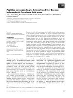
achieved. The following HFOV settings were used and kept
constant throughout the entire protocol (Figure 1): FiO
2
of 1.0,
Available online />Page 3 of 9
(page number not for citation purposes)
an oscillatory frequency of 6 Hz, an inspiration time of 33% of
the respiratory cycle, a bias flow of 30 l/minute, and a pressure
amplitude of 40 mbar.
The protocol for lung volume optimization during HFOV com-
prised three steps: step 1, a lung recruitment maneuver – set-
ting the initial CDP during ongoing oscillations to 45 mbar
(CDP45) for 2.5 minutes; step 2, decrease of the CDP – the
CDP was reduced in a stepwise manner by 5 mbar every 5
minutes from 45 mbar to 40 mbar, 35 mbar, 30 mbar, 25 mbar,
and 20 mbar (CDP20) with simultaneous measurement of the
TPP; and step 3, identification of the optimal TPP – the TPP
level necessary to maintain lung recruitment was defined as
the TPP necessary to prevent a decrease in the PaO
2
/FiO
2
ratio > 25% compared with the PaO
2
/FiO
2
ratio at CDP45
(that is to say, maximum lung recruitment).
Measurements
The following parameters were recorded before and 120 min-
utes after initiation of lung damage during PCV, and at every
CDP level during HFOV: the heart rate, the MAP, the right
atrial pressure, the PAOP, the mean pulmonary artery pres-
sure, and the LVEDP. In addition, the intrathoracic blood vol-
ume, the extravascular lung water, and the cardiac output
(CO), as obtained by the PiCCO
®
-Technology system (Pul-
sion Medical Systems, Munich, Germany), were recorded. For
blood gas analyses, arterial and mixed venous blood samples
were drawn (ABL 500/OSM 3; Radiometer, Copenhagen,
Denmark). The pulmonary vascular resistance, the systemic
arterial vascular resistance, the stroke volume (SV), oxygen
delivery, the oxygenation index, and the pulmonary shunt pro-
portion were calculated according to standard formula.
The TPP was calculated as the difference between the CDP
(measured at the proximal end of endotracheal tube) and the
esophageal pressure.
Statistical analysis
Data are expressed as the median, and 25th and 75th percen-
tiles (interquartile range). Intraindividual differences before and
after induction of lung injury and during the recruitment maneu-
ver (PCV post-lavage and CDP45) were tested nonparametri-
cally using the Wilcoxon signed-rank test. Any differences
during the fast CDP deceleration trial were addressed by a
Friedman repeated-measures analysis of variance on ranks
and multiple comparisons by Tukey's test. P < 0.05 was con-
sidered significant (SigmaStat Version 2.03; SPSS Inc., San
Raphael, California, USA).
Results
All six animals (25 ± 2 kg bodyweight, mean ± standard devi-
ation) completed the entire study protocol. Hemodynamic and
gas exchange variables before and after induction of lung
damage are presented in Table 1. The MAP and PAOP before
initiation of HFOV complied with the predefined requirements;
that is, PAOP of 13 (11–13) mmHg and MAP of 83 (82–85)
mmHg. Repetitive lung lavages decreased the PaO
2
/FiO
2
ratio (PCV pre-lavage, 559 (535–658) Torr vs PCV post-lav-
age, 99 (56–128) Torr; P < 0.05), and increased the pulmo-
nary shunt fraction (PCV pre-lavage, 9.97% (8.8–11%) vs
PCV post-lavage, 51.8% (49–55%); P < 0.05). The oxygena-
tion index increased from PCV pre-lavage (1.4 (1.2–2)) to
PCV post-lavage (16.3 (14.6–21.3)) (P < 0.05). The extravas-
cular lung water increased from PCV pre-lavage (290 (241–
311) ml) to PCV post-lavage (420 (354–463) ml) (P < 0.05).
Figure 1
Illustration of the time course of the study protocolIllustration of the time course of the study protocol. CDP, continuous distending pressure; CDP45, continuous distending pressure of 45 mbar; ∆P,
pressure amplitude; HFOV, high-frequency oscillatory ventilation; PCV, pressure-controlled ventilation; T
insp
, inspiration time.
Critical Care Vol 10 No 5 Karmrodt et al.
Page 4 of 9
(page number not for citation purposes)
Hemodynamics
Hemodynamic variables are presented in Table 1 for the lung
recruitment maneuver and in Table 2 for the stepwise
decrease of the CDP. The MAP, heart rate and intrathoracic
blood volume did not change throughout the entire
experiment. The right atrial pressure, mean pulmonary artery
pressure, PAOP, and LVEDP during the lung recruitment pro-
cedure increased significantly from the PCV post-lavage to
CDP45 (P < 0.05). The CO and SV decreased from PCV
post-lavage (CO, 3.6 (3.1–3.9) l/minute; SV, 32 (31–35) ml)
to CDP45 (CO, 2.6 (2.3–3.1) l/minute; SV, 19 (18–24) ml) (P
= 0.031).
Pulmonary gas exchange and pulmonary shunt fraction
The LVOP increased the PaO
2
/FiO
2
ratio from PCV post-lav-
age (99 (56–128) Torr) to CDP45 (621 (619–660) Torr) (P <
0.05). The pulmonary shunt fraction decreased from PCV
post-lavage (51.8% (49–55%)) to CDP45 (1.03% (0.4–3%))
(P < 0.05). During the stepwise decrease of the CDP, the
shunt fraction increased to 20.2% (7.2–52%) at CDP20 com-
pared with 1.03% (0.4–1.4%) at CDP45 and compared with
Table 1
Hemodynamic data, blood gas data in pressure-controlled ventilation (PCV) pre-lavage, PCV post-lavage, and during lung volume
optimization procedure in high-frequency oscillatory ventilation
PCV pre-lavage PCV post-lavage 45 mbar
Heart rate (beats/minute) 113 (112–124) 112 (102–125) 141 (126–167)
Right atrial pressure (mmHg) 9 (9–9) 10 (8–11) 16 (15–18)**
Mean pulmonary artery pressure (mmHg) 25 (20–37) 34 (30–41) 41 (35.7–52)**
Pulmonary occlusion pressure (mmHg) 10 (7–12) 13 (11–13) 24 (23–26)**
Left ventricular end-diastolic pressure (mmHg) 3 (1–4) 3.5 (2–6) 12 (11–13)**
Mean arterial pressure (mmHg) 89.5 (67–106) 83 (82–85) 85 (66–89)
Cardiac output (l/min) 3.28 (3.1–3.5) 3.6 (3.1–3.9) 2.6 (2.3–3.1)**
Stroke volume (ml) 31 (27–36) 32 (31–35) 19 (18–24)**
Intrathoracic blood volume (ml) 598.5 (548–622) 594 (553–690) 442 (401–469)
Extravascular lung water (ml) 290 (241–311) 420 (354–463)* 355 (315–476)
Systemic vascular resistance (dyn*s/cm
5
) 1817 (1277–2508) 1542 (1450–2074) 2403 (1524–2585)
Pulmonary vascular resistance (dyn*s/cm
5
) 266 (213–775) 545 (275–693) 639 (404–859)
Mean pulmonary artery pressure–right atrial pressure (mmHg) 16 (11–28) 26 (22–30) 28 (19–34)
Transpulmonary pressure (mbar) 4.5 (4–6) 10 (8–11)* 36 (32–43)**
PaO
2
(Torr) 559 (535–658) 99 (56–128)* 621 (557–522)**
PaO
2
/FiO
2
ratio 559 (535–658) 99 (56–128)* 621 (557–522)**
Oxygenation index 1.4 (1.2–2) 16.3 (14.6–21.3)* 7.2 (6.8–7.2)**
Oxygen delivery (ml O
2
/minute) 408.3 (382.5–427.6) 373 (297.6–427.9) 312 (299–466.2)
Shunt proportion 9.9 (8.8–11) 51.8 (49–55)* 1.03 (0.4–3)**
PaCO
2
(Torr) 39.6 (32.8–48.4) 47 (40.7–58.8) 38.5 (28.3–46.6)
Oxygen saturation (%) 99.9 (99.9–99.9) 92 (89.7–96.5)* 99.9 (99–100)**
pH 7.45 (7.4–7.5) 7.38 (7.31–7.4) 7.41 (7.37–7.5)
Standard bicarbonate (mmol/l) 28.2 (27.8–30.2) 28.5 (26.2–30.9) 25 (23.2–27.5)
Base excess 4.8 (2.1–5.9) 2.25 (1–2.9)* 0.7 (-0.12 to 4.95)
Hemoglobin (g/dl) 7.6 (7.5–8) 7.8 (7.4–8.3) 7.9 (7.1–9.7)
Plateau airway pressure (cmH
2
0) 21 (10.8–14.4) 30 (25–35)
Inspiratory tidal volume (ml) 335 (330–450) 390 (310–440)
Dynamic lung compliance (ml/mbar) 12.6 (10.8–14.4) 7.9 (5.3–9.8)*
Data presented as median (25th–75th percentiles). *P < 0.05 vs PCV pre-lavage (Wilcoxon signed-rank test), **P < 0.05 vs PCV post-lavage.
Available online />Page 5 of 9
(page number not for citation purposes)
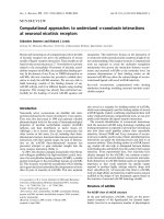
0.7% (0.1–1.4%) at CDP of 40 mbar (P < 0.05). The PaO
2
/
FiO
2
ratio decreased from CDP45 (621 (619–660) Torr) to
CDP20 (429 (52–558) Torr) (P < 0.05) (Figure 2). The
PaCO
2
increased from CDP45 (38.5 (28.3–46) Torr) to
CDP20 (54.4 (40.7–68.6) Torr) (P < 0.001).
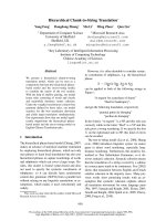
Identification of the required TPP level
The lung optimization procedure in this study required less
than 30 minutes to recruit the lung and to identify the lowest
TPP level to maintain adequate oxygenation and gas
exchange. The TPP increased from PCV post-lavage (10 (8–
11) mbar) to CDP45 (36 (26–43) mbar) (P < 0.05).
A TPP between 32 and 14 mbar prevented a decrease in the
PaO
2
/FiO
2
ratio > 25% compared with the PaO
2
/FiO
2
ratio at
CDP45. These TPP levels were achieved at CDP settings
ranging from 25 to 40 mbar (Figure 3a). In three animals the
PaO
2
/FiO
2
ratio did not decrease more than 25% compared
with the measurement at CDP45, independently of the applied
CDP (Figure 3b). On average, a TPP of 25.5 (17–32) mbar
was required to preserve a difference in the PaO
2
/FiO
2
ratio <
Table 2
Hemodynamic data and blood gas data on every continuous distending pressure in high-frequency oscillatory ventilation study
protocol
Descent continuous distending pressure trial
45 mbar 40 mbar 35 mbar 30 mbar 25 mbar 20 mbar
Heart rate (beats/minute) 141.5 (126–167) 135.5 (112–153) 128 (113–149) 126 (107–147) 115 (102–140) 116 (104.5–128.5)
Right atrial pressure (mmHg) 16 (15–18) 15 (14–15) 14.5 (13–15) 13.5 (12–15) 12.5 (11–13) 11.5 (10–13)
†,**,*
Mean pulmonary artery pressure
(mmHg)
41 (35.7–52) 39 (35–41) 39 (31–44) 41 (35–42) 39 (35–43) 38.5 (36–42)
Pulmonary occlusion pressure
(mmHg)
24 (23–26) 21.5 (20–24) 20 (18–21) 17 (16.2–18.7) 14 (13.5–17)
†
13.5 (12–15)
†,**
Left ventricular end-diastolic
pressure (mmHg)
11.5 (11–13) 9 (9–11) 9.5 (6–10) 7.5 (6–12) 7.5 (5–10)
†
5 (4–6)
†
Mean arterial pressure (mmHg) 85 (66–89) 70 (63–90) 74 (67–104) 74.5 (66–101) 78.5 (77–108) 84 (47–106)
Cardiac output (l/minute) 2.6 (2.3–3.1) 2.6 (2–3) 2.65 (2.28–3.36) 2.52 (2.2–3.6) 2.7 (2.2–4.2) 3.1 (1.8–4.8)
Stroke volume (ml) 19 (18–24) 21 (16.6–24.5) 23.6 (15–29) 23 (15–26.9) 18.2 (16–33) 18.7 (12.8–37)
Intrathoracic blood volume (ml) 442.5 (401–469) 395 (335–495) 485 (431–531) 506.5 (453–542) 552 (412–618) 540 (492–614)
Extravascular lung water (ml) 355 (315–476) 360 (331–441) 405 (287–430) 426 (336–495) 441 (406.5–535) 460 (390–549)
Systemic vascular resistance
(dyn*s/cm
5
)
2,403 (1,524–2,585) 1,598 (1,582–2,271) 1,835 (1,567–1,997) 2,026 (1,442–2,033) 1,933 (1,303–2,469) 1,611 (1,238–2,056)
Pulmonary vascular resistance
(dyn*s/cm
5
)
639 (404–859) 440 (396–663) 457 (376–663) 512 (461–746) 617 (418–970) 678 (415–1,065)
Mean pulmonary artery pressure–
right atrial pressure (mmHg)
28 (19–34) 25.5 (23–26) 25 (18–29) 27.5 (26–28) 27 (25–30) 28.5 (23–31)
Transpulmonary pressure (mbar) 36 (32–43) 31 (27–38) 26.5 (22–33) 21.5 (17–28) 14.5 (12–22)
†,**
10 (7–18)
†,**
PaO
2
(Torr) 621 (619–660) 654 (645–683) 664 (636–694) 644 629–692) 615 (469–680) 429 (52–558)**
PaO
2
/FiO
2
ratio 82.8 (82.5–88) 654 (645–683) 664 (636–694) 644 629–692) 615 (469–680) 429 (52–558)**
Oxygenation index 7.2 (6.8–7.2) 6.1 (5.8–6.1) 5.4 (5–5.5) 4.6 (4.3–4.7) 4 (3.6–5.3) 4.8 (3.5–37.9)
Oxygen delivery (ml O
2
/minute) 312.5 (299–466) 358.2 (305.6–420) 360 (349–418) 357.2 (338–447) 347.4 (292–516) 399.6 (251–548)
Shunt proportion 1.03 (0.4–3) 0.7 (0.1–1.4) 1.5 (0–4.3) 1.6 (1.1–4.9) 4.7 (4.2–11.3) 20.2 (7.2–52)
†,**
PaCO
2
(Torr) 38.5 (28.3–46.6) 40.3 (30.1–51.1) 37.4 (30.7–44.8) 41.2 (31.1548) 46.9 (32.9–61.2) 54.5 (40.7–68.6)
†,*
Oxygen saturation (%) 99.9 (99–100) 99.9 (99.8–100) 99.9 (99.9–100) 99.9 (98.3–100) 99.9 (96–100) 99.8 (83–99)
pH 7.41 (7.37–7.5) 7.4 (7.34–7.53) 7.4 (7.38–7.51) 7.4 (7.38–7.51) 7.38 (7.35–7.48) 7.29 (7.25–7.4)
Standard bicarbonate (mmol/l) 25 (24.2–29.8) 25.8 (25–27) 26 (23.4–27.6) 25.1 (23.5–27.7) 25.3 (24.2–28.5) 24.6 (24.1–28.5)
Base excess 0.95 (0.4–6.2) 1 (-1.5 to 5.2) 02 (-0.65 to 2.97) -0.15 (-2.4 to 1.6) -1.35 (-2.3 to 1.4) 0.2 (-4.25 to 0.47)
Hemoglobin (g/dl) 7.9 (7.1–9.7) 8.8 (7.4–10.1) 8.2 (7.5–10.4) 8 (7.4–9.7) 7.9 (7.1–9.4) 7.8 (7–10)
Data presented as median (25th–75th percentiles). Significant differences in parameters are indicated: *P < 0.05 vs 35 mbar, **P < 0.05 vs 40
mbar,
†
P < 0.05 vs 45 mbar.
Critical Care Vol 10 No 5 Karmrodt et al.
Page 6 of 9
(page number not for citation purposes)
25% related to CDP45; this TPP was achieved at a CDP of
35 (25–40) mbar.
Discussion
The present experimental study investigated the effects upon
gas exchange, TPPs, and hemodynamics of a modified HFOV
initiation protocol to optimize the lung volume in a porcine
model of acute lung injury. The protocol consists of a fast
recruitment maneuver followed by a stepwise decrease of
CDP. The fast stepwise reduction of CDP allows the identifi-
cation of the lowest TPP level required to maintain improve-
ment of oxygenation in each animal. This approach therefore
offers an effective reduction of pulmonary shunt fraction and
improvement in oxygenation without relevant adverse hemody-
namic effects within a very short time.
In most clinical and experimental studies, HFOV is initiated
with a LVOP with an initial CDP level 5 mbar above the mean
airway pressure previously used in conventional ventilation
[5,6,8,12]. The CDP is then increased in a stepwise fashion
(2–5 mbar steps) every 15–30 minutes until a maximum in
PaO
2
or a predetermined CDP is reached. Our study presents
a fast lung optimization procedure that could be technically
applied easily in the clinical scenario. In contrast to recent
studies in animals and in patients, the CDP was directly set to
45 mbar for 2.5 minutes. The lowest possible TPP that still
assures the improved oxygenation is subsequently titrated.
Recruitment maneuvers are beneficial at the initiation of HFOV
to ensure sufficient gas exchange area in the diseased lung
[4]. In the Treatment with Oscillation and an Open Lung Strat-
egy trial, the combination of HFOV and a high initial mean air-
way pressure recruitment maneuver without ongoing HFOV
resulted in a rapid and sustained improvement in oxygenation
[10]. Although sustained inflation pressures up to 55 mbar are
necessary to overcome the opening pressure of collapsed
alveoli [13], the criteria of a fully recruited lung, defined as
pulmonary shunt proportion < 0.1 [14], was achieved at a
CDP of 45 mbar. A CDP of 32 ± 6 mbar in piglets was able to
reduce the shunt fraction < 10% [15]. During the present
study a single pressure step-up for alveolar recruitment was
performed, and therefore no estimate can be made of whether
a lower CDP and TPP would have been adequate to reopen
the lung, and whether the TPP required to open alveolar units
exceeds the TPP required to maintain alveolar patency
[15,16]. Assuming that the TPP stays above a critical closing
pressure, a significant alveolar derecruitment cannot be
expected [17,18]. In other experimental studies with the lav-
age-injury animal model, the mean airway pressures were set
according to the pressure-volume curve for the setting of the
CDP during HFOV [9,16,19]. In their study, Sedeek and col-
leagues repeated recruitment maneuvers with a continuous
positive airway pressure of 50 mbar for 1 minute until the PaO
2
was stable with a CDP set according to the maximal compli-
ance on the pressure-volume curve [9].
We intended to use an alternative method without the pres-
sure-volume curve. The P
a
O
2
/F
i
O
2
ratio was therefore used as
a criterion to identify the lowest TPP preventing alveolar dere-
cruitment during the stepwise decrease of CDP. A TPP below
this threshold was associated with an increased shunt > 10
and with an increased PaCO
2
. An increased PaCO
2
at
unchanged HFOV settings indicates a decreased alveolar sur-
face available for gas exchange.
Previous studies in the lavage animal model showed that CO
decreased at high mean airway pressures. Interestingly, better
oxygenation values in those studies were found at lower mean
airway pressure, suggesting in our study that eventually a
lower CDP eventually would have been sufficient [16,19]. The
lung recruitment maneuver had a marked effect on hemody-
namics. These effects can easily be corrected by volume or by
Figure 2
PaO
2
/FiO
2
ratio and shunt fraction during pressure-controlled ventilation and high-frequency oscillatory ventilationPaO
2
/FiO
2
ratio and shunt fraction during pressure-controlled ventilation and high-frequency oscillatory ventilation. The PaO
2
/FiO
2
ratio and shunt
fraction during pressure-controlled ventilation (PCV) pre-lavage and PCV post-lavage in relation to continuous distending pressure (mbar). CDP,
continuous distending pressure.
Available online />Page 7 of 9
(page number not for citation purposes)
vasoactive drug usage. There was no relevant decrease of the
intrathoracic blood volume as an indicator of reduced venous
preload [20]. Also, the fluid regime before initiation of HFOV
may have attenuated a reduction of venous return.
At a CDP of 45 mbar, the SV and CO were decreased and,
simultaneously, the cardiac filling pressures (LVEDP, right
atrial pressure, and PAOP) were elevated. This can be
explained by a compression of the heart into the cardiac fossa
due to the transmission of high TPPs [21,22]. Impairment of
hemodynamics can therefore be explained by the mechanical
restriction of the heart. Systemic afterload did not have a major
impact on the impaired SV and CO as measured by the sys-
temic vascular resistance. Right ventricular dysfunction may
occur when high airway pressures are applied, and the con-
secutive right ventricular output and left ventricular filling are
impaired leading to SV and CO decreases. As the pulmonary
vascular resistance was unaffected by a CDP of 45 mbar and
the right ventricle was able to generate a pressure gradient
(mean pulmonary artery pressure–right atrial pressure), we
excluded right ventricular failure. During the deceleration CDP
trial, the cardiac filling pressures returned to similar values as
measured before the lung recruitment maneuver. This obser-
vation assures us that the hemodynamic effects are related to
a pressure transmission of the CDP and the TPP.
The safe window for plateau pressures during conventional
ventilation is considered between 30 and 35 mbar, but even
those values can lead to a harmful TPP. Depending on the
elastance of the respiratory system, volutrauma may occur
from cyclic tidal overdistension. No recommendations regard-
ing safe levels for CDP or TPP exist during HFOV. The lowest
TPP levels possible, however, should be applied. The meas-
urement and monitoring of the TPP is therefore helpful and of
increased interest, as the TPP is the effective distending force
of the lung, and ventilator-induced lung injury (VILI) depends
on the TPP [23,24]. By measuring the TPP the mechanical
ventilator settings could be set more individually with respect
to lung and chest wall mechanical characteristics, which ena-
bles the identification of lung recruitment potential in relation
to a potential risk of VILI. Such an individual approach may
reduce the risk for further lung injury in patients with ARDS
undergoing mechanical ventilation [25,26]. In a clinical sce-
nario, a CDP higher than the safe window for plateau pres-
sures under conventional ventilation should be avoided even if
suggested by the LVOP presented in this study. Although the
theoretical advantage of HFOV is the avoidance of volutrauma
caused by tidal overdistension (due to minimal tidal volumes)
in the case of excessive TPP, a compromise between oxygen-
ation and potential VILI should be made to avoid a VILI and to
accept lower but adequate oxygenation.
The esophageal pressure measured by an esophageal balloon
is used as a surrogate parameter of the pleural pressure for
calculation of the TPP. The pleural pressure and therefore the
TPP are dependent on the chest wall elastance in experimen-
tal patients and ARDS patients. For a given mean airway pres-
sure (CDP) the pleural pressure increases if the chest wall
elastance is elevated, and consequently the TPP decreases.
Chest wall elastance, however, depends on the pathophysiol-
Figure 3
Relationship between continuous distending pressure and transpulmo-nary pressure or changes in PaO
2
/FiO
2
ratioRelationship between continuous distending pressure and transpulmo-
nary pressure or changes in PaO
2
/FiO
2
ratio. Relationship (for each ani-
mal) between the continuous distending pressure (CDP) (mbar) and:
(a) the transpulmonary pressure (TPP) (mbar) (X, shunt fraction > 10%;
+, decrease of the PaO
2
/FiO
2
ratio > 25%), and (b) changes in the
PaO
2
/FiO
2
ratio (%) (dotted line, decrease of the PaO
2
/FiO
2
ratio of
25% compared with CDP of 45 mbar (CDP45); X, shunt fraction >
10%).
Critical Care Vol 10 No 5 Karmrodt et al.
Page 8 of 9
(page number not for citation purposes)
ogy of ARDS (for example, high chest wall elastance in
extrapulmonary ARDS). The elevated pleural pressure (due to
increased chest wall elastance) leads to a lower TPP com-
pared with pulmonary ARDS with normal elastance [1,27-29].
We found a pressure difference of about 10 mbar between the
applied CDP and the resulting TPP. The difference in mean
intrathoracic pressure and CDP in HFOV increases with the
oscillatory frequency, decreasing the tracheal tube diameter
and the relative duration of the inspiratory time [30]. The inspi-
ration time:expiration time ratio of 33% in our study results in
an increased tracheal tube resistance and, consequently, in a
decreased mean intrathoracic pressure and TTP.
Limitations
A major limitation of this study is that it was performed in a lav-
age animal model of ARDS and not in patients. Large-animal
models, however, have contributed greatly to the understand-
ing of the basic physiology of HFOV and its clinical application
during severe lung injury [31]. ARDS in patients is rarely and
solely a result of surfactant deficiency only. Early institution of
HFOV, however, and institution of HFOV in patients with high
potential for recruitment is theoretically beneficial to outcome
in ARDS [32-34]. The lavage model is easily recruitable and is
probably the most comparable experimental ARDS model for
early-phase ARDS. Although it is primarily a model of sur-
factant depletion, it has been shown that mechanical ventila-
tion after lavages leads to neutrophil infiltration, cytokine
expression, and capillary-alveolar protein leak (as indicated by
the increased extravascular lung water in this study) [35-37].
The present study only observed immediate changes of hemo-
dynamics and lung oxygenation. High airway pressures and
subsequent lung overdistension may lead to mediator release
and to VILI. These adverse events were not accessible in the
present study. Also, we cannot report whether the optimized
gas exchange remained stable over a longer time period. This
raises the question of how often such recruitment maneuvers
should be applied in clinical practice.
Conclusion
This short-term experimental observation study demonstrates
the feasibility of a fast and safe lung optimization procedure
during uninterrupted HFOV. This lung optimization procedure
was well tolerated and resulted in a dramatic improvement of
oxygenation. No clinically relevant adverse effects on hemody-
namics or barotrauma occurred during the time period of
approximately 1 hour. The stepwise decrease of CDP allowed
the determination of the lowest TPP level necessary to main-
tain adequate oxygenation.
Competing interests
The authors declare that they have no competing interests.
Authors' contributions
JK designed the study protocol, collected data, and drafted
the protocol. MD designed and participated in the study pro-
tocol, collected data, and performed statistical analysis. SY
collected data. KM coordinated the study and revised the
manuscript.
Acknowledgements
This study was funded by the German Research Foundation (DFG)
Grant Ma 2398/3-2. All other sources of financial support for the work
contained in the article have been disclosed. The high-frequency oscil-
latory ventilator was provided by Viasys Healthcare (Hoechberg, Ger-
many). The authors thank Mr Jeffrey Crowder for his editorial assistance.
References
1. Ventilation with lower tidal volumes as compared with tradi-
tional tidal volumes for acute lung injury and the acute respi-
ratory distress syndrome. The Acute Respiratory Distress
Syndrome Network. N Engl J Med 2000, 342:1301-1308.
2. Gattinoni L, Carlesso E, Cadringher P, Valenza F, Vagginelli F,
Chiumello D: Physical and biological triggers of ventilator-
induced lung injury and its prevention. Eur Respir J Suppl
2003, 47:15s-25s.
3. Imai Y, Nakagawa S, Ito Y, Kawano T, Slutsky AS, Miyasaka K:
Comparison of lung protection strategies using conventional
and high-frequency oscillatory ventilation. J Appl Physiol 2001,
91:1836-1844.
4. Froese AB: The incremental application of lung-protective
high-frequency oscillatory ventilation. Am J Respir Crit Care
Med 2002, 166:786-787.
5. Derdak S, Mehta S, Stewart TE, Smith T, Rogers M, Buchman TG,
Carlin B, Lowson S, Granton J: High-frequency oscillatory venti-
lation for acute respiratory distress syndrome in adults: a ran-
domized, controlled trial. Am J Respir Crit Care Med 2002,
166:801-808.
6. Derdak S: High-frequency oscillatory ventilation for acute res-
piratory distress syndrome in adult patients. Crit Care Med
2003, 31:S317-S323.
7. David M, Karmrodt J, Weiler N, Scholz A, Markstaller K, Eberle B:
High-frequency oscillatory ventilation in adults with traumatic
brain injury and acute respiratory distress syndrome. Acta
Anaesthesiol Scand 2005, 49:209-214.
8. David M, Weiler N, Heinrichs W, Neumann M, Joost T, Markstaller
K, Eberle B: High-frequency oscillatory ventilation in adult
acute respiratory distress syndrome. Intensive Care Med 2003,
29:1656-1665.
9. Sedeek KA, Takeuchi M, Suchodolski K, Vargas SO, Shimaoka M,
Schnitzer JJ, Kacmarek RM: Open-lung protective ventilation
with pressure control ventilation, high-frequency oscillation,
and intratracheal pulmonary ventilation results in similar gas
Key messages
• A novel lung optimization procedure in uninterrupted
HFOV is presented.
• The LVOP consists of a single CDP recruitment maneu-
ver during uninterrupted HFOV, followed by a stepwise
decrease of the CDP.
• This LVOP allows the determination of the lowest TPP
that still maintains adequate pulmonary gas exchange.
• This LVOP shows no adverse effects in hemodynamics
and oxygenation during its intervention in a porcine lav-
age ARDS model.
Available online />Page 9 of 9
(page number not for citation purposes)
exchange, hemodynamics, and lung mechanics. Anesthesiol-
ogy 2003, 99:1102-1111.
10. Ferguson ND, Chiche JD, Kacmarek RM, Hallett DC, Mehta S,
Findlay GP, Granton JT, Slutsky AS, Stewart TE: Combining high-
frequency oscillatory ventilation and recruitment maneuvers in
adults with early acute respiratory distress syndrome: the
Treatment with Oscillation and an Open Lung Strategy
(TOOLS) trial pilot study. Crit Care Med 2005, 33:479-486.
11. Baydur A, Cha EJ, Sassoon CS: Validation of esophageal bal-
loon technique at different lung volumes and postures. J Appl
Physiol 1987, 62:315-321.
12. Mehta S, Lapinsky SE, Hallett DC, Merker D, Groll RJ, Cooper AB,
MacDonald RJ, Stewart TE: Prospective trial of high-frequency
oscillation in adults with acute respiratory distress syndrome.
Crit Care Med 2001, 29:1360-1369.
13. Sjostrand UH, Lichtwarck-Aschoff M, Nielsen JB, Markstrom A,
Larsson A, Svensson BA, Wegenius GA, Nordgren KA: Different
ventilatory approaches to keep the lung open. Intensive Care
Med 1995, 21:310-318.
14. Lachmann B: Open up the lung and keep the lung open. Inten-
sive Care Med 1992, 18:319-321.
15. van Genderingen HR, van Vught JA, Jansen JR, Duval EL,
Markhorst DG, Versprille A: Oxygenation index, an indicator of
optimal distending pressure during high-frequency oscillatory
ventilation? Intensive Care Med 2002, 28:1151-1156.
16. Luecke T, Meinhardt JP, Herrmann P, Weisser G, Pelosi P, Quintel
M: Setting mean airway pressure during high-frequency oscil-
latory ventilation according to the static pressure-volume
curve in surfactant-deficient lung injury: a computed tomogra-
phy study. Anesthesiology 2003, 99:1313-1322.
17. Kolton M, Cattran CB, Kent G, Volgyesi G, Froese AB, Bryan AC:
Oxygenation during high-frequency ventilation compared with
conventional mechanical ventilation in two models of lung
injury. Anesth Analg 1982, 61:323-332.
18. Rimensberger PC, Pache JC, McKerlie C, Frndova H, Cox PN:
Lung recruitment and lung volume maintenance: a strategy for
improving oxygenation and preventing lung injury during both
conventional mechanical ventilation and high-frequency
oscillation. Intensive Care Med 2000, 26:745-755.
19. Goddon S, Fujino Y, Hromi JM, Kacmarek RM: Optimal mean air-
way pressure during high-frequency oscillation: predicted by
the pressure-volume curve. Anesthesiology 2001, 94:862-869.
20. Luecke T, Roth H, Herrmann P, Joachim A, Weisser G, Pelosi P,
Quintel M: Assessment of cardiac preload and left ventricular
function under increasing levels of positive end-expiratory
pressure. Intensive Care Med 2004, 30:119-126.
21. Pinsky MR: The hemodynamic consequences of mechanical
ventilation: an evolving story. Intensive Care Med 1997,
23:493-503.
22. Butler J: The heart is not always in good hands. Chest 1990,
97:453-460.
23. Talmor D, Sarge T, O'Donnell CR, Ritz R, Malhotra A, Lisbon A,
Loring SH: Esophageal and transpulmonary pressures in acute
respiratory failure. Crit Care Med 2006, 34:1389-1394.
24. Brander L, Ranieri VM, Slutsky AS: Esophageal and transpulmo-
nary pressure help optimize mechanical ventilation in patients
with acute lung injury. Crit Care Med 2006, 34:1556-1558.
25. Terragni PP, Rosboch GL, Lisi A, Viale AG, Ranieri VM: How res-
piratory system mechanics may help in minimising ventilator-
induced lung injury in ARDS patients. Eur Respir J Suppl 2003,
42:15s-21s.
26. de Chazal I, Hubmayr RD: Novel aspects of pulmonary mechan-
ics in intensive care. Br J Anaesth 2003, 91:81-91.
27. Tobin MJ: Culmination of an era in research on the acute res-
piratory distress syndrome. N Engl J Med 2000,
342:1360-1361.
28. Pelosi P, D'Onofrio D, Chiumello D, Paolo S, Chiara G, Capelozzi
VL, Barbas CS, Chiaranda M, Gattinoni L: Pulmonary and
extrapulmonary acute respiratory distress syndrome are
different. Eur Respir J Suppl 2003, 42:48s-56s.
29. Gattinoni L, Chiumello D, Carlesso E, Valenza F: Bench-to-bed-
side review: chest wall elastance in acute lung injury/acute
respiratory distress syndrome patients. Crit Care 2004,
8:350-355.
30. Pillow JJ, Neil H, Wilkinson MH, Ramsden CA: Effect of I/E ratio
on mean alveolar pressure during high-frequency oscillatory
ventilation. J Appl Physiol 1999, 87:407-414.
31. Kacmarek RM, Malhotra A: High-frequency oscillatory ventila-
tion: what large-animal studies have taught us! Crit Care Med
2005, 33:S148-S154.
32. Higgins J, Estetter B, Holland D, Smith B, Derdak S: High-fre-
quency oscillatory ventilation in adults: respiratory therapy
issues. Crit Care Med 2005, 33:S196-S203.
33. Mehta S, Granton J, MacDonald RJ, Bowman D, Matte-Martyn A,
Bachman T, Smith T, Stewart TE: High-frequency oscillatory
ventilation in adults: the Toronto experience. Chest 2004,
126:518-527.
34. Ferguson ND, Stewart TE: New therapies for adults with acute
lung injury. High-frequency oscillatory ventilation. Crit Care
Clin 2002, 18:91-106.
35. Lachmann B, Robertson B, Vogel J: In vivo lung lavage as an
experimental model of the respiratory distress syndrome.
Acta Anaesthesiol Scand 1980, 24:231-236.
36. Rotta AT, Gunnarsson B, Fuhrman BP, Hernan LJ, Steinhorn DM:
Comparison of lung protective ventilation strategies in a rabbit
model of acute lung injury. Crit Care Med 2001, 29:2176-2184.
37. Sugiura M, McCulloch PR, Wren S, Dawson RH, Froese AB: Ven-
tilator pattern influences neutrophil influx and activation in
atelectasis-prone rabbit lung. J Appl Physiol 1994,
77:1355-1365.