Báo cáo khoa học: "Microcirculatory alterations induced by sedation in intensive care patients. Effects of midazolam alone and in association with sufentanil" potx
Bạn đang xem bản rút gọn của tài liệu. Xem và tải ngay bản đầy đủ của tài liệu tại đây (379.46 KB, 9 trang )
Open Access
Available online />Page 1 of 9
(page number not for citation purposes)
Vol 10 No 6
Research
Microcirculatory alterations induced by sedation in intensive care
patients. Effects of midazolam alone and in association with
sufentanil
Veronique Lamblin, Raphael Favory, Marie Boulo and Daniel Mathieu
Service d'Urgence Respiratoire et Réanimation Médicale et de Médecine Hyperbare, Hôpital Calmette, Centre Hospitalier Universitaire, Boulevard
du Professeur Jules Leclercq, 59037 Lille Cedex, France
Corresponding author: Daniel Mathieu,
Received: 16 Jun 2006 Revisions requested: 5 Jul 2006 Revisions received: 29 Aug 2006 Accepted: 15 Dec 2006 Published: 15 Dec 2006
Critical Care 2006, 10:R176 (doi:10.1186/cc5128)
This article is online at: />© 2006 Lamblin et al.; licensee BioMed Central Ltd.
This is an open access article distributed under the terms of the Creative Commons Attribution License ( />),
which permits unrestricted use, distribution, and reproduction in any medium, provided the original work is properly cited.
Abstract
Introduction Sedation is widely used in intensive care unit (ICU)
patients to limit the risk of pulmonary barotrauma and to
decrease oxygen needs. However, adverse effects of sedation
have not been fully evaluated; in particular, effects of
benzodiazepine and opiates on microcirculation have not been
extensively studied. The aim of this study was to evaluate the
microcirculatory effects of a sedation protocol commonly
prescribed in the ICU.
Methods Ten non-septic patients under controlled ventilation
requiring sedation for therapeutic purposes were enrolled in a
prospective observational study conducted in an ICU of a
university hospital. Sedation was conducted in two successive
steps: first, each patient received midazolam (0.1 mg/kg per
hour after a bolus of 0.05 mg/kg, then adapted to reach a
Ramsay score of between 3 and 5). Second, after one hour,
sufentanil was added (0.1 μg/kg per hour after a bolus of 0.1
μg/kg). Arterial pressure, heart rate, cardiac output determined
by transthoracic impedance, transcutaneous oxygen (tcPO
2
)
and carbon dioxide (tcPCO
2
) pressures, and microcirculatory
blood flow determined by laser Doppler flowmetry at rest and
during a reactive hyperaemia challenge were measured before
sedation (NS period), one hour after midazolam infusion (H
period), and one hour after midazolam-sufentanil infusion (HS
period).
Results Arterial pressure decreased in both sedation periods,
but heart rate, cardiac output, tcPO
2
, and tcPCO
2
remained
unchanged. In both sedation periods, microcirculatory changes
occurred with an increase in cutaneous blood flow at rest (H
period: 207 ± 25 perfusion units [PU] and HS period: 205 ± 25
PU versus NS period: 150 ± 22 PU, p < 0.05), decreased
response to ischaemia (variation of blood flow to peak: H period:
97 ± 16 PU and HS period: 73 ± 9 PU versus NS period: 141
± 14 PU, p < 0.05), and attenuation of vasomotion.
Conclusion Sedation with midazolam or a combination of
midazolam and sufentanil induces a deterioration of vasomotion
and microvascular response to ischaemia, raising the question
of whether this effect may further alter tissue perfusion when
already compromised, as in septic patients.
Introduction
Because of its role in blood-tissue exchanges, the microcircu-
lation is a fundamental element of the vascular network [1,2].
It has been long to recognise. It was only recently recognised
that numerous pathologic conditions like arteriosclerosis, arte-
rial hypertension, or diabetes alter the microcirculation,
explaining, at least in part, the observed tissue hypoxia [3,4].
More recently, sepsis, a major cause of death in the intensive
care unit (ICU), has been shown to induce microcirculatory
dysfunction, even in its early stage and in the absence of
ΔΦ = flow variation during reactive hyperaemia (ΔΦ = Φpeak - Φrest); Φpeak = maximum cutaneous blood flow during the reactive hyperaemia peak;
Φrest = cutaneous blood flow at rest; cGMP = cyclic guanosine monophosphate; CMBC = concentration of moving blood cells; CO = cardiac out-
put; cpm = cycles per minute; H period = set of measurements obtained when the patients were sedated by midazolam; HR = heart rate; HS period
= set of measurements obtained when the patients were sedated by midazolam and sufentanil; ICU = intensive care unit; LDF = laser Doppler flow-
metry; MAP = mean arterial pressure; NO = nitric oxide; NS period = set of measurements obtained when the patients were non-sedated; SpO
2
=
percutaneous oxygen saturation; T
1/2
R = time to half flow normalisation; tcPCO
2
= transcutaneous carbon dioxide pressure; tcPO
2
= transcutaneous
oxygen pressure; Tpeak = time to reactive hyperaemia peak.
Critical Care Vol 10 No 6 Lamblin et al.
Page 2 of 9
(page number not for citation purposes)
circulatory failure [5]. These microcirculatory abnormalities
compromise tissue oxygenation and may contribute to organ
failure development [6-9].
Less attention has been paid to microcirculatory effects of
treatments used in the ICU, in particular sedation. In the ICU,
patients are often sedated in order to limit the risk of pulmo-
nary barotrauma and to decrease oxygen needs. Sedation also
facilitates daily care and diagnostic or therapeutic measures,
often painful for these patients. It guarantees the safety of rest-
less patients and improves their comfort. Due to the impor-
tance of this therapy, guidelines have been issued [10-12].
However, the consequences of drugs used for sedation have
not been fully evaluated; in particular, the microcirculatory
effects of benzodiazepine and opiates, the more commonly
used drugs for ICU sedation, have not been extensively stud-
ied. The aim of our study was to evaluate the microcirculatory
effects of a commonly prescribed sedation protocol, first by
using a benzodiazepine alone (midazolam) and, second, by
using a combination of midazolam and an opiate (sufentanil) in
ICU non-septic patients.
Materials and methods
Patients
This study was prospectively conducted during a six month
period in the ICU of the Calmette University Hospital (Lille,
France) after approval by our local ethics committee. Informed
written consent was obtained from each patient or the closest
relative. The study population included ten non-septic patients
under controlled ventilation for an acute respiratory failure and
requiring sedation in order to optimise mechanical ventilation.
In all patients, hypovolaemia had been either previously
excluded or corrected by a fluid challenge. The haemodynamic
status was stable for at least two hours before the beginning
of the study. Patients treated with drugs known to alter micro-
circulation, such as inotropic, vasopressor, or vasodilator
drugs, were excluded. Other exclusion criteria were sepsis, left
ventricular dysfunction, cardiac arrhythmia, peripheral arterial
disease, haemoglobin level of less than 8 g/dl, renal or hepatic
failure, and all pathologic conditions known to be associated
with microcirculation abnormalities.
Initially, patients were under controlled ventilation without any
sedation for at least 24 hours. Patients were evaluated for
study enrolment when the physician in charge decided to pre-
scribe sedation. Once the patient was included, a complete
set of measurements was obtained before sedation was pre-
scribed (NS period). Then, sedation was conducted accord-
ing to our routine protocol. First, patients received midazolam
(0.1 mg/kg per hour after an intravenous bolus of 0.05 mg/kg)
to reach a level of sedation ranging between 3 and 5 on the
Ramsay scale. If sedation was considered insufficient after 20
minutes, a new bolus of 0.025 mg was injected and the injec-
tion rate was increased by 0.05 mg/kg per hour. In case of
hypotension (systolic arterial pressure of less than 90 mm Hg),
injection rate was decreased by 0.025 mg/kg per hour and
colloids were infused until the hypotension was corrected.
After one hour of sedation and when the targeted level of
sedation was reached, a second set of measurements was
obtained (H period). Second, sufentanil (0.1 μg/kg per hour
after a bolus of 0.1 μg/kg) was added to midazolam (0.1 mg/
kg per hour). If sedation was insufficient after 20 minutes, a
new bolus of 0.05 μg was injected and the injection rate
increased by 0.05 μg/kg per hour. In case of hypotension or
bradycardia, the injection rate was decreased by 0.05 μg/kg
per hour and colloid infusion (25 ml/minute) was started. The
targeted level of sedation and recordings were the same as for
the preceding step (HS period).
Measurements
A complete set of measurements included heart rate (HR),
mean arterial pressure (MAP), percutaneous oxygen saturation
(SpO
2
), cardiac output (CO), transcutaneous oxygen (tcPO
2
)
and carbon dioxide (tcPCO
2
) pressures, and cutaneous blood
flow at rest (Φrest) and during hyperaemia. CO was measured
by transthoracic impedance (Bomed NCCOM 3; Bomed,
Irvine, CA, USA) using a lateral spot electrode configuration
and incorporating the Sramek-Bernstein equation [13]. The
mean of five consecutive determinations of CO was recorded
as CO. A satisfactory agreement between this non-invasive
method and thermodilution had been observed in critically ill
patients under mechanical ventilation, and reproducibility is
comparable to reference techniques [14,15]. tcPO
2
and
tcPCO
2
were continuously recorded (Kontron Instruments,
Basel, Switzerland).
Cutaneous blood flow was measured with a laser Doppler
flowmeter probe and device (Periflux PF4; Perimed AB, Stock-
holm, Sweden). This technique allows real-time and continu-
ous monitoring suitable for cutaneous microcirculation
inquiries [16,17]. Laser Doppler flowmetry (LDF) had been
previously validated in animals and humans by the thermal
clearance technique, in vivo microscopy, and plethysmogra-
phy [18,19]. Different signals are available: velocity, concen-
tration of moving blood cells (CMBC), and their product, flow.
Cutaneous blood flow was measured at rest and during reac-
tive hyperaemia, and values were expressed in perfusion units.
The laser Doppler signal was continuously registered on a per-
sonal computer. The gain was adjusted to 1, the cutoff fre-
quency to 12 Hz, and the time constant to 0.2 seconds. A
constant back-scattered light of at least 30% of the emitted
light indicates an adequate contact of the optical probe with
the tissue surface. Before each patient was studied, a calibra-
tion based on the random Brownian motion of small scatterers
in an emulsion (Periflux motility standard; Perimed AB) was
performed. Φrest, velocity, and CMBC were taken as the
means of a five minute stable LDF recording [20].
Reactive hyperaemia was produced by an arrest of forearm
blood flow with a pneumatic cuff inflated to a suprasystolic
Available online />Page 3 of 9
(page number not for citation purposes)
pressure of 200 mm Hg for three minutes. The signal obtained
during this complete arterial occlusion is flux-independent and
is taken as the biological zero for blood flow measurements
before and during the subsequent reactive hyperaemia
manoeuvre. On completion of the ischaemic period, the
occluding cuff was rapidly deflated to zero. Peak flow was
defined as the highest flow signal during the postocclusive
phase. Reactive hyperaemia is considered to test organ maxi-
mal ability to increase flow on demand, when demand has
been maximally stimulated by a zero flow. This manoeuvre is
widely used as a vascular reactivity test [21,22]. The following
were measured on LDF recordings: maximum flow during the
reactive hyperaemia peak (Φpeak), flow variation (ΔΦ = Φpeak
- Φrest), time to peak (Tpeak), time to half flow normalisation
(T
1/2
R), and time to flow normalisation. On each curve of reac-
tive hyperaemia, the slope of the best-fit line traced using lin-
ear regression associated with the upward portion of
hyperaemia peak (first three seconds: slope 1; second half:
slope 2) was determined. All of these parameters have been
shown to be reproducible [23] and are represented in Figure
1. An example of an LDF recording is shown in Figure 2.
During the whole study period, patients remained in a constant
supine position in comfortable environmental conditions that
were maintained without change. The tcPO
2
and tcPCO
2
probe was placed on the forearm skin distal to the cuff. The
LDF probe remained placed on the same location (left mean
finger pad) without any displacement.
Vasomotion
The small arteries of the microcirculation present rhythmic and
spontaneous variations of their diameter called, by convention,
vasomotion and characterised by a frequency and amplitude
[24]. This low-frequency rhythm is present in the cutaneous
microcirculation and can be studied with LDF. According to
the classification described by Colantuoni and colleagues
[25], frequencies of vasomotion ranging between 0 and 3.5
cycles per minute (cpm) correspond to the A1 medium arter-
ies, those ranging between 2.5 and 4.7 cpm to the A2 small
arteries, those ranging between 4 and 7.6 cpm to the A3 small
arteries, and those ranging between 7.6 and 12 cpm to the
final arteries, A4.
Analysis by Fast Fourier Transformation allows the determina-
tion of the spectra of frequencies and amplitudes contained in
the LDF signal for the frequencies ranging between 0 and 12
cpm at rest and during reactive hyperaemia (PSW Software;
Perimed AB). At each frequency, an amplitude, defined as the
importance of the studied frequency in the portion of LDF
recording analysed, is associated. In each period and for each
frequency (1 to 12 cpm), the median of the vasomotion ampli-
tudes was determined.
Statistical analysis
In our study, each patient acted as his or her own control. All
data are expressed as means ± standard error of the mean
except for the frequencies of vasomotion expressed as a
median. Repeated measures analysis of variance was used to
compare NS, H, and HS periods. When significant, inter-
group comparisons were made by Tukey's multiple compari-
son tests. Vasomotion frequency spectra were compared by a
Kolmogorov-Smirnov test, according to sedation type, and
then a non-parametric repeated measures analysis of variance
(Friedman test) was used to compare NS, H, and HS periods
for each vasomotion frequency studied (0 to 12 cpm). Signifi-
cance was accepted at p < 0.05.
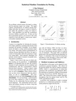
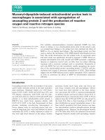
Figure 1
Schematic representation of reactive hyperaemia and measurements realised from laser Doppler recordingSchematic representation of reactive hyperaemia and measurements
realised from laser Doppler recording. 1: Mean blood flow at rest
(Φrest). 2: Peak flow (Φpeak). 3: Time to peak. 4: ΔΦ = Φpeak - Φrest.
5: Time to flow normalisation. 6: Time to half flow normalisation. 7: First
upward slope calculated for the first 3 seconds. 8: Second upward
slope calculated for the second half.
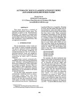
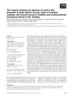
Figure 2
Example of a laser Doppler recording of blood flow during reactive hyperaemia in a patient sedated with midazolamExample of a laser Doppler recording of blood flow during reactive
hyperaemia in a patient sedated with midazolam. 1: Mean blood flow at
rest (Φrest). 2: Peak flow (Φpeak). 3: Time to peak. 4: ΔΦ = Φpeak -
Φrest. 5: Time to flow normalisation. 6: Time to half flow normalisation.
7: First upward slope calculated for the first three seconds. 8: Second
upward slope calculated for the second half. PU, perfusion units.
Critical Care Vol 10 No 6 Lamblin et al.
Page 4 of 9
(page number not for citation purposes)
Results
Ten patients were included in our study. General characteris-
tics are summarised in Table 1. When the H-period data were
collected, 26 ± 13 mg of midazolam had been infused and the
Ramsay score obtained was 4 ± 1. When the HS-period data
were collected, 41 ± 20 mg of midazolam had been infused
during the two hour infusion and 55 ± 36 μg of sufentanil was
added the second hour. The Ramsay score obtained was 5 ±
1.
Pattern of resting parameters
MAP decreased significantly during the sedation periods (H
and HS) compared to the NS period with no difference
between H and HS periods. HR, CO, SpO
2
, tcPO
2
, and
tcPCO
2
remained unchanged in all periods. Mean blood flow
at rest (Φrest) increased during the two sedation periods com-
pared to the NS period. CMBC remained unchanged by seda-
tion, whereas red blood cell velocity increased during H and
HS periods compared to the NS period (Table 2).
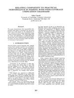
Vasomotion frequency spectra obtained in each sedation
period are represented in Figure 3. Distribution of vasomotion
frequencies was significantly different during the H period
compared to NS and HS periods. There was no difference
Table 1
General characteristics of study population
Patient Age (years) Gender Weight (kg) Temperature
(°C)
Respiratory
failure
SAPS II Outcome
1 74 Male 70 36.9 COPD 34 Alive
2 67 Female 60 37.2 COPD 24 Alive
3 66 Male 84 37.1 Postoperative 35 Alive
4 77 Male 70 36.8 Postoperative 37 Dead
5 19 Female 80 36.8 Asthma 44 Alive
6 50 Female 70 37.4 COPD, obesity 36 Alive
7 76 Female 87 36.8 SAS, obesity 86 Alive
8 73 Male 73 36.9 COPD 38 Alive
9 57 Male 70 37.5 COPD 39 Alive
10 75 Male 80 37.0 COPD 47 Dead
COPD, chronic obstructive pulmonary disease; SAPS II, Simplified Acute Physiology Score II; SAS, sleep apnoea syndrome.
Table 2
Resting parameters
NS period H period HS period
MAP (mm Hg) 94 ± 3 84 ± 4
a
81 ± 4
a
HR (beats per minute) 83 ± 6 81 ± 5 78 ± 5
CO (litres/minute) 5.38 ± 0.82 5.27 ± 0.82 5.33 ± 1.08
SpO
2
(percentage) 96.4 ± 0.5 96.7 ± 0.5 96.1 ± 0.5
tcPO
2
(mm Hg) 67 ± 6 61 ± 6 61 ± 7
tcPCO
2
(mm Hg) 45 ± 2 44 ± 4 42 ± 4
Φrest (PU) 150 ± 22 207 ± 25
a
205 ± 25
a
CMBC (CU) 145 ± 15 152 ± 16 155 ± 15
Velocity (VU) 1.06 ± 0.11 1.39 ± 0.15
a
1.37 ± 0.79
a
a
p < 0.05 versus NS period. Φrest, mean blood flow at rest; CMBC, concentration of moving blood cells in concentration units (CU); CO, cardiac
output; H period, set of measurements obtained when the patients were sedated by midazolam; HR, heart rate; HS period, set of measurements
obtained when the patients were sedated by midazolam and sufentanil; MAP, mean arterial pressure; NS period, set of measurements obtained
when the patients were non-sedated; PU, perfusion units; SpO
2
, percutaneous oxygen saturation; tcPCO
2
, transcutaneous carbon dioxide
pressure; tcPO
2
, transcutaneous oxygen pressure; velocity expressed in velocity units (VU), Φrest (perfusion units)/CMBC.
Available online />Page 5 of 9
(page number not for citation purposes)
between the NS and HS periods. Midazolam significantly
decreased vasomotion, and sufentanil restored the vasomo-
tion. Midazolam acted especially on the low-frequency vaso-
motion, which corresponds to the A1 and A2 small arteries
(Figure 3).
Reactive hyperaemia
Peak blood flow (Φpeak) remained unchanged during seda-
tion periods versus the NS period. ΔΦ decreased significantly
during H and HS periods versus the NS period, whereas no
significant difference existed between sedation periods. Slope
1 associated with the initial upward portion of hyperaemia
peak was not changed by midazolam but increased when suf-
entanil was added to midazolam. Slope 2 associated with the
second upward portion of peak was not influenced by
sedation (Table 3).
In the NS period, vasomotion wave amplitudes were higher
during reactive hyperaemia than at rest. This reinforcement of
vasomotion by reactive hyperaemia has been described in the
literature and proves that the microcirculation of our patients
reacted normally [22]. In vasomotion frequency analysis, this
phenomenon was observed mainly in the low frequencies (1 to
3 cpm) and thus concerned mainly the A1 small arteries. In
contrast, during sedation periods, this inductive role of reac-
tive hyperaemia was not observed. Vasomotion was
depressed and this effect predominated in A1 small arteries
(Figures 3 and 4).
Discussion
Sedation is widely used in ICU patients but its potentially del-
eterious effects, in particular on the microvascular bed, have
not been precisely evaluated. In this study, we found that seda-
tion using midazolam or a combination of midazolam and suf-
entanil induces microcirculatory changes with increased
cutaneous blood flow, decreased response to ischaemia, and
attenuation of vasomotion.
Effects of sedation on cutaneous microcirculation at rest
Microcirculation and midazolam
Sedation with midazolam induces a significant decrease of
MAP. Cardiovascular effects of benzodiazepines are well
known in anaesthesia [26,27]. However, with the subanaes-
thesic dose of benzodiazepine recommended for ICU seda-
tion, MAP and HR decrease only slightly [28], as we have
noted in our study.
Mean cutaneous blood flow increased after one hour of seda-
tion by midazolam. In parallel to blood flow, the red blood cell
velocity increased, whereas CMBC remained stable. These
data are in favour of a cutaneous vasodilation induced by
Figure 3
Distribution of vasomotion frequencies at restDistribution of vasomotion frequencies at rest. Kolmogorov-Smirnov
test: p < 0.05 NS and HS periods versus H period. Friedman test: *p <
0.05 NS period versus H period,
$
p < 0.05 HS period versus H period.
cpm, cycles per minute; H period, set of measurements obtained when
the patients were sedated by midazolam; HS period, set of measure-
ments obtained when the patients were sedated by midazolam and suf-
entanil; NS period, set of measurements obtained when the patients
were non-sedated.
Table 3
Changes in Doppler measurements during reactive hyperaemia according to sedation types
NS period H period HS period
Φpeak (PU) 292 ± 31 304 ± 28 274 ± 25
ΔΦ (PU) 141 ± 14 97 ± 16
a
73 ± 9
a
TP (seconds) 23.5 ± 4.3 43.8 ± 11.5 24.2 ± 8.2
T
1/2
R (seconds) 38.8 ± 13.7 22.9 ± 3.4 23.8 ± 5.5
TR (seconds) 170.1 ± 40.5 117.2 ± 3.4 85.1 ± 14.9
Slope 1 (PU/second) 53.6 ± 13.3 51.5 ± 8.8 74.1 ± 10.9
a,b
Slope 2 (PU/second) 10.8 ± 3.7 8.2 ± 3.0 8.7 ± 0.7
a
p < 0.05 versus NS period;
b
p < 0.05 versus H period. ΔΦ, Φpeak - Φrest; Φpeak, maximal blood flow during reactive hyperaemia; H period, set
of measurements obtained when the patients were sedated by midazolam; HS period, set of measurements obtained when the patients were
sedated by midazolam and sufentanil; NS period, set of measurements obtained when the patients were non-sedated; PU, perfusion units; T
1/2
R,
time to half flow normalisation; TP, time to peak; TR, time to flow normalisation.
Critical Care Vol 10 No 6 Lamblin et al.
Page 6 of 9
(page number not for citation purposes)
midazolam and are in agreement with the literature [29]. Stud-
ies of cutaneous and subcutaneous blood flows after injection
of benzodiazepine show an increase in the surface cutaneous
thermal clearance as well as a stability of the deep thermal
clearance, corresponding to an increase in cutaneous blood
flow with no deterioration of subcutaneous blood flow [30,31].
In another study, LDF also reveals an increase in cutaneous
blood flow among anaesthetised and hypothermic patients
compared to control subjects [32].
The increase in cutaneous blood flow may be explained by the
direct vasodilator effect of benzodiazepines [29]. Midazolam
attenuates the smooth muscle contraction induced by nore-
pinephrine, acting by an inhibition of Ca
2+
influx occurring
through voltage-operated Ca
2+
channels and through agonist-
mediated Ca
2+
channels and by an inhibition of Ca
2+
release
from intracellular storage sites (sarcoplasmic reticulum) [33].
Endothelium-dependent mechanisms also take part in the
vasodilation produced by midazolam through the release of
nitric oxide (NO) from vascular endothelium [34].
Microcirculation and the combination of midazolam and
sufentanil
In our study, the combination of midazolam and sufentanil
worsened hypotension (only slightly) and bradycardia but did
not change CO. Φrest was higher during the HS period than
during the NS period but was not different from that observed
during the H period. Contradictory results concerning the
effects of sufentanil on vascular tone have been described in
the literature. Sufentanil has been shown to decrease periph-
eral vascular resistances through a direct vascular effect [35].
Karasawa and colleagues [36] showed this effect to be due to
an endothelium-independent vasorelaxation mediated by both
an alpha-receptor blockage and a direct effect on smooth
muscle. In addition, Stefano and colleagues [37] reported that
endothelial cells contain opiate receptors called mu3 which
are coupled to NO release and vasodilation. On the other
hand, a direct contractile effect on vascular smooth muscle
has also been described [38]. As shown by Brookes and col-
leagues [39], the discrepancy between these two studies may
be explained by differences in doses. In our study, sufentanil
dose may have been insufficient to induce additional microcir-
culatory disturbances.
Effects of sedation on cutaneous microcirculation
response to ischaemia
Reactive hyperaemia
Reactive hyperaemia is a well-established and widely used
challenge to test microcirculation reactivity. This method has
been largely validated and is reproducible in humans [40,41].
It corresponds to an increase in local blood flow, secondary to
a transient ischaemia, and is thought to exactly reflect the cir-
culatory deficit that has occurred during the vascular
occlusion.
Reactive hyperaemia is the result of the combination of several
phenomena divided into a myogenic phase followed by a met-
abolic phase. The myogenic phase corresponds to the
changes of arteriolar diameter in response to pressure modifi-
cations and is thought to be reflected by the initial upward por-
tion (slope 1) of the hyperaemia peak [42]. At the time of the
metabolic phase thought to be reflected by the second part of
the upward portion (slope 2), the arteriolar vasodilation is the
result of factors acting directly on the vascular smooth muscle
or via the endothelium [42,43]. Engelke and colleagues [44]
showed that prostaglandins, released from the vascular
endothelium, are important determinants of the hyperaemia
peak, in contrast to NO, which takes part only in the mainte-
nance of the vasodilation after the peak [45].
Reactive hyperaemia and midazolam
In our study, midazolam did not influence the blood flow at
hyperaemia peak. On the other hand, ΔΦ (Φpeak - Φrest) was
decreased by 30% compared to the NS period. Peak blood
flow represents the maximum microcirculatory blood flow
obtainable by vasodilation. This explains the stability of Φpeak
and the decrease of ΔΦ during reactive hyperaemia in patients
during the H period, in whom an increased Φrest existed
before the reactive hyperaemia manoeuvre. Time to peak
tended to increase. All of these results show that midazolam
induced a limitation of the vascular response to ischaemia.
Reactive hyperaemia and the combination of midazolam and
sufentanil
During reactive hyperaemia, addition of sufentanil to mida-
zolam did not change peak blood flow compared to NS and H
periods. On the other hand, ΔΦ decreased by 50% during the
HS period compared to the NS period but did not differ from
Figure 4
Distribution of vasomotion frequencies during reactive hyperaemia according to sedationDistribution of vasomotion frequencies during reactive hyperaemia
according to sedation. Kolmogorov-Smirnov test: *p < 0.05 NS period
versus H and HS periods. cpm, cycles per minute; H period, set of
measurements obtained when the patients were sedated by mida-
zolam; HS period, set of measurements obtained when the patients
were sedated by midazolam and sufentanil; NS period, set of measure-
ments obtained when the patients were non-sedated.
Available online />Page 7 of 9
(page number not for citation purposes)
the H period. Time to peak decreased during the HS period
compared to the H period without reaching the threshold of
significance.
The slope 1 was significantly increased compared to the H
period, evoking modification of the myogenic phase of reactive
hyperaemia. During the HS period, small arteries seemed to
vasodilate more easily and more quickly than during the H
period. Sufentanil could induce a decrease in the smooth vas-
cular tonicity by acting directly on the vascular smooth muscle
and making vasorelaxation easier. These results are in agree-
ment with those of Karasawa and colleagues [36], who found
that fentanyl induces vasodilation via a direct action on muscu-
lar smooth cell and by locking alpha-adrenergic receptors.
However, because in our study these changes were observed
during the injection of a combination of midazolam and sufen-
tanil, we cannot determine whether sufentanil was, by itself,
responsible for the decrease in vascular tonicity or only rein-
forced an effect started under midazolam.
Effects of sedation on vasomotion
Blood flow in the microcirculation is not continuous but is sub-
ject to cyclic variations in which periods of high blood flow
alternate with periods of no flow. This phenomenon has been
called vasomotion and is due to changes in lumen diameters
which result from periodic activity of muscle cells in the micro-
vessel wall governed by oscillation of intracellular calcium con-
centration [46].
Vasomotion has been observed since the inception of microv-
ascular studies by intravital microscopy [47,48]. Later, when
LDF appeared, the oscillatory flow patterns observed were
related to the vasomotion activity of the microcirculation. Sub-
sequently, it was shown that frequency analysis of LDF record-
ings was able to discriminate between the types of vessels
from which the signal originates and that low-frequency flow
oscillations were directly related to vasomotion of the arteri-
oles [25,49].
Vasomotion and sedation
In our study, we observed a significant reduction in the impor-
tance of cutaneous vasomotion at rest and during reactive
hyperaemia in the group sedated with midazolam. The combi-
nation of midazolam and sufentanil seemed to restore cutane-
ous vasomotion to its resting level. Anaesthetic drugs have
long been recognised to alter vasomotion [50,51].
Decrease of vasomotion observed during midazolam infusion
is probably due to the benzodiazepine effects on intracellular
calcium concentration: inhibition of Ca
2+
influx and decrease
of Ca
2+
release from sarcoplasmic reticulum [33]. An explana-
tion for the restoration of vasomotion when sufentanil is added
to midazolam is less evident. Stephano and colleagues [37]
have shown that opiates induce NO release through endothe-
lial mu3 receptors. We hypothesise that this increase in NO
could elevate cyclic guanosine monophosphate (cGMP) con-
centration in smooth muscle cells, thereby increasing the
cGMP-dependent Ca
2+
-activated chloride channel, which has
been shown to be responsible for coupling the Ca
2+
oscilla-
tions generated by the sarcoplasmic reticulum to the mem-
brane current that synchronises individual cells [52,53].
In the NS period, we found vasomotion frequency distributions
to be more important during reactive hyperaemia than at rest,
evidence of a potentiation of vasomotion by hyperaemia. Dur-
ing midazolam infusion, an inhibition of this increase of vaso-
motion induced by reactive hyperaemia was noted. Frequency
analysis of the LDF recordings showed that the action of mida-
zolam on vasomotion prevailed on the A1 small arteries (fre-
quency of between 1 and 3 cpm). On the contrary, Colantuoni
and colleagues [25] found that the inhibition of vasomotion by
anaesthesia concerns vessels of all orders. The discrepancy
with our study may be explained by technical reasons. In our
study, 70% of the LDF signal came from the largest arterioles,
A1 and A2, and only 30% of the signal from the smallest arter-
ies, A3 and A4 (Figures 3 and 4). Consequently, it may have
been statistically easier to highlight an effect of sedation on
the A1 small arteries even if sedation deteriorates the vasomo-
tion in all four orders of small arteries.
Our study suffers from some limitations. First, the small
number of patients may have hidden some true variations. Sec-
ond, the study design did not include a randomisation
between the two steps. So, a carry-over effect may interfere
when studying the combination of midazolam and sufentanil. In
accordance with the aim of our study, we designed our seda-
tion protocol following widely accepted guidelines in order to
be closer to routine clinical practice. Doses of sufentanil used
were perhaps not sufficient to induce an additional effect on
cutaneous microcirculation. In clinical practice, the amounts of
opiates used are often higher than those recommended. So,
sufentanil's own effects may have been minimised.
Third, we chose the LDF technique because it is non-invasive
and easy to use in an ICU setting. Numerous techniques have
been proposed to explore the microcirculation, none of which
is without critics. Recently, a new technique, orthogonal polar-
isation spectral imaging, has been used in the ICU. It has sev-
eral advantages, in particular in separating respective changes
in small arteries, capillaries, and venules [8]. However, it gives
semi-quantitative measurements, suffers from an intra/inter-
observer variability of 5% to 10%, and is less suitable for
monitoring short-term microcirculatory blood flow change as
during recruitment manoeuvres. LDF is more suitable for mon-
itoring such rapid microcirculatory blood flow changes but
raises problems of calibration, artifact related to patient move-
ments, inability to separate respective changes between all
the vessels included in the investigated volume, and inter-indi-
vidual flow variations [54]. In our series, we noted great inter-
individual variations of blood flows at rest and during reactive
Critical Care Vol 10 No 6 Lamblin et al.
Page 8 of 9
(page number not for citation purposes)
hyperaemia. However, for the same patient, the signal is repro-
ducible provided that the position of probe and conditions of
measurement remain identical (haemodynamic, temperature)
[23,55]. Cutaneous blood flow varies according to the area
measured. Indeed, in the upper limb, the palms of the hand
and finger pads are better vascularised than the forearm or the
dorsum of the hand. We chose the pad of the mean finger as
the site of recording because this zone is highly vascularised
and, consequently, flow is more easily detectable by LDF [56].
Lastly, we studied the effects of sedation on cutaneous micro-
circulation. Even if skin preparations have often been used as
a model to study microcirculation, extension of our results to
other microcirculations may be made only with caution. Further
studies have to be carried out to determine whether microcir-
culation in other organs reacts in the same way.
Conclusion
Our study is one of the first to examine the effects of a sedation
regimen commonly used in the ICU on cutaneous microcircu-
lation. Benzodiazepine induces an increase in cutaneous
blood flow secondary to vasodilation, a decrease in reactive
hyperaemia, and alterations of vasomotion. Addition of sufen-
tanil does not substantially modify the results obtained.
Clinical studies have clearly established that alterations of nor-
mal microcirculatory control mechanisms may compromise the
tissue nutrient blood flow and may contribute to the develop-
ment of organ failure in septic patients [9,57,58]. Our study
raises the question of whether sedation with benzodiazepine
or a combination of benzodiazepine and sufentanil by deterio-
rating vasomotion and vascular reactivity to ischaemia may
further alter tissue perfusion when already compromised, as in
septic patients.
Competing interests
The authors declare that they have no competing interests.
Authors' contributions
VL conceived the protocol, participated in its design, carried
out bedside measurements and documentation, and drafted
the manuscript. MB and RF conceived the protocol and
helped to interpret the data. DM conceived the protocol, par-
ticipated in its design and coordination, and helped to interpret
the data and to draft the manuscript. All authors read and
approved the final manuscript.
Acknowledgements
The Centre Hospitalier Universitaire de Lille and the Universite de Lille
provided funding for this study.
References
1. Schmid-Schonbein G, Ross J: Structure-function relations in
the peripheral circulation. In Best and Taylor's Physiological
Basis of Medical Practice Edited by: West JB. Baltimore: Williams-
Wilkins; 1990:118-137.
2. Engelson ET, Schmid-Schonbein GW, Zweifach BW: The micro-
vasculature in skeletal muscle. II. Arteriolar network anatomy
in normotensive and spontaneously hypertensive rats. Micro-
vasc Res 1986, 31:356-374.
3. Hauser CJ, Shoemaker WC: Use of a transcutaneous PO
2
regional perfusion index to quantify tissue perfusion in periph-
eral vascular disease. Ann Surg 1983, 197:337-343.
4. Cesarone MR, Laurola G, Belcaro GV: Microcirculation in sys-
temic hypertension. Angiology 1992, 43:899-903.
5. Neviere R, Mathieu D, Chagnon JL, Lebleu N, Millien JP, Wattel F:
Skeletal muscle microvascular blood flow and oxygen trans-
port in patients with severe sepsis. Am J Respir Crit Care Med
1996, 153:191-195.
6. Lush CW, Kvietys PR: Microvascular dysfunction in sepsis.
Microcirculation 2000, 7:83-101.
7. Sair M, Etherington PJ, Peter Winlove C, Evans TW: Tissue oxy-
genation and perfusion in patients with systemic sepsis. Crit
Care Med 2001, 29:1343-1349.
8. De Backer D, Creteur J, Preiser JC, Dubois MJ, Vincent JL: Micro-
vascular blood flow is altered in patients with sepsis. Am J
Respir Crit Care Med 2002, 166:98-104.
9. Sakr Y, Dubois MJ, De Backer D, Creteur J, Vincent JL: Persistent
microcirculatory alterations are associated with organ failure
and death in patients with septic shock. Crit Care Med 2004,
32:1825-1831.
10. Shapiro BA, Warren J, Egol AB, Greenbaum DM, Jacobi J, Nasra-
way SA, Schein RM, Spevetz A, Stone JR: Practice parameters
for intravenous analgesia and sedation for adults patients in
the intensive care unit: an executive summary. Society of Crit-
ical Care Medicine. Crit Care Med 1995, 23:1596-1600.
11. Shafer A: Complications of sedation with midazolam in the
intensive care unit and a comparison with other sedative
regimens. Crit Care Med 1998, 26:947-956.
12. Shelly MP, Sultan MA, Bodenham A, Park GR: Midazolam infu-
sions in critically ill patients. Eur J Anaesthesiol 1991, 8:21-27.
13. Huang KC, Stoddard M, Tsueda KA, Heine MF, Thomas MH,
White M, Wieman TJ: Stroke volume measurements by electri-
cal bioimpedance and echocardiography in healthy
volunteers. Crit Care Med 1990, 18:1274-1278.
14. Shoemaker WC, Belzberg H, Wo CC, Milzman DP, Pasquale MD,
Baga L, Fuss MA, Fulda GJ, Yarbrough K, Van DeWater JP, et al.:
Multicenter study of non invasive monitoring systems as alter-
natives to invasive monitoring of acutely ill emergency
patients. Chest 1998, 114:1643-1652.
15. Salandin V, Zussa C, Risica G, Michielon P, Paccagnella A, Cipol-
otti G, Simini G: Comparison of cardiac output estimation by
thoracic electrical bioimpedance, thermodilution and Fick
methods. Crit Care Med 1988, 16:1157-1158.
16. Bonner RF, Clem TR, Bowen PD: Laser Doppler continuous
real-time monitor of pulsatile and mean blood flow in tissue
microcirculation. In Scattering Techniques Applied to Supramo-
lecular and Non-Equilibrium Systems Edited by: Chen SH, Chu B,
Nossal R. New York: Plenum Press; 1981:685-701.
17. Bonner R, Nossal R: Model for laser Doppler measurements of
blood flow in tissue. Appl Optics 1981, 20:2097-2107.
18. Saumet JL, Dittmar A, Leftheriotis G: Non-invasive measurement
of skin blood flow: comparison between plethysmography,
laser Doppler flowmeter and heat thermal clearance method.
Int J Microcirc Clin Exp 1986, 5:73-83.
19. Fagrell B: Advances in microcirculation network evaluation: an
update. Int J Microcirc Clin Exp 1995, 15(suppl 1):34-40.
20. Bircher A, De Boer EM, Agner T, Wahlberg JE, Serup J: Guide-
lines for measurement of cutaneous blood flow by laser Dop-
pler flowmetry. A report from the Standardization Group of the
European Society of Contact Dermatitis. Contact Dermatitis
1994, 30:65-72.
Key messages
• Sedation with midazolam alone or in association with
sufentanil induces a deterioration of vasomotion and
microvascular response to ischaemia. This raises the
question of whether sedation may further alter tissue
perfusion when already compromised, as in septic
patients.
Available online />Page 9 of 9
(page number not for citation purposes)
21. Moens AL, Goovaerts I, Claeys MJ, Vrints CJ: Flow-mediated
vasodilation: a diagnostic instrument, or an experimental tool?
Chest 2005, 127:2254-2263.
22. Pyke KE, Tschakovsky ME: The relationship between shear
stress and flow-mediated dilatation: implications for the
assessment of endothelial function. J Physiol 2005,
568:357-369.
23. Yvonne-Tee GB, Rasool AH, Halim AS, Rahman AR: Reproduci-
bility of different laser Doppler fluximetry parameters of pos-
tocclusive reactive hyperemia in human forearm skin. J
Pharmacol Toxicol Methods 2005, 52:286-292.
24. Intaglietta M: Arteriolar vasomotion: implication for tissue
ischemia. Blood Vessels 1991, 28(suppl 1):1-7.
25. Colantuoni A, Bertuglia S, Intaglietta M: Quantification of rhyth-
mic diameter changes in arterial microcirculation. Am J
Physiol 1984, 246:H508-H517.
26. Reves JG, Fragen RJ, Vinik R, Greenblatt DJ: Midazolam: phar-
macology and uses. Anesthesiology 1985, 62:310-324.
27. Marty J, Gauzit R, Lefevre P, Coudrec E, Farinotti R, Henzel C, Des-
monts JM: Effects of diazepam and midazolam on baroreflex
control of heart rate and on sympathetic activity in humans.
Anesth Analg 1986, 65:113-119.
28. Ronan KP, Gallager TJ, George B, Hamby B: Comparison of pro-
pofol and midazolam for sedation in intensive care unit
patients. Crit Care Med 1995, 23:286-293.
29. West JM, Estrada S, Heerdt M: Sudden hypotension associated
with midazolam and sufentanil. Anesth Analg 1987,
66:693-694.
30. Saumet JL, Leftheriotis G, Dubost J, Kalfon J, Baurillon V, Freidel
M: Cutaneous and subcutaneous blood flow during general
anaesthesia. Eur J Appl Physiol Occup Physiol 1988,
57:601-605.
31. Saumet JL, Leftheriotis G, Dittmar A, Delhomme G, Degoute CS:
Skin blood flow changes in anaesthetized humans: compari-
son between skin thermal clearance and finger pulse ampli-
tude measurement. Eur J Appl Physiol Occup Physiol 1986,
54:
574-577.
32. Micheels J, Alsbjorn B, Sorensen B: Laser Doppler flowmetry a
new non-invasive measurement of microcirculation in inten-
sive care? Resuscitation 1984, 12:31-39.
33. Yamaguchi S, Kanmura Y, Yoshimura N: Effects of midazolam on
contractions in smooth muscle of rabbit mesenteric artery.
Anesth Analg 1997, 84:199-205.
34. Chang KSK, Feng MG, Davis RF: Midazolam produces vasodil-
atation by mixed endothelium-dependent and independent
mechanisms. Anesth Analg 1994, 78:710-717.
35. White DA, Reitan JA, Kien ND, Thorup SJ: Decrease in vascular
resistance in the isolated canine hindlimb after graded doses
of alfentanil, fentanyl, and sufentanil. Anesth Analg 1990,
71:29-34.
36. Karasawa F, Iwanov V, Moulds RF: Sufentanil and alfentanil
cause vasorelaxation by mechanisms independents of
endothelium. Clin Exp Pharmacol Physiol 1993, 20:705-711.
37. Stefano GB, Hartman A, Bilfinger TV, Magazine HI, Liu Y, Casares
F, Goligorsky MS: Presence of the mu3 opiate receptor in
endothelial cells. Coupling to nitric oxide production and
vasodilation. J Biol Chem 1995, 270:30290-30293.
38. Parra L, Perez-Vizcaino F, Alsasua A, Martin MI, Tamargo J: Mu-
and delta-opioid receptor-mediated contractile effects on rat
aortic vascular smooth muscle. Eur J Pharmacol 1995,
277:99-105.
39. Brookes ZL, Brown NJ, Reilly CS: The dose-dependent effects
of fentanyl on rat skeletal muscle microcirculation in vivo.
Anesth Analg 2003, 96:456-462.
40. Maurel A, Hamon P, Maquin-Mavier I, Lagrue G: Laser-doppler
study of microcirculatory cutaneous bloodflow. A study about
100 human volunteers. Presse Med 1991, 20:1205-1209.
41. Östergren J, Fagrell B: Skin capillary blood cell velocity in man.
Characteristics and reproducibility of reactive hyperemia
response. Int J Microcirc Clin Exp 1986, 5:37-51.
42. Koller A, Kaley G: Role of endothelium in reactive dilation of
skeletal muscle arterioles. Am J Physiol 1990,
259:H1313-H1316.
43. Koller A, Kaley G: Endothelium regulates skeletal muscle
microcirculation by blood flow velocity-sensing mechanism.
Am J Physiol 1990, 258:H916-H920.
44. Engelke KA, Halliwill JR, Proctor DN, Dietz N, Joyner MJ: Contribu-
tion of nitric oxide and prostaglandins to reactive hyperemia in
the human forearm. J Appl Physiol 1996, 81:1807-1814.
45. Meredith IT, Currie KE, Anderson TJ, Roddy MA, Ganz P, Creager
MA: Postischemic vasodilatation in human forearm is depend-
ent on endothelium-derived nitric oxide. Am J Physiol 1996,
270:H1435-H1440.
46. Brekke JF, Jackson WF, Segal SS: Arteriolar smooth muscle
Ca2
+
dynamics during blood flow control in hamster cheek
pouch. J Appl Physiol 2006, 101:307-315.
47. Clark ER, Clark EL: Observations on living preformed blood
vessels as seen in a transparent chamber implanted in the
rabbits ear. Am J Anat 1932, 49:441-474.
48. Nicoll PA, Webb RL: Vascular patterns and active vasomotion
as determiners of flow through minute vessels. Angiology
1955, 6:291-303.
49. Colantuoni A, Bertuglia S, Intaglietta M: Microvascular vascular
vasomotion: origin of laser Doppler flux motion. Int J Microcirc
Clin Exp 1994, 14:151-158.
50. Faber JE, Harris PD, Wiegman DL: Anaesthetic depression of
microcirculation, central hemodynamics, and respiration in
decerebrate rats. Am J Physiol 1982, 243:H837-H843.
51. Colantuoni A, Bertuglia S, Intaglietta M: Effects of anesthesia on
the spontaneous activity of the microvasculature. Int J Micro-
circ Clin Exp 1984, 3:13-28.
52. Peng H, Matchkov V, Ivarsen A, Aalkjaer C, Nilsson H: Hypothesis
for the initiation of vasomotion. Circ Res 2001, 88:810-815.
53. Rahman A, Matchkov V, Nilsson H, Aalkjaer C: Effects of cGMP
on coordination of vascular smooth muscle cells of rat
mesenteric small arteries. J Vasc Res 2005, 42:301-311.
54. Bollinger A, Hoffmann U, Franzeck UK: Evaluation of flux motion
in man by the laser Doppler technique. Blood Vessels 1991,
28:21-26.
55. Hoffmann U, Uckay I, Fisher M, Wen S, Franzeck UK, Bollinger A:
Simultaneous assessment of muscle and skin blood fluxes
with laser Doppler technique. Int J Microcirc Clin Exp 1995,
15:53-59.
56. Wilkin JK: Periodic cutaneous blood flow during post occlusive
reactive hyperemia. Am J Physiol 1986, 250:H765-H768.
57. Wattel F, Mathieu D, Nevière R, Bocquillon N: Role of microcircu-
lation in multiorgan failure of infectious origin. Bull Acad Natl
Med 2000,
184:1609-1619.
58. Lehr HA, Bittinger F, Kirkpatrick CJ: Microcirculatory dysfunction
in sepsis: a pathogenic basis for therapy? J Pathol 2000,
190:373-386.