Core Topics in Operating Department Practice Anaesthesia and Critical Care – Part 8 ppt
Bạn đang xem bản rút gọn của tài liệu. Xem và tải ngay bản đầy đủ của tài liệu tại đây (779.92 KB, 23 trang )
that the dogs displayed a ‘mild inebriation’, noted
after injection.
In 1664, a German scientist named Daniel Meyer
noted that, when needles were inserted into the
tongues of dogs following the injection of IV
opium, the animals exhibited a ‘decreased
response to pain’. Unfortunately the link between
the injection of IV solutions and analgesia was not
recognised on any one of these occasions.
Subsequently, a period of respite followed
together with a reduction in the use of IV
injections.
It is generally assumed that there was no further
research involving IV-related anaesthesia until
1872, when Pierre-Cyprien Ore used IV chloral
hydrate as the sole anaesthetic which was given to
a total of 36 surgical patients (Sykes, 1960).
Unfortunately, because of the high incidence of
mortality linked to Ore’s technique, there was little
further interest, and the idea of using the venous
system to deliver anaesthesia was dismissed until
the late nineteenth century.
Recent research into veterinary anaesthesia has
discovered that almost half a century before Ore’s
work was published, M. Dupy, Director of the
Toulouse Veterinary School, had begun to use the
external jugular veins of horses to administer IV
chemical compounds such as alcohol. Dupy noted,
among other things, that ‘the expired air smelt
strongly of alcohol’ (Anonymous, 1831). This was
the first reference to an IV substance being
excreted by the lungs. The doses of alcohol that
Dupy used during his experiments were not
enough to render the horses unconscious, just to
‘stupefy’ them. If larger doses of alcohol had been
used to induce unconsciousness then the link
between IV induction agents and the loss of
consciousness may have been established much
sooner. The development of IV anaesthesia
might then have taken a different course than it
did at the turn of the century. As it happened the
casual link between loss of consciousness and
the IV injection of a drug was overlooked and the
significance of studying the effects of IV injections
was lost.
Advances in anaesthesia continued with various
inhalational agents able to provide all the compo-
nents of anaesthesia.
The middle of the eighteenth century saw
many technological advances around about the
same time; these advances helped pave the way
for IV therapies.
In 1845 Francis Rynd invented the hollow needle,
while the first syringe was invented in 1853 by
Charles Gabriel Pravaz. None of these develop-
ments were originally designed for IV use. They
were later adapted and refined by Alexander Wood
who used them for injecting morphine directly into
painful joints (Wood, 1855).
The turn of the twentieth century saw something
of a renaissance for TIVA with the development
of a number of IV anaesthetics including hedonal
(Kissin & Wright, 1988), paraldehyde (Noel &
Southar, 1913), magnesium sulphate (Peck &
Meltzer, 1916) and ethol alcohol (Naragwa, 1921;
Carot & Laugier, 1922). Unfortunately the use
of any one of these IV anaesthetics can have
harmful, if not disastrous side effects. During the
same period inhalational anaesthetic agents were
becoming increasingly safer and more established
among early anaesthetists.
The first barbiturates were synthesised in 1903
by Fisher (Fisher and von Mering, 1903), with the
first short-acting, rapid-onset barbiturate (evipan)
being developed almost 30 years later in 1932
(Weese et al., 1932).
The next and most influential advancement in IV
anaesthesia was the synthesis of sodium thiopen-
tone (pentothal) which was first used in 1934
(Dundee, 1980) by Lundy and Waters (Lundy &
Tovell, 1934; Platt et al., 1936).
Originally used as a single 5% infusion, thiopen-
tone was hailed as a wonder drug, the first real IV
monoanaesthetic. Unfortunately, the large doses
that were necessary to maintain anaesthesia had
devastating side effects. The use of thiopentone
as a monoanaesthetic reached its peak during the
Japanese attack on Pearl Harbor in 1941 and led to
the popular myth that more American military
personnel were killed by IV thiopentone than were
146 K. Henshaw
killed as a result of Japanese fire. Regardless of
whether there is any truth to this myth, it became
clear that thiopentone was not a monoanaesthetic
agent, more importantly 5% thiopentone infusions
were linked to high mortality rates.
Nevertheless, as an induction agent, the use of a
reduced-strength thiopentone (2.5%) quickly
became the gold standard that every other induc-
tion agent has since been measured by.
The search for a single anaesthetic agent that
could independently control all components of
anaesthesia began to lose impetus as anaesthesia
quickly changed to become a combination of
inhalational and IV agents.
The term ‘balanced anaesthesia’ has come
to represent the preferred technique of achieving
general anaesthesia by use of a combination of:
• premedication
• IV opioids
• IV muscle relaxants
• inhalational agents
• regional anaesthesia.
Over the last decade the use of IV drugs to
induce and maintain anaesthesia has become a
real alternative when aiming to achieve balanced
anaesthesia.
The arrival of rapid-onset, short-acting opioids
such as remifentanil and alfentanil, with advances
in infusion pump technology, and an increased
understanding of pharmacokinetics has for the first
time, allowed for the development of the tech-
nique of TCI.
Pharmacokinetics and pharmacodynamics
Pharmacokinetics simply means the movement
of a drug in the body. More specifically pharma-
cokinetics describes the relationship between
the dose of a drug and the amount of time
taken for the body to metabolise the drug. This
relationship can be represented and predicted by
the use of complex mathematical models or
algorithms.
The concept of pharmacokinetics was first
used in anaesthesia during the 1950s when Brodie
and Kety first described the process of drug
distribution in the body while researching how
thiopentone and inhalational agents are
metabolised.
They explained the distribution of thiopentone in
vivo and the importance of the role played by lean
tissue (not fat) in the redistribution of thiopentone
in the central nervous system (CNS).
By gradual refinement, physiologic researchers
were able to demonstrate the importance of the
effect-site concentration of a drug during anaes-
thesia. The effect-site concentration of a drug is
that point at which the clinical effect is seen.
In the case of anaesthetic drugs this is when the
blood brain barrier is crossed.
When using inhalational anaesthetics the effect-
site concentration can be monitored by using
capnography. The potency of a defined volatile
agent can then be expressed as the minimum
alveolar concentration (MAC). Each volatile agent
has a potency value which can be expressed
numerically as the MAC. The MAC of an inhala-
tional agent is the amount of anaesthetic needed
to prevent purposeful movement in 50% of the
population at any one time.
The aim of TIVA is to target the effect site
and to adjust appropriate plasma drug levels
accordingly. The problem with using a TIVA
technique in the past was that most IV anaesthetic
drugs that are given as a fixed rate infusion can take
a long time (412 hours) to plateau. Manual
titration meant that too much or too little anaes-
thetic was being infused leading to pain or
awareness or CNS depression and cardiovas-
cular system (CVS) depression. During TIVA it
became apparent that a system that could
employ a rapid response, calculate and respond
to any adverse clinical signs was needed.
That system would have to be flexible enough
to change the concentration of a drug quickly
and be able to recalculate drug concentration
levels in the plasma. This system was first demon-
strated by Schwilden (Schwilden, 1981) who was
Total intravenous anaesthesia 147
able to maintain target plasma levels of a drug by
use of a computer controlled infusion pump.
Any system would need to be able to set a target,
reach the target and then maintain the correct level
of a drug. Such a system became available for
clinical use in 1996 with the introduction of the
Diprifusor
Õ
(Sebel & Lowdown, 1984). The
Diprifusor
Õ
was the first commercially available
TCI device. The introduction of TCIs has allowed
anaesthetists to target and maintain the desired
levels of anaesthetic drugs.
As discussed earlier the problem of maintaining
target levels was that as soon as any drug is
administered the body begins a process of dilution,
distribution and elimination. The time period
of this process is dependent upon a number of
factors such as the patient’s age, weight, sex, type
of drug, the dose, speed of delivery and how the
drug is metabolised. Within the body if the
pharmacokinetic behaviour of a drug is known
then mathematical calculations can be used to
work out exactly how much of the drug is needed to
achieve (and maintain) a pre-set target level. When
a target level has been set the infusion rate of the
drug is then continuously adjusted in order to
maintain the desired target level.
Pharmacodynamics can be defined as ‘what
the drug does to the body’, in other words, the
effects that a drug has on systems of the body.
All inhalational agents, for example, have a depres-
sive effect on the CVS.
How are drug levels maintained
at the correct level?
TCI devises make use of microprocessors to
calculate the concentration of a drug within the
plasma. Calculations are constantly used by the
microprocessor which have been programmed
with an algorithm that uses a bolus elimination
transfer (BET) scheme.
The BET model is used to describe the move-
ment of a drug between two theoretical compart-
ments. It is important to emphasise that these
compartments are theoretical constructs and not
real anatomical compartments.
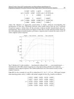
Figure 14.1 Pharmacokinetic model.
148 K. Henshaw
The BET scheme was first proposed by
Kruger-Theimer (Kruger-Theimer, 1968) and was
the first theoretical model to recognise that in order
to achieve a steady-state blood concentration of
a drug then at least three factors need to be
constantly calculated. Any algorithm used by a TCI
device must be able to measure:
• The original loading dose of the drug À this
determines the first phase of distribution. This is
the first phase.
• Any changes to the infusion and be able to
compensate for continuous drug elimination.
This is phase two of the BET Model.
• An infusion rate that can equilibrate drug
concentration in the plasma as the drug is
distributed to the peripheral compartment. This
is the third and final phase.
After the initial loading dose into the central com-
partment (phase 1) is given (Figure 14.1), a con-
stant amount of drug begins to be eliminated in a
fixed period of time. Therefore, if the elimination
times and rates of a defined drug are known then
the blood concentration of that drug can be
predicted and maintained by either increasing or
decreasing the infusion rate to compensate for
elimination (phase 2) and equilibration to other
compartments (phase 3) (Schwilden et al., 1986).
3-Compartment model
All calculations used by current TCI devices
are based on a 3-compartment pharmacokinetic
model. The 3-compartment model consists of a
hypothetic central compartment (V1), a second
compartment (V2), sometimes referred to as ‘fast’
or ‘vessel rich’ and a third compartment (V3)
commonly referred to as the ‘slow’ or ‘vessel poor’
compartment. It is this process of distribution and
elimination of drugs between the compartments
that forms the basis of all current pharmacokinetic
models.
Factors such as the patient’s age, weight and sex
can all effect the drug distribution between
compartments. For this reason it is important
that this information is made available to the
perioperative practitioner when practicable. Once
good venous access has been established and all of
the relevant factors such as age and sex have been
entered into the TCI device, then induction can
begin.
As the drug begins to move down the concentra-
tion gradients between compartments (in an effort
to try to achieve equilibrium) and is simultaneously
being eliminated from the body, the TCI device
calculates the changes between compartments and
compensates by either increasing or decreasing the
infusion rate in order to maintain the desired target
levels.
This ability to adapt infusion rates is the main
difference between a standard syringe driver,
which will deliver a predetermined amount of
drug until the pre-set volume is completed, and a
syringe driver that is target controlled and con-
tinually adjusts itself to maintain a target dose.
Why use TCI systems?
Advances in computer technology, the develop-
ment of fast-acting opioid analgesics and muscle
relaxants, together with more robust pharmacoki-
netic models have allowed anaesthetists to target
the effector site with the minimum amount of
anaesthetic drug to achieve adequate anaesthesia.
An important point to remember here is that TCI
devices are not computerised anaesthetists. All of
the normal clinical observations and decisions
regarding the treatment of a patient still need to
be made during target controlled anaesthesia. In
this sense TCI devices can never replace sound
clinical knowledge and experience.
Advantages Disadvantages
Where the use of high
concentrations of
oxygen are needed
such as:
• Increased IV doses of
anaesthetic agents are
used to compensate
for the lack of N
2
O
• single lung anaesthesia
• hyperbaric medicine
Total intravenous anaesthesia 149
Advantages Disadvantages
In situations where
delivery of inhalational
agent may be restricted:
• Designated target
controlled infusion
devices which may be
initially expensive and
difficult to use
• Difficulty predicting
the end of anaesthesia
as presently there is no
indicator of metabolic
clearance of the drug
that has been infused.
Plasma concentration
estimates are displayed
but are not a direct
measurement of
volatile concentration
as displayed by
end tidal monitors
• Bronchoscopy
• Laryngoscopy
• A reduction in
atmospheric pollution
• A decreased incidence
of post-operative
nausea and vomiting
(PONV)
• A reduced trigger for
malignant
hyperpyrexia
The use of TCVA in
areas where volatile
anaesthetics would be
contraindicated or
difficult to administer.
For example:
• Disconnection, if IV
access is lost either
through
extravasation or
mechanical
disconnection then
anaesthesia is lost.
Difficult to detect
• war zones
• lack of anaesthetic
equipment, i.e. transfer
of the critically
compromised
patients
Other benefits of TIVA
include anaesthesia
for surgery where
the use of N
2
O may be
contraindicated.
For example:
• A second intravenous
infusion line must be
used
• Delayed recovery if
high target plasma levels
are maintained for long
time periods
• inner ear surgery
• long duration bowel
surgery
• pneumothorax
• air embolism
• hepatotoxicity
Principles of TCIs
As already stated, TCIs use a number of factors to
calculate appropriate plasma levels, for example,
the body mass index (BMI) of a young, athletic
male patient would have a very different pharma-
cokinetic profile than that of a patient who might
be older, more sedentary but may share the same
body weight. Depending on which pharmacoki-
netic model is used (newer TCI devices have a
facility to allow selection of specific models) the
microprocessor is able to calculate the appropriate
target by taking into account BMI, gender and age.
Examples of TIVA in clinical use could be:
• propofol which can be used as both an induction
agent and a maintenance drug
• a neuromuscular blocking agent (NMBA) can be
used (in conjunction with a peripheral nerve
stimulator)
• a short-acting opioid such as remifentanil
or alfentanil can be used as a component of
analgesia.
At present the only short-acting opioid that has
an approved algorithm for TCI is remifentanil.
Remifentanil is metabolised anywhere in the body
by non-specific esterases and so doesn’t rely on
hepatic or renal metabolism.
Probably the most well-known TCI device
and the most popular for use in Europe is the
Diprifusor
Õ
which has been available for clinical
use since 1996.
The Diprifusor
Õ
is only able to use pre-filled glass
syringes containing propofol to deliver TCIs. The
syringes are single-use only and contain a magnetic
strip on the flange of the syringe that ‘tells’ the
microprocessors in the infusion pump that the
device is primed with the correct drug and that it is
ready to be used. When the syringe is approaching
empty the magnetic strip is deprogrammed and an
alarm is activated to alert the user that a refill is
needed. Once the metallic strip has been deacti-
vated it can no longer be used or refilled.
A common criticism of TIVA is the high capital
and running costs incurred when compared to
low-flow inhalational anaesthesia. However, since
150 K. Henshaw
the patent for propofol has expired, newer and
cheaper generic propofols have become available
and the development of ‘Open TCI’ devices which
can use generic propofol has reduced the total cost
of TIVA significantly.
It could be argued that the initial expense of
setting up a TCI system can be offset by a reduction
in PONV and a reduced stay in the post-operative
care unit (POCU). Early discharge and faster
patient throughput associated with the use of
TIVA are some of the benefits that are thought to
offset the initial cost of setting up TIVA regimes.
Advocates of TIVA claim that TCIs are best suited
to the modern healthcare system with the empha-
sis on short stay, day case surgery and the growth
of endoscopic and invasive radiological proce-
dures. Opponents of TIVA argue that similar results
(reduced PONV and faster recovery times) can be
achieved using modern volatile agents and
improved methods of post-operative analgesia.
Awareness and depth of anaesthesia
At the present time direct measurement of drug
concentration at the effect site is not a prac-
tical option. Clinical judgement is still needed
to assess, and alter drug target levels both pre-
and intra-operatively. Most clinicians prefer to see
the potency of an anaesthetic agent (the MAC
value) and this can be measured reasonably easily
by sampling the end tidal volume. This option of
not being able to ‘see what’s happening is not
available when using TCIs and is another common
criticism of TIVA.
Depth of anaesthesia is a concern for all anaes-
thetists, but, given the absence of a MAC during
TCIs, many anaesthetists see the lack of a numer-
ical indicator as a real disadvantage.
Awareness and recall can and does occur during
anaesthesia (even when an adequate MAC is dis-
played). The widespread use of NMBAs has
increased occasions where patients have experi-
enced awareness, pain and even explicit recall
during general anaesthesia.
Depth of anaesthesia is notoriously difficult
to quantify. Even when adequate MAC levels are
displayed studies have demonstrated that recall,
learning and even response to commands can
still occur during anaesthesia. Patients have been
able to obey commands while anaesthetised
during surgical procedures for example, but were
unable to recall any of the events of the surgical
procedure.
Movement is a poor indicator of adequate
depth of anaesthesia, as the use of NMBAs prevent
the early detection of purposeful movement. Some
studies have been able to demonstrate purposeful
movement during neuromuscular blockade by
isolating the patient’s forearms from the NMBAs
by use of a tourniquet. Patients were then
instructed to move their hands or fingers in
response to surgical stimulus. This technique has
proved to be a poor indicator of depth of anaes-
thesia not least of all because patient hand move-
ment during a surgical procedure can be
distracting to the surgical staff and a hazard to
the integrity of the sterile field. The maximum
recommended time for this method is 20 minutes
so studies have been limited by time factors.
Adequate depth of anaesthesia has always been
a particular concern for users of TIVA as early
attempts at providing TIVA consisted of manual
infusions that relied on boluses of anaesthetic
drugs in response to surgical stimulus. Since the
availability of TCIs a smoother and more respon-
sive anaesthetic technique is now available to
clinicians.
Depth of anaesthesia monitors goes some
way to address these problems and their use in
anaesthesia has become more widespread.
The majority of depth of anaesthesia monitors
use a variety of electrophysiologic techniques to
monitor responses to stimuli. Commonly used
monitors in the UK are the bispectral index (BIS)
and the auditory evoked potential (AEP).
The perception of auditory stimuli intra-
operatively is well documented and AEP monitors
use a series of high frequency auditory clicks
to stimulate auditory cortical activity which is
Total intravenous anaesthesia 151
then measured as a brainstem response. Auditory
stimuli are administered through the patient’s ears
and so are not suitable for surgical procedures
that involve accessing the ear, or patients who
have pathological hearing disorders.
The bispectral index selectively analyses a
number of EEG waveforms and can help to predict
movement even in the paralysed patient.
Ultimately the most reliable form of depth of
anaesthesia monitor still remains the anaesthetist.
Closed loop systems
Depth of anaesthesia monitors can be used as
‘feed back’ mechanism for computerised TCI
systems. This method has had some limited
success when used to control general anaesthesia
and sedation. When automatic feedback is used the
system is known as closed loop anaesthesia.
Potentially closed loop systems should be able
to provide more accurate feedback which can
then be used to control the level of anaesthesia.
This is already an area where there is a great deal of
research in progress and could lead to computer-
controlled anaesthesia. Most TCI systems currently
in clinical practice rely on an ‘open system’ which
uses clinical judgment to adjust target levels in
response to surgical stimulus.
Components of a TCI system
The principal components of a TCI system must
contain:
• a means of inputting patient data such as age, sex
and weight, and also target drug concentration
• at least one (usually two) microprocessor(s)
and an infusion pump
• a display which shows both the targeted and
current calculated blood concentration
• a means of displaying the infusion rate
• a means of displaying the amount of drug that
has been delivered
• the effect-site concentration (the estimated
amount at the effector site in the brain)
• the estimated time needed to lower the target
concentration at the effector site.
Future developments
As a result of competition from generic versions
of propofol and the introduction of open systems
the overall cost and availability of TCI devices
have started to come down in price. This reduction
in cost of propofol and increased availability has
allowed more anaesthetists access to TCI devices.
The net result has seen a growth of TIVA which is
fast becoming an established technique in today’s
healthcare setting.
The development of new volatile agents has
declined and there is increased pressure from
government and regulatory bodies to reduce
the amount of pollutants in the atmosphere.
This external pressure together with the increased
availability of TCI devices is likely to see a further
decline in the use of volatile anaesthetics.
The search for a monoanaesthetic continues
together with the development of newer, safer IV
drugs. In the meantime the newer hypnotic and
analgesic drugs with their faster acting and more
predictable recovery profiles will enhance anaes-
thetic practice by allowing the clinician even
greater control of the individual components of
anaesthesia. Advocates of TIVA claim that the
quality and speed of reversal from anaesthesia is
greater than with traditional anaesthesia. This is
still an area for future research.
A better understanding of pharmacokinetic and
pharmacodynamic models has led to the develop-
ment of more predictable drugs which can be
simulated in computer programs.
Finally, the improvements and technological
developments associated with drug delivery sys-
tems mean that the safety and reliability of TIVA
techniques can offer a real alternative to traditional
inhalational techniques.
152 K. Henshaw
REFERENCES
Anonymous. (1831). Deals with injection of various
substances intravenously in horses by M. Dupy. Lancet,
2, 76.
Carot, H. & Laugier, H. (1922). Anaaesthesie par
injection intrareineuse d’un produit melange alcool-
chloroform-solution physiologique chez le chien.
CRSeances Soc Biol, 889À92.
Dundee, J. W. (1980). Historical vingettes and classifica-
tion of intravenous anaesthetics. In J.A. Aldrete &
T. H. Stanley, eds., Trends in Intravenous Anaesthesia.
Chicago: Year Book, p. 1.
Fischer, E. & von Mering, J. (1903). Ueber eine
neue klasse von schlafmilteln. Ther Gengenwart, 44,
97À101.
Kissin, I. & Wright, A. J. (1988). The introduction
of Hedonal: a Russian contribution to intravenous
anaesthesia. Anaesthesiology , 69, 242À5.
Kruger-Theimer, E. (1968). Continuous intravenous infu-
sion and multi compartmental accumulation. European
Journal of Pharmacology, 317À34.
Lundy, J. S. & Tovell, R. M. (1934). Some of the newer
local and general anaesthetic agents: methods of
their administration. Northwest Medicine (Seattle), 33,
308À11.
Major, D. J. (1667). Chirugia infusioria placidis CL:
vivorium dubiis impugnata, cun modesta, ad Eadem,
Resposione. Kiloni.
Naragwa, K. (1921). Experimentelle studien uber die
intravenose infusionsnarkose mittles alcohols.
Journal of Experimental Medicine, 2,81À126.
Noel, H. & Southar, H. S. (1913). The anaesthetic effects
of intravenous injection of paraldehyde. Annals of
Surgery, 57,64À7.
Peck, C. H. & Meltzer, S. J. (1916). Anaesthesia in human
beings by intravenous injection of magnesium sulphate.
Journal of the American Medical Association, 67, 1131À3.
Platt, T. W., Tatum, A. L., Hathaway, H. R. & Waters,
R. M. (1936). Sodium ethyl (a-methyl butyl) thiobarbi-
turate: preliminary experimental and clinical study.
American Journal of Surgery, 31, 464À6.
Schwilden, H. (1981). A general method for calculating the
dosage scheme in linear pharmacokinetics. European
Journal of Clinical Pharmacology, 20, 379.
Schwilden, H., Strake, H., Schuttler, J. & Lauven, P. M.
(1986). Pharmacological models and their uses in
clinical anaesthesia. European Journal of Anaesthesiol-
ogy, 3, 175À208.
Sebel, P. S. & Lowdown, J. D. (1989). Propofol: a new
intravenous anaesthetic. Anaesthesiology, 71, 260À77.
Sykes, W. S. (1960). Essays on the First Hundred Years of
Anaesthesia. 3 vols. Edinburgh: Churchill Livingstone.
Weese, H. & Scharpf, W. E. (1932). Ein neuratiges
einschlaffmittel. Deutsche medizinische Wochenschrift,
58, 1205À7.
Wood, A. (1855). A new method of treating neuralgia by
direct application of opiates to the painful points.
Edinburgh Medical & Surgical Journal, 82, 265À81.
Total intravenous anaesthesia 153
15
Anaesthesia and electro-convulsive therapy
Mark Bottell
Key Learning Points
• Explore the history of electro-convulsive therapy
• Reflect on the clinical conditions about electro-
convulsive therapy
• Identify the anaesthetic considerations for the patient
• How to care for the patient having electro-convulsive
therapy
• Discuss current standards in electro-convulsive
therapy and understand the proposed changes in
patient care
The practice of electro-convulsive therapy (ECT)
has often created controversy and disagreement.
It is a dramatic and alarming form of therapy
which is disturbing to watch and equivocal in its
effects. It has enthusiasts on both sides, for and
against. That it is performed on patients who may
be beyond the point of giving fully informed
consent only adds to the uneasiness which many
feel in helping with these procedures.
ECT has been practised over the years both with
and without anaesthesia. The so-called unmodi-
fied ECT or that without anaesthesia was common-
place when the treatment was first discovered.
The shock given to the patient induced uncon-
sciousness and most of the current passed through
the forehead bone.
The main side effect of this treatment was bone
fractures because of uncontrolled seizures, mainly
due to the lack of any suitable muscle relaxants.
Electro-convulsive therapy has been, for many
years, viewed as brutal and barbaric and a
treatment used as an abuse as depicted in
Ken Kesey’s film ‘One Flew Over the Cuckoo’s
Nest’.
Whatever our own perspectives on this practice,
it is nevertheless true to say that ECT is now
performed all over the world, and there are many
practitioners’ patients and carers alike, who
attest to the benefit of this form of treatment.
How ECT came about, how it became popular
with clinicians and specifically, how the patient
undergoing ECT should be cared for during the
anaesthetic phase will be the subject of this
chapter.
How ECT was discovered
Electro-convulsive therapy was first introduced
in Italy in 1938. It is reported that physician
Ugo Cerletti had observed that the electric shocks
passed through the brains of swine queuing for
slaughter made the animals docile and manage-
able. When it was performed on human beings
with intractable mental disorder they too became
more manageable and even improved in their
outlook. How it worked was in many ways as
mysterious then as it is now, though one has to
say that in the early years its use was consid-
ered appropriate in a much wider set of conditions
than it is now. Indeed it was used then for
a range of conditions for which it would now
be considered inappropriate. Nevertheless, half a
Core Topics in Operating Department Practice: Anaesthesia and Critical Care, eds. Brian Smith, Paul Rawling, Paul Wicker and
Chris Jones. Published by Cambridge University Press. ß Cambridge University Press 2007.
154
century on, Alan Bennett (2005) indicates some of
the benefit that carers still report for intractable
depression:
We were told that following a few sessions of ECT, Mam
would be more herself, and progressively so as the
treatment went on. In the event, improvement was more
dramatic. Given her first bout of ECT in the morning, by
the afternoon Mam was walking and talking with my
father as she hadn’t for months. He saw it as a miracle,
as I did, and to hear on the phone the dull resignation
gone from his voice and the old habitual cheerfulness
back was like a miracle, too.
Cerletti specialised in neurology and neuropsy-
chiatry, studying in places such as Paris, Munich
and Heidelberg. In 1924, after his appointment as
the Head of the Neurobiological Institute in Milan,
he took up a post in Bari as lecturer in Neuro-
psychiatry and in 1928 moved to Rome, where he
began to develop ECT practices.
Following his observations on pigs, Cerletti
induced grand mal seizures in animals by subject-
ing them to electric shocks. This built on previous
work which had, in the opinion of some therapists,
suggested that schizophrenia and epilepsy were
antagonistic. In particular, insulin, drugs and even
malaria had been used to induce seizures, in the
belief that this would abate the delusions of
schizophrenia. Nothing however did this as effec-
tually as electric current, especially when it was
applied to the brain directly through the temples by
electrodes placed on either side of the head.
Cerletti’s first promising subject was a
40-year-old man who suffered from schizophrenia.
The man came to Cerletti from Milan and could
barely speak. The noises emanating from him
amounted to gibberish and were incomprehen-
sible, however, after just two treatments, the man
was heard to speak clearly and all signs of his
former gibberish state had been eradicated. The
age of electroshock treatment, as it was then
known, was born.
Treatment developed as the years went by and
in 1949 Larry S. Goldman introduced unilateral
ECT with the electrodes being placed on the right
side of the head only. This was done to minimise
the side effects and in particular the memory loss,
as unilateral ECT has virtually no side effects but is
unfavoured by practitioners due to the fact that the
response to such treatment takes far longer than
with bilateral ECT. Nevertheless, post-ictal excite-
ment in patients who have undergone bilateral or
right unilateral treatment is greater than those
undergoing left-sided unilateral ECT.
Furthermore, variations of these positions were
trialled and bi-frontal ECT was introduced in the
early 1970s.
This was basically a modification of bilateral ECT
but the electrodes were placed on the forehead,
just above the lateral angle of each eye orbit.
It was found to be as effective as bilateral
ECT but it needs higher energy doses to induce
a seizure and therefore to be of any benefit to the
patient’s condition.
It is felt that these doses need to be at least five
times greater than doses associated with bilateral
ECT to be effective.
Pippard and Ellam (1981) describe that the 1970s
saw the greatest decline in the use of ECT from
an estimated 60 000 in Britain in 1972 to 30 000 in
1979. It is felt that one of the main reasons for this
lay in the public’s perception of ECT and how it
was portrayed in the media and on the big screen
in such films as described above.
This all led to people becoming confused about
ECT and its uses and calls for a complete ban were
common. Also development of drug and thera-
peutic treatments became more complex and
apparent.
The use of ECT was also deemed as being used
indiscriminately and utilised as a punishment
instead of a therapeutic intervention.
Civil right groups became concerned and the
issues regarding people being able to give consent
came to the fore.
Nevertheless, despite such concerns it became
apparent that a core group of patients did not
benefit from any chemical or psychological input
and that ECT was the only form of treatment that
would benefit such individuals.
Anaesthesia and electro-convulsive therapy 155
The conditions that ECT is used to treat
Electro-convulsive therapy is not only used to treat
depression, but has been used to treat obsessive
compulsive disorders as well as being used to treat
the distressing symptoms which may accompany
schizophrenia such as extreme lethargy, manic
states and delusional ideas. Nevertheless, its main
focus has been on the treatment of the depressed
patient, including the debilitating effects of post-
natal depression.
The spread of symptoms which ECT is intended
to treat indicates that it is best regarded as a form
of symptom control rather than as a specific cure.
In fact the way that ECT works is still largely
mysterious. That it interferes with the deranged
brain chemistry of the suffering person illuminates
the area hardly at all. The lack of understanding
of how the procedure works only heightens the
controversy relating to its use. It has been likened
by doubters to taking a hammer to a Swiss watch.
Its use is illegal in Slovenia.
Yet the ECT aspects of the procedure are,
relatively speaking, rather safe. The main danger
point comes where the person is given a muscle
relaxant to prevent the convulsions which, in
previous generations, broke bones and pulled
muscles. With the relaxant must also come the
anaesthetic agent which is intended to attenuate
the horror of losing control of one’s muscles
and being subject to the current. Both combined
present the staff with the dangers inherent in
general anaesthesia and muscle relaxation. These
risks and how to reduce them will form the rest
of the chapter.
Anaesthetic considerations for those
undergoing a course of ECT
Electro-convulsive therapy practice has come
a long way since the early years of the treatment.
The pioneers of the therapy gave no anaesthetic
and permitted uncontrolled grand mal seizures.
These were dangerous to the patients and
traumatic to the staff. Restraining patients
often involved enough force to induce injuries
and broken bones. One of these events gave rise
in law to the case which formed the basis of the
Bolam Standard (Bolam v. Friern, 1958). It was the
case that in the late 1940s over 20% of patients
treated with ECT had compression fractures of
the spine.
As time went on, the treatment was given under
a light anaesthetic and by using muscle relaxants
such as curare, which was introduced in 1942, and
suxamethonium which was introduced in 1951.
The two components of the triad of anaesthesia
meant the process became much more humane
and far fewer injuries were sustained (Powell,
2002).
Calvey and Williams (1997) describe the
introduction of methohexitone in 1959 and more
recently propofol in 1985. The anaesthetic treat-
ment of patients has become much smoother
and again has led to a more benevolent type
of treatment. Furthermore the suggestion by
Andersen et al. (2001) that combining methohex-
itone or propofol with remifentanil would produce
a longer seizure in the patient thus offering a more
‘favourable clinical outcome’.
The problem with this approach is that it
introduces other risks which are scarcely less
serious such as airway protection and all of the
range of risks which anaesthesia brings in its wake.
Any person who is considered to require ECT has
exactly the same anaesthetic risks as anyone who
is to have an elective surgical operation.
In order to meet these risks the patient is
thoroughly assessed. The patient is seen prior
to the first treatment by the anaesthetist and a
preoperative assessment is undertaken.
It is generally agreed that preoperative investi-
gations are decided locally, but the recommen-
dation of the ECT Accreditation Service (ECTAS) is
that they should include an ASA grade, cardio-
vascular, respiratory and neurological assessment.
ECTAS is composed of doctors who belong to the
Royal College of Psychiatrists, Royal College of
Anaesthetists and the Royal College of Nursing.
156 M. Bottell
As well as recommending pre-anaesthetic assess-
ment and evaluation measures, ECTAS offers guid-
ance on the number and training of staff required in
a centre, on medico-legal matters such as the
gaining of consent and follow-up, and on the care
of special groups such as children and the elderly.
Today, all patients undergoing ECT are treated
as day cases, indeed some patients come in from
home to undergo treatment. We can now go on
to discuss the care of patients having treatment
and will look at this from the perspective of pre-,
intra- and post-treatment risks.
The care of a patient pre-, intra- and
post-ECT
A person is to having ECT will be assessed prior to
treatment by the anaesthetist and any necessary
investigations will be carried out. All patients will
have a general physical examination and will have
blood taken for a full blood count test. The person’s
medical history will be taken with particular
emphasis placed on previous anaesthetic problems,
and also any family history of anaesthetic problems.
Here special reference will be made to malignant
hyperpyrexia, or sickle cell disease in those patients
who may be susceptible to those conditions.
It is generally felt that chest X-rays only need to
be performed for those patients over 40 years of
age and electrocardiograms are only completed on
those patients over 65 years or with a current
known cardiac complaint.
As with many day cases, patients requiring
general anaesthetic must be fasted from midnight
the night before treatment. Any cardiovascular or
gastrointestinal drugs can still be given at 7 a.m. on
the morning of the treatment.
In November 2005 the Royal College of Nursing
in conjunction with the Royal College of Anaes-
thetists issued guidelines on preoperative fasting
and stated that the intake of water or other clear
fluids, i.e. tea or coffee with no milk can, in adults,
be taken up to 2 hours prior to the induction
of anaesthesia.
The intake of solids, including milk, is restricted
to a minimum of 6 hours before anaesthetic
induction. It also states that the chewing of gum
should not be allowed on the day of treatment.
Consent issues
One of the biggest concerns involving ECT relates
to doubts about the patient’s ability to give
informed consent. It is fair to say that almost
80À90% of patients who undergo treatment do so
freely and voluntarily. Most will be in hospital
informally and will give consent voluntarily,
much like any individual who is to have a surgical
operation. Nevertheless, there will always be a
small number of patients who are detained in
hospital under the Mental Health Act of 1983,
and due to their mental incapacity, will not be able
to give informed consent.
These patients will usually be placed on a
Section 2 for assessment of mental state for up
to 28 days or, more likely, a Section 3, which is
for treatment and can last up to 6 months.
Just because this Section 3 is for treatment, it
does not mean that the treatment can be given
randomly to anyone under the terms of this
section. The person may give their consent for
this treatment, should their mental state allow.
If not, a second opinion will be sought from an
independent psychiatrist who is approved by the
Mental Health Act Commission. Together with the
patient’s nurse and one other individual who is
involved in the patient’s care such as a social
worker or occupational therapist, they will assess
the patient and a decision will be made. The
decision will determine whether the treatment
will be of benefit to the patient and whether it is
in their best interests. Should it be decided that
treatment will aid recovery, then the independent
psychiatrist will complete the necessary paperwork
and will indicate the maximum number of treat-
ments a patient should have. When this number is
complete a further assessment is made if it is felt
that more treatments may be needed. This
Anaesthesia and electro-convulsive therapy 157
safeguard avoids patients having unnecessary
treatments.
A preoperative assessment is made on the ward
by the nursing team responsible and two checks
are completed to ensure the patient undergoing
treatment is the correct person and that all basic
tests, such as blood pressure have been performed
and are within normal limits.
Patients walk down to the ECT suite with the
nurse who will stay with them throughout treat-
ment. Monitoring of blood pressure, pulse, respi-
rations, ECG and pulse oximetry is commenced
and the patient’s details are checked again by the
suite manager.
Electro-encephalogram monitoring is also
applied at this point to measure the brain’s
activity during the seizure; the importance of this
procedure will be explained in more detail a little
further on.
The patient is then anesthetised usually with an
induction dose of propofol at 1.5À2.5 mg/kg or
less in those patients who are over 55 years of
age. This is not the only substance used. Benbow
(2002), describes the virtues of etomidate as an
alternative to propofol, especially for those patients
whose length of seizure is below 25 seconds (see
below). She states that etomidate increased their
mean seizure duration by 245%.
Suxamethonium is then given. This is used
because of its rapid onset of muscle relaxation
and short duration of action. It is given in doses
of 1 mg/kg. When fasciculation or twitching of
muscles indicates that the suxamethonium is
working, the current is applied.
The shock is induced by placing the electrodes
from the machine to either side of the head
just above the temple area. Jelly similar to ECG or
ultrasound jelly is applied to the electrodes to aid
contact with the skin. Once the current is passed,
the seizure begins. There are various stages of
the seizure:
Tonic phase À where muscles become rigid,
though this is greatly reduced due to the adminis-
tration of the muscle relaxant.
Clonic phase À also known as the ictal phase,
where there is movement of the arms, legs/feet and
facial muscles. Again this is controlled due to the
suxamethonium.
Post-ictal phase À when the patient begins to
recover and regains consciousness.
The seizure is paramount in the successful
treatment of the condition, as the therapeutic
effect of the ECT is thought to increase in propor-
tion to the length of the seizures. Many research
studies have found that 25 seconds is the optimum
time for the seizure to last. If this time is increased,
the efficacy of the treatment is not improved.
Electro-encephalogram activity is also moni-
tored. The length of this monitoring should
exceed the seizure by 10À15 seconds. Should
either the seizure or EEG activity continue after
60 seconds, then the seizure is terminated by the
use of intravenous diazemuls given in doses of
10À20 mg at a rate of 0.5 ml every 30 seconds.
During the seizure, the patient’s dentures are
protected by a bite guard placed between the teeth
prior to the seizure taking place.
It was common practice in the early years of
ECT to hold and restrain the patients during
treatment.
Indeed Jefferies and Rakoff (1983) argue that
ECT itself is a form of restraint and describe how
patients who did not respond to chemotherapy
were given ECT to modify their behaviour.
Such restraint during ECT greatly contributed
to the number of fractures that occurred to patients
but at the time it was done in the best interest of
the patient and with good faith. Nevertheless,
it was soon realised that this was not indeed the
case and the practice was eventually stopped.
Within a couple of minutes the patient recovers
consciousness and is transferred to the recov-
ery suite where they are cared for by a trained
recovery practitioner. All vital signs continue to be
monitored and, when fully awake, the patient is
given the option of a cup of tea or coffee. It is vital
that the patient’s nurse remains with them. This is
to ensure patient safety but also so that, once
awake, the person has someone with them who
158 M. Bottell
they know and recognise. This will aid orientation
to time, place and person.
When it is felt that the patient has recovered
enough to return to the ward, he/she is escorted in
a wheelchair with their nurse. Back on the ward,
the patient is allowed to sleep but is carefully
observed until they are fully roused.
Orientation to time, place and person is an
important observation on the ward, as one of the
most common side effects of ECT is short-term
memory loss. A headache is also common. Apart
from these, other than feeling a little weak from
the anaesthetic, the side effects of treatment are
minimal.
The short-term memory loss should disappear
within a couple of hours of treatment and the
headache can be dealt with by simple paracetamol.
It can be seen that anaesthetic considerations
for ECT are relatively simple compared to, for
example, those patients undergoing major ortho-
paedic or cardiac surgery.
Nevertheless, several important elements need
to be emphasised.
The majority of patients having ECT will also
be on other medications which can influence the
seizure threshold. These include diazepam and
other benzodiazepines, anti-convulsants, hypnot-
ics, phenothiazines, to name but a few.
From this it can be assumed that the dose of
anaesthetic as well as the ECT treatment dose
needs to be titrated to enable the optimum seizure
in the shortest amount of anaesthetic time.
Airway management of patients is again kept
fairly uncomplicated due to the short-term dura-
tion of unconsciousness. It is agreed generally that
patients can breathe with the use of a facemask
whilst asleep. It is always essential however that
a supply of laryngeal masks and endotracheal
tubes of various sizes are kept close at hand for
emergency use.
Oxygen is supplied from wall or cylinder outlets
via a Waters or Bains circuit, and as a general
rule, an anaesthetic machine is not part of the
equipment in the ECT suite. Other essential
equipment required is a selection of airways, both
oropharyngeal and nasal, laryngoscopes of various
types and sizes, gum elastic bougies, intravenous
fluids together with the appropriate giving sets and
also a defibrillator which is regularly checked and
maintained by unit staff.
All actions taken during the ECT session are
recorded in the patient’s case notes and this
includes anaesthetic, ECT stimulus dose and moni-
toring parameters of the patient during treatment.
In 2005, the Royal College of Psychiatrists issued
strict guidelines on the use of ECT and how it will
be managed as a practice in the future.
Guidance as to how specific illnesses should be
treated using ECT are discussed. Basic require-
ments in ECT clinics including staffing and training
issues are addressed.
Basic good practice guidelines about consent
and treatment have now been issued and ECT
clinics are now being inspected and audited
both internally and externally by health and
government bodies including the Mental Health
Act Commission and ECTAS.
This inevitably will bring about higher and
better maintained standards for the care of those
undergoing ECT.
The overall aim is to undoubtedly make treat-
ment more acceptable and to banish the stigma of
ECT that still exists today.
Depression and ECT favour no nation, class,
gender or religion in incidence or spread. Many
famous patients have successfully survived depres-
sion having undertaken a course of ECT including
Vivien Leigh, Tammy Wynette, Ernest Hemming-
way, Lou Reed and Yves Saint Laurent, to name
but a few.
It is hoped that this chapter has clarified
any points that the reader was unsure of and it is
hoped that any of the myths surrounding this
‘mysterious’ treatment have been addressed.
ECT continues to be used worldwide to treat
mental health problems and despite numerous
attempts to deny its efficacy, many patients are
able to live their lives in a normal manner and can
go about their daily tasks, thanks to the benefits
of ECT.
Anaesthesia and electro-convulsive therapy 159
REFERENCES
Andersen, F. A., A
˚
rsland, D. & Holst-Larsen, H. (2001).
Effects of combined methohexitone-remifentanil
anaesthesia in electroconvulsive therapy. Acta
Anaesthesiologica Scandinavica , 45(7), 830.
Benbow, S. (2002). Anaesthesia for Electroconvulsive
Therapy: a Role for Etomidate. Available at: http://
pb.rcpshych.org/cgi/content/full/26/9/351 (Accessed
23 February 2006).
Calvey, T. N. & Williams, N. E. (1999). Principles and
Practice of Pharmacology for Anaesthetists, 3rd edn.
Berlin: Blackwell Science Ltd.
Jefferies, J. J. & Rakoff, V. M. (1983). E.C.T. as a form of
restraint. Canadian Journal of Psychiatry, 28(8), 661À3.
Pippard, J. & Ellam, L. (1981). Electroconvulsive
Treatment in Great Britain 1980. London: Gaskell.
Powell, J. (2002). History of anaesthesia, lecture handout.
O.D.P. Course Bristol 18.1.02. Available at: www.john.
powell.net (Accessed 23 November 2005).
Royal College of Nursing. (2005). Perioperative Fasting
in Adults and Children À A Clinical Guideline.
Available at: www.rcn.org.uk/publications/pdf/
guidelines/Perioperative fasting in Adults and
Children À 002779.pdf (Accessed 3 February
2006).
FURTHER READING
Austin, A. T. (1990). Available at: www.23nlpeople.com/
electroshock_history.htm (Accessed 23 November 2005).
Cerletti, U. (1970). Citizens Commission on Human Rights .
(Accessed 16 February 2006.)
Easyweb.easynet.co.uk/simplepsych/ect.html-
electroconvulsive therapy in easy to understand
English.
ECTAS. (2004). Available at: www.rcpsych.ac.uk/cru/
ECTAS Standards (Accessed 23 November 2005).
Hollander, A. B. (2000). Electro-Convulsive Therapy.
Available at: />neuro00/web2/Hollander.html 3À17k (Accessed 1
February 2006).
The E.C.T. Accreditation Service (ECTAS) Standards for
the Administration of E.C.T., 3rd edn, December 2005,
at: www.rcpsych.ac.uk/cru.
The E.C.T. Handbook (2nd edn). The Third Report of
the Royal College of Psychiatrists’ Special Committee
on E.C.T., June 2005, ISBN 1904671225.
Van der Schaar, J. (2005). History of ECT . Available
at: />(Accessed 23 November 2005).
www.aagbi.org/guidelines.html. Consent for Anaesthesia
(2006).
www.dh.gov.uk/-30k-31mar2006 À D H Home: The
Department of Health.
www.medhelp.org/lib/ect.htm-10k À All about E.C.T. À
Electro convulsive therapy.
www.nice.orguk/page.aspx?mode±text &0¼20218À40k À
The clinical effectiveness and cost effectiveness of
electro-convulsive therapy for depressive illness,
schizophrenia, catatonia and mania.
160 M. Bottell
16
Mechanical ventilation of the patient
Jill Nolan
Key Learning Points
• To understand the indications for mechanical
ventilation
• To appreciate the differing regimes of ventilatory
support and their indications
• To be able to describe the patient safety aspects of
mechanical ventilation
• To be able to indicate differing approaches to
weaning from mechanical ventilation
Introduction
Mechanical ventilation is sometimes used within
recovery areas and is commonly used in the
intensive care setting to artificially ventilate
persons who are unable to breathe spontaneously
at all or are unable to provide themselves with
adequate spontaneous ventilation to guarantee
satisfactory gas exchange.
Post operative patients may occasionally require
a short time on mechanical ventilation until they
can be safely extubated, whilst those in an
intensive care setting often require a longer
period of ventilation.
Mechanically ventilated patients may need to be
transported to other departments within the
hospital, for example, to receive a scan, or to a
different hospital for various reasons. Operating
department practitioners (ODPs) play an important
role in maintaining the safety of these patients
during transfer. So, whilst it is imperative for the
intensive care nurse to have a thorough under-
standing of the principles and mechanics of
mechanical ventilation, ODPs and theatre staff
must familiarise themselves with these principles
to ensure the safest care of their patients.
This chapter will examine issues surrounding
mechanical ventilation and the care of patients
who are recovering from surgery.
Mechanical ventilation is the artificial control of
the breathing cycle by means of a machine
(Ashurst, 1997).
Mechanical ventilation has been used for many
years. The ‘iron lung’ was a method of ventilation
that was used in the 1950s. It worked by exerting a
negative pressure to the patient and was used to
treat patients during polio epidemics.
Advances in medical technology over the years
have meant that ventilators have changed beyond
recognition since this time.
Indications for ventilation
The classical indication for ventilatory support is
reversible acute respiratory failure (Tan & Oh, 1997).
Many authors have produced detailed indica-
tions for endotracheal intubation and mechanical
ventilation. Quite often these can appear exhaus-
tive and complicated for many to understand.
Figure 16.1 provides a clear and easy-to-under-
stand list of criteria of the indications for
Core Topics in Operating Department Practice: Anaesthesia and Critical Care, eds. Brian Smith, Paul Rawling, Paul Wicker and
Chris Jones. Published by Cambridge University Press. ß Cambridge University Press 2007.
161
intubation and mechanical ventilation. Many of
these indications will apply to post operative
patients. These patients will require careful evalua-
tion and re-evaluation in the recovery area, and in
some cases may require direct transfer to the
critical care unit if their problems are deemed to
be longer term.
Patients may require intubation at the onset of
anaesthesia. In some patients the circumstances of
the initiation of anaesthesia may not be ideal. In
emergency surgery for instance, the patient may
not have been investigated and/or resuscitated
comprehensively.
Also the baseline health state of the patient may
not be ideal. The patient may have a degree of
lung disease or be in heart failure and may at the
outset be unlikely to be liberated quickly from the
ventilator at the end of surgery. These patients
may need ventilatory support in the recovery area.
Special considerations
The commonest reason for a patient to be
artificially ventilated perioperatively is that they
cannot protect their own airway or breathe spon-
taneously due to the administration of anaes-
thetic and/or muscle relaxants required for the
procedure. ODPs and theatre staff will commonly
observe patients who are intubated immediately
prior to surgery and extubated as soon after as is
possible in the recovery area: ‘If adequate sponta-
neous respiration is not established at the conclu-
sion of surgery, the patient may require a period of
controlled ventilation in the recovery room’
(Eltringham et al., 1998).
It should be remembered however, that many
patients transferred to theatre from an intensive
care unit may have been ventilated for days or even
weeks prior to this transfer.
Types and modes of ventilation
Accepting that a patient may require ventilation in
the recovery area, we will now examine how a
patient is ventilated. The aim here is to enable the
reader to grasp a basic understanding of the modes
of ventilation.
Commonly used abbreviations which will be
used in this part of the chapter are indicated
below. The ability to familiarise yourself with these
abbreviations and their meanings will assist you
in the understanding of the modes of ventilation.
• ASB À Assisted spontaneous breathing
• BIPAP À Bi-phasic positive airway pressure
• CMV À Controlled mechanical ventilation
• CPAP À Continuous positive airway pressure
•MVÀ Mechanical ventilation
• NIPPV À Non-invasive positive pressure
ventilation
• NIV À Non-invasive ventilation
• PCV À Pressure controlled ventilation
• PEEP À Positive end expiratory pressure
• SIMV À Synchronised intermittent mandatory
ventilation
• VCV À Yolume controlled ventilation.
Mechanical ventilationcan be eitherinvasive ornon-
invasive. For invasive ventilation, the patient must
have an artificial airway in place (either a cuffed
endotracheal tube, laryngeal mask or tracheostomy)
which will then be connected to a ventilator.
Non-invasive ventilation requires the patient
to maintain their own airway and is administered
Figure 16.1 Indications for endotracheal intubation and
mechanical ventilation.
162 J. Nolan
through a tight-fitting mask. Non-invasive venti-
lation will be discussed later on in this chapter.
Types of ventilation
Ventilators may be pressure- or volume-
controlled À delivering either a pre-set pressure
or a pre-set tidal volume.
In contrast to the ‘iron-lung’ ventilator which
applied a negative pressure to the thorax, virtually
all ventilators used in intensive care units today,
apply a positive pressure to the airways and lungs
(Lanken, 2001). It is from here the term ‘positive
pressure ventilation’ stems.
Even though negative pressure ventilation stra-
tegies are occasionally used in specialist units, this
chapter will focus on positive pressure ventilation
only.
There are two main types of positive pressure
ventilation:
1. Volume-controlled ventilation (VCV) À this
forces a pre-set volume of air into the lungs.
2. Pressure-controlled ventilation (PCV) À this
inflates the lungs to a pre-set pressure.
The clinical condition of the patient will determine
which type is the safest and most appropriate to be
applied.
Within these two types of ventilation, different
modes may be used. ‘The term ‘‘ventilator mode’’
refers to how the machine ventilates the patient’
(Urden et al., 1998).
Common modes of ventilation which are used
and will be discussed within this chapter are:
• controlled mechanical ventilation (CMV)
• synchronised intermittent mandatory ventilation
(SIMV)
• bi-phasic positive airway pressure (BIPAP)
• assisted spontaneous breathing (ASB).
Settings
When considering positive pressure ventila-
tion, the understanding of the settings on a
ventilator are of immense importance. Common
settings are:
• Mode. CMV/SIMV/BIPAP/CPAP
• FiO
2
. Fraction of inspired oxygen
• PEEP. This applies a positive pressure to the
alveoli and airways to prevent collapse
• Tidal volume. The amount of air that is delivered
with each ventilatory breath
• Frequency. The number of breaths the ventilator
will deliver per minute
• Pressure support. The ventilator will ‘boost’ each
breath initiated by the patient (makes each breath
bigger) which will increase the tidal volume and
reduce the effort needed by the patient
• Inspiratory pressure. Air is delivered by the
ventilator up to this pre-set airway pressure.
Controlled mechanical ventilation (CMV)
This is a commonly used mode of ventilation for
a patient undergoing a surgical procedure (i.e.
sedated and paralysed), however it is rarely used
within the intensive care setting. This mode
provides almost complete respiratory support.
The ventilator will deliver a set frequency and
tidal volume/airway pressure. This can be very
uncomfortable for the patient if they try to initiate
breaths of their own as the ventilator breaths will
not synchronise with their own. This can cause the
patient to ‘fight’ against the ventilator which will
result in inefficient gaseous exchange.
Synchronised mandatory ventilation (SIMV)
This mode of ventilation will deliver a set number
of breaths (mandatory) to the patient. It will also
allow the patient to initiate breaths on their own
between the mandatory breaths but not during
them. The ventilator will synchronise these breaths
with the machine breaths (unlike CMV), which is
a lot more comfortable for the patient.
SIMV can be either volume-controlled
or pressure-controlled. If pressure-controlled
(SIMV-PCV) is chosen, a pressure limit is set and
the mandatory breath will be delivered up to the
pre-set pressure limit. If volume-controlled (SIMV-
VCV) is chosen, a tidal volume is set and the
Mechanical ventilation of the patient 163
mandatory breath will be delivered to achieve a set
tidal volume.
Bi-phasic positive airway pressure (BIPAP)
This is a form of pressure-controlled ventilation
and is sometimes referred to as PCV for this reason.
The ventilator will deliver a set number of breaths
to a set peak airway pressure. This is achieved by
switching between two set continuous positive
airway pressure levels at pre-set time intervals.
As with SIMV, even though there are a set number
of delivered breaths, the patient may also take
spontaneous breaths.
In BIPAP mode, however, these spontaneous
breaths can be taken anywhere within the ventila-
tory cycle.
Assisted spontaneous breathing (ASB)
This is used as a weaning mode of ventilation. This
mode will not allow the machine to deliver any
mandatory breaths therefore it relies on the patient
to be breathing spontaneously. As the patient
initiates a breath a pre-set pressure (pressure
support) will assist the breath. This reduces the
effort of early inspiration and makes breathing
more efficient. The pressure support can be
increased or decreased as required by the patient.
Patients may alternate between fuller modes of
ventilation to ASB during the weaning process, for
example, during periods of fatigue.
Positive end expiratory pressure (PEEP)
‘PEEP remains the most frequent intervention used
to provide airway pressure therapy for atelectasis,
consolidation and pulmonary oedema’ (Park &
Sladen, 2001).
Positive end expiratory pressure may be used
with all forms of ventilation. It applies a
positive pressure to the airways during expiration
which helps with lung expansion and prevents
complete collapse. This facilitates adequate
gaseous exchange and prevents hypoxaemia.
Positive end expiratory pressure may be
increased or decreased depending upon the clinical
need of the patient. There are risks however asso-
ciated with the use of PEEP. Volutrauma, which is
discussed later on in this chapter, can occur as a
consequence of overdistension of the airways and
alveoli caused by high levels of PEEP. Positive
end expiratory pressure will also cause a rise in
intrathoracic pressure which can reduce venous
return. This can cause hypotension which can be
particularly dangerous in the hypovolaemic/
inotropic-dependent patient for example.
Non-invasive ventilation
The justification for mechanical ventilation must
be considered carefully and non-invasive ventila-
tion (NIV) is a means of avoiding intubation either
temporarily, or if successful can be instrumental in
eliminating the need for intubation altogether.
Non-invasive ventilation is used in the clinical
setting when ventilatory support is required but
the need for intubation is not immediate. The use
of NIV may hold advantages not only for gas
exchange, but also by avoiding mechanical venti-
latory induced lung injury, infection and other
complications (Barbas et al., 2005).
Bronchard et al.(1995), suggested that not only
can NIV reduce the need for intubation but it can
also reduce the length of hospitalisation and
mortality rate in selected patients with respiratory
failure. NIV can also be used to avert reintubation
following extubation (Esteban, et al., 2004).
Short term non-invasive ventilation is adminis-
tered through an occlusive mask that fits tightly
over the nose or the nose and mouth. The patient
must be co-operative, be able to maintain their
own airway and remove their own secretions.
Although NIV avoids tracheal intubation and
therefore reduces the complications of artificial
ventilation, air leaks are invariably present and the
airway is unprotected.
The patient must therefore have the ability
to clear his own secretions unless a mini trache-
ostomy can be performed.
164 J. Nolan
There are disadvantages to NIV however, and
these are listed below:
• Tight-fitting mask may be very uncomfortable
for the patient.
• Pressure sores may occur particularly on the
nose and ears.
• Particularly difficult for patients who suffer from
claustrophobia.
• Risk of aspiration of gastric contents, therefore,
very important to have a nasogastric tube in situ
(Woodruff, 2003).
The advantages of NIV are illustrated in
Figure 16.2.
Non-invasive ventilation can be divided into
two groups:
1. Non-invasive positive pressure ventilation
(NIPPV).
2. Continuous positive airway pressure (CPAP).
Non-invasive positive pressure
ventilation (NIPPV)
Non-invasive positive pressure ventilation works in
the same principle as BIPAP does except the
patient must be spontaneously breathing for this
non-invasive form. Two set pressures are used À
expiratory positive airway pressure (EPAP) and
inspiratory positive airway pressure (IPAP) with
the application of PEEP.
Evidence now supports the use of NIPPV in
selected patients and for facilitating the discontin-
uation of ventilatory support (Calfee & Matthay,
2005).
Continuous positive airway pressure (CPAP)
CPAP is often confused with NIPPV. It uses a high
flow oxygen supply and a valve is applied which
creates a positive pressure in the lungs throughout
the respiratory cycle. This is measured in cm H
2
O
and the usual range used is between 5 and 15 cms.
This provides a constant positive pressure during
inspiration and expiration which ‘splints’ open the
alveoli allowing more effective gas exchange to take
place. The result of this is a reduction in the work
of breathing.
Recent developments to reduce the discomfort
of applying CPAP to a patient have seen the
introduction of a CPAP hood. Even though this
method of delivering CPAP has been used infre-
quently in the UK, it has however been successfully
used in Italy for 15 years (Patroniti et al., 2003).
The benefits of the hood have been shown to
include increased patient comfort, less frequent
breaks and skin necrosis of the nasal bridge and the
absence of vomiting (Tonnelier et al., 2003).
Weaning
Because mechanical ventilation can have life-
threatening complications, it should be discontin-
ued at the earliest possible time. The process of
discontinuing mechanical ventilation termed
‘weaning’, is one of the most challenging problems
in any recovery area (Esteban et al., 2000).
Weaning is the gradual withdrawal of mechanical
ventilation and the re-establishment of sponta-
neous breathing.
The length of time spent on a mechanical
ventilator varies among patients. Weaning
should occur without undue delay, in order to
reduce the risk of complications (such as
pneumonia and airway trauma) and their asso-
ciated costs (MacIntyre et al., 2001). For the
majority of patients weaning will be an uncompli-
cated process of stopping sedation, sitting up and
Figure 16.2 Advantages of non invasive ventilation
Mechanical ventilation of the patient 165
extubation. To consider a patient for extubation
they must be able to cough, clear their own
secretions and maintain their own airway. As a
rule of thumb, if a patient does not vigourously
agree when offered extubation then early extuba-
tion should be reconsidered.
It has been recognised that premature weaning
can also have undesirable results such as compro-
mised gas exchange and if the patient must be
reintubated, difficulty in re-establishing an airway
(Lindgreen & Ames, 2005).
Goldhill (2000), produced guidelines for weaning
from a ventilator as illustrated in Figure 16.3.
Guidelines for weaning from a ventilator
It must be remembered however, that
weaning strategies need to be focused on an
individual basis depending upon the patient’s
clinical condition.
Whichever method is chosen, weaning is an
individualised process during which no two
patients will follow the same course. Support,
reassurance and observation of the patient’s vital
signs, especially his/her cardio-respiratory func-
tion and appropriate nursing responses are critical
to success (Carroll, 1996).
Monitoring and alarms
Monitoring patients who are receiving mechanical
ventilation is essential to their safe management.
This monitoring takes many forms that anticipate
potential problems related to:
• the function of the ventilator
• the ventilator-patient interface
• the patient’s physiological status (Lanken,
2001).
Continuous monitoring of the mechanically venti-
lated patient is essential. The nurse and ODP are
responsible for checking and setting the ventilator
alarms, which should alert the nurse to changes in
the desired parameters. Alarms are usually set with
upper and lower limits. However, the importance
of not relying on such alarms continuously must be
Figure 16.3 Guidelines for weaning from a ventilator.
166 J. Nolan
emphasised. The clinical appearance of a patient
is of utmost importance and should not be ignored
at any time.
The nurse and ODP must have a clear under-
standing of the ventilator alarms and their related
problems. This is to ensure that any changes to the
patient’s status may be detected quickly and the
appropriate action is carried out.
Figure 16.4 shows examples of ventilator alarms
and their relevance which has been adapted from
Ashurst (1997).
An alternative method of ventilating the patient,
e.g. Waters circuit with an attachable PEEP valve
should always be available and kept connected to
an oxygen supply.
Monitoring during transport
Consideration needs to be given to the safety of the
patient at all times but particularly during the
transfer of the patient.
Figure 16.4 Examples of ventilator alarms and their relevance which has been adapted from Ashurst, S. (1997).
Mechanical ventilation of the patient 167
Guidelines have been published for the transfer
of critically ill adults. These guidelines apply to
patients transferred between hospitals and those
moved between departments within the same
hospital ICS, 2002.
Guidelines produced by the Intensive Care
Society in 2001 on the transport of the critically ill
patient state that the minimum standards required
for all patients are:
• continuous presence of appropriately trained
staff
• Continous ECG monitoring
• Non invasive blood pressure measurement
(NIBP)
• Measurement of oxbgen saturation (SaO
2
)
• End tidal carbon dioxide (EtCO
2
) in ventilated
patients
• Temperature (preferably core and peripheral).
Intubated patients should normally be
paralysed and sedated. Inspired oxygen concentra-
tion may be guided by SpO
2
and ventilation by
EtCO
2
.
Following stabilisation on the transport ventila-
tor, at least one arterial blood gas analysis should
be performed prior to departure to ensure
adequate gaseous exchange.
Sedation
The ability of a patient to co-operate and comply
well with ventilation is an important aspect of their
treatment. Without this, ventilation may become
ineffective and therefore decrease the patient’s
chances of recovery. The use of sedation in
mechanically ventilated patients can help to
ensure tube tolerance, patient comfort and
reduce levels of anxiety.
Sedation scoring is commonly used to alert
the carer to signs of over/under sedation in order
to provide optimal care to the patient. Analgesia
is also important for the mechanically ventilated
patient. All opioid drugs may produce unwanted
side effects such as respiratory depression and
decreased peristalsis. The respiratory depressant
action may be useful in some patients, i.e. those
with increased respiratory drive or severe cough
reflex, whereas in others it may inhibit sponta-
neous breathing and prevent weaning from the
ventilator (Ashurst, 1997).
Daily sedation breaks are instrumental in not
only the patient’s ability to wean more effectively
but can decrease the incidence of side effects from
the prolonged use of sedative agents.
Risks of mechanical ventilation
A host of complications are associated with
mechanical ventilation. Figure 16.5 illustrates
many of these complications.
A common term used to describe damage caused
to a patient by mechanical ventilation is ventilator-
associated lung injury (VALI). This can manifest
itself in various forms such as pneumothorax or at
the most extreme level, multi-system organ failure
(Cooper, 2004).
Ranien and Zhang (1999) reported that mechan-
ical ventilation cannot only worsen lung injury but
it can actually initiate it.
Dreyfuss and Saumon (1998) characterised VALI
by dividing it into four aspects
1. Barotrauma.
2. Volutrauma.
3. Atelectrauma.
4. Biotrauma.
Figure 16.5 Problems with endotracheal intubation.
168 J. Nolan


