Radiology for Anaesthesia and Intensive Care - Part 3 pdf
Bạn đang xem bản rút gọn của tài liệu. Xem và tải ngay bản đầy đủ của tài liệu tại đây (822.6 KB, 36 trang )
Imaging the chest
1
52
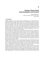
Fig. 1.58 Cystic fibrosis (CF).
The widespread interstitial
pattern shadowing is typical of
CF. Note the right-sided
portacath and a right-sided
apical pneumothorax.
Chap-01.qxd 10/15/02 6:06 PM Page 52
Case illustrations: plain films and CT
1
53
Question 19
Fig. 1.59 Quiz case.
46-year-old smoker.
On surgical waiting list for
hernia surgery (Fig. 1.59).
What is the diagnosis?
Answer
Emphysema
There is hyperexpansion of both lungs with flattening of the diaphragms.
At the level of the diaphragm there are eight anterior ribs (normal is
six to seven). There are decreased lung markings throughout the lungs
particularly affecting the lung bases, a pattern found in alpha-1-antitrypsin
deficiency. Radiological changes of chronic bronchitis and emphysema
chronic obstructive pulmonary disease (COPD) include:
hyperinflation (increased retrosternal clear space on lateral film),
flattening of diaphragms,
reduced lung markings (emphysema),
peribronchial thickening,
bullae (increased risk of pneumothorax),
cardiac enlargement,
enlarged pulmonary arteries (pulmonary hypertension).
Chap-01.qxd 10/15/02 6:06 PM Page 53
Imaging the chest
1
54
Question 20
70-year-old man. Awaiting total hip replacement. Seen in surgical
pre-assessment clinic. History of cough.
Describe the abnormality seen on the chest X-ray (Fig. 1.60).
What are the most likely differential diagnoses?
How would you manage the patient?
Fig. 1.60 Quiz case.
Answer
Solitary pulmonary nodule or mass
There is a solitary nodule seen in the mid-zone of the right lung.
No other pulmonary nodules or rib lesions are seen.
Nipple shadows can often cause confusion on plain films as they may
resemble lung nodules. If any doubt exists then metallic skin markers
should be positioned and further X-rays taken. This case example was
a nipple shadow (see Fig. 1.61).
A common cause of true pulmonary nodule in a patient of this age
would be bronchogenic carcinoma; metastasis, granuloma or infection are
further possibilities.
Solitary pulmonary nodule
With this type of film (especially in a viva situation) you give the most likely
differential diagnosis first and list the others in reducing order of
probability. It is no good giving Wegeners granulomatosis as the opening
differential, although possible, it is not the most likely diagnosis in a
70-year-old smoker (Table 1.9).
Chap-01.qxd 10/15/02 6:06 PM Page 54
Case illustrations: plain films and CT
1
55
Fig. 1.61 Nipple shadow CT.
Although a plain film with
metallic nipple markers is
usually sufficient, this CT
clearly demonstrates the
cause of the ‘nodule’ seen on
the plain film.
Table 1.9 The solitary lung mass
Acquired
Tumour
Malignant Bronchogenic carcinoma (look for rib mets)
Solitary metastasis Breast (mastectomy), renal (surgical clips),
sarcoma, seminoma
Lymphoma
Benign Carcinoid (central position)
Hamartoma (popcorn calcification) (see Fig. 1.62)
Infection
Bacterial Round pneumonias (children)
Parasitic Hydatid (?name, waterlilly sign)
Fungal Histoplasmosis
Infarction
Pulmonary Infarct (peripheral, wedge shaped)
Vascular
Pulmonary avm (Large feeding vessel) (see Fig. 1.63)
Granuloma
TB (Name, satellite lesion)
Wegeners (Old films, ?dialysis line) (Fig. 1.64)
Sarcoid
Rheumatoid nodule (Shoulder erosions, clavicle)
Trauma
Haematoma (Rib fractures)
Congenital
Sequestered segment (Usually basal)
Bronchogenic cyst (Normally mediastinal)
Chap-01.qxd 10/15/02 6:06 PM Page 55
Imaging the chest
1
56
Fig. 1.62 Hamartoma.
‘Popcorn’ calcification is almost
pathonomonic of hamartoma,
usually the lesion is slow
growing and about half
contain fat on CT.
Fig. 1.63 Arterio-venous malformation. These can appear like a suspicious nodule on
chest X-ray. Feeding vessels are sometimes visible on the conventional X-ray but
CT with contrast enhancement will demonstrate these very elegantly.
Chap-01.qxd 10/15/02 6:06 PM Page 56
Case illustrations: plain films and CT
1
57
Fig. 1.64 Wegeners
granulomatosis. This can
appear as patchy alveolar
infiltrate – either single or
multiple which can also be
complicated by cavitation.
You may be pressed for further causes of a solitary nodule by the
examiner. This is a classic example of where to use a surgical sieve.
You must try and narrow the differential by looking for any secondary
signs on the film (look for the signs given in brackets).
Chap-01.qxd 10/15/02 6:06 PM Page 57
Imaging the chest
1
58
Question 21
54-year-old male.
Chronic cough productive of sputum.
Recent fever malaise and haemoptysis.
What is the underlying lung condition (Fig. 1.65)?
What complication has occurred?
Fig. 1.65 Quiz case.
Answer
Bronchiectasis with pulmonary abscess/cavity
Causes of lung cavitation are listed in Table 1.10. In the case of lung
abscesses a solid nodule is the first radiological manifestation. When the
necrotic centre/pus discharges into the bronchial tree, then a fluid level
and the cavity wall are often visible. In addition to pyogenic infections,
a parenchymal lung cavity should raise the possibility of TB. This represents
reactivation disease and classically affects the apical or posterior segments
of the upper lobes. Pulmonary cavities can become complicated by
empyema (Fig. 1.66).
Cavitating malignancy can appear similar to infectious cavities.
These may be primary bronchogenic malignancy or metastatic disease
such as head and neck squamous carcinoma. Cavitating malignancy tends
to have more nodular, thicker walls (more than 15 mm) than infection
(less than 5 mm).
Chap-01.qxd 10/15/02 6:06 PM Page 58
Case illustrations: plain films and CT
1
59
Table 1.10 Causes of lung cavities
Pyogenic abscess Staphlococcus aureus
Beta-haemolytic streptococcus
Klebsiella
Anaerobes
Septic emboli
TB Reactivation (apical or posterior segment of
upper lobes)
Parasitic infection Echinococcus
Malignancy Primary or metastatic (particularly squamous
cell carcinoma)
Rheumatoid nodule
Wegener’s granulomatosis
Cavitating infarct
Fig. 1.66 Infective lung cavity
which has been complicated by
empyema.
Mimics of cavitating lesions include pneumatoceles, emphesematous
bullae and cystic bronchiectasis. Pneumatoceles are thin-walled
intra-parenchymal areas of air trapping which occur in the recovery
phase of staphylococcal pneumonia, contusion or chronic ARDS.
Chap-01.qxd 10/15/02 6:06 PM Page 59
Imaging the chest
1
60
Question 22
Fig. 1.67 Quiz case.
64-year-old patient.
Breathless for 2 months;
three stone weight loss.
What is the most likely
diagnosis (Fig. 1.67)?
Answer
Multiple pulmonary masses
There are multiple pulmonary masses of varying sizes seen in both lungs.
The commonest cause of this appearance in a patient of this age would be
multiple metastases. There are no bony lesions, no mastectomy and
no other clues to suggest a primary site. Old films would help to confirm
the nature of the nodules and rate of growth.
Comment
If pushed, this film is another situation where a surgical sieve will help to
recall causes of multiple pulmonary nodules/masses (Table 1.11).
Chap-01.qxd 10/15/02 6:06 PM Page 60
Case illustrations: plain films and CT
1
61
Table 1.11 Causes of multiple pulmonary nodules/masses
Tumour
Metastasis Breast, renal, thyroid, squamous carcinoma (head
and neck), gastrointestinal tumours, osteosarcoma
Lymphoma
Infection
Bacterial Abscesses, Staph. aureus, Pseudomonas, TB
Parasitic Hydatid
Infarction
Multiple pulmonary infarct
Vascular
Pulmonary avm
Granuloma
Wegeners
Rheumatoid nodule
AIDS
Kaposi sarcoma
Occupation
Caplans, PMF
Others
Amyloid
Papillomatosis of the lung
Chap-01.qxd 10/15/02 6:06 PM Page 61
Imaging the chest
1
62
Question 23
Fig. 1.68 Quiz case.
28-year-old male.
This patient is breathless
febrile and unwell. He has
recently arrived from Africa.
What is the diagnosis
(Fig. 1.68)?
What precautions must you
take with this patient?
If ventilation is required
what further measures
must be taken?
Answer
Miliary nodules
There are multiple miliary nodules in both lungs (see CT Fig. 1.69).
The patient is from Africa and is unwell making miliary TB the most likely
diagnosis. Barrier nursing in isolation should be undertaken.
TB is spread by the respiratory route, so precautions must be taken to
sterilise ventilation equipment following use. Disposable equipment should
be used where possible and bacterial filters should be used to protect
the ITU ventilator. Medical and nursing staff are at risk of infection, full
face masks and eye protection should be worn during procedures involving
the airway. If he was well, consider sarcoid or pneumoconiosis (old films
would help in this latter differential). Any history of primary malignancy is
important, thyroid malignancy, bone sarcoma and trophoblastic disease can
give rise to miliary metastases. Further less common possibilities include
acute extrinsic allergic alveolitis, nodular pulmonary fibrosis and
histiocytosis X.
Haematogenous metastases tend to go to the bases and inhaled dusts to
the apices. Densely calcified tiny nodules have a different differential
diagnosis (Table 1.12).
Chap-01.qxd 10/15/02 6:06 PM Page 62
Case illustrations: plain films and CT
1
63
Table 1.12 Miliary nodules
Miliary TB
Sarcoid
Dust inhalation/pneumoconiosis
Extrinsic allergic alveolitis
Miliary metastases: thyroid, melanoma
Dense miliary nodules
Haemosiderosis
Silicosis
Stannosis
Chicken pox
Fig. 1.69 CT miliary nodules –
miliary TB. There are
innumerable tiny nodules
distributed throughout both
lungs. The clinical setting is
important in making the
diagnosis as dust inhalation,
granulomatous diseases,
infection and metastases can
give a similar appearance.
Chap-01.qxd 10/15/02 6:06 PM Page 63
Imaging the chest
1
64
Question 24
38-year-old patient.
Chronic musculoskeletal
deformity.
What is this deformity
(Figs 1.70 and 1.71)?
How is it likely to affect
respiratory function?
Fig. 1.70 Quiz case.
Fig. 1.71 Quiz case.
Chap-01.qxd 10/15/02 6:06 PM Page 64
Case illustrations: plain films and CT
1
65
Answer
Kyphoscoliosis
Respiratory function can be affected by scoliosis. Breathing may be
impaired by the chest wall deformity, resulting in alveolar hypoventilation.
The chance of impairment in respiratory function is related to the
maximum angle of the curvature (Cobb angle). Atelectasis and segmental
collapse can occur. Chest wall deformity is seen in patients who were
treated for pulmonary tuberculosis with thoracoplasty (see Fig. 1.72).
Thoracoplasty was a treatment used prior to the widespread use of
antibiotics. Marked chest wall deformity occurs often with considerable
volume loss and sometimes with other stigmata of TB such as pleural
calcification. Type II respiratory failure (hypoxia and hypercapnia) with
subsequent cor pulmonale can occur. Selected patients with chronic
ventilatory failure are appropriately treated with long-term domiciliary
ventilation. Patients (with kyphoscoliosis and thoracoplasty) treated with
non-invasive positive pressure ventilation can have improved prognosis.
A nasal mask and continuous positive pressure ventilation is the usual
method employed (Table 1.13).
Fig. 1.72 TB thoracoplasty.
Pre-antibiotic era treatment
for TB.
Chap-01.qxd 10/15/02 6:06 PM Page 65
Imaging the chest
1
66
Table 1.13 Causes of chronic respiratory failure
Type I respiratory failure (hypoxia)
Pulmonary fibrosis
Pulmonary vascular disease
Type II respiratory failure (Hypoxia and Hypercapnoea)
Airways disease
Chronic obstructive pulmonary disease
Cystic fibrosis and bronchiectasis
Obstructive sleep apnoea
Neuromuscular disorders
Motor neurone disease
Muscular dystrophy
Thoracic cage and pleural abnormality
Kyphoscoliosis
Thoracoplasty
Extreme obesity
Chap-01.qxd 10/15/02 6:06 PM Page 66
Case illustrations: plain films and CT
1
67
Question 25
34-year-old drug addict.
Homeless.
Cough and haemoptysis.
Tachypnoea, hypoxia.
You are asked to assess
with view to ventilatory
support.
What does the chest X-ray
(Fig. 1.73) show?
What risk factors should
be considered?
Fig. 1.73 Quiz case.
Answer
Pulmonary tuberculosis
There are confluent ill-defined areas of consolidation in the mid- and upper
zones of both lungs. Given the appearance and distribution of the changes,
pulmonary TB has to be a primary consideration. Altered immunity from
HIV should be considered (Table 1.14).
Table 1.14 Chest X-ray manifestations of TB
Primary pulmonary TB
Air space consolidation 1–7 cm diameter
Lymphadenopathy: hilar, paratracheal
Pleural effusion
Segmental consolidation
Cavitation
Calcified ghon focus
Calcified lymph nodes
Post-primary TB (reactivation or initial infection or infection post-BCG)
Apical and posterior segments of upper lobes
Chronic patchy ill-defined areas of opacification
Cavitation may colonise with Aspergillus
Bronchiectasis
Upper lobe fibrosis (see Fig. 1.74)
Chap-01.qxd 10/15/02 6:06 PM Page 67
Imaging the chest
1
68
Fig. 1.74 TB upper lobe
fibrosis. There is linear
shadowing and volume
loss in both upper zones
with elevation of both hila.
Chap-01.qxd 10/15/02 6:06 PM Page 68
Case illustrations: plain films and CT
1
69
Question 26
68-year-old electrician.
6 months of worsening
chest pain.
What is the diagnosis
(Figs 1.75 and 1.76)?
What further question
will help make a
diagnosis?
Describe the procedure to
confirm the diagnosis.
Fig. 1.75 Quiz case.
Fig. 1.76 Quiz case.
Chap-01.qxd 10/15/02 6:06 PM Page 69
Imaging the chest
1
70
Answer
Mesothelioma
Electricians in the past were frequently exposed to asbestos and this is a
crucial question to ask in the clinical history.
Pleural biopsy will confirm the diagnosis. This has traditionally been
carried out blind, i.e. without image guidance. The procedure is carried
out with the patient seated comfortably and leaning forward with the
posterior of the chest wall exposed. Using the chest X-ray for localisation,
under sterile conditions, the superficial tissues are infiltrated with local
anaesthetic at a suitable rib interspace. A biopsy needle is inserted
adjacent to the superior rib border (to avoid the neurovascular bundle)
and the pleural biopsy taken. Ultrasound can be used to identify pleural
thickening, and thereby guide biopsy and target potentially pathological
areas.
Malignant mesothelioma is often related to previous asbestos exposure
with a lag period of up to 30 years or more. It usually presents as a
multilobulated pleural mass encircling the lung. It involves the mediastinal
pleural reflections and often extends into the lung fissures or out to the
chest wall, particularly, along a biopsy track. Pericardial involvement or
diaphragmatic invasion can occur. Pleural effusions are common and these
may be loculated. Other manifestations of asbestos exposure include
pleural calcification, folded lung (see Fig. 1.77) (which may be mistaken for
a malignant mass) and asbestosis.
Causes of pleural malignancy
Bronchial adenocarcinoma.
Breast carcinoma.
Malignant thymoma.
Subpleural lymphoma.
Malignant mesothelioma.
Fig. 1.77 Asbestos lung disease rounded atelectasis. Also called Folded lung,
is frequently adjacent to a site of pleural thickening – vessels normally
converge towards it in a characteristic way.
Chap-01.qxd 10/15/02 6:06 PM Page 70
Case illustrations: plain films and CT
1
71
Question 27
76-year-old man.
Life-long smoker.
Cough and haemoptysis.
What is the diagnosis
(Fig. 1.78)?
Fig. 1.78 Quiz case.
Table 1.15 Causes of opaque hemithorax
Lung collapse: volume loss, mediastinal shift towards affected side
Pleural fluid: transudate/exudate; empyema; haemothorax; chylothorax
Pneumonectomy: look for surgical clips and thoracotomy
Tumour: fibrous tumour of pleura; pleural metastases; mesothelioma
Extensive consolidation: pneumonia; pulmonary oedema; obstructing
neoplasm
Lung agenesis
Answer
Collapse of the left lung
There is almost total collapse of the left lung, due to an underlying
bronchogenic malignancy. Note the volume loss with shift of
the mediastinum to the affected side (Table 1.15).
Chap-01.qxd 10/15/02 6:06 PM Page 71
Imaging the chest
1
72
Fig. 1.80 The left-sided central venous line is too lateral on the check X-ray.
Question 28
Fig. 1.79 Quiz case.
2-week-old neonate.
What does the chest X-ray
(Fig. 1.79) demonstrate?
Answer
Malpositioned endotracheal tube
If an endotracheal tube is positioned incorrectly within the major airways,
then lobar collapse can follow in the under ventilated lobes. In this
example, the endotracheal tube has been passed along the right main
bronchus distal to the origin of the right upper lobe bronchus with
subsequent collapse of the right upper lobe and the left lung.
Chap-01.qxd 10/15/02 6:06 PM Page 72
Case illustrations: plain films and CT
1
73
Fig. 1.81 On the basis of the X-ray in Fig. 1.17 the central venous line was used for
parenteral nutrition. There is now a large left-sided pleural fluid collection – the line
tip was clearly positioned in the pleural space.
In Figs 1.80 and 1.81 a left-sided central line has been placed in the
pleural space (the line appears too lateral) and intravenous nutrition has
been infiltrated into the pleural cavity compressing the left lung and
the mediastinum.
Chap-01.qxd 10/15/02 6:06 PM Page 73
Imaging the chest
1
74
Question 29
18-hour-old male neonate.
Tachypnoea, sternal and
intercostal rescession.
What does the chest
X-ray (Fig. 1.82) show?
Fig. 1.82 Quiz case.
Answer
Respiratory distress syndrome
This is the most common, serious respiratory disorder in neonates
(Table 1.16). It is caused by lack of surfactant (normally produced by
type II pneumocytes). It is a condition usually associated with prematurity
but may also occur in term neonates of diabetic mothers or following birth
asphyxia. The normal time of onset is at 18–24 hours of life. The radiological
signs are initially of under-inflated lungs which have a granular appearance.
With worsening disease air bronchograms become more prominent
and this can partially obscure the mediastinal contours. Very premature
babies and those with bad respiratory distress syndrome (RDS) are treated
with surfactant (administered via an endotracheal tube) which usually
produces a marked improvement in their condition.
Table 1.16 Possible causes of respiratory distress in a neonate
Respiratory distress syndrome
Transient tachypnoea of the newborn
Aspiration
Pneumothorax
Diaphragmatic hernia
Pneumonia
Congenital heart disease
Chap-01.qxd 10/15/02 6:06 PM Page 74
Case illustrations: plain films and CT
1
75
Complications of respiratory distress
Most of the complications are secondary to mechanical ventilation.
Small airway rupture can lead to air tracking into the pleural space –
pneumothorax or the mediastinum – pneumomediastinum.
Pneumomediastinum usually produces a sharp clearly defined lucency
adjacent to each side of the mediastinum. The mediastinal borders are
usually of increased sharpness as they become outlined by air rather than
lung parenchyma (see Fig. 1.83). A horizontal beam lateral radiograph will
confirm the location of the air. The lungs are poorly compliant in RDS,
so do not collapse. The air can often be seen outlining the thymus as well
as the other mediastinal structures (see Fig. 1.84).
Fig. 1.83 Pneumomediastinum.
Air is present in the anterior
mediastinum outlining the heart.
Fig. 1.84 Neonatal pneumomediastinum
lateral projection. The patient is
positioned supine and a shoot through
lateral projection has been performed.
This is outlining the thymus and other
mediastinal structures.
Chap-01.qxd 10/15/02 6:06 PM Page 75
Imaging the chest
1
76
Question 30
Answer
Diaphragmatic hernia
There are multiple loops of bowel within the chest causing marked shift of
mediastinal structures to the opposite side. The loops of bowel in the
chest are clearly continuous with those in the abdomen.
Most diaphragmatic hernias are symptomatic shortly after birth, they are
more commonly left sided. The abdominal contents occupy the chest cavity
in utero so both lungs (particularly on the side of the hernia) are
hypoplastic. There is a significant mortality following successful surgical
repair (see Table 1.16).
Fig. 1.85 Quiz case.
1-day-old infant with
respiratory difficulties.
What does the X-ray
(Fig. 1.85) show?
Chap-01.qxd 10/15/02 6:06 PM Page 76