Radiology for Anaesthesia and Intensive Care - Part 4 pdf
Bạn đang xem bản rút gọn của tài liệu. Xem và tải ngay bản đầy đủ của tài liệu tại đây (825.81 KB, 36 trang )
Question 4
64-year-old patient.
This patient has a history of diabetes and had reconstructive vascular
surgery for peripheral vascular disease 8 days ago. He received intravenous
broad spectrum antibiotics for a surgical wound infection and now has
bloody diarrhoea.
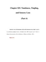
What are the radiological signs (Fig. 2.10)?
What is the diagnosis?
Imaging the abdomen
2
88
Fig. 2.10 Quiz case.
Case illustrations: plain films and CT
2
89
Answer
This case demonstrates colonic wall thickening, thumb printing and
a distended stomach. The diagnosis is pseudomembranous colitis
and diabetic gastroparesis.
Pseudomembranous colitis
In general, the radiological findings are adynamic ileus with moderate
gaseous distension of the small and large bowel. The haustral folds are
frequently shaggy and irregular and ‘thumbprinting’ is often identified
particularly in the transverse colon (as in Fig. 2.10). Diffuse colonic
thickening can be identified on CT. Pseudomembranous colitis is caused by
an overgrowth of the commensal anaerobe Clostridium difficile. Commonly
it is a complication of antibiotic therapy particularly ampicillin, amoxycillin,
clindamycin and the cephalosporins. Antibiotic disturbance of the normal
gut flora appears to allow overgrowth of toxigenic strains of C. difficile.
The clinical and pathological effects are the result of toxin production.
Further predisposing causes include bowel obstruction and co-existent
debilitating disease, e.g. leukaemia. The clinical picture is of profuse
diarrhoea, abdominal cramps and tenderness. A yellow exudative
pseudomembrane, haemorrhagic areas and mucosal ulcers are seen on
colonoscopy.
Diabetic gastroparesis is a recognised complication of diabetes
mellitus when there is gastric retention in the absence of mechanical
obstruction. This can be life threatening. The stomach should be
decompressed and emptied with a nasogastric tube. Other causes
include electrolyte imbalances (diabetic ketoacidosis), drugs, peritonitis
and abdominal trauma.
Question 5
86-year-old female.
The patient has had several episodes of abdominal pain and distension.
She is now vomiting.
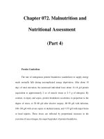
What is the diagnosis (Fig. 2.11)?
Imaging the abdomen
2
90
Fig. 2.11 Quiz case.
Answer
Sigmoid volvulus
This is a rotation of the gut about its own mesenteric axis, which produces
complete intestinal obstruction. It is most commonly seen in the elderly or
those with psychiatric disorders taking medication. Venous congestion
leading to infarction can occur. On the plain abdominal film a hugely
dilated loop of bowel is seen extending from the pelvis. The inverted
‘U’ loop is commonly devoid of haustra and is seen to extend as far as the
liver in the right upper quadrant, and to the 10th thoracic vertebra
superiorly. The inferior convergance of the two limbs of the loop is often
left sided. There may be some secondary loops of dilated large bowel
associated with these appearances. Sigmoidoscopy can be both diagnostic
and therapeutic by releasing flatus. Approximately half of patients have a
further episode of volvulus within 2 years. In caecal volvulus, the caecum
is seen to revolve around its axis to lie across the midline in the
upper/central abdomen Fig. 2.12.
Large bowel obstruction gives rise to distention of the large bowel
down to the level of obstruction sometimes with accompanying small
bowel dilation. The commonest cause is colonic carcinoma. Other causes
include volvulus, intussusception or extrinsic compression.
In paralytic ileus both the large and small bowel can become dilated
which can extend down into the sigmoid colon and rectum (see Fig. 2.13).
Differentiation from low large bowel obstruction may be difficult.
Case illustrations: plain films and CT
2
91
Fig. 2.12 Caecal volvulus.
Imaging the abdomen
2
92
Fig. 2.13 Pseudo-obstruction. This can be difficult to distinguish from distal large
bowel obstruction. Large and small bowel distension is usually present with reduced
small bowel distension on serial films. If concern persists an instant enema can be
performed.
Question 6
51-year-old patient.
Recurrent rectal bleeding, admitted with acute abdominal pain.
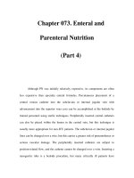
What is the diagnosis?
What are the radiological features (Fig. 2.14)?
Case illustrations: plain films and CT
2
93
Fig. 2.14 Quiz case.
Answer
Pan colitis and perforation
The whole of the colon is distended. There is thickening of the mucosa
which is oedematous. In the centre of the film there are several dilated
loops of small bowel and their inner and outer walls are both visible.
This latter feature indicates free gas within the peritoneal cavity.
The appearances of the bowel are characteristic of a pan colitis
(affecting the whole colon) typical of ulcerative colitis. The bowel has
clearly perforated. The term megacolon is frequently applied in cases of
transmural fulminant colitis when the bowel looses motor tone and dilates
to a transverse diameter of greater than 5.5 cm. The term toxic megacolon
should be reserved for cases of dilatation with systemic toxicity, abnormal
clinical signs (peritonism, fever) and abnormal laboratory indices (raised
inflammatory markers, leukocytosis and left shift). The clinical setting is
usually accompanied by profuse bloody diarrhoea. Mortality is up to 20%,
barium enema is contraindicated. Ulcerative colitis is the commonest cause
but others include Crohn’s disease, amoebiasis, Salmonella,
pseudomembranous and ischaemic colitis.
Extraluminal gas
Normally bowel gas is only present within the bowel lumen. This results in
a clear image of the inner margin of the bowel on the abdominal X-ray.
This is due to the air–mucosa interface which has different densities.
The outer margin, however, is not clearly seen since the serosal surfaces
merge with other adjacent bowel wall loops of similar density. However,
free intra-peritoneal gas will also clearly outline the outer serosal margin
of the bowel. The bowel wall thus appears as a thin ‘pencilled’ line with
gas on either side. This appearance is known as Rigler’s sign. Gas may
be visible under the hemidiaphragms on an erect chest or abdominal film
(Fig. 2.15).
Free gas may be seen after bowel perforation or following laparotomy.
In adults, post-laparotomy pneumoperitoneum persists for up to 7 days but
is absorbed very much more quickly in children, usually by 24 hours.
Imaging the abdomen
2
94
Fig. 2.15 Erect chest X-ray. Pneumoperitoneum – air under the diaphragms.
Question 7
46-year-old male.
This patient has presented with acute right iliac fossa pain.
You have been asked to assess him prior to exploratory laparotomy.
What is the X-ray (Fig. 2.16) abnormality?
What is the likely diagnosis?
Case illustrations: plain films and CT
2
95
Fig. 2.16 Quiz case.
Answer
There is an oval opacity overlying the right sacral ala. The appearances are
typical of a faecolith or appendolith. This calcified faecal material can occur
in the appendix or a large bowel diverticulum. In conjunction with right
iliac fossa pain, appendicitis is the most likely diagnosis. Ultrasound has
high specificity in diagnosing appendicitis but poor sensitivity (see Fig. 2.17).
Abnormal calcification can be used to make a diagnosis in the following
conditions:
calcified aortic aneurysm;
calcified gallstones;
renal/ureteric stones;
pancreas: chronic pancreatitis (Fig. 2.40);
appendolith: appendicitis;
liver calcification: granuloma, old abscess, some metastases;
uterine fibroids.
Imaging the abdomen
2
96
Fig. 2.17 US appendicitis.
The echogenic structure is
an appendicolith.
Question 8
Case illustrations: plain films and CT
2
97
Fig. 2.18 Quiz case.
14-day-old male child.
What is the diagnosis
(Fig. 2.18)?
What are the common
associations?
What co-existent
respiratory problems are
frequently encountered?
Answer
Necrotising enterocolitis
Gas can be seen in the wall of a distended loop of bowel (probably the
transverse and descending colon). It is difficult to differentiate large from
small bowel in the neonate based on bowel distribution alone.
The abdomen is rather featureless elsewhere.
Other recognised radiological signs of necrotising enterocolitis (NEC)
include small and large bowel dilation, a bubbly appearance to the bowel,
gas in the portal venous system and bowel perforation. NEC most
commonly (but not exclusively) affects premature neonates. Barium enema
is contraindicated. In adults, gas in the bowel wall often indicates
bowel infarction and has a poor prognosis. It should not be confused with
pneumatosis cystoides intestinalis.
Associations of NEC:
prematurity,
Hirschsprung’s disease,
bowel obstruction (e.g. meconium ileus or atresia).
It is frequently co-existent with respiratory problems of the ventilated
neonate such as hyaline membrane disease.
Question 9
Imaging the abdomen
2
98
Fig. 2.19 Quiz case.
Aged 5 weeks.
History of infrequent
stools (Fig. 2.19).
Abdominal distension.
What is the
examination?
What are the important
radiological features
and the diagnosis?
Answer
Hirschprung’s disease – barium enema
Hirschsprung’s disease is caused by failure of development of ganglion
cells. The disease starts at the anus and a variable amount of bowel is
affected proximally. Presentation can be with neonatal bowel obstruction
or constipation in later life. The aganglionic segment does not transmit
peristalsis so the proximal bowel dilates and the affected segment
appears normal or small calibre. In the case example, there is a transition
zone with dilated proximal bowel and a normal/small calibre aganglionic
bowel segment. A well-recognised complication (present in the example
above) is necrotising enterocolitis.
Question 10
Age 2.
This child presented with abdominal pain, and blood stained mucus PR.
What do the abdominal film and the ultrasound (US)
(Figs 2.20 and 2.21) show?
What would you request next?
What precautions are necessary?
Case illustrations: plain films and CT
2
99
Fig. 2.20 Quiz case.
Fig. 2.21 Quiz case.
Answer
Intussusception
The abdominal film demonstrates a soft tissue mass in the left upper
quadrant in the region of the transverse colon. This is clearly outlined
on one side by gas in the colon distal to it. This is the lead point of
an intussusception – the clinical history is extremely suggestive in a
child of this age. The ultrasound (US) confirms a mass which is
characteristic of an intussusception.
Air enema/pneumatic reduction is the preferred initial method of
treatment. This requires fluid resuscitation and IV antibiotics prior to the
procedure. This should only be carried out in a centre with paediatric
surgical cover. The procedure fails in a proportion of cases and open
surgical reduction may be necessary. Pneumoperitoneum, peritonitis and
hypovolaemic shock are contraindications to the technique. A large bore
Foley catheter is inserted into the rectum and the buttocks are taped
together. Air is insufflated using a pump with a pressure gauge that has a
valve mechanism to prevent excessive pressures. The lead point of the
intussusception can be followed fluoroscopically and usually reduces fairly
easily (success rate is up to 90%) but there may be some hold up at the
ileo-caecal valve level. When the intussusception reduces, the small bowel
can be seen to suddenly fill with a puff of gas. Bowel perforation is a
potential complication and this may splint the diaphragm compromising
respiration. A large bore needle should be kept to hand and used to
decompress a pneumoperitoneum. Incomplete reduction and recurrence
in up to 10% are further complications.
Imaging the abdomen
2
100
Question 11
28-year-old female.
Dysphagia, chest pains and choking episodes.
What is the diagnosis (Fig. 2.22)?
What is the importance of this condition in anaesthetic practice?
Case illustrations: plain films and CT
2
101
Fig. 2.22 Quiz case.
Answer
Achalasia
This is a condition of middle age caused by a reduced number of ganglion
cells in the myenteric pexus. There is failure of relaxation of the lower
oesophageal sphincter in response to swallowing. There is absence of
peristalsis in the mid- and lower oesophagus which dilates to produce a
megaoesophagus. Symptoms include dysphagia, weight loss, regurgitation
and chest pain. Aspiration can occur. Sometimes, the chest X-ray is
diagnostic and an air–fluid level can be seen in a dilated oesophagus.
On barium examination, there is a characteristic bird beak deformity at
the gastro-oesophageal junction. Manometry will demonstrate an absent
primary peristaltic wave and tertiary contractions.
The differential diagnosis includes infiltrating carcinoma, scleroderma
and Chagas’ disease.
The aim of treatment is to reduce the pressure of the lower oesophageal
sphincter. Medical therapies include long-acting nitrates or calcium channel
blockers. Further options include injection with botulinum toxin, balloon
dilation (which may be repeated if necessary) and surgery –
oesophagomyotomy which can be performed laparoscopically.
Patients are at risk of aspiration and a rapid sequence induction with
cricoid pressure and endotracheal intubation must be performed if
the patient requires general anaesthesia.
Imaging the abdomen
2
102
Question 12
Man aged 78.
Life-long smoker. Dysphagia for solids and liquids.
What are the radiological features (Fig. 2.23)?
What is the diagnosis?
What are the implications for anaesthesia?
Case illustrations: plain films and CT
2
103
Fig. 2.23 Quiz case.
Answer
Malignant oesophageal stricture
There is a long irregular stricture affecting the mid- and lower oesophagus.
The pattern of obstruction is in keeping with an extrinsic infiltrating mass.
On careful inspection, it is clear that the density of the left lung is
increased. The left lung has collapsed due to a mass at the
hilum/mediastinum. The diagnosis is carcinoma of the left lower lobe
bronchus with an associated mediastinal mass. There is extrinsic
compression and invasion of the oesophagus where the mass is invading
the mediastinum. The patient is not ventilating the left lung.
Surgery would be a major undertaking. The patient requires careful
pre-operative assessment with careful consideration of his cardiovascular
and respiratory state, and his ability to survive the procedure, before
surgery is booked.
If surgery is undertaken, one-lung ventilation after placement of a
double lumen tube, would facilitate surgical access. Invasive monitoring
and post-operative ITU care would be required.
Imaging the abdomen
2
104
Question 13
Case illustrations: plain films and CT
2
105
Fig. 2.24 Quiz case.
Age 34.
Recurrent episodes of
right lower quadrant
abdominal pain.
What is this
examination?
What are the
radiological features
(Fig. 2.24)?
What is the
diagnosis?
Answer
Crohn’s disease
This is one of the images taken during a barium follow-through
examination. The small bowel can be examined using a barium
follow-through technique or by intubating the proximal jejunum and
injecting barium – small bowel enema/enteroclysis. A number of abdominal
films are taken (in the prone position to separate the small bowel loops) to
examine the small bowel as the barium passes to the colon. Further views
are taken of the terminal ileum as this is an area particularly affected by
Crohn’s disease. The case example demonstrates a thickened, nodular
(cobblestone-like appearance) of the terminal ileum mucosa – typical of
Crohn’s disease.
Asymmetry, skip lesions, deep ulcers (see Fig. 2.25) and fistula formation
are the hallmark of Crohn’s disease, where as ulcerative colitis is
characterised by a symmetrical disease in continuity, granular mucosa
(see Figs 2.26 and 2.27), superficial ulcers and rectal involvement.
In late stage, there may be inflammatory polypoid changes, narrowing and
shortening of the colon. In ulcerative colitis (Fig. 2.28), the ileo-caecal
valve is gaping (Table 2.1).
Imaging the abdomen
2
106
Fig. 2.25 Crohn’s colon. The double contrast
(air and barium) barium enema shows deep
ulcers in the descending colon and the sigmoid.
Fig. 2.26 Ulcerative colitis.
The entire colon is affected, there
are reduced haustral markings and
the mucosa is abnormal with
a fine mucosal granularity.
Case illustrations: plain films and CT
2
107
Fig. 2.27 Ulcerative colitis.
The descending colon (sigmoid and
rectum are also affected but not
shown) demonstrates multiple
superficial ulcers. Note the normal
appearance to the transverse colon.
Fig. 2.28 Ulcerative
colitis – megacolon. Fulminant
colitis with dilatation of the
transverse colon of more than
5.5 cm; at risk of perforation.
Imaging the abdomen
2
108
Table 2.1 Features of inflammatory bowel disease
Features Crohn’s disease Ulcerative colitis
Distribution Anywhere in Predominantly colon and
gastrointestinal rectum
tract, mainly
terminal ileum
Morphology Skip lesions, Disease involve
aphthous ulcers, continuous segments,
deep (rose thorn) superficial ulceration,
aphthous ulcers (Fig. 2.27)
Fistula and sinuses Common Uncommon
Strictures Yes Relatively more common
Toxic dilatation Uncommon Common (Fig. 2.28)
Rectal involvement Less than 50% Greater than 95%
Anal fistula, sinus Common Uncommon
and fissures
Terminal ileum Narrowed, fissured Dilated with a gaping
ileo-caecal valve
Cancer risk Uncommon High
Pseudodiverticulation Common Not seen
Inflammatory polyps Rare Seen in about 20%
Gallstones Common especially No significant increase
after surgery
Question 14
Case illustrations: plain films and CT
2
109
Fig. 2.29 Quiz case.
Answer
Barium has passed only as far as the proximal transverse colon, there is a
large tubular filling defect within the transverse colon and a small
polypoid lesion is present in the descending colon. The appearance are
characteristic of a colo-colic intussusception. The radiological appearances
of intussusception are said to resemble a coiled spring pattern.
At surgery, this lesion was found to be a caecal tumor at the lead point
of the intussusception. A second 1.5 cm sigmoid colonic polyp was also
present.
Colo-colic intussusception
Further management would include referral to a surgical team to consider
urgent intervention. Staging of the tumour with CT will be required.
In adults, up to 80% of intussusceptions are secondary to a specific
cause and approximately one-fifth are due to malignant neoplasms.
Further causes include benign tumours, lipoma, Meckel’s diverticulum
and idiopathic.
Age 66.
This patient presented
with persistent vomiting
and abdominal pain.
What are the
radiological findings
(Fig. 2.29)?
What is the diagnosis?
What further
management would
you suggest?
Question 15
86-year-old female patient.
Unwell, pyrexia. Elevated white blood count.
What are the therapeutic options for this lesion (Fig. 2.30)?
Imaging the abdomen
2
110
Fig. 2.30 Quiz case.
Answer
Diverticular abscess
There is a 10–12 cm abscess cavity with an air–fluid level in the left flank.
Immediately adjacent to the abscess is a loop of bowel affected by
diverticular disease with signs of direct communication.
Therapeutic options are either to insert a drainage tube under CT or
ultrasound guidance or to proceed to laparotomy. If there is a large
fistulous communication, then the surgical option would be favoured.
Although the origin of the abscess is probably diverticular, the differential
diagnosis includes a perforating colonic tumour and Crohn’s colitis.
Colonic diverticulosis is an acquired herniation of the colonic mucosa
and submucosa through the muscle layers. It is a condition more prevalent
in developed countries with up to 50% of individuals affected by the
seventh decade. The aetiology is linked to a diet lacking in roughage.
The sigmoid is most frequently affected.
Complications include diverticular haemorrhage, colonic diverticulitis
(Table 2.2), abscess formation, perforation, fistula and colonic stricture
formation (see Fig. 2.31).
Case illustrations: plain films and CT
2
111
Table 2.2 Colonic diverticulitis CT features
Diverticula often visible on CT as round oval focus protruding from
bowel wall (may fill with oral contrast)
Streaky peri-colic fat
Bowel wall thickening
Intestinal obstruction
Fluid or gas in peritoneum
Fistulation into
Bladder (look for an air–fluid level)
Small bowel/vagina/cutaneous
Abscess or peri-colic collection
Fig. 2.31 Diverticular disease CT. The sigmoid colon wall is thickened and there are
multiple diverticula. Note oral contrast in sigmoid colon.
Question 16
Imaging the abdomen
2
112
Fig. 2.32 Quiz case.
41-year-old male
patient.
2-day history of right
loin pain.
What does the
film (Fig. 2.32)
demonstrate?
What further film is
required to make the
diagnosis?
Answer
Obstructed right ureter
This film is the post-micturition film of an IVP series. It shows mild
hydronephrosis of the right kidney and a mildly dilated right ureter.
Contrast is seen as far as the right sacral region but no further. This
indicates the level of obstruction of the ureter. The commonest cause of
ureteric obstruction is stone disease. A preliminary/control film (prior to
the administration of contrast) is necessary to check for the presence of
radio-opaque calculus at the level of obstruction.
Causes of intraluminal ureteric obstruction
Opaque calculus (calcium stones).
Non-opaque calculus (uric acid, xanthine stones).
Blood clot.
Papillary necrosis.
Fungus ball.