Báo cáo y học: "Human T-cell leukemia virus type 2 post-transcriptional control protein p28 is required for viral infectivity and persistence in vivo" ppt
Bạn đang xem bản rút gọn của tài liệu. Xem và tải ngay bản đầy đủ của tài liệu tại đây (368.45 KB, 11 trang )
Retrovirology
BioMed Central
Open Access
Research
Human T-cell leukemia virus type 2 post-transcriptional control
protein p28 is required for viral infectivity and persistence in vivo
Brenda Yamamoto1,2,3, Min Li1,2, Matthew Kesic1,2, Ihab Younis1,2,
Michael D Lairmore1,2,3,4 and Patrick L Green*1,2,3,4
Address: 1Center for Retrovirus Research, The Ohio State University, Columbus, OH 43210, USA, 2Department of Veterinary Biosciences, The Ohio
State University, Columbus, OH 43210, USA, 3Department of Molecular Virology, Immunology, and Medical Genetics, The Ohio State, University,
Columbus, OH 43210, USA and 4Comprehensive Cancer Center and Solove Research Institute, The Ohio State University, Columbus, OH 43210,
USA
Email: Brenda Yamamoto - ; Min Li - ; Matthew Kesic - ;
Ihab Younis - ; Michael D Lairmore - ; Patrick L Green* -
* Corresponding author
Published: 12 May 2008
Retrovirology 2008, 5:38
doi:10.1186/1742-4690-5-38
Received: 1 April 2008
Accepted: 12 May 2008
This article is available from: />© 2008 Yamamoto et al; licensee BioMed Central Ltd.
This is an Open Access article distributed under the terms of the Creative Commons Attribution License ( />which permits unrestricted use, distribution, and reproduction in any medium, provided the original work is properly cited.
Abstract
Background: Human T-cell leukemia virus (HTLV) type 1 and type 2 are related but distinct
pathogenic complex retroviruses. HTLV-1 is associated with adult T-cell leukemia and a variety of
immune-mediated disorders including the chronic neurological disease termed HTLV-1-associated
myelopathy/tropical spastic paraparesis. In contrast, HTLV-2 displays distinct biological differences
and is much less pathogenic, with only a few reported cases of leukemia and neurological disease
associated with infection. In addition to the structural and enzymatic proteins, HTLV encodes
regulatory (Tax and Rex) and accessory proteins. Tax and Rex positively regulate virus production
and are critical for efficient viral replication and pathogenesis. Using an over-expression system
approach, we recently reported that the accessory gene product of the HTLV-1 and HTLV-2 open
reading frame (ORF) II (p30 and p28, respectively) acts as a negative regulator of both Tax and Rex
by binding to and retaining their mRNA in the nucleus, leading to reduced protein expression and
virion production. Further characterization revealed that p28 was distinct from p30 in that it was
devoid of major transcriptional modulating activity, suggesting potentially divergent functions that
may be responsible for the distinct pathobiologies of HTLV-1 and HTLV-2.
Results: In this study, we investigated the functional significance of p28 in HTLV-2 infection,
proliferation, and immortaliztion of primary T-cells in culture, and viral survival in an infectious
rabbit animal model. An HTLV-2 p28 knockout virus (HTLV-2Δp28) was generated and evaluated.
Infectivity and immortalization capacity of HTLV-2Δp28 in vitro was indistinguishable from wild type
HTLV-2. In contrast, we showed that viral replication was severely attenuated in rabbits inoculated
with HTLV-2Δp28 and the mutant virus failed to establish persistent infection.
Conclusion: We provide direct evidence that p28 is dispensable for viral replication and cellular
immortalization of primary T-lymphocytes in cell culture. However, our data indicate that p28
function is critical for viral survival in vivo. Our results are consistent with the hypothesis that p28
repression of Tax and Rex-mediated viral gene expression may facilitate survival of these cells by
down-modulating overall viral gene expression.
Page 1 of 11
(page number not for citation purposes)
Retrovirology 2008, 5:38
Background
The human T-cell leukemia viruses (HTLV types 1–4) are
classified as complex retroviruses and members of the
genus Deltaretrovirus [1]. HTLV-1 and HTLV-2 infections
are the most prevalent worldwide, whereas infections
with HTLV-3 and HTLV-4 were discovered only recently in
a very limited number of individuals in Africa [2,3].
Although people infected with HTLV have a persistent
antiviral immune response, these patients fail to clear
virally infected cells. A small percentage of HTLV-1infected individuals develop adult T-cell leukemia (ATL),
a CD4+ lymphocyte malignancy, and various lymphocyte-mediated inflammatory diseases such as HTLV-1
associated myelopathy/tropical spastic paraparesis
(HAM/TSP) [4-7]. However, only a few cases of atypical
hairy cell leukemia or neurologic disease have been associated with HTLV-2 infection [8-12]. HTLV-1 and HTLV-2
have the capacity to promote T-lymphocyte growth both
in cell culture and in infected individuals; however, the
mechanism by which the virus persists in the infected
individual, ultimately resulting in the oncogenic transformation of T-lymphocytes, is not completely understood.
In addition to the gag, pol, and env genes that encode the
structural and enzymatic proteins, HTLV encodes tax/rex
and accessory genes from pX open reading frames (ORFs)
located in the 3' region of the genome. Tax increases the
rate of transcription from the viral long terminal repeat
(LTR) [13-15] and modulates the transcription or activity
of numerous cellular genes involved in cell growth and
differentiation, cell cycle control, and DNA repair [16-20].
Compelling evidence indicates that the pleiotropic effects
of Tax on cellular processes are required for the transforming or oncogenic capacity of HTLV [21-23]. Rex acts posttranscriptionally by preferentially binding, stabilizing and
selectively exporting the unspliced and incompletely
spliced viral mRNAs from the nucleus to the cytoplasm,
thus controlling the expression of the structural and enzymatic proteins as well as virion production [24-26].
Although both Tax and Rex are key positive regulators
essential for efficient viral replication and, ultimately, cellular transformation, it has been hypothesized that the
unregulated expression of these genes would result in the
death of the infected cell in vivo via the induction of apoptosis and/or host immune response.
Growing evidence indicates that the HTLV-1 p30 and the
HTLV-2 p28 accessory proteins encoded by pX ORF II regulate HTLV gene expression and therefore may contribute
to the pathobiology of the virus. The homology between
p30 and p28 is limited with the N-terminal 49 amino
acids of p28 sharing 77% identity with the C-terminal
portion of p30 [27,28]. Using over-expression studies, we
and others reported that the nuclear/nucleolar-localizing
p30 or p28 (p30/p28) specifically bind to and retain tax/
/>
rex mRNA in the nucleus [29,30]. Furthermore, inhibition
of tax/rex mRNA export by p30/p28 appears to be co-transcriptional and requires an interaction between p30/p28
and Tax complexes on the viral promoter, which facilitates
the co-migration of p30/p28 with RNA pol II until the
protein encounters the newly synthesized downstream
RNA binding sequence [31]. In addition, Sinha-Datta et
al. demonstrated that p30 and Rex form a ribonucleoprotein ternary complex specifically on the tax/rex mRNA,
which is consistent with its selective nuclear retention
[32]. Interestingly, p30 also has been shown to interact
with transcriptional co-activators/acetyltransferases,
p300/CBP and TIP60, displaying both positive and inhibitory transcriptional effects on viral and cellular promoters [33-37]. Unlike p30, p28 does not display any
significant transcriptional regulatory activity [29-31] suggesting the possibility of distinct or additional functions.
Together, these findings suggest that p30/p28 facilitates
virus and/or infected cell survival by regulating viral gene
expression.
Under standard cell culture conditions, p30 was dispensable for viral infection, replication and immortalization of
T-lymphocytes in vitro [38]. In vivo studies using a rabbit
model of infection have revealed that p30 is important for
the establishment of persistent infection [39,40]. However, more recent identification of HTLV-1 Hbz, found on
the opposite coding strand partially overlapping p30,
makes precise interpretation of these studies difficult.
HTLV-2 containing a large deletion of the 3' proximal pX
region maintained the capacity to efficiently replicate in
and transform primary T-lymphocytes in culture, but was
significantly attenuated in inoculated rabbits [41,42].
However, the specific contribution of the HTLV-2 accessory gene products, particularly p28, to overall virus biology has not been determined.
In this study, we evaluated the functional role of p28 in
the context of an HTLV-2 infectious molecular clone and
determined its contribution to viral replication and viralinduced immortalization in cell culture as well as viral
replication kinetics and persistence in inoculated rabbits.
Our findings indicate that the loss of p28 and thus its documented repressive post-transcriptional regulatory effect
on Tax/Rex was not sufficient to disrupt the capacity of the
virus to immortalize primary T-lymphocytes in culture.
However, in the in vivo rabbit infection model, a p28defective HTLV-2 had reduced replication and ability to
establish persistent infection. These results suggest that
the posttranscriptional repression of retroviral gene
expression by p28 down-modulates viral replication
thereby directly affecting cell signaling and survival. In
addition, p28 may facilitate immune escape by HTLV
infected cells by preventing their recognition by the host
immune response.
Page 2 of 11
(page number not for citation purposes)
Retrovirology 2008, 5:38
/>
Results
Generation and characterization of the HTLV-2 p28
knockout mutant
As a result of alternative splicing, HTLV-2 p28 has the
potential to be expressed from two distinct singly-spliced
mRNAs (Fig. 1). Both mRNAs also have the potential to
produce the amino terminal truncated p22/p20 Rex proteins [28,43]. It is important to note that the p28 ORF has
complete overlap with Tax exon 3 and partial overlap with
Rex exon 3 (Fig. 1). Using an over-expression system
approach, previous studies revealed that p28 is at least in
part functionally homologous to HTLV-1 p30 and has the
capacity to specifically retain tax/rex mRNA in the nucleus,
thus decreasing Tax and Rex protein and viral replication
via a posttranscriptional mechanism [30]. However, the
specific role of p28 in the context of a proviral clone, and
ultimately on virus biology, has not been investigated. In
order to determine the potential role of p28 in HTLV-2mediated cellular immortalization in cell culture and viral
persistence in inoculated rabbits, a p28-deficient proviral
clone (HTLV-2Δp28) was generated from the HTLV-2
molecular clone pH6neo. To construct HTLV-2Δp28, a
single nucleotide was altered by site directed mutagenesis,
which introduced a stop codon at amino acid 7 of the p28
ORF and had no affect on the overlapping Tax and Rex
amino acid sequence. We initially determined whether
knocking out p28 altered Tax and/or Rex activities. Cotransfection of wild-type HTLV-2 or HTLV-2Δp28, as a
source of Tax, and the LTR-2-Luc reporter revealed that
HTLV-2Δp28 had a consistently lower, but not signifi-
5’LTR 1Kb
2Kb
gag
3Kb
4Kb
5Kb
cantly different LTR-directed gene expression (Fig. 2A).
Moreover, cells transfected with HTLV-2Δp28 produced
levels of p19 Gag in the culture supernatant similar to
wild-type HTLV-2, indicating no significant repression of
Rex function (Fig. 2B). Based on the reported functional
activity of over-expressed p28, we were surprised that
deletion of p28 did not translate into an increase in Tax
activity or p19 Gag expression. Although p28 mRNA is
easily detectable following transient transfection with
proviral clones (Fig. 2C), we have been unable to detect
p28 protein by Western blot [30] (Fig. 2C and data not
shown). We then determined the effect of exogenously
over-expressed p28- and Δp28-AU1 tagged proteins on
Tax-mediated transcription. Our results confirmed previous reports that over-expressed p28 from a CMV-cDNA
expression vector significantly repressed Tax activity in a
dose-dependent manner (Fig. 3A). Importantly, the Δp28
cDNA expression vector failed to repress Tax activity (Fig.
3A). Western blot analysis confirmed the expression of
p28 and that the Δp28 amino terminal truncation mutation resulted in a complete loss of p28 protein expression
(Fig. 3B). Therefore, our results are consistent with the
conclusion that either p28 protein is not expressed from
the proviral clone following transient transfection (48
hours) or that the levels of p28 expressed from the proviral clone are below the threshold concentration required
for detection by Western blot and necessary for repression
of Tax or Rex activity.
6Kb
7Kb
8Kb
3’LTR
pol
Structural &
enzymatic
proteins
pro
env
rex
tax
p28
rex p22/20
p10
Regulatory
proteins
Accessory
proteins
p11
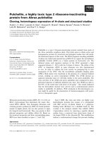
Figure 1
Organization of the HTLV-2 genome and coding regions
Organization of the HTLV-2 genome and coding regions. The complete proviral genome is shown schematically.
Boxes denote long terminal repeats (LTRs). RNAs encoding the various protein ORFs are indicated. p28 has the potential to
be encoded by two distinct singly-spliced mRNAs (gray line in p28 mRNA denotes utilization of two distinct splice acceptor
sites). The arrow above p28 ORF depicts the location of the termination mutation to generate Δp28 (stop codon at amino acid
7).
Page 3 of 11
(page number not for citation purposes)
Retrovirology 2008, 5:38
/>
A
A
24000
18000
16000
20000
14000
16000
RLU
RLU
12000
10000
8000
12000
6000
8000
4000
4000
2000
0
p19 pg/ml
B
0
BC12
HTLV-2
p28
HTLV-2
100
+
-
+
0.2
-
+
0.4
-
+
0.2
+
0.4
80
B
60
40
20
0
C
BC12
p28
-actin
HTLV-2
HTLV-2
104
103
102
101
100
C
p28
105
copy # per 106 gapdh
-
gag/pol
tax/rex
mRNA transcript
p28
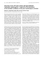
Figure 2
Characterization of proviral clones in vitro
Characterization of proviral clones in vitro. 293 T cells (2 × 10⁵)
were co-transfected with 1 μg of wtHTLV-2 or HTLV-2Δp28 proviral
clones or negative control DNA along with 0.1 μg of LTR-1-Luc and 0.01
μg of TK-Renilla. All transfections were performed in triplicate and normalized to TK-Renilla to control for transfection efficiency. Cell lysates or
supernatants were harvested 48 h post-transfection. (A) Measure of Tax
activity presented as relative luciferase units. Results indicated that loss of
p28 expression from the proviral clone did not significantly alter Tax activity. (B) Rex activity as measured by expression of p19 Gag (virions) in the
cellular supernatants. Results indicated that loss of p28 expression from
the proviral clone did not significantly alter Rex activity. (C) Total RNA
was extracted from 293 T cells transfected with HTLV-2 or HTLV-2pΔ28
as in panels A and B. mRNA copy number was quantified by Taqman realtime RT-PCR. The histogram represents the copy number of gag-pol, tax/
rex, and p28 transcripts normalized to 1 × 10⁶ copies of gapdh. Results
indicated that deletion of p28 protein had no significant affect on tax/rex,
gag/pol, or p28 mRNA expression.
Figure 3 expressed p28, but not Δp28 results in
dependent repression of Tax-mediated transcription doseExogenously
Exogenously expressed p28, but not Δp28 results in
dose-dependent repression of Tax-mediated transcription. 293 T cells (2 × 10⁵) were co-transfected with 1
μg of wtHTLV-2 proviral clone or negative control DNA, 0.1
μg of LTR-2-Luc and 0.01 μg of TK-Renilla, and varying concentrations (0.2–0.4 μg) of CMVp28AU1 or CMVΔp28AU1
expression vectors as indicated. (A) Tax function was measured as firefly luciferase activity from LTR-2-Luc normalized
to Renilla luciferase activity. RLU, relative light units. (B)
Western blot analysis was performed on lysates to confirm
expression of p28 (AU1 antibody) or β-actin as a loading
control. As expected, results indicated that the Δp28 mutation disrupts p28 protein expression.
HTLV-2Δp28 promoted virus-induced proliferation and
immortalization of PBMCs
To determine the capacity of HTLV-2Δp28 to synthesize
viral proteins, direct viral replication, and induce cellular
immortalization, stable 729 cell transfectants expressing
wild-type and p28-deleted HTLV-2 proviral clones were
generated and characterized. Four independent stable
HTLV-2Δp28 transfectants were isolated and found to
contain complete copies of the provirus; the presence of
the expected Δp28 mutation was confirmed by sequencing (data not shown). We quantified the concentration of
p19 Gag produced in the culture supernatant of the four
cell clones by ELISA. Our results showed p19 Gag expression ranging from 250–750 pg/ml (Fig. 4A). The variable
Page 4 of 11
(page number not for citation purposes)
Retrovirology 2008, 5:38
A
/>
900
800
600
500
400
Clone 4
100
Clone 3
200
Clone 2
300
Clone 1
p19 (pg/ml)
700
0
729
B
729
729.
HTLV-2
729.
HTLV-2
729.HTLV-2
Tax-2
-actin
1.7E+0
6.5E+3
2.1E+3
} Copies
p28
mRNA
Figure 4
permanent of p19 Gag and
Expression transfectants Tax protein and p28 mRNA in
Expression of p19 Gag and Tax protein and p28
mRNA in permanent transfectants. (A) Four 729 stable
transfectants (clone 1–4) were isolated for HTLV-2Δp28 as
described in Materials and Methods. Our well-established
729pH6neo (729.HTLV-2) cell clone was used as the wtHTLV-2 stable producer cell line. p19 Gag was quantified by
ELISA from the four independently isolated 729.HTLV-2Δp28
(Clones 1–4), 729.HTLV-2, and the 729 negative control.
Each 729.HTLV-2 producer cell line displayed variable p19
production. (B) Clones indicated by asterisks, which have
been shown to produce similar quantities of p19 Gag, were
further characterized by Western blot for Tax protein
expression using rabbit polyclonal antisera raised against
Tax-2. β-actin was used as a loading control. Numbers below
each lane are the copy number of p28 transcript per 106 copies of GAPDH determined by realtime RT PCR. The results
show similar levels of p28 mRNA expression.
p19 Gag expression from independent stable cell clones
was attributed to chromosomal location of proviral
sequences and overall proviral copy number, consistent
with previous analyses [44,45]. We did not observe a pattern of increased viral gene expression in the absence of
p28. For additional studies, we selected 729.HTLV-
2Δp28Clone 3, a stable producer line with p19 Gag production similar to that of our well-characterized wild-type
HTLV-2-producer cell line 729pH6neo (729.HTLV-2).
Further characterization revealed that, as with transient
transfection, p28 mRNA was detected at similar levels
(approximately 103 copies per 106 copies of cellular gapdh
mRNA) in 729.HTLV-2 and 729.HTLV-2Δp28Clone 3
(Fig. 4B), but Western blot analyses failed to detect p28
protein in 729.HTLV-2 or 729.HTLV-2Δp28 (data not
shown). A similar level of Tax-2 expression in 729.HTLV2 and 729.HTLV-2Δp28 relative to the β-actin loading
control was detected by Western blot and, as expected,
Tax-2 was not detected in the 729 negative control cells
(Fig. 4B). Therefore, as with transient transfection, the
repressive effect of p28 expressed from a stably integrated
provirus on Tax-mediated transcription was not detectable.
We assessed the ability of the HTLV-2Δp28 to induce proliferation and immortalize human PBMCs in co-culture
assays. Freshly isolated human PBMCs co-cultured with
lethally irradiated 729.HTLV-2 or 729.HTLV-2Δp28 in the
presence of 10 U/ml of human IL-2 showed very similar
progressive growth patterns consistent with the HTLV-2
immortalization process, whereas control cells died
within the first few weeks (Fig. 5A). Immortalized PBMCs
expressed similar levels of p19 Gag and harbored the
expected HTLV-2 sequences indicating that viral transmission was responsible for the immortalization of PBMCs
(data not shown). In an effort to obtain a more quantitative measure of the ability of these viruses to infect and
immortalize PBMCs, a fixed number of PBMCs were cocultured with virus-producing cells in a 96-well plate assay
[45]. Since this assay is very stringent as a result of diluting
the cultures 1:3 weekly, slowly growing or non-dividing
cells are eliminated very quickly and the percentage of surviving wells is an accurate measure of the immortalization
efficiency of viruses. A Kaplan-Meier plot of HTLV-2induced T-cell proliferation or survival indicated that the
percentage of wells containing proliferating lymphocytes
was similar between HTLV-2 and two independently isolated HTLV-2Δp28 clones (Fig. 5B). Taken together, our
results are consistent with the conclusion that p28 is not
required for efficient infectivity or HTLV-2-mediated
immortalization of primary human T-lymphocytes in culture.
In vivo rabbit inoculation results
To evaluate the role of p28 in vivo, we compared the abilities of 729, 729.HTLV-2, or 729.HTLV-2Δp28 cell lines to
transmit virus to rabbits, which is an established model to
investigate HTLV infection and persistence [46]. Rabbits
were inoculated with lethally irradiated cell lines (cell
inocula were equilibrated based on their p19 Gag production) and on weeks 0, 1, 2, 4, 6, 8, and 11, whole blood
Page 5 of 11
(page number not for citation purposes)
Retrovirology 2008, 5:38
A
/>
80
729
HTLV-2
Cell Number (x105)
70
60
50
40
30
20
10
0
1
2
3
4
5
6
7
8
9
10
11
12
14
Week
B
100
% Surviving Wells
80
C
HTLV-2
60
40
20
0
0
1
2
3
4
5
6
7
8
Weeks
p28 is dispensable for HTLV-2-mediated proliferation and immortalization of primary T-lymphocytes
Figure 5
p28 is dispensable for HTLV-2-mediated proliferation and immortalization of primary T-lymphocytes. (A)
Human PBMCs were isolated by Ficoll/Paque and co-cultivated with irradiated (10,000 rads) 729, 729.HTLV-2, or 729.HTLV2Δp28 stable cell lines. PBMCs (2 × 10⁶) were cultured with irradiated donor cells (1 × 10⁶) in 24 well plates as indicated. A
representative growth curve of HTLV-2 infected cells is shown. Cell viability was determined weekly by trypan blue exclusion
(0–14 wks post co-cultivation). The mean and standard deviation of each time point was determined from three independent
samples. (B) Pre-stimulated PBMCs (104) were co-cultured with 2 × 10³ irradiated 729 stable producer cells in 96 well plates.
The percentages of proliferating wells were plotted as a function of time (wks). Representative Kaplan-Meir plots for wtHTLV2, HTLV-2Δp28, and 729 uninfected control cells are shown. Results indicated that the percentage of wells containing proliferating lymphocytes was similar between wtHTLV-2 and HTLV-2Δp28 infected cells.
was collected and processed for isolation of plasma and
PBMCs. Antibody response to viral antigens was detectable by Western blot in all rabbits inoculated with cells
expressing either wild type HTLV-2 or HTLV-2Δp28, and
the antibody titers in the majority of the rabbits increased
over the time course of the study (data not shown). Moreover, quantitative comparison of antibody responses
between each rabbit was performed using an HTLV-specific ELISA (Fig. 6). Statistical analysis of titers at six, eight,
and eleven weeks post-inoculation revealed a significantly
lower antibody response to HTLV-2 antigens in the
729.HTLV-2Δp28-inoculated rabbits as compared to the
wild-type HTLV-2 control group. Consistent with our antibody data, HTLV-2 proviral DNA sequences were detected
in all wild type HTLV-2 and five of six HTLV-2Δp28infected rabbits at two weeks post inoculation (Table 1).
However, over time, HTLV-2Δp28 failed to persist and
quantitative real-time Taqman PCR revealed that at eleven
weeks post inoculation, proviral loads in rabbits infected
with HTLV-2Δp28 were below the level of detection.
Page 6 of 11
(page number not for citation purposes)
Retrovirology 2008, 5:38
/>
1.2
1
O.D.
0.8
0.6
0.4
0.2
0
Wks 0
1
2
4
6
8
11
0
1
729
2
4
6
8
11
0
1
2
4
6
8
11
729.HTLV-2
Figure 6
Assessment of HTLV-2 infection in rabbits
Assessment of HTLV-2 infection in rabbits. Antibody response against HTLV-2 from each rabbit was measured by antiHTLV commercial ELISA assay, using both HTLV Gag and envelope proteins as antigens. Each dot represents the absorbance
value of a single inoculated rabbit at 0, 2, 4, 6, 8, and 11 wks post inoculation within each group. Inocula used for the rabbits
were 729.HTLV-2 (n = 6), 729.HTLV-2Δp28 (n = 6), or 729 (n = 2). The horizontal line represents the average of the rabbit
group at each weekly time point and the dotted line represents three times the standard deviation of uninfected control values.
Taken together, our results indicated that p28, while dispensable for HTLV-2 infection, attenuated virus replication as measured by antibody response to viral antigens
and proviral loads. This attenuation was apparent within
two weeks post inoculation, suggesting that p28 is
required early for efficient replication and survival in the
host.
Discussion
The importance of the HTLV-2 nonstructural or accessory
proteins in virus biology either in cell culture or in inoculated animals has not been investigated thoroughly. A previous study evaluated an HTLV-2 molecular clone
containing a large deletion within the proximal pX region,
which at the time was thought to delete the coding
sequences for all the known accessory proteins. Results
from this study indicated that this region, which later was
shown to contain open reading frames (ORFs) for p10
and p11 [28], was dispensable for viral infection and cellular transformation in vitro [41]. Subsequently, it was
demonstrated that this deletion resulted in reduced proviral load and maintenance of infection in vivo [42]. However, the role of the HTLV-2 p28 accessory protein
encoded by ORF II located in exon 3 of tax/rex was not
addressed directly in these studies. We previously demonstrated that exogenously over-expressed p28 functions as
a negative regulator of viral replication by binding to and
retaining tax/rex mRNA in the nucleus, thus repressing Tax
and Rex protein production and overall viral gene expression [30,31]. In this study, a site directed mutation was
introduced in an infectious clone of HTLV-2 that severely
truncated p28 (HTLV-2Δp28) while maintaining the ability of the virus to express other gene products. Subsequently, we examined the expression of p28 and
determined its biological significance for the infectivity
and immortalization of primary T-lymphocytes in cell culture and viral infectivity and persistence in vivo.
Data from our transient transfection studies revealed that,
in the context of a proviral clone, the repressive effects of
p28 on Tax-mediated transcription and Rex function were
not apparent (Fig. 2A &2B). In fact, the loss of p28
resulted in a reproducible, but not significant decrease in
Tax activity (75–90%). Consistent with the functional
reporter assays, quantitative real-time RT-PCR revealed
that the levels of tax/rex and gag/pol mRNA were not dramatically different in cells transfected with HTLV-2 and
HTLV-2Δp28 proviral clones (Fig. 2C). Although we could
detect p28 encoding mRNA (approximately 103-104 total
copies per 106 copies of gapdh), p28 protein was below
the limit of detection by Western blot. Due to alternative
splicing, p28 has the potential to be expressed from two
distinct singly-spliced mRNAs (both of these mRNAs also
have the potential to produce the truncated p22/p20rex).
Studies by Li and Green showed that these two mRNAs
have significantly different expression levels in newly
infected PBMCs (105 vs 103 copies per 106 copies of cellular gapdh) [43]. Although nearly impossible to definitively confirm experimentally, we hypothesize that the
low copy number mRNA is the primary transcript utilized
to encode p28, thus resulting in low protein expression
(below our limit of detection). To date, with the exception
Page 7 of 11
(page number not for citation purposes)
Retrovirology 2008, 5:38
/>
Table 1: Detection of HTLV-2 sequences in PBMCs from
inoculated rabbitsa
Weeks Post Inoculation
Inoculum and Rabbit
729.HTLV-2
R27
R28
R29
R30
R31
R32
729.HTLV-2Δp28
R20
R21
R22
R23
R24
R25
729
R1
R6
0
2
6
8
11b
-
+
+
+
+
+
+
+
+
+
+
+
+
+
+
+
+/-
+ (12.0)
+ (8.3)
+ (5.3)
+ (32.8)
+ (10.7)
+ (4.2)
-
+
+/+/+/+/-
+/-
+
-
- (0.2)
- (0.1)
- (0.3)
- (0.3)
- (1.1)
- (1.2)
-
-
-
-
- (0.1)
- (0.3)
aGenomic DNA was isolated from rabbit PBMCs and subjected to
standard PCR (40 cycles) using HTLV-2 specific primers (TRE-pH6-S/
TRE-pH6-AS). -, no amplified PCR fragment; +, amplified PCR
fragment.
bNumbers in parentheses at wk 11 denote copy number per 1000
cells of rabbit PBMC as determined by real-time RT PCR. Copy
numbers in rabbits inoculated with 729.HTLV-2Δp28 at wk 11 were
significantly different than 729.HTLV-2 as determined by ANOVA
followed by Turkey's test (p<0.00032)
of the HTLV-1 HBZ protein, none of the HTLV-1 or HTLV2 accessory proteins have been detected in transfected or
infected cells. Interestingly, the mRNA copy number of
HBZ in infected cells was 10- to 100-fold higher than the
other accessory gene mRNAs, which was consistent with
its detection [43]. However, we did confirm that overexpression of p28 from a cDNA expression plasmid, but
not Δp28, down-regulated Tax-mediated viral transcription in a dose-dependent manner (Fig 3A). Furthermore,
we demonstrated that the repressive effects of p28 on Taxmediated transcription and Rex activity were not detectable in stable cell lines as represented by variable p19 production less than or equal to wild-type HTLV-2
production levels (Fig 4A). Therefore, we speculate that
p28 protein expression is temporally regulated and not
expressed following transient proviral DNA plasmid
delivery or in stable transfectants and/or a threshold level
of p28 is required for the repressive activity.
Results from our short-term proliferation and immortalization assays indicated that the reported repressive effects
of the HTLV-2 p28 on Tax and Rex [30,31] were not sufficient to disrupt the capacity of the virus to infect, induce
proliferation, and/or immortalize primary T lymphocytes
in vitro (Fig 5A and 5B). Therefore, similar to the HTLV-1
and other HTLV-2 pX ORF-encoded accessory proteins
[38,41,47], p28 appears to be dispensable for efficient
viral infectivity, replication and primary T-lymphocyte
immortalization capacity in vitro.
Based on the efficient infectivity and immortalization of
cells in vitro and the transient infection observed in
729.HTLV-2Δp28-inoculated rabbits, we hypothesize that
the function of p28 and its role in HTLV-2 biology
involves early virus/host interactions that may include
virus spread and/or survival of the infected cell. We
observed reduced proviral load as early as two weeks post
inoculation as compared to that in the wild type virusinfected rabbits (Table 1). By four weeks, p28 mutant
inoculated rabbits showed a significant reduction in the
antibody response to viral gene products, which continued for the duration of the study (Fig. 6). By week eleven,
we failed to detect a visible PCR amplified band or realtime PCR proviral loads in all HTLV-2Δp28-inoculated
rabbits. All wild-type HTLV-2-inoculated rabbits showed
variable but significant proviral loads. To date, p28 has
been documented to repress Tax-mediated transcription
and Rex activity; based on our results, we speculate that
p28 might function in concert with other viral gene products to tightly regulate viral replication and/or influence
virus expression in the infected lymphocyte to promote
infected cell survival (apoptosis vs cell proliferative signals), viral spread, and establishment of persistent infection. It remains possible that p28 may have multiple
activities that function at different stages of the infection
process. Future experiments designed to quantitatively
assess viral infectivity of rabbits at 1–2 days post inoculation will be required to definitively rule out an early block
in infection in vivo. Interestingly, the gross phenotype of
HTLV-2Δp28 in vivo showed significant similarities to
HTLV-1 HBZ, p30 and p13 virus mutants. More detailed
comparative studies will be required to dissect mechanistic differences which may provide important insight
regarding how viral proteins function causing the distinct
pathobiology between HTLV-1 and HTLV-2.
Conclusion
In summary, our data confirmed that over-expression of
p28 in cell culture repressed viral gene expression, but in
the context of a replicating virus, was completely dispensable for efficient cellular immortalization. Utilizing a
rabbit model of infection, these are the first biological
studies to demonstrate the critical requirement of the p28
accessory protein in the establishment of HTLV-2 infection in vivo. It is likely that p28, as a negative regulator of
Tax and Rex, is critical in the temporal regulation of gene
expression upon infection and promotes cell survival.
This importance is not seen without the selective pressure
applied by the presence of a functional immune system.
Page 8 of 11
(page number not for citation purposes)
Retrovirology 2008, 5:38
These biological studies have led the way for future studies
that are needed to understand the function of p28. Such
studies will entail identifying the functional domains of
the protein involved in localization, protein interactions,
and RNA binding as well as precisely identifying the viral
mRNA response element. In addition, gene array studies
may provide clues as to whether p28 expression by itself
has any direct or indirect cellular effects that facilitate the
survival of the T-lymphocyte, the natural target for HTLV
infection and cellular transformation.
Methods
Cells
293T cells and 729 B cell lines were maintained in Dulbecco's modified Eagle and Iscove medium, respectively,
supplemented with 10% fetal bovine serum (FBS), 2 mM
glutamine, penicillin (100 U/mL), and streptomycin (100
ug/mL). Human and rabbit peripheral blood mononuclear cells (PBMCs) were isolated using Ficoll Hypaque
(Amersham, Piscataway, NJ) and Percoll® (Amersham,
Piscataway, NJ), respectively, and cultured in RPMI 1640
medium supplemented with 20% FBS, glutamine and
antibiotics as above, plus 10 U/mL of recombinant interleukin-2 (IL-2; Roche Applied Biosciences, Indianapolis,
IN).
Plasmids
The p28 cDNA expression vector (CMV-p28-AU1) and the
wild type (wt) HTLV-2 infectious proviral clone (pH6neo)
were described previously [30,48]. Using PCR mutagenesis and CMV-p28-AU1 as a template, a single nucleotide
mutation (C to A) was introduced in the p28 reading
frame. This change (nt 7333 of the pH6neo proviral
sequence) resulted in a stop codon in the seventh amino
acid (aa) of p28, designated Δp28. This specific mutation
was designed to not alter the aa sequence of either Tax or
Rex, both of which share overlapping reading frames with
p28. The Δp28 mutation expressed in the context of the
proviral clone pH6neo, was designated HTLV-2Δp28. The
mutation in all mutant plasmids was confirmed by DNA
sequencing. The Tax reporter plasmid, LTR-2-Luc, and the
transfection efficiency control plasmid, TK-Renilla, were
described previously [30,31].
Transfection, reporter assays, and p19 Gag ELISA
293T cells (2 × 10⁵) were transfected using Lipofectamine®
(Invitrogen, Carlsbad, CA) as recommended by the manufacturer. For p28 protein detection, cells were transfected
with 1 μg of cDNA expression plasmids and 10 ng of TKRenilla. Cell lysates were prepared at 48 h post transfection and normalized for transfection efficiency prior to
Western blot analysis. To assess the repressive effects of
p28 or Δp28 by Tax reporter assays, cells were transfected
with 1 μg wtHTLV-2 in the presence or absence of variable
concentrations (0.2–0.4 μg) of p28 or Δp28 cDNA expres-
/>
sion vector and 0.1 μg of LTR-2-Luc, and 10 ng of TKRenilla or 1 μg HTLV-2Δp28 and 0.1 μg of LTR-2-Luc, and
10 ng of TK-Renilla. Cell lysates were harvested at 48 h
post transfection and dual luciferase activity was measured. The data represent average luciferase activity values
after normalization for transfection efficiency for three
independent experiments. To generate the 729HTLV2Δp28 stable transfectant, the proviral plasmid clone containing neor gene was introduced into cells by nucleofection using the Nucleofector kit V (Amaxa Biosystems,
Gaithersburg, MD). Stable transfectants containing the
desired proviral clone were isolated following incubation
in 24-well culture dishes in medium containing 1 mg/ml
Geneticin (Gibco, Carlsbad, CA). Following a 4–5 weeks
selection period, viable cells were expanded and maintained in culture for further analysis. The well-characterized wtHTLV-2 729 producer cell line (729pH6neo) used
in this study was described previously [46,49].
Western Blot
To detect p28, 50 μg of total cell lysates from transfected
cells was separated by SDS-PAGE and transferred to a
nitrocellulose membrane (Amersham, Piscataway, NJ).
Rabbit polyclonal antibodies against p28 or a monoclonal antibody to AU1 (Covance Research Products,
Denver, PA,) was used for p28 detection. Rabbit polyclonal antibody to β-actin (Novus Biological, Littleton, CO)
was used as a loading control. Proteins were visualized
using the ECL western blotting analysis system (Santa
Cruz Biotechnology, Santa Cruz, CA).
DNA isolation, standard PCR, and Taqman real-time PCR
DNA was isolated from 729 producer cells and rabbit
peripheral blood mononuclear cells (PBMCs) using the
PURGENE DNA purification system (Gentra, Minneapolis, MN). Rabbit DNA (1 μg) was subjected to a standard
40-cycle PCR amplification for detection of integrated
provirus and the product was visualized on a 2% agarose
gel stained with ethidium bromide. The primer pair used
in the PCR, based on the pH6neo sequence, was TRE-PHS (5'-41GAG TCA TCG ACC CAA AAG G59-3') and TREPH-AS (5'-298TGC GCT TTT ATA GAC TCG GC279-3'),
which amplified a 257 bp product in the HTLV-2 LTR.
Taqman real-time PCR (Applied Biosystems, Foster City,
CA) using 500 ng of rabbit DNA and 40 cycle amplification was performed in a 25 ul reaction to quantify the proviral copy number per cell in infected rabbit PBMCs using
primers and probes directed towards Gag sequences [43].
The reaction contained 100 ng (25 ng/μL) of each primer
and the probe at a concentration of 100 pmol/μL. A standard curve was generated for each run using duplicate samples of log10 dilutions of a plasmid containing the Gag
sequences. The copy number for each sample was determined from the standard curve, and the copy number per
Page 9 of 11
(page number not for citation purposes)
Retrovirology 2008, 5:38
cell for each sample calculated based on the estimate that
1 μg PBMC DNA is equal to 67,300 cells.
Short-term proliferation and long-term immortalization
coculture assays
Short term microtiter proliferation assays were performed
as detailed previously with modifications [45,50]. Briefly,
freshly isolated human PBMCs were pre-stimulated with 2
μg/ml PHA and 10 U/ml IL-2 (Roche, Indianapolis, IN)
for three days. 729 producer cells (2 × 10³) were irradiated
(100 Gy) and co-cultured with 104 pre-stimulated PBMCs
in the presence of IL-2 in 96-well round bottom plates.
Wells were enumerated for growth and split 1:3 at weekly
intervals. Cell proliferation was confirmed by MTS assay
using CellTiter 96® Aqueous One Solution Reagent as recommended by the manufacturer (Promega, Madison,
WI). For the long-term immortalization assays, 106 irradiated producer cells were co-cultivated with 2 × 10⁶ freshly
isolated PBMCs with 10 U/ml IL-2 in 24-well culture
plates [41]. HTLV expression was confirmed by detection
of p19 Gag protein in the culture supernatant measured at
weekly intervals using a commercially available ELISA
(Zeptometrix, Buffalo, NY). Viable cells were counted
weekly by trypan blue exclusion. Cells inoculated with
HTLV-2 that continued to produce p19 Gag antigen and
proliferate 12 weeks post co-culture in the presence of
exogenous interleukin-2 (IL-2) were identified as HTLV
immortalized. For each assay, at least three independent
experiments were performed using PBMCs from distinct
healthy donors.
Rabbit inoculation, ex vivo culture, and serologic analysis
Twelve week-old specific pathogen-free New Zealand
White rabbits (Harlan, Indianapolis, IN) were inoculated
with approximately 1 × 10⁷ gamma-irradiated (100 Gy)
729 viral producer cells (6 rabbits per group) or 729 uninfected control cells (2 rabbits) via the lateral ear vein. The
virus-containing inocula were equilibrated based on
HTLV-2 p19 Gag production (ELISA). At weeks 0, 1, 2, 4,
6, 8, and 11 after inoculation, 10 ml of blood was drawn
from the central auricular artery from each animal and
rabbit plasma and PBMCs were isolated. HTLV Western
blot assay (HTLV Blot 2.4 Western Blot Assay; MP Diagnostics, Singapore) was used to examine serum reactivity
to specific viral antigenic determinants. Serum showing
reactivity to Gag (p24 or p19) and Env (gp21 or gp46)
antigens was classified as positive for HTLV-2 seroreactivity. A commercial HTLV ELISA kit (Vironostika HTLV-I/II
Microelisa System; bioMerieux, Durham, NC) was used to
quantitate HTLV-2 serum antibody using plasma diluted
1:100 to obtain values within the linear range of the assay.
Data is shown as absorbance values. DNA was isolated
from rabbit PBMCs using the PURGENE DNA purification system (Gentra, Minneapolis, MN) and subjected to
proviral load analysis by realtime PCR.
/>
Authors' contributions
BY generated mutant clones, carried out functional assays,
virus replication and immortalization assays, the in vivo
studies, and drafted the manuscript. ML developed the
realtime PCR primers and performed or assisted with all
the assays and quantitation. MK helped with the collection and processing of in vivo samples, and assisted with
the Western blot analysis. IY helped with the generation of
mutant clones and the development of the functional
assays. MDL has helped in finalizing the manuscript and
has provided important input on the design of the rabbit
portion of the study. PLG conceived the study, participated in its coordination, helped in drafting and finalizing the manuscript. All authors read and approved the
final manuscript.
Acknowledgements
We thank Kate Hayes for editorial comments on the manuscript and Tim
Vojt for figure preparations. This work was supported by a grant from the
National Institutes of Health (CA100730) to PLG.
References
1.
2.
3.
4.
5.
6.
7.
8.
9.
10.
11.
12.
13.
Lairmore M, Franchini G: Human T-Cell Leukemia Virus Types
1 and 2. In Fields Virology Volume 5th. Edited by: Fields BN, Knipe DM,
Howley PM, Chanock RM, Monath TP, Melnick JL, Roizman B, Straus
SE. Philadelphia, PA USA. , Lippincott Williams, and Wilkins;
2007:Chapter 56, 2071-2106.
Wolfe ND, Heneine W, Carr JK, Garcia AD, Shanmugam V, Tamoufe
U, Torimiro JN, Prosser AT, Lebreton M, Mpoudi-Ngole E,
McCutchan FE, Birx DL, Folks TM, Burke DS, Switzer WM: Emergence of unique primate T-lymphotropic viruses among central African bushmeat hunters. Proc Natl Acad Sci U S A 2005,
102(22):7994-7999.
Calattini S, Chevalier SA, Duprez R, Bassot S, Froment A, Mahieux R,
Gessain A: Discovery of a new human T-cell lymphotropic
virus (HTLV-3) in Central Africa. Retrovirology 2005, 2(1):30.
Takatsuki K: Discovery of adult T-cell leukemia. Retrovirology
2005, 2:16.
Osame M, Igata A, Matsumoto M, Usuku K, Izumo S, Kosaka K:
HTLV-I associated myelopathy: A report of 85 cases. Ann
Neurol 1987, 22:116.
Yoshida M: Discovery of HTLV-1, the first human retrovirus,
its unique regulatory mechanisms, and insights into pathogenesis. Oncogene 2005, 24(39):5931-5937.
Gallo RC: History of the discoveries of the first human retroviruses: HTLV-1 and HTLV-2.
Oncogene 2005,
24(39):5926-5930.
Rosenblatt JD, Giorgi JV, Golde DW, Ezra JB, Wu A, Winberg CD,
Glaspy J, Wachsman W, Chen IS: Integrated human T-cell leukemia virus II genome in CD8 + T cells from a patient with
"atypica" hairy cell leukemia: evidence for distinct T and B
cell lymphoproliferative disorders. Blood 1988, 71:363-369.
Berger JR, Svenningsson A, Raffanti S, Resnick L: Tropical spastic
paraparesis-like illness occurring in a patient dually infected
with HIV-1 and HTLV-II. Neurology 1991, 41:5-7.
Hjelle B, Appenzeller O, Mills R, Alexander S, Torrez-Martinez N,
Jahnke R, Ross G: Chronic neurodegenerative disease associated with HTLV-II infection. Lancet 1992, 339:645-646.
Harrington Jr WJ, Sheremata W, Hjelle B, Dube DK, Bradshaw P,
Foung SK, Snodgrass S, Toedter G, Cabra L, Poiesz B: Spastic ataxia
associated with human T-cell lymphotropic virus type II
infection. Annals Neurol 1993, 7:1031-1034.
Poiesz B, Dube D, Dube S, Love J, Papsidero L, Uner A, Hutchinson
R: HTLV-II-associated cutaneous T-cell lymphoma in a
patient with HIV-1 infection.
N Engl J Med 2000,
342(13):930-936.
Cann AJ, Rosenblatt JD, Wachsman W, Shah NP, Chen ISY: Identification of the gene responsible for human T-cell leukemia
virus transcriptional regulation. Nature 1985, 318:571-574.
Page 10 of 11
(page number not for citation purposes)
Retrovirology 2008, 5:38
14.
15.
16.
17.
18.
19.
20.
21.
22.
23.
24.
25.
26.
27.
28.
29.
30.
31.
32.
33.
Felber BK, Paskalis H, Kleinman-Ewing C, Wong-Staal F, Pavlakis GN:
The pX protein of HTLV-I is a transcriptional activator of its
long terminal repeats. Science 1985, 229:675-679.
Inoue JI, Yoshida M, Seiki M: Transcriptional (p40x) and posttranscriptional (p27xIII) regulators are required for the
expression and replication of human T-cell leukemia virus
type I genes. Proc Natl Acad Sci USA 1987, 84:3653-3657.
Leung K, Nabel GJ: HTLV-I transactivator induces interleukin2 receptor expression through an NFkB-like factor. Nature
1988, 333:776-778.
Mulloy JC, Kislyakova T, Cereseto A, Casareto L, LoMonico A, Fullen
J, Lorenzi MV, Cara A, Nicot C, Giam C, Franchini G: Human T-cell
lymphotropic/leukemia virus type 1 Tax abrogates p53induced cell cycle arrest and apoptosis through its CREB/
ATF functional domain. J Virol 1998, 72(11):8852-8860.
Ressler S, Morris GF, Marriott SJ: Human T-cell leukemia virus
type 1 Tax transactivates the human proliferating cell
nuclear antigen promoter. J Virol 1997, 71:1181-1190.
Schmitt I, Rosin O, Rohwer P, Gossen M, Grassmann R: Stimulation
of cyclin-dependent kinase activity and G1- to S-phase transition in human lymphocytes by the human T-cell leukemia/
lymphotropic virus type 1 Tax protein.
J Virol 1998,
72(1):633-640.
Siekevitz M, Feinberg MB, Holbrook N, Wong-Staal F, Greene WC:
Activation of interleukin 2 and interleukin 2 receptor (Tac)
promoter expression by the trans-activator (tat) gene product of human T-cell leukemia virus, type I. Proc Natl Acad Sci
USA 1987, 84:5389-5393.
Robek MD, Ratner L: Immortalization of CD4+ and CD8+ Tlymphocytes by human T-cell leukemia virus type 1 Tax
mutants expressed in a functional molecular clone. J Virol
1999, 73:4856-4865.
Ross TM, Narayan M, Fang ZY, Minella AC, Green PL: Tax transactivation of both NFkB and CREB/ATF is essential for Human
T-cell leukemia virus type 2-mediated transformation of primary human T-cells. J Virol 2000, 74:2655-2662.
Wycuff DR, Marriott SJ: The HTLV-1 Tax Oncoprotein:Hypertasking at the molecular level. Front Biosci 2005, 10:620-642.
Ballaun C, Farrington GR, Dobrovnik M, Rusche J, Hauber J, Bohnlein
E: Functional analysis of human T-cell leukemia virus type I
Rex-response element: Direct RNA binding of Rex protein
correlates with in vivo binding activity.
J Virol 1991,
65:4408-4413.
Kusuhara K, Anderson M, Pettiford SM, Green PL: Human T-cell
leukemia virus type 2 Rex protein increases stability and promotes nuclear to cytoplasmic transport of gag/pol and env
RNAs. J Virol 1999, 73:8112-8119.
Narayan M, Kusuhara K, Green PL: Phosphorylation of two serine residues regulates human T-cell leukemia virus type 2
Rex function. J Virol 2001, 75(18):8440-8448.
Koralnik IJ, Gessain A, Klotman ME, Lo Monico A, Berneman ZN,
Franchini G: Protein isoforms encoded by the pX region of
human T-cell leukemia/lymphotropic virus type I. Proc Natl
Acad Sci U S A 1992, 89(18):8813-8817.
Ciminale V, D'Agostino DM, Zotti L, Franchini G, Felber BK, ChiecoBianchi L: Expression and characterization of proteins produced by mRNAs spliced into the X region of the human Tcell leukemia/lymphotropic virus type II. Virology 1995,
209(2):445-456.
Nicot C, Dundr JM, Johnson JR, Fullen JR, Alonzo N, Fukumoto R,
Princler GL, Derse D, Misteli T, Franchini G: HTLV-1-encoded
p30II is a post-transcriptional negative regulator of viral replication. Nat Med 2004, 10(2):197-201.
Younis I, Khair L, Dundr M, Lairmore MD, Franchini G, Green PL:
Repression of human T-cell leukemia virus type 1 and 2 replication by a viral mRNA-encoded posttranscriptional regulator. J Virol 2004, 78:11077-11083.
Younis I, Boris-Lawrie K, Green PL: Human T-cell leukemia virus
ORF II p28 encodes a post-transcriptional repressor that is
recruited at the level of transcription. J Virol 2006, 80:181-191.
Sinha-Datta U, Datta A, Ghorbel S, Dodon MD, Nicot C: Human Tcell Lymphotrophic Virus Type I Rex and p30 Interactions
Govern the Switch between Virus Latency and Replication.
J Biol Chem 2007, 282(19):14608-14615.
Zhang W, Nisbet JW, Albrecht B, Ding W, Kashanchi F, Bartoe JT,
Lairmore MD: Human T-lymphotropic virus type 1 p30II reg-
/>
34.
35.
36.
37.
38.
39.
40.
41.
42.
43.
44.
45.
46.
47.
48.
49.
50.
ulates gene transcription by binding CREB binding protein/
p300. J Virol 2001, 75(20):9885-9895.
Zhang W, Nisbet JW, Bartoe JT, Ding W, Lairmore MD: Human Tlymphotropic virus type 1 p30II functions as a transcription
factor and differentially modulates CREB-responsive promoters. J Virol 2000, 74(23):11270-11277.
Michael B, Nair AM, Datta A, Hiraragi H, Ratner L, Lairmore MD:
Histone acetyltransferase (HAT) activity of p300 modulates
human T lymphotropic virus type 1 p30II-mediated repression of LTR transcriptional activity.
Virology 2006,
354(2):225-239.
Awasthi S, Sharma A, Wong K, Zhang J, Matlock EF, Rogers L, Motloch P, Takemoto S, Taguchi H, Cole MD, Luscher B, Dittrich O, Tagami H, Nakatani Y, McGee M, Girard AM, Gaughan L, Robson CN,
Monnat RJ Jr., Harrod R: A Human T-Cell Lymphotropic Virus
Type 1 Enhancer of Myc Transforming Potential Stabilizes
Myc-TIP60 Transcriptional Interactions. Mol Cell Biol 2005,
25(14):6178-6198.
Harrod R, Kuo YL, Tang Y, Yao Y, Vassilev A, Nakatani Y, Giam CZ:
p300 and p300/cAMP-responsive element-binding protein
associated factor interact with human T-cell lymphotropic
virus type-1 Tax in a multi-histone acetyltransferase/activator-enhancer complex. J Biol Chem 2000, 275(16):11852-11857.
Derse D, Mikovits J, Ruscetti F: X-I and X-II open reading frames
of HTLV-I are not required for virus replication or for
immortalization of primary T-cells in vitro. Virology 1997,
237:123-128.
Bartoe JT, Albrecht B, Collins ND, Robek MD, Ratner L, Green PL,
Lairmore MD: Functional role of pX open reading frame II of
human T-lymphotropic virus type 1 in maintenance of viral
loads in vivo. J Virol 2000, 74(3):1094-1100.
Silverman LR, Phipps AJ, Montgomery A, Ratner L, Lairmore MD:
Human T-cell lymphotropic virus type 1 open reading frame
II-encoded p30II is required for in vivo replication: evidence
of in vivo reversion. J Virol 2004, 78:3837-3845.
Green PL, Ross TM, Chen ISY, Pettiford S: Human T-cell leukemia
virus type II nucleotide sequences between env and the last
exon of tax/rex are not required for viral replication or cellular transformation. J Virol 1995, 69:387-394.
Cockerell GL, Rovank J, Green PL, Chen ISY: A deletion in the
proximal untranslated pX region of human T-cell leukemia
virus type II decreases viral replication but not infectivity in
vivo. Blood 1996, 87(3):1030-1035.
Li M, Green PL: Detection and quantitation of HTLV-1 and
HTLV-2 mRNA species by real-time RT-PCR. J Virol Methods
2007, 142(1-2):159-168.
Arnold J, Yamamoto B, Li M, Phipps AJ, Younis I, Lairmore MD, Green
PL: Enhancement of infectivity and persistence in vivo by
HBZ, a natural antisense coded protein of HTLV-1. Blood
2006, 107(10):3976-3982.
Xie L, Yamamoto B, Haoudi A, Semmes OJ, Green PL: PDZ binding
motif of HTLV-1 Tax promotes virus-mediated T-cell proliferation in vitro and persistence in vivo.
Blood 2006,
107(5):1980-1988.
Xie L, Green PL: Envelope is a major viral determinant of the
distinct in vitro cellular transformation tropism of human Tcell leukemia virus type 1 (HTLV-1) and HTLV-2. J Virol 2005,
79(23):14536-14545.
Robek M, Wong F, Ratner L: Human T-cell leukemia virus type
1 pX-I and pX-II open reading frames are dispensable for the
immortalization of primary lymphocytes.
J Virol 1998,
72:4458-4462.
Chen ISY, McLaughlin J, Gasson JC, Clark SC, Golde DW: Molecular
characterization of genome of a novel human T-cell leukaemia virus. Nature 1983, 305:502-505.
Green PL, Xie Y, Chen ISY: The internal methionine codons of
the human T-cell leukemia virus type-II rex gene are not
required for p24Rex production or virus replication and
transformation. J Virol 1990, 64:4914-4921.
Persaud D, Munoz JL, Tarsis SL, Parks ES, Parks WP: Time course
and cytokine dependence of human T-cell lymphotropic
virus type 1 T-lymphocyte transformation as revealed by a
microtiter infectivity assay. J Virol 1995, 69:6297-6303.
Page 11 of 11
(page number not for citation purposes)