Báo cáo y học: " HuR interacts with human immunodeficiency virus type 1 reverse transcriptase, and modulates reverse " pps
Bạn đang xem bản rút gọn của tài liệu. Xem và tải ngay bản đầy đủ của tài liệu tại đây (687.57 KB, 14 trang )
BioMed Central
Page 1 of 14
(page number not for citation purposes)
Retrovirology
Open Access
Research
HuR interacts with human immunodeficiency virus type 1 reverse
transcriptase, and modulates reverse transcription in infected cells
Julie Lemay
1,2,5
, Priscilla Maidou-Peindara
1,2
, Thomas Bader
1,2
, Eric Ennifar
3
,
Jean-Christophe Rain
4
, Richard Benarous*
1,2,6
and Lang Xia Liu*
1,2,7
Address:
1
Institut Cochin, Université Paris Descartes, CNRS (UMR8104), Paris, France,
2
Inserm, U567, Paris, France,
3
Architecture et réactivité de
l'ARN, UPR 9002 CNRS, 15 rue René Descartes, 67084 Strasbourg, France,
4
Hybrigenics S.A., F-75014 Paris, France,
5
current address : University
Children's Hospital, Division of Immunology, Steinwiesstrasse 75, CH-8032, Zürich, Switzerland,
6
current address : CellVir, 4 rue Pierre Fontaine,
9100 Evry, France and
7
Current Address: Institutes of Life and Health Engineering, Jinan University, 601 Huang Pu Avenue West, Guangzhou
510632, China.
Email: Julie Lemay - ; Priscilla Maidou-Peindara - ;
Thomas Bader - ; Eric Ennifar - ; Jean-Christophe Rain - ;
Richard Benarous* - ; Lang Xia Liu* -
* Corresponding authors
Abstract
Reverse transcription of the genetic material of human immunodeficiency virus type 1 (HIV-1) is a
critical step in the replication cycle of this virus. This process, catalyzed by reverse transcriptase
(RT), is well characterized at the biochemical level. However, in infected cells, reverse transcription
occurs in a multiprotein complex – the reverse transcription complex (RTC) – consisting of viral
genomic RNA associated with viral proteins (including RT) and, presumably, as yet uncharacterized
cellular proteins. Very little is known about the cellular proteins interacting with the RTC, and with
reverse transcriptase in particular. We report here that HIV-1 reverse transcription is affected by
the levels of a nucleocytoplasmic shuttling protein – the RNA-binding protein HuR. A direct
protein-protein interaction between RT and HuR was observed in a yeast two-hybrid screen and
confirmed in vitro by homogenous time-resolved fluorescence (HTRF). We mapped the domain
interacting with HuR to the RNAse H domain of RT, and the binding domain for RT to the C-
terminus of HuR, partially overlapping the third RRM RNA-binding domain of HuR. HuR silencing
with specific siRNAs greatly impaired early and late steps of reverse transcription, significantly
inhibiting HIV-1 infection. Moreover, by mutagenesis and immunoprecipitation studies, we could
not detect the binding of HuR to the viral RNA. These results suggest that HuR may be involved
in and may modulate the reverse transcription reaction of HIV-1, by an as yet unknown mechanism
involving a protein-protein interaction with HIV-1 RT.
Introduction
HIV-1 reverse transcriptase (RT) is a DNA- and RNA-
dependent DNA polymerase responsible for converting
the virion ssRNA genome into a dsDNA genome once the
virus has entered the cell [1]. HIV-1 RT also displays RNA
degradation activity (RNase H), independent of its
polymerase activities. Both activities are essential for the
reverse transcription process in vivo.
HIV-1 reverse transcriptase is incorporated into virions,
during their assembly, as part of the Gag-Pol precursor. It
is processed into two subunits by the viral protease, during
Published: 10 June 2008
Retrovirology 2008, 5:47 doi:10.1186/1742-4690-5-47
Received: 9 January 2008
Accepted: 10 June 2008
This article is available from: />© 2008 Lemay et al; licensee BioMed Central Ltd.
This is an Open Access article distributed under the terms of the Creative Commons Attribution License ( />),
which permits unrestricted use, distribution, and reproduction in any medium, provided the original work is properly cited.
Retrovirology 2008, 5:47 />Page 2 of 14
(page number not for citation purposes)
particle maturation, after budding. The p66 subunit
includes domains responsible for the RNase H and DNA
polymerase activities, whereas the p51 subunit bears only
the polymerase domain. The two subunits dimerize within
the viral particle, and form the p66/p51 heterodimer, the
active form of the enzyme [2]. Reverse transcription occurs
essentially in the cytoplasm once the virus has entered the
cell. It is mediated by a complex formed by two copies of
the viral RNA, associated viral proteins, including RT, and,
presumably, cellular proteins that have yet to be character-
ized. This reverse transcription complex (RTC) is gradually
transformed into the preintegration complex (PIC), during
its progressive migration to the nucleus. The PIC is respon-
sible for ensuring the integration of the proviral genomic
DNA generated by reverse transcription into the host
genome (recently reviewed in [3]).
Recent studies point towards the importance of cellular co-
factors for an efficient reverse transcription of HIV-1 in vivo
[4,5]. However, the cellular factors involved in this reac-
tion have not yet been identified. Moreover, there have
been very few reports of cellular proteins interacting with
HIV-1 RT. Hottiger et al. showed that the HIV-1 p66 mon-
omer interacts directly with beta-actin [6]. Olova et al. have
shown that eRF1 interacts directly with the reverse tran-
scriptase of the murine retrovirus, M-MuLV [7], but not
with HIV-1 RT. We searched for other molecules poten-
tially interacting with HIV-1 RT, by carrying out yeast two-
hybrid screening with HIV-1 p66 as the bait and a CEMC7
cell line cDNA library as the prey. We identified HuR (or
ELAVL1) as potentially interacting with HIV-1 RT.
HuR is a ubiquitous protein involved essentially in stabi-
lizing mRNAs by binding to adenylate/uridylate-rich ele-
ments (AREs). HuR is mostly found in the nucleus, but
can shuttle to the cytoplasm, and has also been found
associated with stress granules [8,9]. There is a direct cor-
relation between the capacity of HuR to stabilize mRNA
and its shuttling to the cytoplasm. HuR shuttling can be
observed in the HIV cell targets, T lymphocytes, following
their activation, by the binding of ICAM-1 to the LFA-1
integrin, for example [10]. Furthermore, HuR levels vary
during the cell cycle and are maximal during the G2 phase
[11,12].
We show here that HuR interacts with HIV-1 RT in the
RNase H region, and that HuR silencing, using specific
siRNAs, or overexpression, through the transient transfec-
tion of an HuR expression vector, greatly affects the
reverse transcription process.
Materials and methods
Yeast two-hybrid screening
Two-hybrid screens were carried out with a cell-to-cell
mating protocol, as previously described [13,14]. Ran-
dom cDNA librairies from CEMC7 cells were constructed
into the pP6 plasmid derived from the original pACT2, by
blunt-end ligation of an SfiI linker. E. coli DH10B (Invit-
rogen, Carlsbad, California) was transformed with these
libraries, giving over 50 million clones. S. cerevisiae was
transformed with these libraries, by the classical lithium
acetate protocol. Ten million independent colonies were
collected, pooled, and stored at -80°C as aliquots of the
same library. The HIV-1 reverse transcriptase gene was
amplified with appropriate primers from the YU2 proviral
DNA plasmid and inserted into pB27 [15]. For the
rebound screening, HuR was inserted into pB27, using
appropriate primers, and the HIV genomic library used
was as previously described [13,15].
Plasmids
The prokaryotic expression vector, p6H-RT-PR, was kindly
provided by Dr Giovanni Maga and has been described
elsewhere [16]. GST-HuR was constructed by PCR ampli-
fication of the HuR gene from the image clone #
IMGCLO2901220 (accession # BC003376) bought from
GeneService (Cambridge, UK), using the following prim-
ers: sense: 5'-GCG GCG GAA TTC TCT AAT GGT TAT GAA
GAC CAC A-3', antisense: 5'-GCG GCG GTC GAC TTA
TTT GTG GGA CTT GTT GG-3'. The resulting fragment
was inserted between the EcoRI and SalI sites of pGEX4T1
(GE healthcare). pCMV-HuR was constructed by introduc-
ing this fragment into pcDNA3 (Invitrogen). pNL4-
3AREmut was generated by site-directed mutagenesis on
pNL4-3 [17], using the "overlap extension PCR" method
with pfu polymerase (Stratagene), as described elsewhere
[18]. The following primers were used: sense: 5'-CAC TAC
TTC GAC TGC TTC TCC GAG TCT GCT ATA AGA AAT
ACC ATA TTA GGA CGT AT-3', antisense: 5'-AGA CTC
GGA GAA GCA GTC GAA GTA GTG CAG ATG AAT TAG
TTG GTC TGC-3'. The Flag-p66 construct was generated
by PCR amplification of the HIV-1 NL4-3 p66 region and
its insertion into the pSG5 vector (Stratagene).
Production and purification of recombinant proteins
6xHis-tagged RT was produced from E. coli DH5α trans-
formed with the p6H-RT-PR expression vector. GST-HuR
was produced from E. coli BL21 transformed with
pGEX4T1-HuR. Overnight cultures of bacteria were
diluted to an OD of 0.05 in LB media (50 μg/ml ampicil-
lin) and cultured to an OD of 0.4. Then, 1 mM isopropyl-
1-thio-β-D-galactopyranoside (IPTG) was added to the
cultures, which were incubated for 3 hours to induce pro-
tein production. The His-RT bacterial pellet was weighed
and ground for 2 minutes in a chilled mortar with 2.5
parts of type A-5 aluminum oxide (Sigma), at 4°C. The
extract was then resuspended in extraction buffer (300
mM NaCl, 50 mM sodium phosphate) and centrifuged at
12,000 g for 20 minutes at 4°C. His-tagged recombinant
proteins were purified from the supernatant, using BD-
Retrovirology 2008, 5:47 />Page 3 of 14
(page number not for citation purposes)
TALON IMAC Resin (Clontech), according to the manu-
facturer's instructions. The GST-HuR bacterial pellet was
resuspended in lysis buffer (20 mM Tris-Cl pH 7.5, 2 mM
DTT, 1 mM EDTA, 10% glycerol, 1 M NaCl, 1 μg/ml lyso-
syme, 100 μg/ml chloramphenicol, 0.1 mM PMSF) sup-
plemented with protease inhibitor cocktail (Sigma), and
subjected to 3 15-second sonication pulses, on ice. The
lysate was centrifuged for 30 minutes, at 15,700 g and
4°C. The supernatant was incubated with Glutathione-
Sepharose 4B beads (GE Healthcare) for 1 hour at 4°C.
The beads were washed several times in lysis buffer and
proteins were eluted in 20 mM reduced glutathione
(Roche).
HTRF assay
GST-HuR or GST was serially diluted in the following
buffer: 50 mM phosphate buffer, 0.8 M potassium phos-
phate, 0.0075% Tween-20 and 2 mM MgCl
2
. RT-His was
diluted in the same buffer such that the final reaction mix-
ture contained 10 ng/ml. Anti-GST-TBPEu
3+
and anti-
HisXL665 antibodies were reconstituted as recommended
by the manufacturer. The proteins were incubated with
both antibodies and readings were taken in a black 384
half-well plate (Greiner). The plate was read with the
PHERAstar apparatus from BMG LABTEC at 665 nm
(XL665 fluorescence) and 620 nm (europium cryptate flu-
orescence) after excitation at 337 nm. This dual measure-
ment made it possible to calculate the signal ratio. The
specific signal was obtained as follows:
Fluorescence ratio R = [signal 665 nm/signal 620 nm] ×
10,000. ΔR = [R
sample
- R
negative
] and ΔF (%) = [ΔR/R
negative
]
× 100.
Cells, viruses, and transfections
HEK293T, HeLa, HeLa P4.2 and HeLa R7 Neo cells were
grown in DMEM (Invitrogen) supplemented with 10%
fetal calf serum (FCS; Invitrogen) and antibiotics (100
units/ml penicillin, 100 mg/ml streptomycin; Invitrogen).
HeLa P4.2 (CD4+, LTR-LacZ) cells were cultured in the
presence of 200 μg/ml G418 [19]. HeLa R7 Neo (stably
infected with the HIV-1 neo Δenv virus) cells were cultured
in the presence of 500 μg/ml G418, and were kindly pro-
vided by Dr. Pierre Sonigo [20]. Jurkat cells were grown in
RPMI 1640 (Invitrogen), supplemented with 10% FCS
and antibiotics (100 units/ml penicillin, 100 mg/ml strep-
tomycin). For the overexpression and immunofluores-
cence assays, HeLa cells were tranfected with Fugene-6
reagent (Roche), according to the manufacturer's proto-
col. Virus stocks were generated by transfecting HEK293T
cells with the provirus pNL4-3 or pNL4-3AREmut, using
the calcium phosphate technique (Stratagene). Single
round pseudotyped viruses were obtained by cotransfect-
ing cells with pNL4-3Δenv and a VSV-G envelope expres-
sion vector, as previously described [21]. Viral particle
production in the cell culture supernatant was evaluated
with the anti-p24 ELISA kit from Beckman Coulter. Puri-
fied viral particles were obtained by passing the cell cul-
ture supernatant through a filter with 0.45 μM pores, and
centrifuging the filtrate on a 20% sucrose cushion at
27,000 rpm for 90 minutes at 4°C in an SW28 rotor. For
infected cell quantification, HeLa P4.2 cells were fixed in
0.5% glutaraldehyde (Sigma) in phosphate-buffered
saline (PBS) and stained overnight at 4°C in 4 mM potas-
sium ferrocyanide, 4 mM potassium ferricyanide, 2 mM
MgCl
2
and 400 μg/ml X-Gal (Roche) in PBS.
siRNA assays
siRNA HuR1 (HuR1.1: GCCUGUUCAGCAGCAUUGGTT
and HuR1.2: CCAAUGCUGCUGAACAGGCTT) was syn-
thesized by Eurogentec and annealed according to the
manufacturer's instructions. siRNA HuR2 and HuR3 were
obtained from Qiagen (cat.no. SI00300139 and
SI03246887 respectively). The negative control, a non tar-
geting siRNA (siCONTROL) was obtained from Dhar-
macon. HeLa or HeLa P4.2 cells were transfected twice
with 30 nM of siRNA, using Oligofectamine reagent (Inv-
itrogen).
Quantification of early and late RT products in infected
HeLa cells
HeLa cells were transfected either twice with 30 nM
siHuR1 (or siCtrl) siRNA during a 24-hour period, using
Oligofectamine reagent (Invitrogen), or with 1 μg/mL
pCMV-HuR (or the empty vector), using FUGENE-6
(Roche Applied Science). Cells were incubated for 24
hours and then washed three times with PBS and infected
with NL4.3(ΔEnv) VSV-G-pseudotyped virus at a multi-
plicity of infection (MOI) of 0.1. About 16 hours after
infection, cells were harvested, washed in PBS and treated
with 500 units of DNase I (Roche Diagnostics) for 1 h at
37°C. Total DNA was then extracted, using a QIAamp
blood DNA minikit (Qiagen), and early and late RT prod-
ucts (minus-strand stop DNA and full-length DNA,
respectively) were quantified by real-time PCR. DNA sam-
ples were assayed in duplicate, using the LC FastStart DNA
hybridization probes kit (Roche Diagnostics). Fluores-
cence was measured on a LightCycler
®
2.0 Instrument
(Roche Applied Science). The following primers and
probes were used: early RT forward primer: 5'-TAACTAG-
GGAACCCACTG-3'; early RT reverse primer: 5'-CACT-
GACTAAAAGGGTCT-3'; early RT probe1:
GCTTGCCTTGAGTGCTCA (Fluo); early RT probe2:
(Red640) GTAGTGTGTGCCCGTCT (Phosphate); late RT
forward primer: 5'-CGTCTGTTGTGTGACT-3'; late RT
reverse primer: 5'-TTTTGGCGTACTCACC-3'; late RT
probe1: ATCTCTCGACGCAGGAC (Fluo); late RT probe2:
(Red640) GGCTTGCTGAAGCGCG (Phosphate). DNA
copy numbers were determined from standard curves
obtained using DNA samples extracted from HeLa R7 Neo
Retrovirology 2008, 5:47 />Page 4 of 14
(page number not for citation purposes)
cells, which were estimated to contain 1.24 ± 0.03 copies
of proviral cDNA per cell [20]. Results were normalized by
dividing by the number of cells, using the Light Cycler
control kit according to the manufacturer's instructions
(Roche Diagnostics).
Western blot analysis
Cells were lysed in lysis buffer (20 mM Tris pH 7.5, 50
mM NaCl, 2 mM EDTA, 1% Triton X-100). The protein
concentration of the extract was determined by Bradford
assay, using the Coomassie Protein Assay Reagent
(Pierce). Equal amounts of protein were loaded into each
well of a polyacrylamide gel, subjected to SDS-PAGE and
transferred to PVDF membranes for immunoblotting.
Membranes were exposed to X-ray films or revealed by the
Fuji LAS-3000 video acquisition device.
Antibodies
Anti-GST-TBPEu
3+
and anti-HisXL665 antibodies were
purchased from Cisbio Intl. Rabbit anti-HuR antibody
was obtained from Upstate. Goat anti-actin, mouse mon-
oclonal anti-HuR and rabbit anti-His antibodies were
obtained from Santa Cruz Biotechnology. Rabbit polyclo-
nal anti-p24, mouse monoclonal EVA3019 anti-HIV-1 RT
and rabbit anti-HIV-1 p24 antibodies were obtained from
the NIBSC Centralised Facility for AIDS Reagents sup-
ported by the EU program EVA/MRC (contract QLKZ-CT-
1999-00609) and the UK Medical Research Council, and
were kindly provided by Dr D. Helland and Dr A.M. Szil-
vay (anti-RT) and Dr G Reid (anti-p24). Mouse mono-
clonal anti-FLAG M2, rabbit polyclonal anti-FLAG, and
mouse monoclonal anti-HA antibodies were obtained
from Sigma. Horseradish peroxidase (HRP)-coupled anti-
mouse, anti-rabbit and anti-goat secondary antibodies
were obtained from Dako. Fluorescent secondary anti-
bodies directed against rabbit FITC, rabbit Cy3, mouse
FITC and mouse Cy3 were obtained from Jackson Immu-
noResearch.
Computational analysis
ARE-containing mRNA sequences were aligned, using the
AlignX program of VectorNTI AdvanceTM software (Invit-
rogen). RNA secondary structures were determined, using
the MFOLD program [22]. Accelrys Discovery Studio soft-
ware was used to visualise the binding site of HuR on the
RT heterodimer (PDB 1D 1HMI). Quantitative analysis of
the siRNA silencing of HuR by Western blot was done
with the Multi-Gauge software associated with the Fuji
LAS-3000 video acquisition device.
Immunoprecipitation assays
The protocol used to detect mRNAs bound to HuR has
been described elsewhere [23,24]. HeLa cells (10
6
cells)
were lysed in a lysis buffer (50 mM Tris pH 7.5; 150 mM
NaCl, 1% Nonidet P40, 0.5% sodium deoxycholate). The
supernatant was precleared with 2 μg of IgG1 (Santa Cruz
Biotechnology) and 50 μl of protein G-agarose (Roche).
The cleared supernatant was then incubated with 2 μg of
mouse anti-HuR or mouse anti-HA antibody for 1 hour at
4°C. We then added 50 μl of protein G-agarose and incu-
bated the mixture overnight at 4°C. Beads were washed
five times in lysis buffer and treated with RNase-free DNa-
seI and proteinase K. RNA was extracted with phenol/
chloroform, precipitated, and reverse-transcribed using
MLV RT and random primers (Invitrogen). Precipitated
mRNA was detected by qPCR, using the protocol and
primers described by Lal et al. [23]. The primers used to
detect Gag-Pol mRNA were the same as those used to
detect the full-length HIV cDNA (late RT product).
Results
HuR is a cellular protein interacting with HIV-1 p66 reverse
transcriptase
We used a yeast two-hybrid screening system to identify
cellular proteins able to interact with HIV-1 p66 reverse
transcriptase. HIV-1 p66 fused to the LexA binding
domain (LexA BD) was used as a bait to screen random
primed cDNA libraries of CEMC7 lymphocytes, fused to
the Gal4 activator domain. HuR fragments interacting
with p66 HIV-1 RT were identified. All the fragments
obtained contained the region of HuR between amino
acids 286 and 326, which overlaps the third RNA recogni-
tion motif (RRM) in the C-terminal region of HuR (fig.
1A). This region constitutes the binding site of HIV-1 RT
on HuR.
We assessed the specificity of HuR interaction with HIV-1
RT and mapped the HuR binding site on HIV-1 RT, by car-
rying out a yeast two-hybrid rebound screening, using
HuR fused to LexA BD as the bait and a library of random
fragments of HIV-1 DNA as the prey. This library of ran-
dom HIV-1 DNA fragments was obtained from DNA
sheared by nebulization, and then repaired and fused to
Gal4 AD, as previously described [25]. All the random
fragments of HIV-1 DNA that interacted with HuR
included part of the RT sequence – the RNAse H region, in
particular (Fig. 1B). No HIV-1 fragment interacting with
HuR was found outside the RT sequence. The results of
this rebound screen confirmed the specificity of the inter-
action between the two proteins, and allowed us to map
the site of interaction with HuR between amino acids 482
and 539 in the C-terminal region of p66, corresponding to
the domain with RNase H activity (fig. 1B).
Mapping of the predicted binding site for HuR on the RT
heterodimer bound to a primer-template DNA revealed
that it is freely accessible and extends to the vicinity of the
primer-template. This observation leaves open the possi-
bility of a simultaneous interaction of HuR with both RT
and viral RNA (fig 1C).
Retrovirology 2008, 5:47 />Page 5 of 14
(page number not for citation purposes)
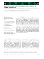
Identification of HuR as a partner of HIV-1 p66 reverse transcriptaseFigure 1
Identification of HuR as a partner of HIV-1 p66 reverse transcriptase. A. A yeast two-hybrid screen was carried out
with HIV-1 RT-p66 as the bait, and a CEMC7 cDNA library as the prey. Amino-acid sequence of HuR and its predicted binding
site to HIV-1 p66. RRM: RNA recognition motif. B. Alignment of the different fragments of HIV-1 interacting with HuR in the
yeast two-hybrid rebound screen, using HuR as the bait and random fragments of HIV-1 YU-2 isolate as the prey. Numbers in
brackets indicate the occurrence of each fragment. C. Mapping of the HuR interaction site on HIV-1 RT bound to a primer-
template. Solvent accessible surface (probe radius 1.4 A) of the protein is represented in two different views (PDB 1D 1HMI)
[53]. The p51 is shown in blue and p66 in pink. The DNA primer-template is represented in grey. The putative HuR binding
site on p66 is represented in red.
A.
B.
RRM1
RRM2
RRM3
HIV p66 binding site
RT INPR
C.
1-MSNGYEDHMA EDCRGDIGRT NLIVNYLPQN MTQDELRSLF SSIGEVESAK LIRDKVAGHS
61-LGYGFVNYVT AKDAERAINT LNGLRLQSKT IKVSYARPSS EVIKDANLYI SGLPRTMTQK
121-DVEDMFSRFG RIINSRVLVD QTTGLSRGVA FIRFDKRSEA EEAITSFNGH KPPGSSEPIT
181-VKFAANPNQN KNVALLSQLY HSPARRFGGP VHHQAQRFRF SPMGVDHMSG LSGVNVPGNA
241-SSGWCIFIYN LGQDADEGIL WQMFGPFGAV TNVKVIRDFN TNKCKGFGFV TMTNYEEAAM
301-AIASLNGYRL GDKILQVSFK TNKSHK
Retrovirology 2008, 5:47 />Page 6 of 14
(page number not for citation purposes)
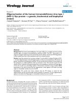
The HIV-1 RT heterodimer interacts directly with GST-HuRFigure 2
The HIV-1 RT heterodimer interacts directly with GST-HuR. A. Coomassie blue staining of the purified RT het-
erodimer. NP: non purified, NF: non fixed, E1–E3: elutions. B. Coomassie blue staining of the purified GST and GST-HuR. NP:
non purified, P: purified. C. Schematic representation of our HTRF assay (adapted from Cisbio Intl.). Europium trisbipyridine
cryptate (TBPEu
3+
) was coupled to anti-GST antibodies, acting as the FRET energy donor, following excitation at 337 nm.
Cross-linked allophycocyanin (XL665) was coupled to anti-His antibodies acting as the FRET energy acceptor and emitting a
sustained signal at 665 nm. D. Serial dilutions of purified GST-HuR or GST alone were incubated with a constant concentration
of RT-His (10 ng/ml), in the presence of constant amounts of anti-His-XL665 and anti-GST-TBPEu
3+
antibodies. The Fret signal
was measured after 24 hours of incubation at 4°C. These results are representative of those obtained in four independent
experiments.
A.
D.
B.
C.
HuR
RT p66
Anti-
GST
G
S
T
H
i
s
Anti-
His
TBPEu
3+
XL665
-50
0
50
100
150
200
250
300
0.1 1 10 100
concentration of GST-proteins (ng/ml)
GST-HuR GST
175
83
62
47.5
32.5
25
NP NF E1 E2 E3
P66-His
p51
RNaseH-His
NP P NP P MW
GST GST-HuR
83
62
47.5
32.5
175
GST
GST-HuR
Retrovirology 2008, 5:47 />Page 7 of 14
(page number not for citation purposes)
Purified GST-HuR and HIV-1 reverse transcriptase interact
together in an in vitro assay
We produced and purified the recombinant proteins, to
confirm the interaction between the two predicted part-
ners in vitro. We used p6H-RT-PR, a vector allowing the
simultaneous production of a C-ter 6xHis-tagged form of
HIV-1 p66 reverse transcriptase together with the HIV-1
protease [16]. The products of C-ter 6xHis-tagged p66
cleavage by HIV-1 protease are untagged p51 and C-ter
6xHis-tagged RNaseH. The simultaneous production of
cleaved and uncleaved p66 favors the formation of a well
folded, fully functional p66/p51 RT heterodimer. Purified
6xHis-proteins were separated by reducing SDS-PAGE and
stained with Coomassie blue to assess their purity (fig.
2A). Recombinant RT production was also checked by
western blotting (data not shown). As expected, anti-RT
monoclonal antibodies detected both RT chains, whereas
anti-6xHis antibodies recognized only p66. As the affinity
between p66 and p51 is strong, the detection of the p51
chain by Coomassie blue staining results from its copre-
cipitation with purified p66-His, rather than its binding to
the affinity beads.
We also inserted the HuR gene into pGEX4T1, to produce
a GST-HuR fusion protein. Purified GST-proteins were
separated by SDS-gel electrophoresis and stained with
Coomassie blue, to assess their purity (fig. 2B).
The purified recombinant p66-His and GST-HuR proteins
were used in an HTRF interaction assay (reviewed in
[26,27]). A schematic representation of the principle
underlying this assay is shown in figure 2C. GST-HuR and
C-ter 6xHis tagged RT-p66 are incubated with anti-GST
antibodies conjugated with a fluorescence energy donor
TBPEu3+, and anti-6His antibodies conjugated with a flu-
orescence energy acceptor XL665. Upon TBPEu
3+
excita-
tion at 337 nm, a fluorescence resonance energy transfer
signal emitted at 665 nm by the XL665 conjugate can be
detected if an interaction occurs between the two recom-
binant proteins. The magnitude of this signal depends on
the respective concentrations of the two interacting pro-
teins.
Serial dilutions of the purified GST-HuR or GST alone
were incubated for 24 hours at 4°C in the presence of con-
stant amounts of antibodies against the 6xHis and GST
tags, and a constant concentration of RT-His (20 ng/ml in
total present in the reaction mixture, corresponding to
about 10 ng/ml of p66-His, as evaluated by densitome-
try). As expected, a bell-shaped curve was obtained (fig.
2D). At lower concentrations, too little GST-HuR was
present in the complex with RT-His and, at higher concen-
trations, some of the anti-GST antibodies were captured
by the excess GST-HuR not associated with RT-His,
thereby diminishing the signal. We obtained a signal with
GST-HuR, but not with GST alone, consistent with a spe-
cific interaction. The two peaks obtained may result from
the interaction of GST-HuR with both the full-length C-ter
6xHis p66 and the C-ter 6xHis RNaseH copurified on
IMAC resin (fig. 2A). These results confirm that the RT-
p66 and HuR recombinant proteins can interact in vitro
and that this interaction is specific, as it does not take
place with GST alone used as a control.
HuR is important for the early steps of the HIV-1
replication cycle
We evaluated the potential role of HuR in the HIV-1 rep-
lication cycle, using RNA interference techniques for gene
silencing. We first monitored the early steps of the viral
replication cycle, using an assay dependent on the correct
entry, reverse transcription and integration of HIV into the
cell genome. Reporter HeLa P4.2 cells (CD4+, LTR-LacZ,
endogenous CXCR4) were independently transfected with
three different siRNAs targeting different regions of the
HuR mRNA, a negative control siRNA or no siRNA. Three
days later, cells were infected with the X4 tropic strain
HIV-1
NL4.3
. An aliquot of the transfected cells was lysed at
the time of infection and HuR silencing was assessed by
western blotting (fig. 3A, upper panel). A 90% decrease in
HuR levels was observed. Cells were fixed 24 hours after
infection, and stained with X-Gal, as previously described
[19]. An aliquot of cells was collected, lysed and analyzed
by western blotting. HuR knockdown was maintained
throughout the experiment, as 90% silencing of HuR was
still observed at the time of fixation (fig. 3A, lower panel).
Tat-activated LTR was used for β-galactosidase production
and the counting of successfully infected cells (fig. 3B).
These results show significant impairment of the infection
of HeLa P4.2 cells treated with the three different siRNAs.
The similar levels of downregulation obtained with all
three siRNAs, despite differences in the regions of the HuR
mRNA targeted, and the similar phenotypic effects of
these three siRNAs in our assay suggest that HuR may be
involved in the early steps of the HIV-1 replication cycle.
We further assessed the importance of HuR in the early
steps of HIV infection, by studying the reverse transcrip-
tion products generated in infected cells in the presence
and absence of HuR. We transfected HeLa cells with
siRNA HuR1 or a control siRNA and infected them 48
hours later with non-replicative HIV-1ΔEnv-luciferase
VSV-G pseudotyped viruses. The viral DNA produced by
reverse transcriptase during this single cycle of infection
were quantified by quantitative real-time PCR, using
primers specific for early products (minus-strand, strong
stop DNA) or late products (full-length DNA), as
described in Materials and Methods. In cells treated with
the HuR1 siRNA, the levels of both transcription products
were much lower than those in cells treated with the con-
trol siRNA (fig. 3C). We also investigated the effects on
Retrovirology 2008, 5:47 />Page 8 of 14
(page number not for citation purposes)
HuR is involved in the early steps of HIV-1 replication cycleFigure 3
HuR is involved in the early steps of HIV-1 replication cycle. A. siRNA silencing of HuR revealed by Western blot.
HeLa P4.2 cells (CD4
+
, LTR-LacZ) were transfected with 30 nM of siRNAs directed against HuR (HuR1, HuR2, HuR3), H
2
O, or
a non targeting siRNA (Ctrl). For each siRNA, five wells were infected with the HIV-1
NL4.3
strain, 72 hours after transfection
with the siRNA. The contents of one well were collected and lysed, to check that HuR expression was silenced at the time of
infection (A, upper panel). B. Effect of HuR silencing on the infection of cells by wild type HIV-1. 24 hours post-infec-
tion, infected cells were counted after the fixation and X-Gal staining of triplicate wells. The contents of one well were col-
lected and lysed, to check that HuR expression was effectively silenced at the time of fixation (A, lower panel). The results
presented are a compilation of six independent experiments, normalized as a function of the results obtained with the control
siRNA (Ctrl). C. Effect of HuR silencing on HIV-1 reverse transcription. HeLa cells were treated with siRNA HuR-1 or a
non targeting siRNA (Ctrl), then infected with HIV-1ΔEnv-luciferase VSV-G pseudotyped viruses at an MOI of 1. Total DNA
was extracted from the infected cells and RT products were quantified by quantitative real-time PCR, 16 hours after infection.
ssRT: minus-strand strong stop DNA, flRT: full-length HIV DNA, HBB, human beta-globin. D. Effect of HuR overexpression
on HIV-1 reverse transcription. As in C, except that the cells were transfected with a vector allowing HuR overexpression
(pCMV-HuR) or an empty vector (pcDNA3), before infection.
B.
C. D.
0
20
40
60
80
100
120
140
160
H2O HuR-1 HuR-2 HuR-3 Ctrl
siRNA
0
50
100
150
200
250
ssRT/HBB flRT/HBB
RT products
pcDNA3 pCMV-HuR
A.
H
2
OHuR-1
HuR-2 CtrlHuR-3
siRNA 30 nM
Actin
HuR
Actin
HuR
72h
Time of
infection
96h
Time of
fixation
0
20
40
60
80
100
120
ssRT/HBB flRT/HBB
RT products
siRNA Ctrl siRNA HuR1
D1:siRNA D2:siRNA D4:infection D5:staining
Retrovirology 2008, 5:47 />Page 9 of 14
(page number not for citation purposes)
reverse transcription of increasing HuR levels, by transfec-
tion with a vector allowing the overexpression of HuR
(pCMV-HuR). In the presence of HuR overproduction, by
contrast with what was observed with HuR silencing, both
early and late products of reverse transcription were more
abundant than in mock-transfected cells (fig. 3D). These
results suggest a potential role for HuR in reverse tran-
scription.
HuR is not required for the post-integration steps of the
HIV-1 replication cycle
As the RNAse H domain found in our yeast two-hybrid
screens is also a part of the Gag-Pol precursor, we investi-
gated whether HuR also affected other steps of the viral
replication cycle. We analyzed the impact of knocking
down HuR levels in the producer cells. HeLa cells were
treated with siRNA HuR1 or control siRNA. The cells were
then transfected with the pNL4.3 provirus, making it pos-
sible to bypass the reverse transcription step. The silencing
of HuR 48 hours after transfection with the HuR1 siRNA
was assessed by western blotting (fig. 4A). No difference
in virus production was detected between cells expressing
and not expressing HuR, as identified by ELISA quantifi-
cation of the Gag CA-p24 antigen in the supernatant (fig.
4B). We investigated whether HuR affected the infectivity
of the viral particles, by using the supernatant of the cells
in fig. 4B to infect HeLa P4.2 cells. No significant differ-
ence was observed in the number of infected cells (= infec-
tious particles) (fig. 4C) or in the infectivity of these
particles normalized on the basis of equal amounts of
released p24 (data not shown). This result is consistent
with the lack of detection of any HuR incorporated into
viral particles produced from cells producing normal
amounts of HuR (fig. 4D). Thus, HuR is unlikely to play a
role in the late steps of the HIV-1 replication cycle, such as
viral protein production, budding and maturation.
Instead, it seems to act only in the target cell, following
viral entry.
Mutagenesis of a putative ARE sequence found in the HIV-
1 genome
HuR has been reported to interact with ARE sequences
found in the RNAs of several distantly related viruses, and
is thought to be involved in their stabilization or expres-
sion [28-32]. We therefore investigated whether a similar
phenomenon was also observed with HIV. We investi-
gated in more detail the possible effects of HuR on the
reverse transcription process, taking into account that
HuR is generally considered to stabilize ARE-containing
mRNAs, by checking HIV-1 RNAs for the presence of such
ARE elements. Alignment analysis identified a sequence in
HIV-1
NL4.3
displaying significant similarity to known ARE
sequences, and particularly to that of the prothymosin
alpha (PTMA) mRNA (data not shown). An identical
"hairpin" structure was predicted for both sequences (data
not shown) [24]. The putative HIV-1 ARE sequence is sit-
uated in the coding sequence of vif and is remarkably con-
served between HIV-1 isolates.
To verify the importance of this putative HIV-1 ARE
sequence, we inserted several silent mutations into the
coding sequence of pNL4.3, to deplete this region of U
residues without affecting the amino acid sequence of vif
(fig. 5A). HEK293T cells were transfected with this viral
construct, to produce the mutated virus (AREmut). This
virus was produced in similar amounts to the WT,
although the viral particles were slightly less infectious
(figure 5B). This mutated virus was used to infect Jurkat
cells, and virus production was followed over time by
quantifying HIV Gag CA-p24 antigen in the cell culture
supernatant. No significant difference was observed
between the replication kinetics of the WT and AREmut
viruses (fig. 5C). These results are consistent with an
absence of a role for the ARE motif or even with the pres-
ence of such a motif in this Vif sequence region of the HIV-
1 RNA, although we cannot rule out the possibility that
such a motif is present elsewhere in the HIV-1 genome.
The role of HuR in HIV-1 reverse transcription does not
seem to be mediated by binding to the HIV-1 RNA
We investigated whether HuR bound to a non typical class
III ARE sequence elsewhere in the HIV-1 RNA, as for c-Jun
[33], by determining the possible association of any HIV-
1 RNA transcript with HuR, in an RNA-immunoprecipita-
tion experiment using anti-HuR antibodies, as previously
described [23]. We used HeLa R7 Neo cells stably infected
with the HIV-1 neo Δenv virus [20], constituting a homo-
geneous population with similar levels of HIV-1 tran-
scripts. RNA-immunoprecipitation experiments were
carried out with anti-HuR antibodies or irrelevant anti-HA
antibodies as the negative control. The immunoprecipi-
tated proteins were detected by western blotting, showing
the specific immunoprecipitation of HuR with anti-HuR
antibodies and not with anti-HA antibodies (fig. 5D). As
a positive control, PTMA mRNA, which is known to bind
to HuR [23,24], was found associated with the immuno-
precipitated HuR protein, as revealed by RT-PCR with the
anti-HuR immunoprecipitate, using primers specific for
PTMA mRNA (fig. 5E). The association of PTMA mRNA
with HuR was specific, as the irrelevant immunoprecipi-
tate obtained with anti-HA antibodies was not enriched in
this RNA. The PTMA mRNAs precipitated with the anti-
HuR antibody were 3.5 times more abundant than the
negative control, the mRNA of the housekeeping gene
gapdh. In contrast, the HIV-1 Gag-Pol transcript was not
greatly enriched compared to PTMA mRNAs, since only a
1.5 folds increase was observed. This difference could be
due to the relative abundance of the two mRNA species as
well as a difference in the affinity of the interaction
between HuR and the different mRNAs.
Retrovirology 2008, 5:47 />Page 10 of 14
(page number not for citation purposes)
HuR is not involved in the late steps of the HIV-1 replication cycleFigure 4
HuR is not involved in the late steps of the HIV-1 replication cycle. HeLa cells were transfected with an siRNA
directed against HuR or a non-targeting siRNA (Ctrl). 24 hrs later, cells were transfected with HIV-1 provirus pNL4.3. A.
Western blot confirming the silencing of HuR 48 hours after transfection with the siRNA. B. Quantification, by ELISA for Gag
CA-p24 antigen, of the virions produced in the supernatant, 30 hours after transfection with pNL4.3. C. The virions produced
in B were used to infect HeLa P4.2 cells (CD4
+
, LTR-LacZ). 24 hours post-infection, infected cells were fixed, stained with X-
Gal and counted. D. 2 × 10
6
HEK293T cells were transfected with HIV-1 provirus pNL4.3. 48 hours later, the cell culture
supernatant was collected, filtered and ultracentrifuged to collect the virions. Producer cells and virion pellets were lysed and
analyzed by western blotting, to check their contents and HuR incorporation.
Ctrl
HuR1
siRNA 30 nM
Actin
HuR
A.
C.
0
200
400
600
800
1000
1200
Ctrl HuR1
siRNA
0
20
40
60
80
100
120
140
160
180
200
Ctrl HuR1
siRNA
D.
B.
HuR
p55
p24
++
Cells Virions
pNL4.3
Retrovirology 2008, 5:47 />Page 11 of 14
(page number not for citation purposes)
HuR does not seem to bind to HIV-1 RNAFigure 5
HuR does not seem to bind to HIV-1 RNA. A. Sequence of HIV WT ARE sequence and silent mutations introduced in
the AREmut virus. B. HEK293T cells were transfected with WT or AREmut pNL4.3 proviruses. Quantification, by ELISA, of
the virions produced in the cell culture supernatant, based on the detection of Gag CA-p24. C. Jurkat cells were infected with
WT or AREmut NL4.3 viruses. Viral replication was monitored by ELISA quantification of HIV Gag CA-p24 antigen in the cell
culture supernatant. D. Immunoprecipitation was carried out with anti-HuR antibodies or unrelated anti-HA antibodies. West-
ern blot analysis, showing the immunoprecipitated protein. E. Coimmunoprecipitated mRNAs were detected by quantitative
RT-PCR, using primers against HIV-1 gag-pol, ptma (as a positive control for HuR binding), and the housekeeping gene gapdh (as
a negative control).
0
50000
100000
150000
200000
250000
300000
350000
400000
02468101214
days post-infection
p24 (ng/ml)
wt AREmut
B. C.
0
2
4
6
8
10
12
14
16
18
20
WT AREmut
infectious particles/ng p24
E.
0
0.5
1
1.5
2
2.5
3
3.5
4
Gag-Pol PTMA GAPDH
mRNA amounts
(fold, relative to anti-HA
anti-HuR anti-HA
D.
Anti-
HuR
Anti-
HA
HuR
*
*
* Non-specific bands
IP
*
IgG
IgG
A.
Retrovirology 2008, 5:47 />Page 12 of 14
(page number not for citation purposes)
Discussion
We performed a yeast two-hybrid screen, using the HIV-1
p66 RT subunit as the bait, to characterize cellular cofac-
tors involved in the reverse transcription step of the HIV-
1 replication cycle. We identified and validated an interac-
tion between HIV-1 RT and the RNA-binding protein
HuR. The HuR interaction site was mapped to the C-ter-
minal part of the p66 RT subunit. This region, belonging
to the RNase H domain, is freely accessible on the RT and
extends to the vicinity of the primer-template. The p66
RT-HuR interaction was confirmed in vitro by an HTRF
assay, suggesting that there was a direct interaction
between HuR and p66 RT. However, since both HuR and
RT are RNA binding proteins it could be possible that their
interaction be mediated by RNA. Indeed, other interac-
tions involving HuR have been shown to be RNA depend-
ent, like the interaction between HuR and APOBEC3G
[34]. HTRF assays conducted in the presence of RNAse did
not allowed us to draw clear conclusions, since upon this
treatment we obtained a slight and inconstant inhibition
of the interaction signal (data not shown). Therefore, this
question remains an open question that will need further
investigations to be solved.
By silencing HuR expression with three different siRNAs
targeting three different sites in the HuR mRNA sequence,
we demonstrated that HuR expression was required for an
optimal HIV-1 replication cycle and for both the early and
late steps of reverse transcription, in particular. The
enhancement of the reverse transcription reaction
observed when HuR was overexpressed is consistent with
these results. The absence of HuR affected wild type HIV-
1, but also a non-replicative HIV-1ΔEnv-luciferase virus
pseudotyped with the VSV-G envelope glycoprotein. As
previously described, the entry pathways of these viruses
are clearly differents [35]. While the wild type virus, bear-
ing gp41/gp120, enters by fusion at the cell surface, VSV-
G targets the virus to endocytosis and fusion in the endo-
somes. Although one cannot exclude this possibility, an
effect of HuR on both entry pathways, in addition to its
effect on reverse transcription, would be very unlikely. The
effect of HuR seemed to be specific to the reverse tran-
scription step in HIV target cells, as HuR silencing in HIV-
1 producer cells had no effect on the production of viral
particles or the infectivity of these newly released particles.
Moreover, no incorporation of HuR into virions was
observed, indicating that the HuR protein affecting reverse
transcription was that present in the target cell, and not
that in the producer cell.
The major role of HuR is to stabilize ARE-containing mes-
senger RNAs (reviewed in [36,37]). This property of HuR
seems to be related to its nucleocytoplasmic shuttling
[8,38,39], following cellular stresses such as heat shock,
exposure to UV light or infection [40]. Indeed, previous
studies have reported the binding of HuR to the RNAs of
various viruses, including HPV-1, HPV-16, Herpesvirus
saimiri and HCV [28,31,32,41,42]. However, no interfer-
ence of HuR with HIV-1 RNA has been reported in previ-
ous studies.
We identified a putative HuR binding motif, based on
recent studies by Lopez de Silanes et al. [24]. We mutated
this motif to disrupt the U-rich region. No effect on HIV
replication was observed. Moreover, RNA-immunoprecip-
itation studies provided no evidence of an association
between the HIV-1 RNA and HuR. This suggests that the
mode of action of HuR in HIV-1 reverse transcription is
based on its interaction with p66 RT rather than its inter-
action with the HIV-1 RNA. HuR plays a major role in sta-
bilizing mRNAs, by binding to ARE elements, but
previous studies have demonstrated protein-protein inter-
actions involving HuR and playing an important role in
the regulation of HuR activity [43-45]. One such interac-
tion – with the RanGTP-binding nuclear transport recep-
tor transportin 2 – was recently highlighted [46]. This
interaction probably occurs in the cytoplasm, mediating
the nuclear import of HuR. This interaction is optimal in
the absence of RNA bound to HuR, suggesting that HuR is
imported into the nucleus only when not bound to
mRNA. The nucleocytoplasmic shuttling of HuR that
seems to be responsible for mRNA stabilization was
observed by Wang et al. upon T-cell activation, following
the engagement of the integrin leukocyte function-associ-
ated molecule-1 (LFA-1) [10]. Several groups have previ-
ously reported the importance of LFA-1 for HIV infection
and transmission to T cells [47-51]. As activated T cells are
the preferred target cells for HIV infection, whereas unac-
tivated T cells are very poorly infected by HIV, it is tempt-
ing to speculate that an absence of nucleocytoplasmic
shuttling of HuR in unactivated T cells is correlated with
the refractory state of these cells to HIV infection, together
with other important recently discovered factors, such as
the low molecular weight form of APOBEC 3G in these
cells [52]. HuR has also been found in stress granules [9],
together with APOBEC 3G [34], and is now considered to
be a marker of these bodies. Is the ability of HuR to bind
to p66 RT, positively affecting the reverse transcription of
HIV-1 related to the nucleocytoplasmic shuttling property
of HuR? Further work will be required to answer this
important question.
In conclusion, we have identified a new cellular partner of
HIV-1 reverse transcriptase: HuR. By modulating HuR lev-
els, we were able to affect the infection of cells by HIV.
However, the mechanism by which HuR influences the
reverse transcription process remains to be elucidated.
Retrovirology 2008, 5:47 />Page 13 of 14
(page number not for citation purposes)
Abbreviations
HIV-1: human immunodeficiency virus type 1; RT: reverse
transcriptase; siRNA: short interfering RNA; MOI: multi-
plicity of infection; GST: glutathione S-transferase; WT:
wild type; HTRF: homogenous time-resolved fluorescence
assay; VSV-G: vesicular stomatitis virus glycoprotein.
Authors' contributions
JL designed and performed the siRNA experiments for
analysis of infection, viral production and infectivity, pro-
duced the RT proteins, performed the HTRF experiments,
and wrote the manuscript, PMP constructed and pro-
duced GST-HuR protein and the ARE-mutant, TB designed
the HTRF experiments, EE mapped the HuR binding site
on RT, JCR performed the yeast two-hybrid screening, RB
conceived the study, participated to data analysis and con-
tributed to the writing of the manuscript, LXL designed
and performed the siRNA experiments for analysis of RT
products by qPCR, analysed HuR incorporation into viral
particles and its interaction with HIV-1 mRNA, and con-
tributed to the writing of the manuscript. All authors read
and approved the final manuscript.
Acknowledgements
We thank G. Maga for kindly providing the plasmid encoding the 6xHis-
tagged recombinant HIV-1 RT, C. Berlioz-Torrent and S. Emiliani for helpful
discussions and support, and L. Boutin for technical assistance.
J.L was supported by doctoral fellowships from ANRS and FRM. L.L was
supported by a postdoctoral fellowship from EC project TRIoH (LSHB-CT-
2003-503480). This work was supported by grants from the ANRS, the
FRM, SIDACTION, ANR, and by grants from the European 6
th
Framework
Program for Research and Development via the Integrated Project TRIOH
n° LSHB-CT-2003-503.
References
1. Telesnitsky A, Goff SP: Reverse Transcriptase and the Genera-
tion of Retroviral DNA. In Retroviruses Edited by: Coffin JM,
Hughes SH, Varmus HE. Plainview, NY 11803: Cold Spring Harbor
Press; 1997.
2. di Marzo Veronese F, Copeland TD, DeVico AL, Rahman R, Oroszlan
S, Gallo RC, Sarngadharan MG: Characterization of highly immu-
nogenic p66/p51 as the reverse transcriptase of HTLV-III/
LAV. Science 1986, 231:1289-1291.
3. Nisole S, Saib A: Early steps of retrovirus replicative cycle. Ret-
rovirology 2004, 1:9.
4. Hooker CW, Harrich D: The first strand transfer reaction of
HIV-1 reverse transcription is more efficient in infected cells
than in cell-free natural endogenous reverse transcription
reactions. J Clin Virol 2003, 26:229-238.
5. Warrilow D, Meredith L, Davis A, Burrell C, Li P, Harrich D: Cell
Factors Stimulate HIV-1 Reverse Transcription In Vitro. J
Virol 2008.
6. Hottiger M, Gramatikoff K, Georgiev O, Chaponnier C, Schaffner W,
Hubscher U: The large subunit of HIV-1 reverse transcriptase
interacts with beta-actin. Nucleic Acids Res 1995, 23:736-741.
7. Orlova M, Yueh A, Leung J, Goff SP: Reverse transcriptase of
Moloney murine leukemia virus binds to eukaryotic release
factor 1 to modulate suppression of translational termina-
tion. Cell 2003, 115:319-331.
8. Fan XC, Steitz JA: Overexpression of HuR, a nuclear-cytoplas-
mic shuttling protein, increases the in vivo stability of ARE-
containing mRNAs. Embo J 1998, 17:3448-3460.
9. Kedersha N, Anderson P: Stress granules: sites of mRNA triage
that regulate mRNA stability and translatability. Biochem Soc
Trans 2002, 30:963-969.
10. Wang JG, Collinge M, Ramgolam V, Ayalon O, Fan XC, Pardi R,
Bender JR: LFA-1-dependent HuR nuclear export and
cytokine mRNA stabilization in T cell activation. J Immunol
2006, 176:2105-2113.
11. Wang W, Caldwell MC, Lin S, Furneaux H, Gorospe M: HuR regu-
lates cyclin A and cyclin B1 mRNA stability during cell pro-
liferation. Embo J 2000, 19:2340-2350.
12. Atasoy U, Watson J, Patel D, Keene JD: ELAV protein HuA (HuR)
can redistribute between nucleus and cytoplasm and is
upregulated during serum stimulation and T cell activation.
J Cell Sci 1998, 111(Pt 21):3145-3156.
13. Emiliani S, Mousnier A, Busschots K, Maroun M, Van Maele B, Tempe
D, Vandekerckhove L, Moisant F, Ben-Slama L, Witvrouw M, et al.:
Integrase mutants defective for interaction with LEDGF/p75
are impaired in chromosome tethering and HIV-1 replica-
tion. J Biol Chem 2005, 280:25517-25523.
14. Fromont-Racine M, Rain JC, Legrain P: Building protein-protein
networks by two-hybrid mating strategy. Methods Enzymol
2002, 350:513-524.
15. Vojtek AB, Hollenberg SM: Ras-Raf interaction: two-hybrid anal-
ysis. Methods Enzymol 1995, 255:331-342.
16. Maga G, Amacker M, Ruel N, Hubscher U, Spadari S: Resistance to
nevirapine of HIV-1 reverse transcriptase mutants: loss of
stabilizing interactions and thermodynamic or steric barri-
ers are induced by different single amino acid substitutions.
J Mol Biol 1997, 274:738-747.
17. Adachi A, Gendelman HE, Koenig S, Folks T, Willey R, Rabson A, Mar-
tin MA: Production of acquired immunodeficiency syndrome-
associated retrovirus in human and nonhuman cells trans-
fected with an infectious molecular clone. J Virol 1986,
59:284-291.
18. Ho SN, Hunt HD, Horton RM, Pullen JK, Pease LR: Site-directed
mutagenesis by overlap extension using the polymerase
chain reaction. Gene 1989, 77:51-59.
19. Clavel F, Charneau P: Fusion from without directed by human
immunodeficiency virus particles. J Virol 1994, 68:1179-1185.
20. Brussel A, Sonigo P: Analysis of early human immunodeficiency
virus type 1 DNA synthesis by use of a new sensitive assay for
quantifying integrated provirus. J Virol 2003, 77:10119-10124.
21. Maroun M, Delelis O, Coadou G, Bader T, Segeral E, Mbemba G, Petit
C, Sonigo P, Rain JC, Mouscadet JF, et al.
: Inhibition of early steps
of HIV-1 replication by SNF5/Ini1. J Biol Chem 2006,
281:22736-22743.
22. Zuker M: Mfold web server for nucleic acid folding and hybrid-
ization prediction. Nucleic Acids Res 2003, 31:3406-3415.
23. Lal A, Kawai T, Yang X, Mazan-Mamczarz K, Gorospe M: Antiapop-
totic function of RNA-binding protein HuR effected through
prothymosin alpha. Embo J 2005, 24:1852-1862.
24. Lopez de Silanes I, Zhan M, Lal A, Yang X, Gorospe M: Identification
of a target RNA motif for RNA-binding protein HuR. Proc Natl
Acad Sci USA 2004, 101:2987-2992.
25. Fromont-Racine M, Rain JC, Legrain P: Toward a functional anal-
ysis of the yeast genome through exhaustive two-hybrid
screens. Nat Genet 1997, 16:277-282.
26. Bazin H, Preaudat M, Trinquet E, Mathis G: Homogeneous time
resolved fluorescence resonance energy transfer using rare
earth cryptates as a tool for probing molecular interactions
in biology. Spectrochim Acta A Mol Biomol Spectrosc 2001,
57:2197-2211.
27. Bazin H, Trinquet E, Mathis G: Time resolved amplification of
cryptate emission: a versatile technology to trace biomo-
lecular interactions. J Biotechnol 2002, 82:233-250.
28. Cook HL, Mischo HE, Steitz JA: The Herpesvirus saimiri small
nuclear RNAs recruit AU-rich element-binding proteins but
do not alter host AU-rich element-containing mRNA levels
in virally transformed T cells. Mol Cell Biol 2004, 24:4522-4533.
29. Esclatine A, Taddeo B, Roizman B: Herpes simplex virus 1
induces cytoplasmic accumulation of TIA-1/TIAR and both
synthesis and cytoplasmic accumulation of tristetraprolin,
two cellular proteins that bind and destabilize AU-rich
RNAs. J Virol 2004, 78:8582-8592.
30. McInerney GM, Kedersha NL, Kaufman RJ, Anderson P, Liljestrom P:
Importance of eIF2alpha phosphorylation and stress granule
Publish with BioMed Central and every
scientist can read your work free of charge
"BioMed Central will be the most significant development for
disseminating the results of biomedical research in our lifetime."
Sir Paul Nurse, Cancer Research UK
Your research papers will be:
available free of charge to the entire biomedical community
peer reviewed and published immediately upon acceptance
cited in PubMed and archived on PubMed Central
yours — you keep the copyright
Submit your manuscript here:
/>BioMedcentral
Retrovirology 2008, 5:47 />Page 14 of 14
(page number not for citation purposes)
assembly in alphavirus translation regulation. Mol Biol Cell
2005, 16:3753-3763.
31. Spangberg K, Wiklund L, Schwartz S: HuR, a protein implicated in
oncogene and growth factor mRNA decay, binds to the 3'
ends of hepatitis C virus RNA of both polarities. Virology 2000,
274:378-390.
32. Sokolowski M, Furneaux H, Schwartz S: The inhibitory activity of
the AU-rich RNA element in the human papillomavirus type
1 late 3' untranslated region correlates with its affinity for
the elav-like HuR protein. J Virol 1999, 73:1080-1091.
33. Chen CY, Shyu AB: Selective degradation of early-response-
gene mRNAs: functional analyses of sequence features of the
AU-rich elements. Mol Cell Biol 1994, 14:8471-8482.
34. Gallois-Montbrun S, Kramer B, Swanson CM, Byers H, Lynham S,
Ward M, Malim MH: Antiviral protein APOBEC3G localizes to
ribonucleoprotein complexes found in P bodies and stress
granules. J Virol 2007, 81:2165-2178.
35. Aiken C: Pseudotyping human immunodeficiency virus type 1
(HIV-1) by the glycoprotein of vesicular stomatitis virus tar-
gets HIV-1 entry to an endocytic pathway and suppresses
both the requirement for Nef and the sensitivity to
cyclosporin A. J Virol 1997, 71:5871-5877.
36. Brennan CM, Steitz JA: HuR and mRNA stability. Cell Mol Life Sci
2001, 58:266-277.
37. Mitchell P, Tollervey D: mRNA stability in eukaryotes. Curr Opin
Genet Dev 2000, 10:193-198.
38. Fan XC, Steitz JA: HNS, a nuclear-cytoplasmic shuttling
sequence in HuR. Proc Natl Acad Sci USA 1998, 95:15293-15298.
39. Gallouzi IE, Steitz JA: Delineation of mRNA export pathways by
the use of cell-permeable peptides. Science 2001,
294:1895-1901.
40. Wang W, Furneaux H, Cheng H, Caldwell MC, Hutter D, Liu Y, Hol-
brook N, Gorospe M:
HuR regulates p21 mRNA stabilization
by UV light. Mol Cell Biol 2000, 20:760-769.
41. Carlsson A, Schwartz S: Inhibitory activity of the human papil-
lomavirus type 1 AU-rich element correlates inversely with
the levels of the elav-like HuR protein in the cell cytoplasm.
Arch Virol 2000, 145:491-503.
42. Koffa MD, Graham SV, Takagaki Y, Manley JL, Clements JB: The
human papillomavirus type 16 negative regulatory RNA ele-
ment interacts with three proteins that act at different post-
transcriptional levels. Proc Natl Acad Sci USA 2000, 97:4677-4682.
43. Brennan CM, Gallouzi IE, Steitz JA: Protein ligands to HuR mod-
ulate its interaction with target mRNAs in vivo. J Cell Biol 2000,
151:1-14.
44. Malek SN, Katumuluwa AI, Pasternack GR: Identification and pre-
liminary characterization of two related proliferation-associ-
ated nuclear phosphoproteins. J Biol Chem 1990,
265:13400-13409.
45. Mencinger M, Panagopoulos I, Contreras JA, Mitelman F, Aman P:
Expression analysis and chromosomal mapping of a novel
human gene, APRIL, encoding an acidic protein rich in
leucines. Biochim Biophys Acta 1998, 1395:176-180.
46. Guttinger S, Muhlhausser P, Koller-Eichhorn R, Brennecke J, Kutay U:
Transportin2 functions as importin and mediates nuclear
import of HuR. Proc Natl Acad Sci USA 2004, 101:2918-2923.
47. Hioe CE, Chien PC Jr, Lu C, Springer TA, Wang XH, Bandres J, Tuen
M: LFA-1 expression on target cells promotes human immu-
nodeficiency virus type 1 infection and transmission. J Virol
2001, 75:1077-1082.
48. Groot F, Kuijpers TW, Berkhout B, de Jong EC: Dendritic cell-
mediated HIV-1 transmission to T cells of LAD-1 patients is
impaired due to the defect in LFA-1. Retrovirology 2006, 3:75.
49. Tardif MR, Tremblay MJ: LFA-1 is a key determinant for prefer-
ential infection of memory CD4+ T cells by human immuno-
deficiency virus type 1. J Virol 2005, 79:13714-13724.
50. Tardif MR, Tremblay MJ: Regulation of LFA-1 activity through
cytoskeleton remodeling and signaling components modu-
lates the efficiency of HIV type-1 entry in activated CD4+ T
lymphocytes. J Immunol 2005, 175:926-935.
51. Fortin JF, Cantin R, Tremblay MJ: T cells expressing activated
LFA-1 are more susceptible to infection with human immu-
nodeficiency virus type 1 particles bearing host-encoded
ICAM-1. J Virol 1998, 72:2105-2112.
52. Chiu YL, Soros VB, Kreisberg JF, Stopak K, Yonemoto W, Greene
WC: Cellular APOBEC3G restricts HIV-1 infection in resting
CD4+ T cells. Nature 2005, 435:108-114.
53. Ding J, Das K, Hsiou Y, Sarafianos SG, Clark AD Jr, Jacobo-Molina A,
Tantillo C, Hughes SH, Arnold E: Structure and functional impli-
cations of the polymerase active site region in a complex of
HIV-1 RT with a double-stranded DNA template-primer and
an antibody Fab fragment at 2.8 A resolution. J Mol Biol 1998,
284:1095-1111.