Báo cáo khoa học: UXT interacts with the transcriptional repressor protein EVI1 and suppresses cell transformation ppt
Bạn đang xem bản rút gọn của tài liệu. Xem và tải ngay bản đầy đủ của tài liệu tại đây (641.08 KB, 12 trang )
UXT interacts with the transcriptional repressor protein
EVI1 and suppresses cell transformation
Roger McGilvray
1
, Mark Walker
2
and Chris Bartholomew
1
1 Department of Biological & Biomedical Sciences, Glasgow Caledonian University, UK
2 CRUK Beatson Laboratories, Beatson Institute for Cancer Research, Glasgow, UK
EVI1 is a member of the PR domain family of pro-
teins [1], which include PRDM1 (PRDI-BF1) [2],
PRDM2 (RIZ) [3] and PRDM16 (MEL1) [4],
and share structural similarities with N-terminal PR
domains, functional similarities with transcriptional
repressor activities, have multiple cys2 ⁄ his2 zinc-finger
motifs, and roles in cell differentiation and tumorigen-
esis. The complex phenotype observed in Evi1 knock-
out mice suggests that it has a role in a range of
biological processes including general cell proliferation,
vascularization and cell-specific developmental signal-
ling [5]. The pleiotropic phenotype reflects Evi1’s func-
tions as a transcription factor [6] as well as its ability
to interfere with several signalling pathways through
interactions with Smad [7,8] and JNK [9] proteins.
The Evi1 transcriptional repressor protein is essen-
tial for normal development [5] and when inappropri-
ately expressed participates in the progression of a
subset of leukaemias and myelodysplasias [10]. In vitro
studies have shown that a number of biological prop-
erties can be attributed to the Evi1 protein including:
(a) deregulation of cell proliferation [11]; (b) inhibition
of transforming growth factor-b, bone morphogenic
protein and activin signalling [7,8]; and (c) inhibition
of stress-induced apoptosis [9]. These activities require
the DNA-binding domains ZF1 ⁄ ZF2 [12,13] and a
Keywords
ART-27; cell transformation; EVI1; leukemia;
ubiquitously expressed transcript
Correspondence
C. Bartholomew, Glasgow Caledonian
University, Department of Biological &
Biomedical Sciences, City Campus,
Cowcaddens Road, Glasgow G4 OBA, UK
Fax: +44 (0)141 331 3208
Tel: +44 (0)141 331 3213
E-mail:
(Received 30 June 2006, revised 10 May
2007, accepted 8 June 2007)
doi:10.1111/j.1742-4658.2007.05928.x
The EVI1 transcriptional repressor is critical to the normal development of
a variety of tissues and participates in the progression of acute myeloid leu-
kaemias. The repressor domain (Rp) was used to screen an adult human
kidney yeast two-hybrid library and a novel binding partner designated
ubiquitously expressed transcript (UXT) was isolated. Enforced expression
of UXT in Evi1-expressing Rat1 fibroblasts suppresses cell transformation
and UXT may therefore be a negative regulator of Evi1 biological activity.
The Rp-binding site for UXT was determined and non-UXT-binding Evi1
mutants (Evi1D706–707) were developed which retain the ability to bind
the corepressor mCtBP2. Evi1D706–707 transforms Rat1 fibroblasts, show-
ing that the interaction is not essential for Evi1-mediated cell transforma-
tion. However, Evi1D706–707 produces an increased proportion of large
colonies relative to wild-type, showing that endogenous UXT has an inhibi-
tory effect on Evi1 biological activity. Exogenous UXT still suppresses
Evi1D706–707-mediated cell transformation, indicating that it inhibits cell
proliferation and ⁄ or survival by both Evi1-dependent and Evi1-independ-
ent mechanisms. These observations are consistent with the growth-
suppressive function attributed to UXT in human prostate cancer. Our
results show that UXT suppresses cell transformation and might mediate
this function by interaction and inhibition of the biological activity of cell
proliferation and survival stimulatory factors like Evi1.
Abbreviations
CtBP, C-terminal binding protein; DBD, DNA-binding domain; GST, glutathione S-transferase; Rp, repressor domain; SD, synthetic dropout
medium; UXT, ubiquitously expressed transcript.
3960 FEBS Journal 274 (2007) 3960–3971 ª 2007 The Authors Journal compilation ª 2007 FEBS
200-amino acid transcriptional repressor domain
(Rp) [6].
Rp is critical to the biological activity of Evi1. We,
and others, have shown that Rp binds the C-terminal
binding protein (CtBP) family of corepressor proteins
that interact via two conserved PLDLS-like motifs
[14,15]. Critically, the ability of Evi1 to repress tran-
scription, deregulate cell growth [14] and inhibit trans-
forming growth factor-b, bone morphogenic protein
and activin signalling [7,8,15], all rely on the recruit-
ment of CtBP.
Several lines of evidence suggest that Rp is involved
in additional protein interactions that may also con-
tribute to Evi1’s broad spectrum of biological activit-
ies. C-terminal deletion mutants of Rp, which retain
CtBP-binding sites, create more effective repressors [6],
suggesting that this activity is regulated. Naturally
occurring splice variants in both human and murine
Evi1 exist that insert nine amino acids (FP ⁄
QLPDQRTW) into Rp [16,17], which may either
create or disrupt novel or pre-existing interactions,
respectively. Inspection of aligned human and murine
Rp primary amino-acid sequences with the correspond-
ing region of the related MEL1 protein [4] reveal signi-
ficant stretches of conservation in addition to the
CtBP-binding sites, suggesting additional common
activities might have been conserved during evolution
for these two proteins.
To date, several Evi1-binding proteins have been
identified using a candidate protein approach. It
remains possible that Evi1 interacts with as yet
unknown cellular proteins responsible for regulating
biological activity. To investigate this, we screened a
yeast two-hybrid library to identify new Rp-binding
partners.
Results
Isolation of a new Evi1 Rp-domain binding
protein
Rp was subcloned from pGBT9Rp [14] into the kana-
mycin-selectable yeast vector pKGI [18] to create
pKGIRp (see Experimental procedures). As expected,
pKGIRp produces a GAL4 DNA-binding domain
(DBD) ⁄ Rp protein which interacts with mCtBP2 in
yeast AH109 cells, confirming its suitability for use
in screening a yeast two-hybrid library (Fig. 1A–C;
pKGIRp, pGAD10mCtBP2).
In total, 1 · 10
6
independent clones from a human
adult kidney yeast two-hybrid library (Clontech) were
screened with pKGIRp in AH109 cells. Initially, 37
potentially interacting clones were identified of which
only 17 grew under more stringent conditions. Target
plasmid DNAs recovered from these clones were intro-
duced into AH109 cells with pKGIRp and growth was
reassessed. Six recombinant plasmids contained genes
encoding putative Rp-interacting proteins (Table 1,
secondary screen). Sequencing of the plasmid DNA
inserts revealed that five were HuCtBP2 (A6, A7, A10,
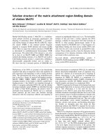
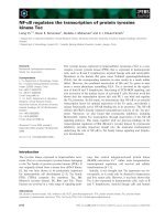
Fig. 1. Interaction of Rp and A1 in yeast
cells. (A,D) Yeast AH109 cells (Clontech)
were transformed with the combination of
plasmids shown. The growth of single yeast
colonies containing these plasmids are
shown in (B) and (E) on SD lacking
L-histi-
dine and adenine and (C) on SD plus
L-histi-
dine and adenine.
R. McGilvray et al. Evi1 binding proteins
FEBS Journal 274 (2007) 3960–3971 ª 2007 The Authors Journal compilation ª 2007 FEBS 3961
A14 and A37). The remaining clone, A1, contained a
novel gene. This gene was isolated repeatedly from
additional independent library screens (data not
shown) and together with HuCtBP2 represents the
only Rp-interacting proteins identified in the human
adult kidney library.
AH109 cells only grow under stringent conditions
when they contain both Rp and A1 expressed as
GAL4DBD and GAL4AD fusion proteins, respectively
(Fig. 1A–C; pKGIRp, pGAD10 A1), showing that
these proteins interact. To see if A1 and Rp interact
irrespective of their fusion partners, GAL4AD or
GAL4DBD, a domain swap was undertaken. A1 and
Rp were inserted into pGBT9 (pGBT9 A1) and
pGADT7 (pGADT7 Rp), respectively, and shown to
still interact in AH109 cells (Fig. 1A–C; pGBT9 A1,
pGADT7 Rp). The yeast two-hybrid assay also
revealed that A1 can homodimerize in AH109 cells
(Fig. 1D,E; pGBT9 A1, pGAD10 A1). Furthermore,
the interaction of A1 with Rp is specific because A1
does not interact with laminin, p53 (Fig. 1A–C;
pGBKT7Lam, pGAD10 A1 and pGBKT753,
pGAD10 A1) or mCtBP2 (Fig. 1D,E; pGBT9 A1 ⁄
pGAD10mCtBP2).
A1 is identical to a novel gene called UXT
The sequence of A1 is shown in Fig. 2A. It consists of
a 546-nucleotide cDNA, which includes a partial
poly(A) tail. There is an unbroken reading frame of
162 amino acids that is continuous with the vector
GAL4DBD and terminates with TGA (Fig. 2A) at
nucleotide 487. Inspection of the sequence shows that
the first ATG (Fig. 2A) fits the Kozak consensus for
translation initiation [19], suggesting that the gene nor-
mally encodes a putative protein of 157 amino acids
with a predicted molecular mass of 18.2 kDa. Interro-
gation of the NCBI nucleotide database revealed the
identity of A1 with a novel gene designated UXT
(AF092737) that encodes a 157-amino-acid protein
(Fig. 2A). A1 is subsequently referred to as UXT.
The tissue distribution of UXT was examined using
northern blot analysis. This shows that UXT produces
an abundant transcript of 750 bp in all tissues exam-
ined, with the highest expression levels in heart, skel-
etal muscle, pancreas, peripheral blood leukocytes,
thyroid and lymph node (Fig. 2B). The transcript size
is consistent with A1 being almost full length, allowing
for an additional 39 5¢ nucleotides described for UXT
and a 200 nucleotide poly(A) tail.
The tissue distribution of UXT shows that it is
expressed in the same tissues as Evi1, including lung,
kidney, ovary and heart. RT-PCR was performed to
confirm that both genes are expressed simultaneously
in the same cells. Evi1 is abundantly expressed in the
leukaemia cell line DA-3 [20] and primary mouse
embryo fibroblasts (MEFS), but not in lymphoma-
derived monocytic U937 cells (Fig. 2C). UXT is
expressed in all cell types examined (Fig. 2C), confirm-
ing that transcripts for both genes coexist in cells in
which Evi1 is expressed, including leukaemia cells
where Evi1 has been activated and in cells where Evi1
is normally expressed such as fibroblasts.
UXT binds full-length Evi1
UXT ⁄ Evi1 binding was confirmed using a glutathi-
one S-transferase (GST)-pull down assay. GST–Rp and
GST–UXT fusion proteins were expressed and purified
from Escherichia coli strain pLysS cells using bacterial
expression vectors (see Experimental procedures).
35
S-Methionine-labelled in vitro-translated UXT and
Evi1 proteins were produced using the expression
vectors pCDNA3–UXT (Experimental procedures) and
pRC ⁄ CMV FL [6], respectively (Fig. 3A). GST pull-
down assays were performed with combinations of
Table 1. Isolation of Rp-interacting proteins in yeast. The number of colonies obtained (clone A1 to A37), their growth on various selective
media (SM), and their production of a- and b-galactosidase from both the primary and secondary library screening are shown. ND, not done.
Assay Primary screen Secondary screen
Growth on SM -Trp ⁄ -Leu ND A1, A6, A7, A10, A11, A14, A15, A17
A20, A24, A25, A26, A29, A32, A33
Growth on SM -Trp ⁄ -Leu ⁄ -His Clones A1 to A37 ND
Growth on SM -Trp ⁄ -Leu ⁄ -His ⁄ -Ala A1, A6, A7, A10, A11, A14, A15, A17
A1, A6, A7, A10, A14, A37
A20, A24, A25, A26, A29, A32, A33
A36, A37
b-galactosidase activity ND A1, A6, A7, A10, A14, A37
a-galactosidase activity ND A1, A6, A7, A10, A14, A37
Evi1 binding proteins R. McGilvray et al.
3962 FEBS Journal 274 (2007) 3960–3971 ª 2007 The Authors Journal compilation ª 2007 FEBS
Fig. 2. (A) Sequence of clone A1. The nuc-
leotide sequence of clone A1 is shown.
Below is the sequence of UXT (lower case)
and regions of identity are indicated by
Below the nucleotide sequence is shown
the primary amino acid sequence using the
single letter code. The bold nucleotide and
amino acid sequence is partial pGBT9 GAL4
DBD vector nucleotide and amino acid
sequence. Bold boxed nucleotide sequences
show the predicted translation initiation site
for UXT ⁄ clone A1 and the translation termin-
ation site. The boxed amino acid sequence
represents translation of predicted 5¢ UXT
noncoding leader sequence that maintains
the reading frame of GAL4 AD and UXT. (B)
Northern blot analysis of UXT expression.
Human MTN
TM
blots (Human, Human II and
Human III; Clontech) containing 2 lg per
lane of the indicated poly(A
+
) RNAs were
hybridized to a
32
P-labelled UXT (A1) probe.
Filters were washed stringently, 0.1· NaCl ⁄
Cit, 0.1% SDS, 65 °C and bands were visu-
alized by autoradiography. (C) RT-PCR analy-
sis of total cellular RNA derived from the
indicated cells using human ⁄ mouse-specific
Evi1 (HME1 ⁄ HME2), Uxt (HMUXT5 ⁄
HMUXT3) and Gapdh (GAPDH5¢⁄GAPDH3¢)
primers. The expected size fragments for all
three genes: Evi1 (467 bp); Uxt (278 bp) and
Gapdh (451 bp) are indicated by arrows.
M indicates the 1kb hyperladder (Bioline).
R. McGilvray et al. Evi1 binding proteins
FEBS Journal 274 (2007) 3960–3971 ª 2007 The Authors Journal compilation ª 2007 FEBS 3963
bacterially expressed and
35
S-methionine-labelled pro-
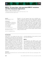
teins and the results are shown in Fig. 3. These con-
firm that UXT binds Rp (Fig. 3B, lane 5) and
furthermore also interacts with full-length Evi1
(Fig. 3B, lane 7). Formation of the complexes are spe-
cific as they do not occur with GST alone (Fig. 5B,
lanes 2 and 3).
The UXT ⁄ Evi1 interaction was also confirmed in
mammalian cells using coimmunoprecipitation. UXT
was inserted inframe with three copies of the HA epi-
tope tag into the expression vector pcDNA3 to create
pcDNA33HAUXT (Experimental procedures). Cells
from the human embryonic kidney cell line BOSC-23
were transiently transfected with various combinations
of the C-myc epitope-tagged Evi1-encoding vector
pcDNA3EVI1myc (A. Coyle & C. Bartholomew, unpub-
lished) and pcDNA33HAUXT, and portions of the
cell extracts were examined either directly by western
blot analysis or immunoprecipitated prior to western
blotting. Western blot analysis of whole-cell extracts
with either HA-specific a-12CA5 or C-myc-specific
a-9E10 shows the expected size epitope-tagged 21 kDa
UXT (HAUXT) and 145 kDa Evi1 (EVI1myc)
proteins, respectively, in cells transfected with the
corresponding expression vectors (Fig. 3C). Immuno-
precipitation of cell extracts and western blot analysis
with a-myc reveals EVI1myc in cells transfected with
pcDNA3EVI1myc, as expected (Fig. 3; IP a-9E10). In
addition, western blot analysis of a-myc-immunopre-
cipitated cell extracts with a-HA shows HAUXT only
in those extracts that also contain EVI1myc, confirm-
ing that these two proteins form a complex. Examina-
tion of the UXT–Evi1 interaction at the endogenous
level using the same method must await new reagents
to be developed.
UXT suppresses Evi1-mediated transformation
Next we investigated whether UXT has an effect on
Evi1 biological activity by examining the impact of
Fig. 3. (A) SDS–PAGE of in vitro translated products showing
35
S-labelled UXT (lane 1) and Evi1 (lane 2). (B) Analysis of GST pull-down
assays by SDS–PAGE. White box indicates bacterially derived GST protein, stippled box represents in vitro translated UXT and bacterially
derived GST–UXT fusion protein. Grey box represents bacterially derived GST–Rp fusion protein. Evi1 zinc-finger domains and repressor
domains are indicated by black and grey boxes, respectively. Hatched box shows acidic domain. (C) Interaction of Evi1 and UXT (A1) in
mammalian cells. Various combinations of pcDNA3Evi1myc (4 lg) and pcDNA33HAUXT (1 lg), indicated by +, were transiently transfected
into BOSC-23 cells as described previously [2]. One-tenth of whole-cell extracts were utilized directly for western blot analysis and the
remainder was immunoprecipitated with a-myc (Santa Cruz Biotechnology, IP a-9E10) prior to western blot analysis. Whole-cell extracts and
immunoprecipitation a-9E10 extracts were resolved by SDS–PAGE (10%) and sequentially probed with a-haemagglutinin (Boehringer Mann-
heim, Mannheim, Germany; WB a-12CA5) and a-myc (WB a-9E10) mAb. Proteins were visualized by ECL
TM
(Amersham Pharmacia Biotech).
Protein size was estimated by comparison with Full Range Rainbow
TM
molecular mass markers (Amersham Pharmacia Biotech, not shown).
Evi1myc and HAUXT fusion proteins are indicated by arrows.
Evi1 binding proteins R. McGilvray et al.
3964 FEBS Journal 274 (2007) 3960–3971 ª 2007 The Authors Journal compilation ª 2007 FEBS
UXT expression on Rat1 and RatFL (Evi1-trans-
formed cells) cell transformation. The retroviral
expression vector p50MHAUXTzeo, containing UXT
fused inframe to a HA tag, was created. Recombinant
retrovirus produced in BOSC-23 cells was used to
infect Rat1 and RatFL cells and cell populations
expressing UXT were selected. Neither UXT expres-
sion in Rat1 cells nor empty vector controls were
transforming (Fig. 4A, lanes 1,3,4), whereas Evi1 was
(Fig. 4A, lane 2). However, enforced expression of
UXT reduced the number of transformed colonies pro-
duced by RatFL cells by 50% (Fig. 4A, lane 5). The
empty vector control had no effect in RatFL cells
(Fig. 4A, lane 5). Cell extracts prepared from the var-
ious cell populations confirmed that they each produce
the expected UXT HA-tagged fusion protein (Fig. 4B).
The effect of UXT on Evi1 transcriptional repressor
activity was also examined but no significant changes
were observed (data not shown).
An Evi1 Rp domain mutant lacking UXT-binding
activity enhances Rat1 transformation activity
The UXT-binding region of Rp was determined using
a series of deletion mutants (Experimental procedures)
using the yeast two-hybrid assay. Binding was retained
when the Rp fragment (514–724) was deleted from the
N-terminus to amino acid 634 (fragment 634–724), but
lost upon C-terminal deletions between 715 and 695
(data not shown). A series of refined deletion mutants
were created from the C-terminus of the 634–715 frag-
ment to determine the minimum deletion required to
lose UXT-binding activity. Yeast two-hybrid assays
revealed that Rp UXT binding is lost upon deletion
of amino acids 706–707 (Fig. 5A–C, quadrant 7–9).
Western blot analysis with a-GAL4DBD confirmed
expression of equally abundant correct size proteins
(fragments were subcloned from pGBT9 to pGBKT7
for this purpose; data not shown).
The ability of an Rp-deletion mutant lacking only
amino acids 706 and 707 (Rp D706–707) to bind UXT
and mCtBP2 was examined using the yeast two-hybrid
assay. Results confirm that this mutant is unable to
bind UXT (Fig. 5D–F, quadrant 2) but retains the
ability to bind mCtBP2 (Fig. 5D–F, quadrant 5).
The transforming activity of Evi1 containing the
UXT-binding mutant Rp domain was investigated in
Rat1 cells. The Rp domain of the previously described
vector, p50MRpWTneo, which has identical trans-
forming activity to p50M4.6neo [14], was substituted
for RpD706–707 to create p50MRpD706–707neo and
populations of Rat1 cells expressing this gene were
selected and tested for transformation using a soft agar
colony assay. The results show that Evi1D706–707 gen-
erates the same number of transformed Rat1 cell col-
onies as wild-type Evi1 (data not shown). However,
the mutant protein produces a higher proportion of
larger colonies (Fig. 6A). Figure 6B shows the percent-
age of total colonies that are > 0.3 mm in diameter,
generated from populations of Rat1 cells expressing
either WT or mutant forms of Evi1. Approximately
14% of colonies generated by Evi1D706–707 are
> 0.3 mm in size, whereas only 3–4% of Evi1 trans-
formed colonies achieve these dimensions.
To see if enforced expression of UXT inhibits the
transforming activity of Evi1D706–707, the colony-
Fig. 4. (A) Colony formation of Rat1 and RatFL cell populations
infected with the indicated retroviral vectors. Numbers were deter-
mined for colonies > 0.1 mm derived from plating 10
3
cells. Error
bars indicate the average number of colonies observed from three
independent assays. Schematic representation of Evi1 and UXT
proteins are as described in the legend to Fig. 3. The HA epitope
tag is shown as a striped box. (B) Western blot analysis with
a-haemagglutinin (as described in Fig. 3) of whole-cell extracts
derived from Rat1 cells (1), p50MHAUXTzeo infected BOSC 23 (2),
Rat1 (3) and RatFL (4) cells. The HAUXT fusion proteins are indica-
ted by an arrow.
R. McGilvray et al. Evi1 binding proteins
FEBS Journal 274 (2007) 3960–3971 ª 2007 The Authors Journal compilation ª 2007 FEBS 3965
forming activity of Rat1 cells expressing both exo-
genous proteins was examined. Populations of Rat1
cells infected with both p50MRpD706–707neo and
p50MHAUXTzeo were selected and the production of
macroscopic colonies in soft agar assessed. The results
show that transformation is suppressed by 50% in
cells containing Evi1D706–707 and UXT (Fig. 6C).
The empty vector control, p50MXZEO, has no effect
on Evi1D706–707 transforming activity.
Discussion
We have shown that Evi1 can interact with at least
two proteins present in human adult kidney cells via
the Rp domain. One interaction is with the human
homologue of mCtBP2, HuCtBP2, and its significance
has already been documented [14]. Surprisingly, our
library screens did not isolate HuCtBP1, which recog-
nizes and binds the same conserved PLDLS motif as
HuCtBP2, despite PCR analysis of the human kidney
library demonstrating that HuCtBP1 was present (data
not shown). Evi1 has previously been shown to associ-
ate with both HuCtBP1 [21] and mCtBP1 (E. Ritchie
and C. Bartholomew, unpublished). The most likely
explanation is that no appropriate HuCtBP1 clones in
the library are expressed inframe with GAL4AD.
Evi1’s interactions with UXT, an 18.2 kDa protein,
have not been described previously. Enforced UXT
expression in RatFL cells moderately suppresses cell
transformation. The Evi1 UXT-binding mutant
(Evi1D706–707) retains the ability to bind mCtBP2
and Rat1 cell-transformation activity is enhanced. This
shows that UXT binding: (a) is not required for Evi1
interaction with mCtBP2; (b) is not required for Evi1-
mediated cell transformation; and (c) has an inhibitory
effect on Evi1 biological activity. Interestingly,
enforced expression of UXT with Evi1D706–707 in
Rat1 cells still moderately suppresses cell transforma-
tion. The data suggest that at least part of the suppres-
sor activity is mediated by its interaction with UXT
but that enforced UXT expression has a general
growth-suppressive activity that is independent of
Evi1. In this regard, it would be interesting to see if
UXT expression suppresses cell transformation by
other oncogenes too.
Recent studies suggest that Evi1 is a survival factor.
It is able to protect cells against chemically induced
apoptosis [22] and promote survival of haemopoietic
stem cells [23]. Therefore, negative regulation of Evi1
biological activity might compromise its survival func-
tion, reducing cell transformation and ⁄ or proliferation
by decreasing the ability of cells to proliferate optimal-
ly in new environments, for example, the anchorage-
independent growth of Rat1 fibroblasts in soft agar
displayed in the transformation assay.
UXT is located on human chromosome Xp11 and
was originally identified when searching for the
X-linked genes responsible for Renpenning syndrome,
Prieto syndrome and Sutherland–Haan syndrome that
map to this region, although it does not appear to be
involved in their development [24]. As shown here,
UXT is abundantly and ubiquitously expressed in all
tissues examined. Based on an EST database search it
has been suggested that UXT is overexpressed in
Fig. 5. Interaction of UXT deletion mutants
with Rp and mCtBP2 in yeast cells. (A,D)
Yeast AH109 cells were transformed with
the combination of plasmids shown. The
growth of single yeast colonies containing
these plasmids are shown in (B) and (E) on
SD plus
L-histidine and adenine and (C) and
(F) on SD lacking
L-histidine and adenine.
Evi1 binding proteins R. McGilvray et al.
3966 FEBS Journal 274 (2007) 3960–3971 ª 2007 The Authors Journal compilation ª 2007 FEBS
tumour cells [24] and might have a role in tumorigen-
esis. In this regard, it is interesting to note that acute
basophilic leukaemia is associated with translocation
t(X,6)(p11,q23) [25] and therefore UXT itself is a can-
didate for the disease-associated Xp11 gene or alternat-
ively its ubiquitously active promoter may deregulate
expression of a novel leukaemia gene located on 6q23.
The function of UXT is not known, but it has previ-
ously been shown to associate with several other pro-
teins, each of which has a role in the regulation of
gene transcription. These include the leucine-rich pen-
tatricopeptide repeat-motif-containing (LRPPRC) pro-
tein [26] and the transcriptional coactivator CITED2
[27]. UXT is also known as ART-27, a transcriptional
coactivator that interacts with the N-terminus of the
androgen receptor [28]. Interestingly, enforced expres-
sion of UXT ⁄ ART-27 in LNCaP prostate cancer cells
inhibits proliferation and furthermore its expression is
downregulated in human prostate cancer [29]. This
proliferation-suppressive activity is consistent with the
inhibition of Evi1-mediated Rat1 cell transformation
mediated by UXT in our studies. Both these experi-
mental observations are in direct contradiction to the
earlier interpretation derived from interrogation of the
EST database [24]. Furthermore, UXT expression has
also been shown to be elevated in bladder, breast,
ovary and thyroid tumours suggesting it may be a
tumour marker [30]. This apparent discrepancy may be
reconciled if UXT effects on cell transformation are
tissue specific or that either up- or downregulation
contributes to oncogenesis. Alternatively, there are at
least two naturally occurring UXT splice variants,
transcript variant 1 encoding a 157-amino acid protein
(NM153477) and transcript variant 2 encoding an
N-terminal truncated protein of 145 amino acids
(NM004182), which might have opposing effects on
cell transformation. In this regard, it will be interesting
to investigate both the form of UXT and the structural
integrity of the gene coding sequences in tumour cells
where its expression is elevated.
Several lines of evidence indicate that UXT regulates
cell proliferation. UXT is a target gene for the E2F
family of transcription factors that regulate transition
through the G
1
⁄ S phase boundary of the cell cycle
[31]. Both E2F1 and E2F6 inhibit UXT gene expres-
sion [31,32]. Furthermore, E2F6 corepresses UXT and
other genes with common functions in tumour sup-
pression, suggesting that it might have a similar activ-
ity [32], consistent with its ability to inhibit cell
transformation.
UXT has also been isolated as STAP1 and classified
as a member of the a class prefoldin family [33]. UXT
(STAP1) is a component of a large protein complex
that can regulate transcription in HeLa cells, consistent
with its interaction with the Evi1 transcription factor
observed here. UXT has also been shown to be located
Fig. 6. (A) Photograph of colonies formed
by Rat1 cell populations expressing the
indicated Evi1 proteins using a Leica GZ6
microscope. (B) Histogram showing the
percentage of soft agar colonies > 0.3 mm
generated by populations of Rat1 cells
expressing either wild-type (Evi1) or mutant
(Evi1D706–707) Evi1. The percentage was
determined by counting total number of col-
onies generated and the number of colonies
> 0.3 mm. Colony numbers were deter-
mined from two independent experiments
(1 & 2). Control 1 and 2 are parental Rat1
cells. Error bars indicate variation between
three independent assays for each experi-
ment. (C) Histogram showing total number
of soft agar colonies > 0.3 mm produced
per 1000 Rat1 cells expressing the indicated
proteins. Control is empty p50MXZEO retro-
viral vector. Error bars indicate the average
number of colonies observed from three
independent assays. No colonies are
observed in Rat1 cells in the absence of
Evi1 (not shown).
R. McGilvray et al. Evi1 binding proteins
FEBS Journal 274 (2007) 3960–3971 ª 2007 The Authors Journal compilation ª 2007 FEBS 3967
in centrosomes and its overexpression disrupts centro-
some structure in human U2OS cells [30]. It has been
suggested that UXT abnormality may cause dysfunc-
tion of the centrosome contributing to malignant
transformation [30]. Centrosomes play a key role in
cell proliferation, serving both to nucleate polarized
microtubule arrays for mitotic spindle organization
and cytokinesis and providing a multiplatform scaffold
with protein-docking sites for integrating cellular regu-
latory events [34]. This raises the possibility that Evi1
might contribute to the regulation of cell proliferation
by interacting with a component of centrosomes.
Our results show that UXT inhibits Evi1-mediated
cell transformation in addition to the previously des-
cribed inhibition of cell-proliferation activity and
therefore may be a negative regulator of cell growth.
UXT interacts with Evi1 and may be a direct negative
regulator of its biological activity. The precise molecu-
lar mechanism by which UXT reduces Evi1 activity is
unknown. UXT might mediate its negative control of
cell proliferation and transformation by directly inter-
acting with and regulating the activity of factors that
stimulate this biological activity such as Evi1. In
addition, UXT mediates its negative activity on cell
proliferation and transformation indirectly, by an
independent mechanism.
Experimental procedures
Construction of plasmids
pKGIRp was created by inserting a EcoRI ⁄ BamHI Rp
domain fragment from pGBT9Rp [14] into pKG1 [18].
pGBT9A1 was created by excising A1 as a BglII fragment
from pGAD10 A1and inserting into a BamHI site of
pGBT9. pGADT7Rp was created by excising a EcoRI ⁄
BamHI Rp fragment from pKG1Rp and inserting into the
corresponding site of pGADT7 (Clontech, Mountain View,
CA). pGEXUXT was created by PCR amplification with
5¢- and 3¢-oligonucleotides CGCTGGATCCCGGGAGG
AGCCCATCATG and GGAAGAATTCTCAAATTCCA
GGAAAAAACCA, respectively, and insertion of UXT
fragments into BamHI ⁄ EcoRI of pGEX2 (Promega, Madi-
son, WI). pGEXRp was created by PCR amplification with
5¢- and 3¢-oligonucleotides AAGCGGATCCCGCATTCT
CTCAATCAATG and AAGCTGAATTCGTAGCGCTC
TTTCCCCT and insertion of Rp fragments into BamHI ⁄
EcoRI of pGEX1 (Promega). pcDNA3UXT was created by
inserting a HindIII ⁄ BamHI UXT PCR fragment amplified
from pGAD10 A1 with oligonucleotides AATTCAA
GCTTGCGCAATGAAGGTGAAGG and AATTCGGAT
CCTCAATGGTGAGGCTTCTC. pcDNA33HAUXT was
created by simultaneous ligation of a NcoI ⁄ NotI-digested
UXT PCR fragment, amplified from pGAD10 A1 with
5¢- and 3¢-oligonucleotides GAATCCATGGCGACGAC
GCCCCCTAAGCG and GAATTGCGGCCGCCTCAAT
GTGAGGCTTC, respectively, with a NcoI ⁄ EcoRI frag-
ment containing three copies of the HA epitope from S3H-
ERK2 (gift from W. Kolch, Beatson Institute, Glasgow,
UK) and EcoRI ⁄ NotI-digested pcDNA3 (Invitrogen, Carls-
bad, CA). p50MHAUXTzeo was created by PCR amplifi-
cation of pGAD10 A1 with 5¢- and 3¢-oligonucleotides
AGCTTGCGGCCGCATCAT GTACCCATACGATGTTC
CAGATTACGCTGCGACCCCCCTAAGCG and GCTG
AATTCTCAATGGTGAGGCTTC which was digested
with NotI ⁄ EcoRI and inserted into the corresponding site
of p50Mxzeo [14]. Rp domain deletion mutants were cre-
ated by inserting EcoRI ⁄ NotI-digested PCR fragments gen-
erated using the following 5¢-oligonucleotide (E634)
AGCTGAAATTCCCCTTCTTCATGGACCCCATT and
3¢-oligonucleotides (N724) AATTGCGGCCGCTCAGTA
GCGCTCTTTCCCCTT (N715) AATTGCGGCCGCTCA
GTTCTCTGGCAGGGTGTT or EcoRI
⁄ BamHI-digested
PCR fragments generated with 3¢-oligonucleotides (B713)
AGCTTGGATCCTATGGCAGGGTGTTGGGAGGAGC,
(B711) AGCTTGGATCCTAGGTGTTGGGAGGAGCTC
GGAA (B709) AGCTTGGATCCTAGGGAGGAGCTC
GGAAGCTGAA (B707) AGCTTGGATCCTAAGCTCG
GAAGCTGAACATGGA (B705) AGCTTGGATCCTA
GAAGCTGAACATGGAGGGCAC, into EcoRI ⁄ NotI-
digested pGBT9N [14] or EcoRI ⁄ BamHI-digested pGBT9.
pGBT9RpD706–707 was created by site-directed mutagen-
esis (QuickChange XL system, Stratagene, La Jolla, CA) of
pGBT9Rp [14] with oligonucleotides 5¢-CCCTCCATGTT
CAGCTTCCCTCCCAACACCCTGCC and 3¢-GGCAG
GGTGTTGGGAGGGAAGCTGAACATGGAGGG. The
same primers were used for site directed mutagenesis of
p50MRpWTneo [14] to create p50MRpWTD706–707neo.
Cell culture, transfections, CAT and
b-galactosidase assays
RatFL cells have been described previously [11]. Rat1, Rat-
FL, Bosc-23, HEK293 and primary mouse embryo fibro-
blasts were all maintained in high glucose Dulbecco’s
modified Eagle’s medium supplemented with 10% fetal
bovine serum, sodium pyruvate and glutamine. U937 cells
were maintained in RPMI-1640 supplemented with 10%
heat-inactivated fetal bovine serum, sodium pyruvate and
glutamine. DA-3 cells were similarly maintained in the pres-
ence of 10% WEHI-3-conditioned medium. Procedures for
transfections, production of helper-free recombinant retrovi-
rus, retroviral infections and growth in soft agar, CAT and
b-galactosidase assays have all been described previously [6].
Cells infected with zeocin containing retroviral vectors were
selected and maintained in 1 mgÆmL
)1
zeocin
TM
(Invitro-
gen).
Evi1 binding proteins R. McGilvray et al.
3968 FEBS Journal 274 (2007) 3960–3971 ª 2007 The Authors Journal compilation ª 2007 FEBS
Yeast two-hybrid assay
For the primary screen, AH109 competent cells were pre-
pared and transformed as described by the suppliers
(Clontech) with 1 mg pKGIRp and 0.8 mg human kidney
cDNA plasmid library (Clontech). Total numbers of trans-
formed colonies were estimated by growing an aliquot of
transformed cells for 3 days, at 30 °C on synthetic dropout
medium (SD), 0.67% yeast nitrogen base without amino
acids (Difco Laboratories, Sparks, MD), 0.06% CSM-HIS-
LEU-TRP (Bio101, Inc., Irvine, CA), 2% glucose, pH 5.8,
1.5% select agar (Life Technologies, Grand Island, NY)
and 20 lgÆmL
)1
l-histidine HCl (Sigma, St. Louis, MO;
H-9511). The remaining cells were grown for 14 days on
SD without l-histidine. Growth of any colonies was subse-
quently examined under stringent conditions on SD lacking
both l-histidine and adenine by substituting CSM for DO
supplement (-ADE,-HIS,-LEU,-TRP; Clontech).
Plasmid DNA was isolated from yeast colonies by
scraping into 1 mL TE buffer, pelleting cells and resus-
pending them in 0.5 mL 10 mm K
2
HPO
4
pH 7.2, 10 mm
EDTA, 50 mm 2-mercaptoethanol, 0.25 mgÆmL
)1
zymo-
lase, at 37 °C for 30 min, mixing with 0.1 mL 25 mm
Tris ⁄ HCl pH 7.5, 25 mm EDTA, 2.5% SDS, at 65 °C for
30 min followed by 10 min at 0 °C with 166 lL3m
KOAc. E. coli strain DH5a electroporation-competent
cells were prepared according to Sambrook et al. [35],
electroporated as described by Clontech and transformed
colonies selected on Luria–Bertani plates containing
50 lgÆmL
)1
ampicillin. Plasmid DNAs were prepared
using a NucleoSpinÒ plus miniprep plasmid extraction kit
(Clontech). a- and b-galactosidase assays, respectively,
were performed as described by Clontech.
GST pull-down assay
Bacterial cultures containing pGEX expression vectors were
induced with 1 mm isopropylthio-b-d-galactoside for 3 h
and cells were resuspended and sonicated in NETN buffer
(20 mm Tris pH 8.0, 100 mm NaCl, 1 mm EDTA, 0.5%
NP40). Extracts were cleared in a microfuge at 4 °C and
GST-fusion proteins bound to glutathione Sepharose by
mixing with glutathione Sepharose slurry equilibrated with
NETN at 20 °C for 30 min followed by centrifugation and
washing three times in NETN.
35
S-Labelled in vitro transla-
ted proteins were produced using TNT-coupled reticulocyte
lysates (Promega). GST pull-down assays were performed
by incubating GST-fusion protein ⁄ glutathione Sepharose
conjugate (5 mg), 100 mg E. coli cell extract and in vitro
translated protein in NETN, 4 °C, overnight. Extracts were
washed three times in excess NETN, 1 · in excess MTPBS
(150 mm NaCl, 16 mm Na
2
HPO
4
,4mm NaH
2
PO
4
, pH 7.3)
and bound protein eluted from complex in 50 mm
Tris ⁄ 5mm reduced glutathione.
Immunoprecipitation
Cells were scraped into 0.25 mL of immunoprecipitation
buffer [36], rapidly frozen, thawed at 0 °C for 1 h, then
microfuged at 11 000 g for 10 min at 4 °C. Supernatant
was removed, and 25 lL was aliquoted as whole-cell extract
for western blotting and the remainder incubated o ⁄ n with
anti-(c-myc a-9E10) serum (Santa Cruz Biotechnology,
Santa Cruz, CA) at 4 °C and subsequently incubated with
50 lL of 50% slurry of rabbit anti-(mouse IgG)-coated
protein A Sepharose beads for 2 h at 4 °C. Beads were
washed three times in immunoprecipitation buffer and pre-
pared for western blot analysis.
RT-PCR
Total RNA (0.2 lg, prepared using the RNazol
TM
B method)
was amplified using the Calypso
TM
RT-PCR system
(BioGene Ltd., Kimbolton, UK) according to the manufac-
turers instructions. The coupled reaction was performed in a
MJ Scientific thermal cycler at 50 °C for 30 min, followed by
amplification by 30 cycles of 30 s at 94 °C, 30 s at 55 °C,
1 min at 72 °C, and a final 10 min at 72 °C extension
time using the following human ⁄ mouse-specific primers:
HME1 CCAGATGTCACATGACAGTGGAAAGCACTA;
HME2 CCGGGTTGGCATGACTCATATTAACCATGG;
UXT 5¢-GACAAGGTATATGAGCAGCTG; UXT 3¢-TTG
ATATTCATGGAGTCCTTG; Gapdh5 ACCACAGTCCA
TGCCATCAC; Gapdh3 TCCACCACCCTGTTGCTGTA.
PCR products were resolved by agarose gel electrophoresis
(NuSieveÒ GTGÒ agarose, FMC).
Sequencing
A Licor automated sequencer was used for sequence
determ\ination using SequiTherm EXCELII (Cambio,
Cambridge, UK) and appropriate IRD-800 labelled primers.
Site-directed mutagenesis
The QuickChange XL system (Stratagene) was used accord-
ing to the manufacturer’s instructions.
Western blot analysis
Whole-cell extracts were prepared as described previously
[14]. Proteins were examined by SDS ⁄ PAGE, transferred to
Hybond
TM
-ECL nitrocellulose, incubated with appropriate
antibodies and visualized with an ECL western blotting
detection system (Amersham Pharmacia Biotech, Piscata-
way, NJ). Protein sizes were estimated by comparison with
Full Range Rainbow
TM
molecular mass markers (Amer-
sham Pharmacia Biotech).
R. McGilvray et al. Evi1 binding proteins
FEBS Journal 274 (2007) 3960–3971 ª 2007 The Authors Journal compilation ª 2007 FEBS 3969
Acknowledgements
This study was funded by Glasgow Caledonian Uni-
versity PhD studentship (RM) and The Leukaemia
Research Fund (98⁄ 10).
References
1 Huang S (1999) The retinoblastoma protein-interacting
zinc finger gene RIZ in 1p36-linked cancers. Front Bio-
sci 4, D528–D532.
2 Huang S (1994) Blimp-1 is the murine homolog of the
human transcriptional repressor PRDI-BF1. Cell 78,9.
3 Buyse I, Shao G & Huang S (1995) The retinoblastoma
protein binds to RIZ, a zinc finger protein that shares
an epitope with the adenovirus E1A protein. Proc Natl
Acad Sci USA 92, 4467–4471.
4 Mochizuki N, Shimizu S, Nagasawa T, Tanaka H, Tan-
iwaki M, Yokota J & Morishita K (2000) A novel gene,
MEL1, mapped to 1p36.3 is highly homologous to the
MDS1 ⁄ EVI1 gene and is transcriptionally activated in
t(1;3)(p36;q21)-positive leukemia cells. Blood 96, 3209–
3214.
5 Hoyt PR, Bartholomew C, Davis AJ, Yutzey K, Gomer
LM, Potter SS, Ihle JN & Mucenski ML (1997) The
Evi1 proto-oncogene is required at midgestation for
neural, heart, and paraxial mesenchyme development.
Mech Dev 65, 55–70.
6 Bartholomew C, Kilbey A, Clark A-M & Walker M
(1997) The Evi-1 proto-oncogene encodes a transcrip-
tional repressor activity associated with transformation.
Oncogene 14, 569–577.
7 Kurokawa M, Mitani K, Irie K, Matsuyama T,
Takahashi T, Chiba S, Yazaki Y, Matsumoto K &
Hirai H (1998) The oncoprotein Evi-1 represses TGF-
beta signalling by inhibiting Smad3. Nature 394, 92–96.
8 Alliston T, Ko TC, Cao Y, Liang YY, Feng XH,
Chang C & Derynck R (2005) Repression of bone mor-
phogenetic protein and activin-inducible transcription
by Evi-1. J Biol Chem 280, 24227–24237.
9 Kurokawa M, Mitani K, Yamagata T, Takahashi T,
Izutsu K, Ogawa S, Moriguchi T, Nishida E, Yazaki Y
& Hirai H (2000) The evi-1 oncoprotein inhibits c-Jun
N-terminal kinase and prevents stress-induced cell
death. EMBO J 19, 2958–2968.
10 Morishita K, Parganas E, Willman CL, Whittaker MH,
Drabkin H, Oval J, Taetle R, Valentine MB & Ihle JN
(1992) Activation of EVI1 gene expression in human
acute myelogenous leukemias by translocations span-
ning 300–400 kilobases on chromosome band 3q26.
Proc Natl Acad Sci USA 89, 3937–3941.
11 Kilbey A, Stephens V & Bartholomew C (1999) Loss of
cell cycle control by deregulation of cyclin-dependent
kinase 2 kinase activity in Evi-1 transformed fibroblasts.
Cell Growth Differ 10, 601–610.
12 Kurokawa M, Ogawa S, Tanaka T, Mitani K, Yazaki
Y, Witte ON & Hirai H (1995) The AML1 ⁄ Evi-1 fusion
protein in the t(3;21) translocation exhibits transforming
activity on Rat1 fibroblasts with dependence on the
Evi-1 sequence. Oncogene 11, 833–840.
13 Kilbey A & Bartholomew C (1998) Evi-1 ZF1 DNA
binding activity and a second distinct transcriptional
repressor region are both required for optimal
transformation of Rat1 fibroblasts. Oncogene 16,
2287–2291.
14 Palmer S, Brouillet J-P, Kilbey A, Fulton R, Walker
M, Crossley M & Bartholomew C (2001) Evi-1
transforming and repressor activities are mediated
by CtBP co-repressor proteins. J Biol Chem 276,
25830–25840.
15 Izutsu K, Kurokawa M, Imai Y, Maki K, Mitani K &
Hirai H (2001) The corepressor CtBP interacts with
Evi-1 to repress transforming growth factor beta signa-
ling. Blood 97, 2815–2822.
16 Morishita K, Parganas E, Douglass EC & Ihle JN
(1990) Unique expression of the human Evi-1 gene in an
endometrial carcinoma cell line: sequence of cDNAs
and structure of alternatively spliced transcripts. Onco-
gene 5, 963–971.
17 Alzuherri H, McGilvray R, Kilbey A & Bartholomew C
(2006) Conservation and expression of a novel alternat-
ively spliced Evi1 exon. Gene 383, 154–162.
18 Bannasch D & Schwab M (1999) A versatile bait vector
allowing rapid isolation of prey vectors in Gal4-depend-
ent yeast two-hybrid screens. Plasmid 42, 139–143.
19 Kozak M (1989) The scanning model for translation: an
update. J Cell Biol 108, 229.
20 Morishita K, Parker DS, Mucenski ML, Jenkins NA,
Copeland NG & Ihle JN (1988) Retroviral activation of
a novel gene encoding a zinc finger protein in IL-3-depen-
dent myeloid leukemia cell lines. Cell 54, 831–840.
21 Senyuk V, Chakraborty S, Mikhail FM, Zhao R, Chi Y
& Nucifora G (2002) The leukemia-associated transcrip-
tion repressor AML1 ⁄ MDS1 ⁄ EVI1 requires CtBP to
induce abnormal growth and differentiation of murine
hematopoietic cells. Oncogene 21, 3232–3240.
22 Liu Y, Chen L, Ko TC, Fields AP & Thompson EA
(2006) Evi1 is a survival factor which conveys resistance
to both TGFb- and taxol-mediated cell death via
P13K ⁄ AKT. Oncogene 25, 3565–3575.
23 Yuasa H, Oike Y, Iwama A, Nishikata I, Sugiyama D,
Perkins A, Mucenski ML, Suda T & Morishita K
(2005) Oncogenic transcription factor Evi1 regulates
hematopoietic stem cell proliferation through GATA-2
expression. EMBO J 24, 1976–1987.
24 Schro
¨
er A, Schneider S, Ropers H & Nothwang HG
(1999) Cloning and characterization of UXT, a novel
gene in human Xp11, which is widely and abundantly
expressed in tumor tissue. Genomics 56, 340–343.
Evi1 binding proteins R. McGilvray et al.
3970 FEBS Journal 274 (2007) 3960–3971 ª 2007 The Authors Journal compilation ª 2007 FEBS
25 Dastugue N, Duchayne E, Kuhlein E, Rubie H,
Demur C, Aurich J, Robert A & Sie P (1997) Acute
basophilic leukaemia and translocation t(X;6)(p11;q23).
Br J Haematol 98, 170–176.
26 Liu L & McKeehan WL (2002) Sequence analysis of
LRPPRC and its SEC1 domain interaction partners
suggests roles in cytoskeletal organization, vesicular
trafficking, nucleocytosolic shuttling, and chromosome
activity. Genomics 79, 124–136.
27 Liu L, Vo A, Liu G & McKeehan WL (2002) Novel
complex integrating mitochondria and the microtubu-
lar cytoskeleton with chromosome remodeling and
tumor suppressor RASSF1 deduced by in silico
homology analysis, interaction cloning in yeast, and
colocalization in cultured cells. In Vitro Cell Dev Biol
Anim 38, 582–594.
28 Markus SM, Taneja SS, Logan SK, Li W, Ha S, Hittel-
man AB, Rogatsky I & Garabedian MJ (2002) Identifi-
cation and characterization of ART-27, a novel
coactivator for the androgen receptor N-terminus. Mol
Biol Cell 13, 670–682.
29 Taneja SS, Ha S, Swenson NK, Torra IP, Rome S,
Walden PD, Huang HY, Shapiro E, Garabedian MJ &
Logan SK (2004) ART-27, an androgen receptor coacti-
vator regulated in prostate development and cancer.
J Biol Chem 279, 13944–13952.
30 Zhao H, Wang Q, Zhang H, Du Liu QX, Richiter M &
Greene MI (2005) UXT is a novel centrosomal protein
essential for cell viability. Mol Biol Cell 16, 5857–5865.
31 Weinmann AS, Yan PS, Oberley MJ, Huang TH &
Farnham PJ (2002) Isolating human transcription factor
targets by coupling chromatin immunoprecipitation and
CpG island microarray analysis. Genes Dev 16, 235–244.
32 Oberley MJ, Inman DR & Farnham PJ (2003) E2F6
negatively regulates BRCA1in human cancer cells with-
out methylation of histone H3 on lysine 9. J Biol Chem
278, 42466–42476.
33 Gstaiger M, Luke B, Hess D, Oakeley EJ, Wirbelauer C,
Blondel M, Vigneron M, Peter M & Krek W (2003) Con-
trol of nutrient-sensitive transcription programs by the
unconventional prefoldin URI. Science 302, 1208–1212.
34 Anderson SJ, Wilkinson CJ, Mayor T, Mortensen P,
Nigg EA & Mann M (2003) Proteomic characterization
of the human centrosome by protein correlation profil-
ing. Nature 426, 570–574.
35 Sambrook J, Fritsch EF & Maniatis T (1989) Molecular
Cloning. A Laboratory Manual, 2nd edn. Cold Spring
Harbor Laboratory Press, Cold Spring Harbor, NY.
36 Matsushime H, Quelle DE, Shurtleff SA, Shibuya M,
Sherr CJ & Kato JY (1994) D-type cyclin-dependent
kinase activity in mammalian cells. Mol Cell Biol 14,
2066–2076.
R. McGilvray et al. Evi1 binding proteins
FEBS Journal 274 (2007) 3960–3971 ª 2007 The Authors Journal compilation ª 2007 FEBS 3971