Báo cáo y học: " Reconstitution of the myeloid and lymphoid compartments after the transplantation of autologous and genetically modified CD34+ bone marrow cells, following gamma irradiation in cynomolgus macaques" potx
Bạn đang xem bản rút gọn của tài liệu. Xem và tải ngay bản đầy đủ của tài liệu tại đây (891.34 KB, 15 trang )
BioMed Central
Page 1 of 15
(page number not for citation purposes)
Retrovirology
Open Access
Research
Reconstitution of the myeloid and lymphoid compartments after
the transplantation of autologous and genetically modified CD34
+
bone marrow cells, following gamma irradiation in cynomolgus
macaques
Sonia Derdouch
1,2
, Wilfried Gay
1,2
, Didier Nègre
3,4,5
, Stéphane Prost
1,2
,
Mikael Le Dantec
1,2
, Benoît Delache
1,2
, Gwenaelle Auregan
1,2
,
Thibault Andrieu
1,2
, Jean-Jacques Leplat
6,7
, François-Loïc Cosset
3,4,5
and
Roger Le Grand*
1,2
Address:
1
CEA, service d'Immuno-Virologie, Institut des Maladies Emergentes et Thérapies Innovantes, Direction des Sciences du Vivant, Fontenay
aux Roses, France,
2
Université Paris-Sud, UMR-E01, Orsay, France,
3
Université de Lyon, (UCB-Lyon1), IFR128, Lyon, F-69007, France,
4
INSERM,
U758, Lyon, F-69007, France,
5
Ecole Normale Supérieure de Lyon, Lyon, F-69007, France,
6
CEA, DSV, IRCM, SREIT, Laboratoire de Radiobiologie
et d'Etude du Génome, Jouy-en-Josas, F-78352 France and
7
INRA, DGA, Laboratoire de Radiobiologie et d'Etude du Génome, Jouy-en-Josas, F-
78352 France
Email: Sonia Derdouch - ; Wilfried Gay - ; Didier Nègre - ;
Stéphane Prost - ; Mikael Le Dantec - ; Benoît Delache - ;
Gwenaelle Auregan - ; Thibault Andrieu - ; Jean-Jacques Leplat - ;
François-Loïc Cosset - ; Roger Le Grand* -
* Corresponding author
Abstract
Background: Prolonged, altered hematopoietic reconstitution is commonly observed in patients
undergoing myeloablative conditioning and bone marrow and/or mobilized peripheral blood-derived stem
cell transplantation. We studied the reconstitution of myeloid and lymphoid compartments after the
transplantation of autologous CD34
+
bone marrow cells following gamma irradiation in cynomolgus
macaques.
Results: The bone marrow cells were first transduced ex vivo with a lentiviral vector encoding eGFP, with
a mean efficiency of 72% ± 4%. The vector used was derived from the simian immunodeficiency lentivirus
SIVmac251, VSV-g pseudotyped and encoded eGFP under the control of the phosphoglycerate kinase
promoter. After myeloid differentiation, GFP was detected in colony-forming cells (37% ± 10%). A
previous study showed that transduction rates did not differ significantly between colony-forming cells and
immature cells capable of initiating long-term cultures, indicating that progenitor cells and highly immature
hematopoietic cells were transduced with similar efficiency. Blood cells producingeGFP were detected as
early as three days after transplantation, and eGFP-producing granulocyte and mononuclear cells persisted
for more than one year in the periphery.
Conclusion: The transplantation of CD34
+
bone marrow cells had beneficial effects for the ex vivo
proliferation and differentiation of hematopoietic progenitors, favoring reconstitution of the T- and B-
lymphocyte, thrombocyte and red blood cell compartments.
Published: 19 June 2008
Retrovirology 2008, 5:50 doi:10.1186/1742-4690-5-50
Received: 8 February 2008
Accepted: 19 June 2008
This article is available from: />© 2008 Derdouch et al; licensee BioMed Central Ltd.
This is an Open Access article distributed under the terms of the Creative Commons Attribution License ( />),
which permits unrestricted use, distribution, and reproduction in any medium, provided the original work is properly cited.
Retrovirology 2008, 5:50 />Page 2 of 15
(page number not for citation purposes)
Background
Gene therapy strategies hold promise for the treatment of
hematopoietic disorders. All hematopoietic lineages,
including polymorphonuclear cells, monocytes, lym-
phocytes and natural killer cells, and hematopoietic stem
cells (HSC) – which are capable of self-renewal and
pluripotent differentiation – have been targeted for trans-
duction with therapeutic genes. Most diseases for which
gene therapy could be proposed require stable and long-
lasting transgene expression for efficacy. Retroviral vectors
present the major advantage of integrating the transferred
DNA stably into the genome of target cells, which is then
passed on to progeny. However, they cannot infect and
integrate into non dividing cells[1]. Most HSC are quies-
cent [2], respond slowly to stimulation [3-7] and tend to
differentiate and lose their repopulating capacity upon
stimulation[3,8-11]. Lentiviral vectors can be used to
transduce cells in growth arrest [12]in vivo and ex vivo[13],
thanks to interaction of the preintegration complex –
composed of viral VPX and integrase proteins – with the
nuclear pore complex[14]. Vectors derived from HIV-
1[15,16], HIV-2[17], FIV[18] and equine infectious ane-
mia virus (EIAV)have been tested[19].
Methods for transferring genes into hematopoietic cells
must be tested in relevant animal models before their
application to humans [20,21]. Studies in nonhuman pri-
mates (NH)P provide an ideal compromise, because these
species are phylogenetically closely related to humans and
a high level of nucleotide sequence identity is observed
between the genes encoding many hematopoietic growth
factors and cytokines in these mammals and their coun-
terparts in humans[22]. Moreover, hematopoiesis in
macaques is very similar to that in humans, and the HSC
biology of macaques is much more similar to that of
humans than is that of rodents, making macaques good
candidates for hematopoietic stem cell engraftment stud-
ies [23-26]. In addition, testing lentiviral based gene trans-
fer strategies need to be assessed in species that are
susceptible to lentivirus induced disease. Or particular
interest are the Feline immunodeficiency virus (FIV) infec-
tion which causes a clinical disease in cats that is remark-
ably similar to HIV disease in human [27-30] and
experimental infection of macaques with the simian
immunodeficiency virus (SIV) reproducing both chronic
infection and an AIDS-like disease very similar to those
observed in human patients infected with HIV. Despite
the theoretical advantages of lentiviral vectors over
oncoretroviral vectors, non human primate lentiviruses
clearly have pathogenic properties [31]. The use of lentivi-
ral vectors derived from potentially pathogenic primate
lentiviruses, such as SIV, therefore continues to raise seri-
ous clinical acceptance concerns. SIV-based vectors, such
as SIVmac239[31,32] and SIVmac251[33,34], may pro-
vide a unique opportunity to test the safety and efficacy of
primate lentiviral vectors in vivo.
Recent improvements in the efficiency of gene transfer to
NHP repopulating cells[11,35,36] have provided new
opportunities to follow the progeny of each primitive pro-
genitor and stem cells directly in vivo, using retroviral
marking to track individual progenitor or stem cell
clones[37]. Clinically relevant levels (around 10%) of
genetically modified cells in the peripheral blood have
been achieved by ex vivo gene transfer into HSC and the
autologous transplantation of these cells into
macaques[37]. Successful and persistent engraftment (up
to six months) has also been reported in non human pri-
mates with primitive CD34
+
progenitors genetically mod-
ified with a murine retrovirus vector encoding the murine
CD24 gene as a reporter gene[38]. In both trials, marked
cells of multiple hematopoietic lineages were identified in
the blood: granulocytes, monocytes and B and T cells,
including naive T lymphocytes[37,38]. The efficacy of
HSC gene transfer could theoretically be improved by the
use of newly developed retroviral or lentiviral vectors. Par-
ticles bearing an alternative envelope protein, such as that
of the feline endogenous virus (RD114), have been shown
to be superior to amphotropic vectors for the transduction
of NHP stem cells followed by autologous transplantation
[39,40].
We report here the results obtained in vitro and in vivo in
an experiment assessing the efficacy and safety of a gene
transfer protocol based on the transduction of simian
CD34
+
bone marrow cells with a minimal SIVmac251-
derived lentiviral vector. This system is based on the VSVg-
pseudotyped SIV vector encoding enhanced green fluores-
cent protein (eGFP) under control of the phosphoglycer-
ate kinase (PGK) promoter. Most immature CD34
+
hematopoietic cells capable of initiating long-term culture
(LTC-IC) were efficiently transduced, and eGFP-positive
cells were detectable in vivo in all animals more than one
year after transplantation.
Methods
Animals
Male cynomolgus macaques (Macaca fascicularis), weigh-
ing between 3 and 6 kg were imported from Mauritius and
housed in single cages within level 3 biosafety facilities,
according to national institutional guidelines (Commission
de génie génétique, Paris, France). All experimental proce-
dures were carried out in accordance with European
guidelines for primate experiments (Journal Officiel des
Communautés Européennes, L358, December 18 1986).
Retrovirology 2008, 5:50 />Page 3 of 15
(page number not for citation purposes)
Immunoselection of non human primate CD34
+
bone
marrow progenitor cells
Bone marrow mononuclear cells were obtained from the
iliac crest or by aspiration from the humerus and isolated
by standard Ficoll density-gradient centrifugation
(MSL2000, Eurobio, Les Ulis, France). Cells were washed
twice in phosphate-buffered saline (PBS, Eurobio, Les
Ulis, France) and resuspended in 1% FCS (Fetal Calf
Serum; Bio West, France) in PBS. The cellular fraction was
then enriched in CD34
+
cells by positive immunomag-
netic selection, using beads coupled to a specific antibody
(clone 561; Dynabeads M-450 CD34, Progenitor Cell
Selection System, Dynal, Oslo, Norway), according to the
manufacturer' s instructions. Immunoselected CD34
+
cells were stained with a specific PE-conjugated anti-CD34
antibody (clone 563; Pharmingen, Becton Dickinson,
California, USA) and analyzed by flow cytometry (LSR,
Becton Dickinson, California, USA) to evaluate the level
of enrichment. All preparations contained more than95%
CD34
+
cells, with a mean value of 97% ± 1% (n = 12) for
in vitro assays and 96% ± 1% (n = 4) for in vivo assay.
Lentiviral vector
Two SIV-derived vectors were produced, one for in vitro
studies and the other for in vivo studies: 1) pRMES8 is a
minimal packaging-competent SIVmac251-based vec-
tor[34]. It contains the enhanced green fluorescent pro-
tein (eGFP) marker gene under control of the mouse
phosphoglycerate kinase (PGK) promoter, placed
between the SIVmac251 LTRs and leader sequences. It car-
ries the SIVmac251 RRE region and minimal sequences of
the gag and pol genes encompassing central polypurine
tract/central termination sequence (cPPT/CTS) regions
(figure 1A). pRMES8 was used for in vitro assays investigat-
ing the susceptibility of CD34
+
cells from primate bone
marrow to transduction with SIVmac251-derived vectors.
2) For in vivo assays, we used pGASE; this plasmid is an
optimised version of pRMES8, with a 3'-SIN-LTR for safety
and insertion of an exon splicing enhancer (ESE)
upstream the PGK promoter to increase titer [41]
pSIV3
+
is the packaging plasmid derived from the BK28
molecular clone of SIVmac251, as described else-
where[33]. Briefly, the pSIV3
+
gag/pol expression plasmid
Schematic representation of SIV-derived SIN vector, helper construct and VSV-g encoding plasmidFigure 1
Schematic representation of SIV-derived SIN vector, helper construct and VSV-g encoding plasmid. An SIVmac251-derived vec-
tor was produced by cotransfecting 293T cells with three plasmids: A. a plasmid pGASE containing the eGFP gene under con-
trol of the PGK promoter; B. a plasmid pSIV3+ containing viral genes; C. a plasmid pGREV containing the VSV envelope gene.
Cis genetic elements are symbolized with white boxes, whereas promoters and genes are depicted by shadowed boxes. pCMV,
early cytomegalovirus promoter; pPGK, mouse phosphoglycerate kinase-1 promoter; RRE, REV-responsible element; SA, SIV
Rev/Tat splice acceptor; cPPT and PPT, central and 3' polypurine tracks, respectively; GFP, the gene encoding the enhanced
green fluorescent protein; LTRsin, partially U3 deleted 3'LTR; LG, leader and a 5' GAG region.
pCMV
A
pGASE
RU5 RRE
GFP
SAL
G
cPPT
pPGK
PPT
LTRsin
pCMV
B
GAG
POL
Tat
Rev
pSIV3
+
Vif
Vpx Vpr
RRE
polyA
pCMV
C
VSV-G RevIRES
polyA
pGREV
pCMV
Retrovirology 2008, 5:50 />Page 4 of 15
(page number not for citation purposes)
was obtained by replacing the 5' LTR of SIVmac251
(nucletotides 1 to 506) by the human cytomegalovirus
(CMV) early-immediate promoter and enhancer region.
The 5' half of the env gene (nt 6582 to 7981) was also
removed, leaving the RRE (REV-responsive element)
sequence and the 5' and 3' exons of the tat and rev regula-
tory genes intact. The 3' LTR (nt 9444 to 10249) was
replaced by a SV40 polyadenylation sequence, resulting in
deletion of the 3' end of the nef gene. Finally, the nef ini-
tiation codon was inactivated to prevent translation (fig-
ure 1B).
pGREV was used for pseudotyping. It is a bicistronic
expression construct encoding the vesicular stomatitis
virus glycoprotein (VSV-g) and the REV regulatory pro-
tein, linked by an EMCV IRES. Expression of this cassette,
which contains the rabbit β-globin intron II and polyade-
nylation (pA) sequences (figure 1C), is driven by the con-
stitutive CMV promoter.
Production of SIV vectors
293T cells were plated at a density of 4.0 × 10
5
cells per
well (in 6-well plates) on the day before transfection. Cells
were transfected as previously described[42]. SIV vectors
were produced by cotransfection with three plasmids: the
SIV plasmid vector (pRMES8 or pGASE)(1.7 μg), the
helper plasmid, pSIV3
+
, encoding Gag-Pol and regulatory
proteins other than Env and Nef (1.7 μg) and the enve-
lope glycoprotein-encoding plasmid pGREV (2.2 μg). The
transfection medium was replaced after 16 hours of incu-
bation. Virus-containing medium was collected 40 hours
after transfection, clarified by centrifugation for 5 minutes
at 800 g, and passed through a filter with 0.45 μm pores.
For high-titer preparations, SIV vectors were concentrated
by ultracentrifugation at 110,000 g for 2 hours. The viral
pellet was resuspended by incubation for 2 hours at 4°C
in phosphate-buffered saline supplemented with 1% glyc-
erol[34].
For determination of the infectious titer, sMAGI cells were
seeded at a density of 4 × 10
5
cells/ml in six-well plates
one day before transduction in DMEM medium (Life
Technologies Inc., Berlin, Germany) supplemented with
10% fetal bovine serum (FBS) (Gibco BRL, Grand Island,
New York, USA), polybrene (6 μg/ml) (Sigma, Saint
Louis, USA) and an antibiotic mixture (5 mg/ml penicil-
lin; 5 mg/ml streptomycin; 10 mg/ml neomycin; Gibco
BRL, Grand Island, New York, USA). The cells were cul-
tured for one day, and we then added serial dilutions of
virus preparations and incubated the plates for a further
four hours. Cells were then washed in DMEM (Life Tech-
nologies Inc., Berlin, Germany). Transduction rates was
determined 48 hours after infection, as the percentage of
GFP-positive sMAGI cells (%GFP
+
c), by flow cytometry
(FACScan, Becton Dickinson, San Jose, Mountain View,
California, USA) after transducing 4 × 10
5
cells with 1 ml
of diluted viral supernatant (dilution factor = d). The
infectious titer (IT), expressed as transducing units/ml,
was calculated as: IT = %GFP
+
cells × 4 × 10
5
/100 × d.
Transduction of immunoselected CD34
+
cells
Following immunoselection, CD34
+
cells were cultured in
a proliferation medium composed of Iscove's MDM sup-
plemented with 1% bovine serum albumin (BSA), bovine
pancreatic insulin (10 μg/ml), human transferrin (200 μg/
ml), 2-mercaptoethanol (10
-4
M) and L-glutamine (2 mM;
Stemspan, Stem Cell Technologies, Meylan, France). The
medium was supplemented with 50 ng/ml recombinant
human (rh) SCF (Stem Cell Technologies, Meylan,
France), 50 ng/ml rh Flt3-L (Stem Cell Technologies, Mey-
lan, France), 10 ng/ml rh IL-3 (R&D Systems, Minneapo-
lis, USA),10 ng/ml rh IL-6 (R&D Systems, Minneapolis,
USA) and 4 μg/ml polybrene (Sigma, Saint Louis, USA) in
plates coated with retronectin (Cambrex Bio Science,
Paris, France). The CD34
+
cells were then transduced by 24
hours of coculture with the vector (multiplicity of infec-
tion (MOI) = 100).
Myeloid differentiation of CD34
+
cells
Following the coculture of CD34
+
cells with lentiviral vec-
tor, part of the cell culture was fixed in CellFix solution
(Becton Dickinson, Erembodegem, Belgium) for evalua-
tion of the rate of transduction of undifferentiated CD34
+
cells. Part of the cell culture was cultured for 14 days in 35
mm Petri dishes containing semi-solid medium (Methoc-
ult GF H4434, Stem Cell Technologies, Meylan, France)
composed of Iscove's MDM medium supplemented with
1% methylcellulose, 30% fetal bovine serum, 10
-4
M 2-
mercaptoethanol, 2 mM L-glutamine, 50 ng/ml rhSCF, 10
ng/ml rhGM-CSF, 10 ng/ml rhIL-3 and 3 IU/ml rhEPO.
Cells were cultured at a density of 10
4
cells/ml (in tripli-
cate) at 37°C, under an atmosphere containing 5% CO
2
,
to allow the myeloid differentiation of colony-forming
cells (CFC).
The remaining cells were cocultured in 96-well plates for
35 days at 37°C, under an atmosphere containing 5%
CO
2
, on a layer of stromal cells of the murine fibroblastic
cell line M2-10B4, in a medium composed of αMEM sup-
plemented with 12.5% horse serum (HS), 12.5% FBS, 2
mM L-glutamine, 10
-4
M 2-mercaptoethanol, 0.16 M I-
inositol and 16 μM folic acid (Myelocult H5100, Stem
Cell Technologies, Meylan, France) and 10
-6
M hydrocor-
tisone. Cells were cultured at a concentration of 10
3
cells
per well (24 wells per condition per monkey), to allow
long-term culture-initiating cells (LTC-IC) to undergo
myeloid differentiation to generate progenitor cells or
CFC. The CFC were cultured for 14 days on semi-solid
medium, as described above, to allow their myeloid dif-
ferentiation into more mature cells.
Retrovirology 2008, 5:50 />Page 5 of 15
(page number not for citation purposes)
AZT pretreatment of immunoselected CD34
+
cells
CD34
+
cells were treated with AZT before transduction, to
inhibit transduction due to reverse transcription of the
lentiviral vector genome. Immunoselected CD34
+
cells
were cultured overnight in the proliferation medium
described above, with AZT concentrations of 0, 10
-7
, 10
-6
and 10
-5
molar. The cells were washed twice and trans-
duced with the lentiviral vector, according to the protocol
described above. The real percentage of GFP-positive cells
resulting from reverse transcription of the lentiviral vector
was thus determined by subtracting the percentage of
GFP-positive cells obtained after treatment with a saturat-
ing dose of AZT, from the percentage of GFP-positive cells
obtained in the absence of AZT treatment.
Fluorescence microscopy
After transduction and myeloid differentiation in semi-
solid medium, the colonies formed by AZT-treated CFC
were observed by fluorescence microscopy (Axiovert
S100, Zeiss) using a magnification factor of 100. Fluores-
cence microscopy was used to detect GFP in each colony
subtype, making it possible to determine the percentage
of the colonies positive for GFP. We considered all colo-
nies containing GFP-producing cells to be GFP-positive.
Images were analyzed with Adobe Premiere and Adobe
Photoshop software (Adobe Systems Inc., San Jose, CA,
USA).
Gamma irradiation
Eight animals were sedated with ketamine (Imalgène; 10
mg/kg, i.m.), Rhône-Mérieux, France) and placed in a
restraint chair. They received myeloablative conditioning,
in the form of total body exposure to
60
Co gamma rays
with an anterior unilateral direction. A total midline tissue
dose of 6 Gy was delivered at a rate of 25.92 cGy/minute.
Dosimetry was performed, with 100 μL ionization cham-
bers placed in paraffin wax cylindrical phantoms of a sim-
ilar size and orientation to the seated animal.
Transplantation of modified CD34
+
bone marrow cells
After the coculture of CD34
+
cells with the lentiviral vec-
tor, four animals underwent intramedullary infusion, of
whole immunoselected CD34
+
cells into both humeri
(Table 1).
Clinical support
All animals received clinical support in the form of antibi-
otics and fresh irradiated whole blood, as required. An
prophylactic antibiotic regimen was initiated when leuko-
cyte count fell below 1,000/μl and continued daily until it
exceeded 1,000/μl for three consecutive days: 1 ml/10 kg/
day Bi-Gental
®
(Schering-Plough Santé Animale) and 1
ml/10 kg Terramycin
®
(Pfizer). Fresh, irradiated (25 Gy;
137
Cs gamma radiation) whole blood (approximately 50
ml/transfusion) from a random donor pool was adminis-
tered if platelet count fell below 20,000/μl and hemo-
globin concentration was less than 6 g/dl.
Flow cytometry analysis
Peripheral blood and bone marrow mononuclear cells
were incubated for 30 min at 4°C with 10 μl of selected
monoclonal antibodies for single- or triple-color mem-
brane staining. The following antibodies were used: APC-
conjugated anti-CD3 (SP34-2, Becton Dickinson), PE-
conjugated anti-CD14 (clone M5E2, BD Pharmingen),
PE-conjugated anti-CD11b (BEAR-1, Beckman Coulter),
PerCP-conjugated anti-CD20 (clone B9E9, Immunotech),
PE-conjugated anti CD8 (clone RPA-T8, Becton Dickin-
son) and PerCP-conjugated antiCD4 (clone L200, BD
Pharmingen). Cells were washed twice and fixed in Cell-
Fix solution (Becton Dickinson, Erembodegem, Belgium)
for 3 days before analysis on a Becton Dickinson FACS
apparatus with CellQuest Software (Becton Dickinson).
eGFP fluorescence was detected in the isothiocyanate
(FITC) channel. Negative controls from normal macaques
were run with every experimental sample and were used
to establish gates for eGFP quantification.
Polymerase chain reaction (PCR) assays
Cellular DNA was extracted from peripheral blood mono-
nuclear cell (PBMC) samples, using the High Pure PCR
Template Preparation Kit according to the manufacturer's
instructions (Roche Mannheim, Germany). DNA was
quantified by measuring optical density (Spectra Max
190; Molecular Devices, California, USA). The eGFP
sequence was analyzed by quantitative real-time PCR on
250 ng of DNA run on an iCycler real-time thermocycler
(Bio-Rad, California, USA). Primers were as follows: for-
ward primer, 5'ACGACGGCAACTACAAGACC3'; reverse
primer, 5'GCCATGATATAGACGTTGTGG3'. Amplifica-
tion was performed in a final volume of 50 μl, with IQ™
Table 1: Reconstitution with transduced autologous CD34
+
cells in irradiated cynomolgus macaques
Monkeys CD34
+
cells purity CD34
+
cells collected CD34
+
cells transduced CD34
+
cells infused/kg
6653 96.42% 8.8 × 10
6
76.54% 2.96 × 10
6
6833 95.85% 8.0 × 10
6
67.74% 1.50 × 10
6
6896 95.46% 7.3 × 10
6
67.76% 1.47 × 10
6
7036 97.08% 5.5 × 10
6
74.22% 1.46 × 10
6
Retrovirology 2008, 5:50 />Page 6 of 15
(page number not for citation purposes)
SYBR
®
Green Supermix (Bio-Rad, California, USA), in
accordance with the manufacturer's instructions. Amplifi-
cation was carried out over 40 cycles of denaturation at
95°C, annealing at 59°C and elongation at 72°C. Stand-
ard curves for the eGFP sequence were generated by serial
10-fold dilutions of duplicate samples of the eGFP plas-
mid in DNA from untransduced PBMC, with 250 ng of
total DNA in each sample. Samples from animals were
run in duplicate, and the values reported correspond to
the means for replicate wells.
Statistical analysis
Paired and unpaired comparisons were performed using
non parametric Kruskal Wallis, Wilcoxon rank and Mann
& Whitney tests, respectively, both of which can be used
for the analysis of small samples when normal distribu-
tion is uncertain or not confirmed. Tests were performed
using StatView 5.01 sofware (Abacus Concepts, Berkeley,
CA).
Results
Efficient transduction of cynomolgus macaque CD34
+
bone marrow cells
We first assessed, in vitro, the efficiency with which a
SIVmac251-derived vector transduced CD34
+
hematopoi-
etic cells from macaque bone marrow (BM). We harvested
BM cells from the iliac crests of 12 different animals.
CD34
+
cell preparations with a purity of 97% ± 1% were
obtained by immunomagnetic purification. The CD34
+
cells were then transduced by coculture for 24 h with the
lentiviral vector (MOI = 100) in medium supplemented
with SCF, Flt3-L, IL-3 and IL-6. The vector used (pRMES8)
was derived from SIVmac251 and contains the eGFP
reporter gene under control of the phosphoglycerate
kinase (pGK) promoter (Figure 1). Transduction effi-
ciency (Figure 2A and 2B), as evaluated by flow cytometry
analysis of eGFP expression at 24 h, was 41% ± 9% on
average (n = 12). After 24 hours of culture with the lenti-
viral vector, some of the purified CD34
+
cells were cul-
tured for 14 days in semi-solid medium containing SCF,
GM-CSF, IL-3 and EPO to allow the myeloid differentia-
tion of colony-forming cells (CFC), whereas some cells
were cocultured for 35 days on a layer of murine fibrob-
lasts of the M2-10B4 cell line and were then cultured for
14 days on semi-solid medium containing SCF, GM-CSF,
IL-3 and EPO, for the identification of long-term culture-
initiating cells (LTC-IC). Transduction had no effect on
the clonogenic capacity of CD34
+
cells: the mean number
of colonies was 41 ± 10 for non transduced cells and 44 ±
12 for pRMES8-transduced cells (12 animals tested, P =
0.60 (Mann & Whitney test)). Similar results were
obtained for LTC-IC, with 19 ± 3 colonies obtained for
non transduced cells and 19 ± 3 for transduced cells (n =
12; P = 0.79 (Mann & Whitney test)). Transduction rates
did not differ significantly between CFC and LTC-IC (P =
0.4884 (Wilcoxon test), n = 12), with 18% ± 7% and 19%
± 7% of colonies, respectively, eGFP-positive. However, in
both cases, the percentage of eGFP-positive cells was sig-
nificantly lower than that observed 24 hours after trans-
duction (P < 0.0001 (Wilcoxon test)). This apparent
discrepancy between analyses carried out at 24 h and anal-
yses on CFC or LTC-IC may be due to the eGFP protein
present in viral particles and incorporated into the cell
cytoplasm during the coculture period. The proportion of
cells producing eGFP shortly after transduction was
reduced by 25% ± 15% (Figure 2C) if 10
-6
M AZT was
added to cocultures of CD34
+
BM cells and lentiviral vec-
tor (MOI = 100). Untreated CFC cultures gave percentages
of eGFP-producing cells similar to those observed before
differentiation (26% ± 5%) (Figure 2D). No fluorescence
was detected after myeloid differentiation of the AZT-
treated CFC (n = 3), confirming that eGFP detection
resulted from the production of this protein from inte-
grated vector.
Mosaicism was observed in eGFP gene expression in sev-
eral colonies (Figure 3). Indeed, eGFP was detected in
56% ± 4% of colonies, whereas only 26% ± 5% of individ-
ual cells were eGFP-positive. These results suggest that, on
average, only 47% of cells from a single colony contained
the SIV vector.
Transplantation of autologous BM CD34
+
cells transduced
by SIV-based vector into cynomolgus macaques
We explored the capacity of autologous CD34
+
BM cells
transduced ex vivo with a lentiviral vector to engraft effi-
ciently into macaques after total body irradiation (TBI)
with a gamma source at the sublethal dose of 6 Gy. Three
groups of 4 animals were used: 1) In Group 1, macaque
CD34
+
BM cells (96% ± 1% pure on average) were
obtained from the two humeri before gamma irradiation
(Table 1). These cells were cocultured, as described above,
with pGASE, which is an improved version of pRMES8.
Indeed, a mean transduction efficiency of 72% ± 4% was
obtained (n = 4) at 24 hours and 37% ± 10% of CFC pro-
duced eGFP. Two days after gamma irradiation, 1.4 × 10
6
to 2.9 × 10
6
CD34
+
cells per kg were injected into both
humeri of macaques (Table 1); 2) Group 2 included irra-
diated (6 Gy) macaques that did not undergo cell trans-
plantation: 3) Group 3 included 4 non irradiated animals,
which were used as controls, with a similar bleeding fre-
quency.
Reconstitution of hematopoietic cells in vivo
Following total-body irradiation with 6 Gy, transfusion
and an antibiotic regimen were required to ensure that all
the animals survived. However, one animal from group 1
(7036) died on day 40 due to profound pancytopenia
(Figure 4). This macaque received the smallest number of
autologous and transduced CD34
+
BM cells. All other ani-
Retrovirology 2008, 5:50 />Page 7 of 15
(page number not for citation purposes)
mals from groups 1 and 2 were studied from days -1 to
471 after gamma irradiation. Controls were followed over
the same period.
Radiation rapidly induced severe anemia in all animals
(data not shown). A significant decrease in the number of
polymorphonuclear cells in the periphery was observed,
starting on day 1 after irradiation (Figure 4). No signifi-
cant difference was observed between the animals of
groups 1 and 2 in terms of the minimum number of cells
(821 ± 226 cells/μl for group 1 and 658 ± 107 cells/μl for
group 2, P = 0.3768 (Mann & Whitney test)) or the time
at which that minimum occurred (6 ± 5 days for group 1
and 7 for group 2, P = 0.4795 (Mann & Whitney test)).
Lymphocyte counts also decreased in all macaques by day
1 after gamma irradiation (Figure 4), falling to a mini-
mum of 220 ± 107 lymphocytes/μl on day 18 ± 12 in
group 2 and of 347 ± 62/μl on day 11 ± 12 in transplanted
animals (group 1). Animals undergoing transplantation
tended to display less severe lymphopenia, but no statisti-
cal difference was observed between the two groups of
irradiated animals in terms of the day on which minimum
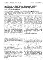
Efficiency of transduction of cynomologus macaque primitive hematopoietic cells with SIV-based lentiviral vectorsFigure 2
Efficiency of transduction of cynomologus macaque primitive hematopoietic cells with SIV-based lentiviral vectors. A: Non
transduced cells were used as a control for each animal. B: Transduction of bone marrow progenitor cells with an SIV-based
vector. CD34
+
cells were cultured in the presence of cytokines (see materials and methods) and exposed to vector particles at
an MOI of 100 for 24 hours before FACS analysis for eGFP production. C: CD34
+
cells were cultured overnight in a prolifera-
tion medium supplemented with various concentrations of AZT (100 nM, 1 mM, 10 mM). Cells were then washed twice and
transduced with various multiplicities of infection (MOI) of the lentiviral vector (0, 1, 10, 100). After 24 hours of coculture with
lentiviral vector, some of the CD34
+
cells were used to evaluate the rate of transduction of undifferentiated CD34
+
cells (C); *
indicate statistically significant differences (Kruskal Wallis test) between cultures with and without AZT treatment for MOI = 1
(p = 0,0378), MOI = 10 (p = 0,0224) and MOI = 100 (p = 0,0247). Some of the cells were cultured for 14 days, to allow the
myeloid differentiation of CFC. Cells were then resuspended, washed and fixed for three days. They were analyzed by flow
cytometry, to evaluate the percentage of eGFP-positive cells and determine the rate of transduction (D); * indicates a statisti-
cally significant difference (p = 0,0237(Kruskal Wallis test)) between cultures with and without AZT treatment for MOI = 100.
The results shown are the mean values for the three monkeys, each studied in triplicate.
D
0
20
40
60
0 1.E-07 1.E-06 1.E-05
moi 0
moi 1
moi 10
moi 100
AZT Doses (M)
% of eGFP positive cells
moi 0
moi 1
moi 10
moi 100
0 1.E-07 1.E-06 1.E-05
0
20
40
60
AZT Doses (M)
% of eGFP positive cells
C
B
A
10
0
10
1
10
2
10
10
4
10
0
10
1
10
2
10
3
10
4
FL1-eGFP
FL2-Height
0%
10
0
10
1
10
2
10
3
10
4
10
0
10
1
10
2
10
3
10
4
FL1-eGFP
43%
FL2-Height
*
*
*
*
P=0,0378
P=0,0224
P=0,0247
P=0,0237
Retrovirology 2008, 5:50 />Page 8 of 15
(page number not for citation purposes)
lymphocyte count was reached (P = 0.1939 (Mann &
Whitney test)) or the level of that minimum (P = 0.3805
(Mann & Whitney test)). A significant decrease in platelet
counts, beginning by day 10 (Figure 4), was observed in
all irradiated animals. Thrombocytopenia (platelet count
< 20,000/μl) was characterized in non transplanted ani-
mals by a minimum value of 3.75 ± 2.49 × 10
3
platelets/
μl on day 18 ± 3. Thrombocytopenia tended to be less
severe in transplanted animals, but this difference was not
significant for the minimum number of platelets (10.33 ±
5.25 × 10
3
platelets/μl; P = 0.1124 (Mann & Whitney
test)) or for the day on which that minimum occurred
(14.33 ± 0.94; P = 0.3123 (Mann & Whitney test)). This
thrombocytopenia required one transfusion in all ani-
mals (other than animal 7036, which needed two transfu-
sions) of both groups. However, platelet reconstitution
seemed to be correlated with the dose of CD34
+
cells
infused, the speed of reconstitution increasing with the
number of CD34
+
cells injected (macaque 6653).
Reconstitution of bone marrow clonogenic activity
We determined the effects of CD34
+
bone marrow cell
transplantation following gamma irradiation on the ex
vivo proliferation and differentiation of hematopoietic
progenitors. Before gamma irradiation, a mean of 40 ± 9
and 38 ± 6 colonies was observed for groups 1 and 2,
respectively (Figure 5). Colony number decreased signifi-
cantly (P < 0.0001 (Wilcoxon test)) by day 7 in all ani-
mals. In both groups, clonogenic activity was detected by
day 43 after gamma irradiation with reconstitution signif-
icantly better in the animals undergoing transplantation
than in those that did not undergo transplantation (P =
0.0009 (Mann & Whitney test)).
Presence of eGFP-positive cells in bone marrow and
peripheral blood
Cells with integrated SIV-vector DNA were detected by
PCR (Table 2) as early as day 3 after transplantation, in at
least two animals (6653 and 6833). These two animals
had received the largest numbers of transduced CD34
+
bone marrow cells. Monkey 7036, which died within 40
Fluorescence microscopy after myeloid differentiation of CFC (×100)Figure 3
Fluorescence microscopy after myeloid differentiation of CFC (×100). Freshly isolated CD34
+
cells were transduced or not
with the lentiviral vector (24 hours of culture with lentiviral vector at MOI = 100). Cells were then cultured for 14 days in the
presence of cytokines, to allow myeloid differentiation of transduced (A) and not transduced (B) CD34+ cells. Abbreviations:
CFU-GEMM, Colony-Forming Unit-Granulocytes, Erythroid, Macrophage, Megakaryocyte; BFU-E, Burst-Forming Unit-Eryth-
roid; CFU-GM, Colony-Forming Unit-Granulocytes, Macrophage; CFU-G, Colony-Forming Unit-Granulocytes; CFU-M, Col-
ony-Forming Unit-Macrophage.
CFU-GEMM
BFU-E
CFU-GM
CFU-G CFU-M
CFU-GEMM BFU-E CFU-GM CFU-G CFU-M
A
B
Phase contrast
Green
fluorescence
Phase contrast
Green
fluorescence
Retrovirology 2008, 5:50 />Page 9 of 15
(page number not for citation purposes)
days of gamma irradiation had very few transduced cells
in the bone marrow and SIV-DNA was not detected in
peripheral blood cells. In the three remaining animals,
vector DNA was detected in peripheral blood cells (up to
500 copies per million cells) and in the bone marrow (up
to 6250 copies per million cells) more than one year after
transplantation (day 471).
Effect of irradiation and transplantation on polymorphonuclear cell, lymphocyte and thrombocyte countsFigure 4
Effect of irradiation and transplantation on polymorphonuclear cell, lymphocyte and thrombocyte counts. All animals were fol-
lowed during the weeks preceding the study, and for more than 240 days after the irradiation. We carried out hematological
analysis including blood cell counts with an automated hemocytometer (Coulter Corporation, Miami, USA).
5825
5887
6122
6297
6487
6508
6547
6630
6653
6833
6896
7036
Polymorphonuclear Lymphocytes Thrombocytes
Day of the experiment
1,E+01
1,E+02
1,E+03
1,E+04
-60 -10 40 90 140 190 240
1,E+01
1,E+02
1,E+03
1,E+04
-60 -10 40 90 140 190 240
1,E+01
1,E+02
1,E+03
1,E+04
-60 -10 40 90 140 190 240
1,E+00
1,E+01
1,E+02
1,E+03
-60 -10 40 90 140 190 240
1,E+00
1,E+01
1,E+02
1,E+03
-60 -10 40 90 140 190 240
1,E+00
1,E+01
1,E+02
1,E+03
-60 -10 40 90 140 190 240
1,E+02
1,E+03
1,E+04
-60 -10 40 90 140 190 240
1,E+02
1,E+03
1,E+04
-60 -10 40 90 140 190 240
1,E+02
1,E+03
1,E+04
-60 -10 40 90 140 190 240
Cells / l
Cells / l
X10
3
Cells / l
Controls
Irradiated
Irradiated and engrafted
10
4
10
3
10
2
10
4
10
3
10
2
10
4
10
3
10
2
Polymorphonuclear (cells/ l) Lymphocytes (cells/ l) Thrombocytes (x10
3
cells/ l)
10
4
10
3
10
2
10
1
10
4
10
3
10
2
10
1
10
4
10
3
10
2
10
1
10
3
10
2
10
1
10
0
10
3
10
2
10
1
10
0
10
3
10
2
10
1
10
0
Controls
Irradiated
Irradiated
And
engrafted
Day of the experiment
-60 -10 40 90 140 190 240 -60 -10 40 90 140 190 240 -60 -10 40 90 140 190 240
-60 -10 40 90 140 190 240
-60 -10 40 90 140 190 240 -60 -10 40 90 140 190 240 -60 -10 40 90 140 190 240
-60 -10 40 90 140 190 240
-60 -10 40 90 140 190 240
Table 2: Number of DNA copies per million mononuclear cells in peripheral blood (PB) and bone marrow (BM)
Monkey
6653 6833 6896 7036
Days post transplantation PB BM PB BM PB BM PB BM
-3 0000 0 000
3 500 ND 250 ND 0 ND 0 15
5 250 500 ND 250 ND ND 0 0
108 250 ND 250 ND 1250 ND * *
121 750 ND 250 ND 250 ND * *
128 250 ND 250 ND 250 ND * *
142 250 ND 250 ND 1750 3250 * *
471 ND 250 250 250 500 6250 * *
ND: not determined
*: 7036 died on day 40
Retrovirology 2008, 5:50 />Page 10 of 15
(page number not for citation purposes)
Flow cytometry analysis demonstrated the presence of
eGFP-producing cells among peripheral blood mononu-
clear cells in myeloid and lymphoid lineges of monkey
6896 (Figure 6). Peripheral blood cells were sorted on the
basis of eGFP production, with the aim of characterizing
the phenotype of populations of cells expressing the trans-
gene in more detail. We found that 61% of eGFP-positive
cells were CD11b-positive,5% of these cells appeared to
be CD14+ monocytes, 14% were CD20
+
B cells and 10%
were CD3+ T cells, 23% of which expressed CD8 and 77%
expressed CD4 (data not shown).
Discussion
The aim of this work was to study reconstitution of the
myeloid and lymphoid compartments after the autolo-
gous transplantation of genetically modified CD34
+
bone
marrow cells into cynomolgus macaques previously sub-
jected to gamma irradiation.
We first assessed, in vitro, the efficiency with which a
SIVmac251-derived vector transduced macaque CD34
+
hematopoietic bone marrow cells. These vectors are simi-
lar to those derived from HIV. However, SIV-derived vec-
tors clearly outperform HIV-derived vectors in simian
cells. In fact, HIV-1 fails to replicate in simian cells
because of an early postentry block [43,44], and Kootstra
et., al showed that the viral determinant involved in
postentry restriction of HIV-1 replication in simian cells is
located at or near the cyclophilin A (CyPA) binding region
of the capside protein [45]. The hydrophobic pocket of
cyclophilin A (CypA) makes direct contact with an
exposed, proline-rich loop on HIV-1 capsid (CA) and
renders reverse transcription complexes resistant to an
antiviral activity in human cells. A CypA fusion with
TRIM5 (a member of the tripartite motif family) that is
unique to New World owl monkeys also targets HIV-1 CA,
but this interaction potently inhibits infection. A similar
block to HIV-1 infection in Old World monkeys is attrib-
utable to the α isoform of the TRIM5 orthologue in these
Recovery of bone marrow clonogenic activityFigure 5
Recovery of bone marrow clonogenic activity. Bone marrow-derived colony-forming units following sublethal irradiation of
cynomolgus monkeys transplanted (black bars) or not transplanted with CD34
+
cells (open bars). Mean ± SD of CFC number
(triplicate). The results of statistical test are indicates; * indicates a statistically significant difference (p < 0,0001 (Wilcoxon
test)) between day 0 and day 7 for the both group; ** indicates a statistically significant difference (p = 0,0009 (Mann & Whitney
test)) at day 43 between animals undergoing transplantation and those that did not undergo transplantation.
0
10
20
30
40
50
60
Day-1 Day7 Day146
Days after gamma-radiation
Number of colonies per 5.10
4
CMMOs
Not transplanted
Transplanted
P<0,0001
**
*
Day43
P=0,0009
Retrovirology 2008, 5:50 />Page 11 of 15
(page number not for citation purposes)
species and using RNA interference techniques, Berthoux
et., al demonstrated that CypA inhibits HIV-1 replication
in these cells because it is required for CA recognition by
TRIM5α [46]. SIV vectors can also efficiently transduce
human cells[33,47], and may therefore prove a useful
alternative to HIV-1-based vectors, at least in the early
phase of preclinical testing of lentivirus vectors. We found
that the proportion of eGFP-positive cells obtained before
myeloid differentiation (mean value of 30%) was similar
to that obtained with CD34
+
cells from human donors
transduced with lentiviral [48-51], retroviral [52-54],
AAV[55], or adenovirus/AAV-derived [56] vectors. How-
ever, it is possible to increase the transduction rate, such
that 90% transduced human CD34
+
cells are obtained
from cord blood, 80% from bone marrow and 75% from
G-CSF mobilized peripheral blood [57]. We analyzed
transduction in two types of assay, based on committed
(CFC) and primitive (LTC-IC) hematopoietic progenitors,
as analyses of the transduction of committed progenitors
only bear little relation to the transduction efficiency for
stem cells and less differentiated cells in the long term.
After myeloid differentiation, eGFP
+
cells were detected, in
similar proportions, in CFC on day 15 and LTC-IC on day
50 after transduction, indicating that the vector was able
to transduce progenitor cells and most immature hemat-
opoietic cells with a similar efficiency. Similar results have
been reported for stimulated human CFC and LTC-IC,
which were found to be transduced with similar efficiency
by a lentiviral vector based on HIV-1[58]. In this previous
study, significant resistance to lentiviral transduction was
reported in unstimulated primitive human cells. These
results may explain why, in our study, the use of cytokines
during transduction made possible the genetic modifica-
tion of LTC-IC, which are quiescent. Cytokine treatment
may have led to these cells entering the cell cycle, facilitat-
ing transduction. This result confirms the greater effi-
ciency of lentiviral vectors than of retroviral vectors for the
transduction of CD34
+
cells. Nevertheless, in our study,
only half as many eGFP-positive cells were obtained after
differentiation as were obtained from undifferentiated
CD34
+
cells. Similar observations have been made with
MLV-transduced progenitor cells from human
Flow cytometry analysis of hematopoiesis reconstitutionFigure 6
Flow cytometry analysis of hematopoiesis reconstitution. Animal transplanted with autologous CD34
+
bone marrow cells
transduced with an SIV-based vector. eGFP-positive cells present in P1 and P2 were analyzed by immuno-staining to identify
the subpopulations of eGFP-positive cells in peripheral blood. CD20-PerCP-Cy5, CD14-PE, CD11b-APC and CD3-APC stain-
ing were used to identify the B-lymphocyte, monocyte, granulocyte and T-lymphocyte subpopulations.
5%
10
2
10
3
10
4
10
5
10
5
10
4
10
3
10
2
monocytes
GFP
61%
10
2
10
3
10
4
10
5
10
5
10
4
10
3
10
2
CD11b
granulocytes
14%
10
2
10
3
10
4
10
5
10
5
10
4
10
3
10
2
CD20
B lymphocytes
10%
10
2
10
3
10
4
10
5
10
5
10
4
10
3
10
2
CD3
T lymphocytes
P1
50 100 150 200 250
250
200
150
100
50
(X 1000)
(X 1000)
SSC
FSC
CD14
P2
P2
10
2
10
3
10
4
10
5
250
200
150
100
50
(X 1000)
SSC
GFP
Retrovirology 2008, 5:50 />Page 12 of 15
(page number not for citation purposes)
donors[59]. We demonstrate here that these differences
may be accounted for by the pseudotransduction detected
at 24 h of incubation with the vector, confirming the
results reported with CD34
+
cells in studies using VSVg-
pseudotyped MLV-derived[60] or lentivirus-derived vec-
tors[51]. It has been suggested that pseudotransduction
may result from VSVg-pseudotyping due to membrane
fusion efficiency being higher than the rate of integration
of the transgene[61]. Nevertheless, most lentiviral vectors
have been generated with VSV-G, as this glycoprotein
makes it easy to recover and concentrate the pseudotyped
vectors [62].
We also showed that eGFP was produced in all colony
subtypes. Clusters of eGFP production were observed on
fluorescence microscopy, indicating that not all the cells
of a given positive colony – theoretically derived from a
single cell – produced eGFP. This result is consistent with
those of Mikkola et al. concerning murine HSC transduc-
tion by a VSVg-pseudotyped lentiviral vector, in which a
mismatch was reported between the transduction rate of
cells (almost 25%) and the transduction rate of myeloid
colonies (almost 60%). These authors highlighted the
occurrence of mosaicism in GFP gene expression in colo-
nies obtained following the myeloid differentiation of
CD34
+
cells[63], possibly due to a delay in the integration
of the transgene during differentiation, resulting in the
formation of clusters of GFP-positive cells within a single
myeloid colony.
In our in vivo study, autologous HSC were injected into
the bone marrow, whereas intravenous injection is cur-
rently the most frequently used transplantation method.
We aimed to increase seeding efficiency and homing, as
only a limited number of stem cells were theoretically
available. However, 2.5 × 10
6
to 5.0 × 10
6
CD34
+
cells is
generally sufficient to ensure engraftment, and we found
that less than 2.0 × 10
6
cells were sufficient for long-term
reconstitution in macaques. As predicted[64,65], total-
body gamma irradiation leads to a drastic decrease in the
number of hematopoietic progenitors, preventing the
development of mature cells [66]. Despite the occurrence
of severe pancytopenia, a positive correlation has been
found between the number of CD34
+
cells infused and
time required for immune reconstitution [42,67,68].
However, hematopoietic recovery may take longer if fewer
than 2.0 × 10
6
CD34
+
cells/kg are infused. This notion is
consistent with our observation that CD34
+
cell transplan-
tation decreases both the severity and duration of irradia-
tion-induced cytopenia. Clonogenic activity also
reappeared more strongly in transplanted animals. We
also showed that the animals recovered B cells, T cells,
monocytes and granulocytes. Nevertheless, the functional
activity of these cells requires confirmation, particularly
for lymphocytes. However, although we observed long-
term reconstitution with lentiviral vector-transduced cells
of different lineages, its proportion remained below 1%.
Hanawa et al., provided the first evidence that SIV-based
vectors can successfully transduce rhesus macaque repop-
ulating hematopoietic stem cells, with an average of 16%
of peripheral blood leukocytes containing the SIV vector
genome. However, this study was carried out with HSC
from mobilized peripheral blood cells, making it possible
to obtain larger numbers of HSC than can be harvested
from bone marrow. Nevertheless theoretically, these cells
contained more progenitors that were already committed
and fewer pluripotent stem cells capable of long-term
reconstitution than medullary HSC[69]. The small num-
bers of eGFP-producing cells observed in our study may
be due to an anti-eGFP immune response. Some reports
have suggested that such reactions do not generally occur
after irradiation[70], but two reports described the induc-
tion of cytotoxic T-lymphocyte responses to enhanced
green (GFP) or yellow (YFP) fluorescent proteins after
myeloablative conditioning. One of these reports con-
cerned baboons that had received primitive hematopoi-
etic cells transduced with HIV-1-based lentiviral
vectors[71] and the other concerned rhesus macaques that
had received CD34
+
stem cells transduced with a retroviral
vector[72].
Lentiviruses, like retroviruses, can be used to integrate
transgenes into the host genome. Two severe adverse
events occurred in two patients in the SCID-X1 gene ther-
apy trial 30 to 34 months after injection of the autologous
CD34
+
cells corrected using a retroviral vector. In these
patients, an uncontrolled clonal T lymphoproliferative
syndrome, similar to acute lymphoblastic leukemia, was
observed [73,74]. This study highlights the risk of inser-
tional mutagenesis restricted to retroviral and lentiviral
gene transfer. In the future, additional safety measures
could be considered, such as the use of self-inactivating
LTRs (as in our study) to reduce enhancer activity, the
addition of insulators to reduce the risk further, and the
insertion of a second transgene encoding a "suicide" prod-
uct, such as herpes thymidine kinase, making it possible
to kill the transduced cells with ganciclovir. Unlike studies
in mice, in which the follow-up period is necessarily lim-
ited, studies in large animals, with a longer life span, are
compatible with more extensive follow-up. The develop-
ment of linear amplification-mediated PCR (LAM-PCR), a
sensitive and robust approach to molecular clonal analy-
sis, has made it possible to identify and analyze the con-
tribution of individual transduced clones to
hematopoiesis. Clonal analysis may provide information
about the dominance of transduced clones, potentially
predicting possible progression or the propensity to
develop clonal hematopoiesis and leukemia. Moreover,
replication-competent retrovirus (RCRs), recombinant
retrovirus and interaction with endogenous retroviruses
Retrovirology 2008, 5:50 />Page 13 of 15
(page number not for citation purposes)
should also be investigated, when evaluating the biosafety
of retrovirus and lentivirus.
Conclusion
The results reported here provide the first evidence that
gene transfer into medullary hematopoietic stem cells and
long-term expression of the transgene are possible, using
an SIV-based lentiviral vector in non human primates,
which provide the best clinical models for in vivo evalua-
tion of the feasibility and safety of gene therapy strategies.
Competing interests
The authors never received reimbursements, fees, funding,
or salary from an organization that may in any way gain
or lose financially from the publication of this paper. The
authors never have any stocks or shares in an organization
that may in any way gain or lose financially from the pub-
lication of this paper. The authors have no competing
interests to declare in relation to this paper.
Authors' contributions
SD was the main contributor to this paper. This work is
part of her PhD project. She carried out transduction of
CD34
+
cells, transplantation of animals, PCR for identifi-
cation of cells expressing the transgene in vivo, flow cytom-
etry analysis, WG Have improved assays for transduction
of macaque bone marrow CD34
+
cells with SIV derived
vector, DN constructed and produced the SIV derived vec-
tor, SP technical assistance to cell sorting, MLD technical
assistance to transplantation, BD technical assistance to
cell culture, flow cytometry and irradiadion of NHP, GA
technical assistance to molecular biology, TA technical
assistance to flow cytometry and cell sorting, JLL irradia-
tion of animals and dosimetry, FLC supervises vector
design and production, RLG supervisor of SD.
Acknowledgements
We would like to thank M. Ripaux, A. Fort, S. Jacquin, D. Mérigard, P.
Pochard, D. Renault, J. C. Wilks and R. Rioux for excellent technical assist-
ance. This work was supported by the Agence Nationale de Recherches sur
le SIDA (ANRS, Paris, France), the Centre de Recherches du Service de
Santé des Armées Emile Pardé (CRSSA, La Tronche, France), and the Com-
missariat à l'Energie Atomique (CEA, Fontenay aux Roses, France).
References
1. Miller DG, Adam MA, Miller AD: Gene transfer by retrovirus
vectors occurs only in cells that are actively replicating at the
time of infection. Mol Cell Biol 1990, 10:4239-4242.
2. Jones RJ, Wagner JE, Celano P, Zicha MS, Sharkis SJ: Separation of
pluripotent haematopoietic stem cells from spleen colony-
forming cells. Nature 1990, 347:188-189.
3. Gothot A, Loo JC van der, Clapp DW, Srour EF: Cell cycle-related
changes in repopulating capacity of human mobilized
peripheral blood CD34(+) cells in non-obese diabetic/severe
combined immune-deficient mice. Blood 1998, 92:2641-2649.
4. Hao QL, Thiemann FT, Petersen D, Smogorzewska EM, Crooks GM:
Extended long-term culture reveals a highly quiescent and
primitive human hematopoietic progenitor population.
Blood 1996, 88:3306-3313.
5. Ploemacher RE, Sluijs JP van der, Voerman JS, Brons NH: An in vitro
limiting-dilution assay of long-term repopulating hematopoi-
etic stem cells in the mouse. Blood 1989, 74:2755-2763.
6. Ploemacher RE, Sluijs JP van der, van Beurden CA, Baert MR, Chan
PL: Use of limiting-dilution type long-term marrow cultures
in frequency analysis of marrow-repopulating and spleen col-
ony-forming hematopoietic stem cells in the mouse. Blood
1991, 78:2527-2533.
7. Traycoff CM, Kosak ST, Grigsby S, Srour EF: Evaluation of ex vivo
expansion potential of cord blood and bone marrow hemat-
opoietic progenitor cells using cell tracking and limiting dilu-
tion analysis. Blood 1995, 85:2059-2068.
8. Bodine DM, Crosier PS, Clark SC: Effects of hematopoietic
growth factors on the survival of primitive stem cells in liquid
suspension culture. Blood 1991, 78:914-920.
9. Larochelle A, Vormoor J, Hanenberg H, Wang JC, Bhatia M, Lapidot
T, Moritz T, Murdoch B, Xiao XL, Kato I, et al.: Identification of
primitive human hematopoietic cells capable of repopulat-
ing NOD/SCID mouse bone marrow: implications for gene
therapy. Nat Med 1996, 2:1329-1337.
10. Peters SO, Kittler EL, Ramshaw HS, Quesenberry PJ: Ex vivo expan-
sion of murine marrow cells with interleukin-3 (IL-3), IL-6,
IL-11, and stem cell factor leads to impaired engraftment in
irradiated hosts. Blood
1996, 87:30-37.
11. Tisdale JF, Hanazono Y, Sellers SE, Agricola BA, Metzger ME, Dona-
hue RE, Dunbar CE: Ex vivo expansion of genetically marked
rhesus peripheral blood progenitor cells results in dimin-
ished long-term repopulating ability. Blood 1998, 92:1131-1141.
12. Lewis PF, Emerman M: Passage through mitosis is required for
oncoretroviruses but not for the human immunodeficiency
virus. J Virol 1994, 68:510-516.
13. Naldini L: Lentiviruses as gene transfer agents for delivery to
non-dividing cells. Curr Opin Biotechnol 1998, 9:457-463.
14. Fouchier RA, Malim MH: Nuclear import of human immunode-
ficiency virus type-1 preintegration complexes. Adv Virus Res
1999, 52:275-299.
15. Naldini L, Blomer U, Gallay P, Ory D, Mulligan R, Gage FH, Verma IM,
Trono D: In vivo gene delivery and stable transduction of non-
dividing cells by a lentiviral vector. Science 1996, 272:263-267.
16. Richardson JH, Kaye JF, Child LA, Lever AM: Helper virus-free
transfer of human immunodeficiency virus type 1 vectors. J
Gen Virol 1995, 76(Pt 3):691-696.
17. Arya SK, Zamani M, Kundra P: Human immunodeficiency virus
type 2 lentivirus vectors for gene transfer: expression and
potential for helper virus-free packaging. Hum Gene Ther 1998,
9:1371-1380.
18. Poeschla EM, Wong-Staal F, Looney DJ: Efficient transduction of
nondividing human cells by feline immunodeficiency virus
lentiviral vectors. Nat Med 1998, 4:354-357.
19. Mitrophanous K, Yoon S, Rohll J, Patil D, Wilkes F, Kim V, Kingsman
S, Kingsman A, Mazarakis N: Stable gene transfer to the nervous
system using a non-primate lentiviral vector. Gene Ther 1999,
6:1808-1818.
20. Hu J, Dunbar CE: Update on hematopoietic stem cell gene
transfer using non-human primate models. Curr Opin Mol Ther
2002, 4:482-490.
21. Donahue RE, Dunbar CE: Update on the use of nonhuman pri-
mate models for preclinical testing of gene therapy
approaches targeting hematopoietic cells. Hum Gene Ther
2001, 12:607-617.
22. Villinger F, Brar SS, Mayne A, Chikkala N, Ansari AA: Comparative
sequence analysis of cytokine genes from human and nonhu-
man primates. J Immunol 1995, 155:3946-3954.
23. Farese AM, MacVittie TJ, Roskos L, Stead RB: Hematopoietic
recovery following autologous bone marrow transplantation
in a nonhuman primate: effect of variation in treatment
schedule with PEG-rHuMGDF. Stem Cells 2003, 21:79-89.
24. Dunbar CE, Takatoku M, Donahue RE: The impact of ex vivo
cytokine stimulation on engraftment of primitive hemat-
opoietic cells in a non-human primate model. Ann N Y Acad Sci
2001, 938:236-244. discussion 244-235.
25. Wagemaker G, Neelis KJ, Hartong SCC, Wognum AW, Thomas GR,
Fielder PJ, Eaton DL: The efficacy of recombinant TPO in
murine And nonhuman primate models for myelosuppres-
sion and stem cell transplantation. Stem Cells 1998, 16(Suppl
2):127-141.
Retrovirology 2008, 5:50 />Page 14 of 15
(page number not for citation purposes)
26. Shi PA, Hematti P, von Kalle C, Dunbar CE: Genetic marking as an
approach to studying in vivo hematopoiesis: progress in the
non-human primate model. Oncogene 2002, 21:3274-3283.
27. Burkhard MJ, Dean GA: Transmission and immunopathogene-
sis of FIV in cats as a model for HIV. Curr HIV Res 2003, 1:15-29.
28. Willett BJ, Flynn JN, Hosie MJ: FIV infection of the domestic cat:
an animal model for AIDS. Immunol Today 1997, 18:182-189.
29. Talbott RL, Sparger EE, Lovelace KM, Fitch WM, Pedersen NC, Luciw
PA, Elder JH: Nucleotide sequence and genomic organization
of feline immunodeficiency virus. Proc Natl Acad Sci USA 1989,
86:5743-5747.
30. Olmsted RA, Barnes AK, Yamamoto JK, Hirsch VM, Purcell RH, John-
son PR: Molecular cloning of feline immunodeficiency virus.
Proc Natl Acad Sci USA 1989, 86:2448-2452.
31. Schnell T, Foley P, Wirth M, Munch J, Uberla K: Development of a
self-inactivating, minimal lentivirus vector based on simian
immunodeficiency virus. Hum Gene Ther 2000, 11:439-447.
32. Wagner R, Graf M, Bieler K, Wolf H, Grunwald T, Foley P, Uberla K:
Rev-independent expression of synthetic gag-pol genes of
human immunodeficiency virus type 1 and simian immuno-
deficiency virus: implications for the safety of lentiviral vec-
tors. Hum Gene Ther 2000, 11:2403-2413.
33. Negre D, Mangeot PE, Duisit G, Blanchard S, Vidalain PO, Leissner P,
Winter AJ, Rabourdin-Combe C, Mehtali M, Moullier P, et al.: Char-
acterization of novel safe lentiviral vectors derived from sim-
ian immunodeficiency virus (SIVmac251) that efficiently
transduce mature human dendritic cells. Gene Ther 2000,
7:1613-1623.
34. Mangeot PE, Negre D, Dubois B, Winter AJ, Leissner P, Mehtali M,
Kaiserlian D, Cosset FL, Darlix JL: Development of minimal len-
tivirus vectors derived from simian immunodeficiency virus
(SIVmac251) and their use for gene transfer into human den-
dritic cells. J Virol 2000, 74:8307-8315.
35. Kiem HP, Andrews RG, Morris J, Peterson L, Heyward S, Allen JM,
Rasko JE, Potter J, Miller AD: Improved gene transfer into
baboon marrow repopulating cells using recombinant
human fibronectin fragment CH-296 in combination with
interleukin-6, stem cell factor, FLT-3 ligand, and megakary-
ocyte growth and development factor. Blood
1998,
92:1878-1886.
36. Wu T, Kim HJ, Sellers SE, Meade KE, Agricola BA, Metzger ME, Kato
I, Donahue RE, Dunbar CE, Tisdale JF: Prolonged high-level detec-
tion of retrovirally marked hematopoietic cells in nonhuman
primates after transduction of CD34+ progenitors using clin-
ically feasible methods. Mol Ther 2000, 1:285-293.
37. Kim HJ, Tisdale JF, Wu T, Takatoku M, Sellers SE, Zickler P, Metzger
ME, Agricola BA, Malley JD, Kato I, et al.: Many multipotential
gene-marked progenitor or stem cell clones contribute to
hematopoiesis in nonhuman primates. Blood 2000, 96:1-8.
38. Rosenzweig M, MacVittie TJ, Harper D, Hempel D, Glickman RL,
Johnson RP, Farese AM, Whiting-Theobald N, Linton GF, Yamasaki G,
et al.: Efficient and durable gene marking of hematopoietic
progenitor cells in nonhuman primates after nonablative
conditioning. Blood 1999, 94:2271-2286.
39. Kelly PF, Donahue RE, Vandergriff JA, Takatoku M, Bonifacino AC,
Agricola BA, Metzger ME, Dunbar CE, Nienhuis AW, Vanin EF: Pro-
longed multilineage clonal hematopoiesis in a rhesus recipi-
ent of CD34 positive cells marked with a RD114
pseudotyped oncoretroviral vector. Blood Cells Mol Dis 2003,
30:132-143.
40. Hu J, Kelly P, Bonifacino A, Agricola B, Donahue R, Vanin E, Dunbar
CE: Direct comparison of RD114-pseudotyped versus
amphotropic-pseudotyped retroviral vectors for transduc-
tion of rhesus macaque long-term repopulating cells. Mol
Ther 2003, 8:611-617.
41. Mangeot PE, Duperrier K, Negre D, Boson B, Rigal D, Cosset FL, Dar-
lix JL: High levels of transduction of human dendritic cells
with optimized SIV vectors. Mol Ther 2002, 5:283-290.
42. Chen C, Okayama H: High-efficiency transformation of mam-
malian cells by plasmid DNA. Mol Cell Biol 1987, 7:2745-2752.
43. Munk C, Brandt SM, Lucero G, Landau NR: A dominant block to
HIV-1 replication at reverse transcription in simian cells.
Proc Natl Acad Sci USA 2002, 99:13843-13848.
44. Besnier C, Takeuchi Y, Towers G: Restriction of lentivirus in
monkeys. Proc Natl Acad Sci USA 2002, 99:11920-11925.
45. Kootstra NA, Munk C, Tonnu N, Landau NR, Verma IM: Abroga-
tion of postentry restriction of HIV-1-based lentiviral vector
transduction in simian cells. Proc Natl Acad Sci USA 2003,
100:1298-1303.
46. Berthoux L, Sebastian S, Sokolskaja E, Luban J: Cyclophilin A is
required for TRIM5{alpha}-mediated resistance to HIV-1 in
Old World monkey cells. Proc Natl Acad Sci USA 2005,
102:14849-14853.
47. Sandrin V, Boson B, Salmon P, Gay W, Negre D, Le Grand R, Trono
D, Cosset FL: Lentiviral vectors pseudotyped with a modified
RD114 envelope glycoprotein show increased stability in
sera and augmented transduction of primary lymphocytes
and CD34+ cells derived from human and nonhuman pri-
mates. Blood 2002, 100:823-832.
48. Sutton RE, Wu HT, Rigg R, Bohnlein E, Brown PO: Human immu-
nodeficiency virus type 1 vectors efficiently transduce
human hematopoietic stem cells. J Virol 1998, 72:5781-5788.
49. Evans JT, Kelly PF, O'Neill E, Garcia JV: Human cord blood
CD34+CD38- cell transduction via lentivirus-based gene
transfer vectors. Hum Gene Ther 1999, 10:1479-1489.
50. Piconi S, Trabattoni D, Fusi ML, Milazzo F, Dix LP, Rizzardini G,
Colombo F, Bray D, Clerici M: Effect of two different combina-
tions of antiretrovirals (AZT+ddI and AZT+3TC) on
cytokine production and apoptosis in asymptomatic HIV
infection. Antiviral Res 2000, 46:171-179.
51. Case SS, Price MA, Jordan CT, Yu XJ, Wang L, Bauer G, Haas DL, Xu
D, Stripecke R, Naldini L, et al.: Stable transduction of quiescent
CD34(+)CD38(-) human hematopoietic cells by HIV-1-based
lentiviral vectors. Proc Natl Acad Sci USA 1999, 96:2988-2993.
52. Poznansky MC, La Vecchio J, Silva-Arietta S, Porter-Brooks J, Brody
K, Olszak IT, Adams GB, Ramstedt U, Marasco WA, Scadden DT:
Inhibition of human immunodeficiency virus replication and
growth advantage of CD4+ T cells and monocytes derived
from CD34+ cells transduced with an intracellular antibody
directed against human immunodeficiency virus type 1 Tat.
Hum Gene Ther 1999, 10:
2505-2514.
53. Rosenzweig M, Marks DF, Hempel D, Lisziewicz J, Johnson RP:
Transduction of CD34+ hematopoietic progenitor cells with
an antitat gene protects T-cell and macrophage progeny
from AIDS virus infection. J Virol 1997, 71:2740-2746.
54. Davis BR, Saitta FP, Bauer G, Bunnell BA, Morgan RA, Schwartz DH:
Targeted transduction of CD34+ cells by transdominant
negative Rev-expressing retrovirus yields partial anti-HIV
protection of progeny macrophages. Hum Gene Ther 1998,
9:1197-1207.
55. Chatterjee S, Li W, Wong CA, Fisher-Adams G, Lu D, Guha M, Macer
JA, Forman SJ, Wong KK Jr: Transduction of primitive human
marrow and cord blood-derived hematopoietic progenitor
cells with adeno-associated virus vectors. Blood 1999,
93:1882-1894.
56. Shayakhmetov DM, Carlson CA, Stecher H, Li Q, Stamatoyannopou-
los G, Lieber A: A high-capacity, capsid-modified hybrid aden-
ovirus/adeno-associated virus vector for stable transduction
of human hematopoietic cells. J Virol 2002, 76:1135-1143.
57. Amsellem S, Ravet E, Fichelson S, Pflumio F, Dubart-Kupperschmitt A:
Maximal lentivirus-mediated gene transfer and sustained
transgene expression in human hematopoietic primitive
cells and their progeny. Mol Ther 2002, 6:673-677.
58. Zielske SP, Gerson SL: Cytokines, including stem cell factor
alone, enhance lentiviral transduction in nondividing human
LTCIC and NOD/SCID repopulating cells. Mol Ther 2003,
7:325-333.
59. Yam PY, Yee JK, Ito JI, Sniecinski I, Doroshow JH, Forman SJ, Zaia JA:
Comparison of amphotropic and pseudotyped VSV-G retro-
viral transduction in human CD34+ peripheral blood progen-
itor cells from adult donors with HIV-1 infection or cancer.
Exp Hematol 1998, 26:962-968.
60. Liu ML, Winther BL, Kay MA: Pseudotransduction of hepato-
cytes by using concentrated pseudotyped vesicular stomati-
tis virus G glycoprotein (VSV-G)-Moloney murine leukemia
virus-derived retrovirus vectors: comparison of VSV-G and
amphotropic vectors for hepatic gene transfer. J Virol 1996,
70:2497-2502.
61. Gallardo HF, Tan C, Ory D, Sadelain M: Recombinant retrovi-
ruses pseudotyped with the vesicular stomatitis virus G glyc-
oprotein mediate both stable gene transfer and
Publish with BioMed Central and every
scientist can read your work free of charge
"BioMed Central will be the most significant development for
disseminating the results of biomedical research in our lifetime."
Sir Paul Nurse, Cancer Research UK
Your research papers will be:
available free of charge to the entire biomedical community
peer reviewed and published immediately upon acceptance
cited in PubMed and archived on PubMed Central
yours — you keep the copyright
Submit your manuscript here:
/>BioMedcentral
Retrovirology 2008, 5:50 />Page 15 of 15
(page number not for citation purposes)
pseudotransduction in human peripheral blood lym-
phocytes. Blood 1997, 90:952-957.
62. Burns JC, Friedmann T, Driever W, Burrascano M, Yee JK: Vesicular
stomatitis virus G glycoprotein pseudotyped retroviral vec-
tors: concentration to very high titer and efficient gene
transfer into mammalian and nonmammalian cells. Proc Natl
Acad Sci USA 1993, 90:8033-8037.
63. Mikkola H, Woods NB, Sjogren M, Helgadottir H, Hamaguchi I, Jacob-
sen SE, Trono D, Karlsson S: Lentivirus gene transfer in murine
hematopoietic progenitor cells is compromised by a delay in
proviral integration and results in transduction mosaicism
and heterogeneous gene expression in progeny cells. J Virol
2000, 74:11911-11918.
64. Bolus NE: Basic review of radiation biology and terminology.
J Nucl Med Technol 2001, 29:67-73. test 76–67.
65. MacVittie TJ, Farese AM, Herodin F, Grab LB, Baum CM, McKearn JP:
Combination therapy for radiation-induced bone marrow
aplasia in nonhuman primates using synthokine SC-55494
and recombinant human granulocyte colony-stimulating fac-
tor. Blood 1996, 87:4129-4135.
66. Dainiak N: Hematologic consequences of exposure to ionizing
radiation. Exp Hematol 2002, 30:513-528.
67. Nieboer P, de Vries EG, Vellenga E, Graaf WT van der, Mulder NH,
Sluiter WJ, de Wolf JT: Factors influencing haematological
recovery following high-dose chemotherapy and peripheral
stem-cell transplantation for haematological malignancies;
1-year analysis. Eur J Cancer 2004, 40:1199-1207.
68. Jillella AP, Ustun C: What is the optimum number of CD34+
peripheral blood stem cells for an autologous transplant?
Stem Cells Dev 2004, 13:598-606.
69. Hanawa H, Hematti P, Keyvanfar K, Metzger ME, Krouse A, Donahue
RE, Kepes S, Gray J, Dunbar CE, Persons DA, Nienhuis AW: Effi-
cient gene transfer into rhesus repopulating hematopoietic
stem cells using a simian immunodeficiency virus-based len-
tiviral vector system. Blood 2004, 103:4062-4069.
70. Persons DA, Allay JA, Riberdy JM, Wersto RP, Donahue RE, Sorren-
tino BP, Nienhuis AW: Use of the green fluorescent protein as
a marker to identify and track genetically modified hemat-
opoietic cells. Nat Med 1998, 4:1201-1205.
71. Morris JC, Conerly M, Thomasson B, Storek J, Riddell SR, Kiem HP:
Induction of cytotoxic T-lymphocyte responses to enhanced
green and yellow fluorescent proteins after myeloablative
conditioning. Blood 2004, 103:492-499.
72. Rosenzweig M, Connole M, Glickman R, Yue SP, Noren B, DeMaria
M, Johnson RP: Induction of cytotoxic T lymphocyte and anti-
body responses to enhanced green fluorescent protein fol-
lowing transplantation of transduced CD34(+)
hematopoietic cells. Blood 2001, 97:1951-1959.
73. Hacein-Bey-Abina S, von Kalle C, Schmidt M, Le Deist F, Wulffraat N,
McIntyre E, Radford I, Villeval JL, Fraser CC, Cavazzana-Calvo M,
Fischer A: A serious adverse event after successful gene ther-
apy for X-linked severe combined immunodeficiency. N Engl
J Med 2003, 348:255-256.
74. Hacein-Bey-Abina S, Von Kalle C, Schmidt M, McCormack MP, Wulf-
fraat N, Leboulch P, Lim A, Osborne CS, Pawliuk R, Morillon E, et al.:
LMO2-associated clonal T cell proliferation in two patients
after gene therapy for SCID-X1. Science 2003, 302:415-419.