Báo cáo y học: "Oral keratinocytes support non-replicative infection and transfer of harbored HIV-1 to permissive cells" ppsx
Bạn đang xem bản rút gọn của tài liệu. Xem và tải ngay bản đầy đủ của tài liệu tại đây (805.2 KB, 14 trang )
BioMed Central
Page 1 of 14
(page number not for citation purposes)
Retrovirology
Open Access
Research
Oral keratinocytes support non-replicative infection and transfer of
harbored HIV-1 to permissive cells
Anjalee Vacharaksa
1,2
, Anil C Asrani
1,2
, Kristin H Gebhard
1,2
,
Claudine E Fasching
2
, Rodrigo A Giacaman
1,2
, Edward N Janoff
2,3
,
Karen F Ross
1,2
and Mark C Herzberg*
1,2
Address:
1
Department of Diagnostic and Biological Sciences, School of Dentistry, University of Minnesota, Minneapolis, MN 55455, USA,
2
Mucosal and Vaccine Research Center, Minneapolis VA Medical Center, Minneapolis, MN 55417, USA and
3
Division of Infectious Diseases,
Colorado Center for AIDS Research, and the Mucosal and Vaccine Research Program Colorado, University of Colorado Denver, and the Denver
Veterans Affairs Medical Center, Denver, CO 80220, USA
Email: Anjalee Vacharaksa - ; Anil C Asrani - ; Kristin H Gebhard - ;
Claudine E Fasching - ; Rodrigo A Giacaman - ; Edward N Janoff - ;
Karen F Ross - ; Mark C Herzberg* -
* Corresponding author
Abstract
Background: Oral keratinocytes on the mucosal surface are frequently exposed to HIV-1 through
contact with infected sexual partners or nursing mothers. To determine the plausibility that oral
keratinocytes are primary targets of HIV-1, we tested the hypothesis that HIV-1 infects oral
keratinocytes in a restricted manner.
Results: To study the fate of HIV-1, immortalized oral keratinocytes (OKF6/TERT-2; TERT-2
cells) were characterized for the fate of HIV-specific RNA and DNA. At 6 h post inoculation with
X4 or R5-tropic HIV-1, HIV-1gag RNA was detected maximally within TERT-2 cells. Reverse
transcriptase activity in TERT-2 cells was confirmed by VSV-G-mediated infection with HIV-NL4-
3Δenv-EGFP. AZT inhibited EGFP expression in a dose-dependent manner, suggesting that viral
replication can be supported if receptors are bypassed. Within 3 h post inoculation, integrated HIV-
1 DNA was detected in TERT-2 cell nuclei and persisted after subculture. Multiply spliced and
unspliced HIV-1 mRNAs were not detectable up to 72 h post inoculation, suggesting that HIV
replication may abort and that infection is non-productive. Within 48 h post inoculation, however,
virus harbored by CD4 negative TERT-2 cells trans infected co-cultured peripheral blood
mononuclear cells (PBMCs) or MOLT4 cells (CD4+ CCR5+) by direct cell-to-cell transfer or by
releasing low levels of infectious virions. Primary tonsil epithelial cells also trans infected HIV-1 to
permissive cells in a donor-specific manner.
Conclusion: Oral keratinocytes appear, therefore, to support stable non-replicative integration,
while harboring and transmitting infectious X4- or R5-tropic HIV-1 to permissive cells for up to 48
h.
Published: 17 July 2008
Retrovirology 2008, 5:66 doi:10.1186/1742-4690-5-66
Received: 30 April 2008
Accepted: 17 July 2008
This article is available from: />© 2008 Vacharaksa et al; licensee BioMed Central Ltd.
This is an Open Access article distributed under the terms of the Creative Commons Attribution License ( />),
which permits unrestricted use, distribution, and reproduction in any medium, provided the original work is properly cited.
Retrovirology 2008, 5:66 />Page 2 of 14
(page number not for citation purposes)
Introduction
During oral-sexual contacts and breast feeding, oral kerat-
inocytes of the stratified squamous epithelium represent
the most abundant cell type exposed to infectious HIV-1
[1-5]. Since HIV-1
gag
RNA is detected in cytokeratin-posi-
tive cells of mucosal biopsies [6] and shedding buccal
cells [7], HIV-1 could infect and persist in oral keratinoc-
ytes during primary infection or secondary to systemic dis-
semination. HIV-1 that is harbored in keratinocytes could
be transferred to proximal immature dendritic (Langer-
hans) cells of the mucosal epithelium. These Langerhans
cells present HIV-1 to permissive CD4+ T lymphocytes.
Alternatively, permissive lymphoid cells could access virus
at inter-epithelial spaces where HIV-1 particles have been
visualized by electron microscopy [7].
In infant [8,9] and adult primates [10], cell-free simian
immunodeficiency virus (SIV) infects intact oral mucosa
within one day after non-traumatic exposure and viral
RNA is detected in the proximal epithelium. About four
days later, signs of SIV infection appear in the gut, fol-
lowed by viremia and simian AIDS. Hence, the pathogen-
esis of SIV-infection in primates is consistent with the
possibility that clinical exposures of HIV-1 to the oral and
oropharyngeal mucosa result in primary infections of the
keratinocytes in the squamous epithelium. Primary
human infections from an oral epithelial focus, therefore,
could result in systemic dissemination of HIV-1.
Oral keratinocytes use an atypical mechanism to facilitate
entry of HIV-1. In permissive cells, which express CD4,
HIV-1 efficiently enters cells using gp120-mediated mem-
brane fusion [11-13]. Since oral keratinocytes do not
express CD4 [14], HIV-1 entry into keratinocytes is
expected to be less efficient than other permissive cells.
Galactosylceramide (GalCer) [15] and heparin sulfate
proteoglycans (HSPGs) [16,17] have been suggested to be
alternate receptors for HIV-1 on CD4-negative cells
including keratinocytes, enabling HIV-1 to enter host cells
in an envelope-independent manner [18]. After internali-
zation, HIV-1 may be mobilized intracellularly by selec-
tive and rapid transcellular vesicular trafficking [19].
Based on in vitro studies, it is unclear if HIV-1 replicates in
oral keratinocytes or if the cells harbor and transfer infec-
tious particles (trans infect) to permissive cells such as
peripheral blood mononuclear cells [20-22]. Suggestive of
viral integration, HIV-1
LTR/gag
DNA has been isolated from
primary gingival keratinocytes [20], but HIV-1
LTR/gag
PCR
primers could have amplified unintegrated linear HIV-1
DNA. HIV-1 propagated in permissive producer cells is
contaminated by integrated human HIV-1 DNA
sequences [23]. These sequence contaminants are poten-
tially mistaken for new integration events when detected
by PCR. To remove contaminating DNA, HIV-1 has been
treated with DNase before infection of keratinocytes, but
the efficacy of this approach was not reported [24]. Other
studies of oral keratinocytes [20-22] have not reported
expression of integrated HIV DNA or two-LTR circles [25].
To determine the fate of HIV-1 in oral keratinocytes, we
investigated key life cycle events reported in permissive
cells [26,27], including viral entry, integration, and the
expression of HIV-specific genes. To eliminate interper-
sonal variability that can confound studies of primary
cells in culture, we studied immortalized OKF6/TERT-2
(TERT-2) cells as a genetically and phenotypically consist-
ent oral keratinocyte [28] target for HIV-1 infection. Orig-
inally isolated from the floor of a human mouth, TERT-2
cells show a normal phenotype and an extended replica-
tive life span [28]. We hypothesized that HIV could inte-
grate and replicate in TERT-2 oral keratinocytes, produce
sufficient HIV-1 to infect neighboring permissive cells,
and that key steps in the life cycle are demonstrable. Since
receptive transmission by an oral route occurs infre-
quently [29], HIV-1 infection and viral production were
expected to be of low abundance in TERT-2 cells. To show
convincingly that HIV-1 integrates into the genome of
keratinocytes, albeit at low levels, highly sensitive nested
PCR was utilized. To eliminate contaminating integrated
human HIV-1 DNA sequences derived from producer
cells, genomic DNA was isolated directly from the nuclei
of HIV-1 inoculated TERT-2 cells and the fate of HIV-spe-
cific RNA was followed over time.
Results
Oral keratinocytes capture and transfer HIV-1 to infect
peripheral blood mononuclear cells
Primary tonsil epithelial (TE) cells from six donors were
compared for the ability to transfer (trans infect) HIV-1 to
peripheral blood mononuclear cells (PBMCs) in vitro (Fig.
1A). After incubation with HIV-1 (IIIb or BaL) for 6 h, TE
cells from some donors (nos. 144, 195, 196, and 1101)
appeared to capture and transfer the lab-adapted HIV
strains; exceptions included TE cells from donor tissues
193 and 233 (Fig. 1A). To avoid the subject-to-subject var-
iability seen in primary TE cell cultures, we evaluated
TERT-2 cells for further study of capture, infection, repli-
cation and transfer of HIV-1 to permissive cells.
TERT-2 cells appeared to transfer both HIV-1 strains to
PBMCs (Fig. 1B), with average effectiveness when com-
pared to the TE cells from different donors (Fig. 1A). Per-
formed in parallel with TERT-2 cells, trans infection by TE
cells (tissue no. 233) was consistent with the previous
experiment and similar to non-permissive mouse fibrob-
lasts (NIH 3T3) (Fig. 1B). At similar levels to TERT-2 cells,
several other keratinocyte cell lines, including TR146 [30]
and KB [31], also trans infected HIV-1 IIIb and BaL to per-
missive cells (data not shown).
Retrovirology 2008, 5:66 />Page 3 of 14
(page number not for citation purposes)
To determine the time course of uptake and transfer, HIV-
1 was incubated with TERT-2 cells (MOI 0.01),
trypsinized to remove extracellular virus, and co-cultured
with PBMCs at indicated times for up to 24 h. After incu-
bation with HIV-1 for up to 6 h, TERT-2 cell internalized
HIV-1 appeared to be maximally transferable to PBMCs.
Trans infection of internalized HIV-1 from TERT-2 cells
decreased to the limits of detection by 24 h post inocula-
tion (Fig. 1C).
Putative HIV receptor expression on TERT-2 oral
keratinocytes and TE primary cells
Since oral keratinocytes are negative for CD4 [21], we ana-
lyzed TERT-2 cells for alternative HIV receptors and co-
receptors by flow cytometry (Table 1) and immunofluo-
rescence staining (data not shown). In preliminary exper-
iments, candidate molecules of interest were cleaved from
TERT-2 cells when harvested using trypsin (data not
shown). Consequently, TERT-2 cells were harvested with-
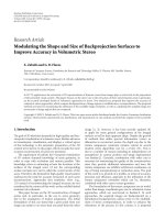
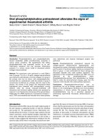
Oral keratinocytes trans infect HIV-1 to permissive PBMCsFigure 1
Oral keratinocytes trans infect HIV-1 to permissive PBMCs. TERT-2 or TE monolayers were inoculated and incubated
for 6 h with lab-adapted HIV-1, IIIb or BaL. Tonsils were obtained from six donors (tissues 144, 193, 195, 196, 1101, and 223).
Cells from each donor were propagated separately and TE cells were cultured as described in Materials and Methods. After
incubation, cells were trypsinized, washed to remove non-internalized particles, and then co-cultured with PHA-activated
PBMCs (2 × 10
5
cells) in PBMC growth media. To estimate HIV-1 trans infection from keratinocytes, PBMCs supernatants were
collected on day 9 post inoculation and p24
gag
expression was estimated using ELISA. (A) TE cells from each donor differentially
trans infect HIV-1 to PBMCs. (B) TERT-2 and TE 223 cells were tested side-by-side in the same experiments to compare HIV
uptake and transfer. Mouse fibroblast cells (NIH 3T3) were included as a negative control. (C) To investigate the rate of HIV-1
trans infection over time, TERT-2 cells were trypsinized and washed to remove extracellular HIV-1 at indicated times post
inoculation. TERT-2 cells from each time point were then co-cultured with PBMCs and p24
gag
production was analyzed. TERT-
2 cells incubated with media only (no virus; NV) or heat-inactivated HIV-1 BaL (HV) were included as negative controls. Data
in panel A represent the mean ± standard deviation of triplicate determinations in one experiment since the availability of pri-
mary tonsil cells from each donor was limited. Data in panel B and C are reported as the mean ± standard deviation from three
independent experiments each performed in triplicate.
Retrovirology 2008, 5:66 />Page 4 of 14
(page number not for citation purposes)
out trypsin for flow cytometry analysis. TERT-2 cells were
negative for CD4 as expected (Table 1) and 80% of the
cells were positive for CD104, a β4-integrin chain gener-
ally expressed by epithelial cells [32] (Table 1). TERT-2
cells were also positive for HSPGs (91 ± 1%) and less fre-
quently positive for the HIV-1 co-receptor CXCR4 (3.5 ±
2%) and galactosylceramide (GalCer) (<1%). Unlike sali-
vary gland epithelial cells [20], less than 1% of TERT-2
cells expressed the CCR5 co-receptor for HIV-1. TE cells
(tissue nos. 164, 193 and 196) were also analyzed for
putative HIV-1 receptors and co-receptors (Table 1). TE
cells did not express CD4, or CXCR4 or CCR5 (< 1%).
When compared to TERT-2 cells, TE cells express GalCer
(4 ± 0.1%) with similar frequency, but HSPGs (13 ± 7%)
are expressed less frequently.
TERT-2 oral keratinocytes support HIV-1 reverse
transcription and integration
To demonstrate reverse transcriptase activity, we infected
TERT-2 cells with pseudotype HIV-NL4-3Δenv-EGFP par-
ticles, which express the vesiculostomatitis virus glycopro-
tein (VSV-G) envelope (virus-like particles; VLPs). When
infected, cells express EGFP as a reporter for HIV-1 LTR
promoter activity and expression of viral-specific proteins.
When TERT-2 cells were inoculated with VLPs at a MOI of
10, EGFP was expressed at a high level, confirming reverse
transcriptase activity, LTR promoter activity and expres-
sion of new viral-specific proteins (reported by EGFP)
(Fig. 2A). When the cells were pre-treated with increasing
amounts of the viral inhibitor AZT (5 to 2500 μM), EGFP
expression was inhibited in a dose-dependent manner
(Fig. 2A). Integration and expression of HIV-1 specific
proteins was stable since EGFP was expressed after 10 pas-
sages of TERT-2 cells (data not shown).
To estimate the kinetics of the HIV LTR promoter activity,
EGFP expression was analyzed at indicated times post
inoculation with VLPs (Fig. 2B). EGFP expression was first
detected at approximately 18 h post inoculation and max-
imized at 48 h, reflecting the time course of activation of
the HIV LTR promoter in infected cells.
To confirm HIV-1 integration in TERT-2 cells, we infected
TERT-2 monolayers with HIV-1 strains IIIb or BaL and
then performed a nested PCR with HIV- and human alu-
specific primers (Table 2). These PCR reactions amplify
HIV-1 sequences integrated in human genomic DNA. In
preliminary experiments, we showed that laboratory
stocks of HIV-1 are contaminated with DNA that is
acquired from PBMCs during viral propagation (data not
shown). The contaminating DNA was substantially resist-
ant to DNase treatment of the HIV-1 stocks and could be
amplified as a false-positive indication of integration. To
eliminate contaminating sources on the plasma mem-
brane or in the cytoplasm, integrated HIV-1 DNA was
extracted directly from TERT-2 cell nuclei.
Infected TERT-2 nuclei contained integrated copies of
HIV-1 from HIV strains IIIb and BaL, but only IIIb is
shown (Fig. 3). Nested-PCR products were detectable in
TERT-2 cell nuclei between 3 and 72 h post inoculation.
An attenuated signal persisted after subculturing the cells
for 1 to 3 passages, showing that integration is stable.
Nuclei extracted from ACH-2 cells, an HIV
LAV
latent T cell
clone [33], and HIV-infected PBMCs also contained HIV
integrated DNA and served as positive controls. In con-
trast, integrated HIV-1 DNA was not detected in TERT-2
nuclei incubated in the absence of HIV-1 (NV), when HIV-
1 was heat-inactivated (HV), or when cells were pre-
treated with AZT (500 μM) or colchicine (500 μM).
From 3 to 72 h post inoculation, but not after subculture,
total linear HIV DNA was detected in the nuclei of TERT-
2 cells but not in the negative controls. Consistent with
the low level of integration, HIV DNA two-LTR circles
were not detected in TERT-2 cells except for a weak signal
at 6 h post inoculation and not detected in the negative
controls. When TERT-2 cells were pre-treated with increas-
Table 1: Putative HIV receptor expression on oral keratinocytes
Receptor Function TE
(Mean ± SD)
a
TERT-2
(Mean ± SD)
b
CD104 (β4 integrin) transmembrane protein expressed predominantly in
epithelial cells [32]
80 ± 11 83 ± 4
HSPGs HIV gp120 binding [70] 13 ± 7 91 ± 20
GalCer HIV gp120 binding [71] 4 ± 0.1 < 1
CD4 HIV gp120 binding [72] < 1.0 < 1
CXCR4 X4-tropic chemokine co-receptor [73] < 1.0 3.5 ± 2
CCR5 R5-tropic chemokine co-receptor [73] < 1.0 < 1
CD3, CD11a/LFA-1, CD32, CD64, CD89, DC-SIGN,
Macrophage Mannose Receptor
< 1.0 Not tested
Human fibroblast 4 ± 4 Not tested
a
Mean ± SD of four independent experiments (1 experiment from tissue no. 164 and 193, and 2 experiments from tissue no. 196)
b
Mean ± SD of three independent experiments
Retrovirology 2008, 5:66 />Page 5 of 14
(page number not for citation purposes)
ing doses of AZT for 2 h followed by incubation with HIV-
1 for 6 h, integration of HIV-1 DNA was inhibited. Results
with BaL were similar (not shown).
New HIV RNA transcripts in TERT-2 cells
Using RT-PCR, we attempted to detect new HIV-specific
transcripts, including multiply spliced HIV-1 RNA,
unspliced HIV-1 RNA, and U3-U5 HIV-1 RNA in TERT-2
cells. TERT-2 cells were incubated with HIV-1 IIIb or BaL
for 6 h, trypsinized, washed, and incubated for up to 72 h.
Some cells were sub-cultured after infection. Although we
detected multiply spliced and unspliced products when
using specific primers, these transcripts could not be dis-
tinguished clearly from contamination (data not shown).
Multiply spliced and unspliced HIV-1 RNA appeared to
degrade and were not detected after 12 h post inoculation.
HIV-1 RNA species were also undetected after cells were
sub-cultured and U5-U3 HIV-1 RNA was not detected at
any time (data not shown).
New HIV-specific transcript levels were also estimated by
SYBR real time RT-PCR relative to the level in the viral
inoculum (data not shown). Relative to levels in the viral
inoculum, multiply spliced and singly spliced HIV-1
RNAs were barely detectable.
HIV-1
gag
-specific RNA, however, was detectable. Using
real time RT-PCR, HIV
gag
-specific RNA was quantified and
the expression relative to 0 h was determined (Fig. 4). In
TERT-2 cells, HIV
gag
-specific RNA appeared to increase up
to 6 h post inoculation, suggesting that HIV-1 binds and
enters TERT-2 cells. By 24 h post inoculation, however,
the amount of HIV
gag
-specific RNA declined below the
level of detection. If replication occurred, HIV-1
gag
-specific
RNA was expected to increase during the 72 h incubation.
The HIV-1
gag
-specific RNA decayed over time, however,
and was not a product of new transcriptional events. In
TERT-2 cells, therefore, RNA products of the HIV replica-
tion cycle were not prominent and replication appeared to
abort.
HIV-1 harboring in TERT-2 keratinocytes
To study harbored HIV-1, TERT-2 monolayers were incu-
bated with HIV-1 for 6 h, then trypsinized, and washed to
eliminate non-internalized viral particles. TERT-2 monol-
ayers were maintained in culture for the indicated times
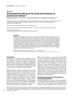
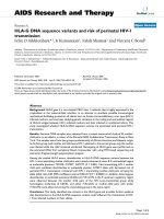
Replication-incompetent HIV-NL4-3Δenv virus-like particles infect TERT-2 keratinocytesFigure 2
Replication-incompetent HIV-NL4-3Δenv virus-like particles infect TERT-2 keratinocytes. Replication-incompe-
tent HIV-NL4-3Δenv virus-like particles (VLPs) were packaged in 293T cells to express VSV-G protein as described in Materi-
als and Methods. The TCID
50
of VLPs was determined by titration in TZM-bl cells, and TERT-2 monolayers were then
incubated for 6 h with VLPs at a MOI 10 (TCID
50
per cell). Cells were then washed, trypsinized to remove unincorporated
VLPs, and incubated for up to 48 h. Post inoculation, cells were fixed in 2% paraformaldehyde and nuclei were stained with
DAPI (blue). (A) TERT-2 cultures were pre-incubated with AZT (0 to 2500 μM) before inoculation with VLPs. The expression
of EGFP reporter gene was analyzed at 48 h post inoculation. (B) Kinetics of EGFP expression from 18 h to 48 h post inocula-
tion. TERT-2 cells incubated with envelope-deficient particles were included as a negative control (48 h post inoculation).
Arrows indicate EGFP expressing TERT-2 cells (green). Scale bar represent 50 μm. Images are representative of three inde-
pendent experiments.
Retrovirology 2008, 5:66 />Page 6 of 14
(page number not for citation purposes)
up to 120 h post inoculation. To determine release of
infectious virions, TERT-2 cell supernatants were aspirated
(contains HIV-1 released from TERT-2 cells) and inocu-
lated into PHA-activated PBMCs. After the infectious
supernatants were aspirated, the TERT-2 cells were co-cul-
tured separately with PHA-activated PBMCs to determine
harbored virus available for direct transfer. TERT-2 cells
appeared to harbor and trans infect X4- and R5 HIV-1 to
permissive PBMCs (Fig. 5A). In contrast, supernatants
from TERT-2 monolayers were barely infectious (Fig. 5B).
HIV-1 trans infection by TERT-2 cells decreased to unde-
tectable levels at 48 h (Fig. 5A and 5B), suggesting that
harbored virus had decayed.
MOLT-4/CCR5 cells acquire VLPs from infected TERT-2
cells
To confirm that PBMCs acquire HIV-1 primarily by cell-
to-cell interactions, TERT-2 cell monolayers were inocu-
lated with replication-incompetent HIV-NL4-3Δenv-
EGFP particles (VLPs) for 6 h at MOI 100. These VLPs rep-
licate for a single round in the cell that ultimately
becomes infected. TERT-2 cells with harbored, non-repli-
cating HIV VLPs do not express EGFP. TERT-2 cells were
then co-cultured with permissive MOLT-4/CCR5 T cells
(MOLT-4/CCR5). VLP trans infection was determined by
EGFP expression in MOLT-4/CCR5 at 48 h after co-cul-
ture. At 6 h post inoculation, TERT-2 cells captured and
transferred 56% of the VLP inoculum to infect MOLT-4/
CCR5 (Fig. 6). When TERT-2 cells were treated with
trypsin at 6 h and washed to inactivate and remove extra-
cellular virus, 18% of the VLP inoculum was transferred
from within the TERT-2 cells to infect co-cultured MOLT-
4/CCR5 cells. When TERT-2 cells were treated with colch-
icine (500 μM for 30 min before inoculation) to uncouple
the tubulin cytoskeleton, 18% of the VLP inoculum was
harbored and transferable to MOLT4 cells. At 4°C, 7%
was harbored by untreated TERT-2 cells. A small percent-
age of VLPs were resistant to trypsin and colchicine treat-
Table 2: Primer sequences and PCR conditions
Target Primer Sequences (5'-3') PCR conditions
Integrated HIV-1
DNA
a
• First round PCR L-M667 ATGCCACGTAAGCGAAACTCTGGCTAACT
AGGGAACCCACTG
95°C, 8 min and 95°C, 10 s, 60°C, 10 s, 72°C,
170 s for 12 cycles
Alu 1 TCCCAGCTACTGGGGAGGCTGAGG
Alu 2 GCCTCCCAAAGTGCTGGGATTACAG
• Second round PCR Lambda T ATGCCACGTAAGCGAAACT 95°C, 8 min and 95°C, 10 s, 60°C, 10 s, 72°C, 9 s
for 40 cycles
AA55M GCTAGAGATTTTCCACACTGACTAA
Linear HIV DNA
a
MH531 TGTGTGCCCGTCTGTTGTGT 95°C, 8 min and 95°C, 10 s, 60°C, 10 s, 72°C, 6 s
for 40 cycles
MH532 GAGTCCTGCGTCGAGAGAGC
2-LTR circle
a
HIV F GTGCCCGTCTGTTGTGTGTGACT 95°C, 8 min and 95°C, 10 s, 60°C, 10 s, 72°C, 10
s for 40 cycles
HIV R ACTGGTACTAGCTTGTAGCACCATCCA
U5-U3 RNA
a
HIV F GTGCCCGTCTGTTGTGTGTGACT 95°C, 2 min and 95°C, 5 s, 60°C, 10 s, 72°C, 10 s
for 40 cycles
HIV R ACTGGTACTAGCTTGTAGCACCATCCA
Gag For CCCATAGTGCAGAACATCCA 50°C, 2 min, 95°C, 2 min, and 95°C, 15s and
60°C, 30s, for 50 cycles
Rev GGGCTGAAAGCCTTCTCTTC
Singly spliced
b
M669 GTGTGCCCGTCTGTTGTGTGACTCTGGTA
AC
50°C, 2 min, 95°C, 2 min, and 95°C, 15s and
60°C, 30s, for 50 cycles
La 23 GCCTATTCTGCTATGTCGACACC
Multiply spliced HIV RNA
a
P659 GACTCATCAAGTTTCTCTATCAAA 95°C, 4 min and 95°C, 5 s, 54°C, 10 s, 72°C, 8 s
for 40 cycles
P413MOD AGTCTCTCAAGCGGTGGT
Unspliced HIV RNA
a
La 9 GACGCTCTCGCACCCATCTC 95°C, 2 min and 95°C, 10 s, 60°C, 40 s for 40
cycles
La 8.1 CTGAAGCGCGCACGGCAA
β-actin Actin F ATGGCCACGGCTGCTTCCAGC 95°C, 15 s, 55°C, 30 s, 72°C, 15 s for 30 cycles
Actin R CATGGTGGTGCCGCCAGACAG
GAPDH GAPDH F GAGTCAACGGATTTGGTCGT 95°C, 15 s, 60°C, 30 s, 72°C, 15 s for 30 cycles
GAPDH R TTGATTTTGGAGGGATCTCG
a
Primer sequences and PCR conditions were modified from [40]
b
Primer sequences and PCR conditions were modified from [74]
Retrovirology 2008, 5:66 />Page 7 of 14
(page number not for citation purposes)
ments, infecting MOLT-4/CCR5 cells (4%) when co-
cultured with TERT-2 cells. VLPs could bind TERT-2 cells
at 4° or 37°C, but VLPs could be internalized efficiently
only at 37°C suggesting that microtubule activity was
required for internalization. The fraction of VLPs that
resisted trypsinization and were sensitive to cold and col-
chicine appeared to be harbored within TERT-2 cells and
transferred to MOLT4/CCR5 cells by direct cell-to-cell
interactions.
Discussion
Lining the oral and oropharyngeal mucosal surfaces, oral
keratinocytes are potential targets for primary HIV-1 infec-
tion, harboring and dissemination. We now show for the
first time that oral keratinocytes harbor and transfer viable
HIV-1 to infect permissive cells for up to 48 h. Therefore,
HIV-1 internalization by oral keratinocytes in vitro
[18,20,22,34] may model an overlooked mechanism for
HIV transmission and dissemination in vivo.
Although conventional wisdom suggests that primary
human infection of permissive cells actually occurs in the
gut [9,35], the oral mucosa in primate models becomes
infected within a day of atraumatic oral mucosal exposure
to SIV-1 [8,36]. Infection becomes marked in the GI tract
four days after initial exposure, suggesting that virus dis-
seminates from an oral focus.
Among oral mucosal sites, palatine tonsils are likely to
disseminate HIV-1 since tonsil epithelial cells express
appropriate receptors in situ [37,38], and trans infect HIV-
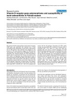
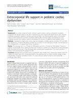
Integrated HIV-1 DNA detected in TERT-2 nucleiFigure 3
Integrated HIV-1 DNA detected in TERT-2 nuclei. TERT-2 cells were grown in monolayers and inoculated with HIV-1
(IIIb or BaL). Some cells were sub-cultured after infection. At 0.5 to 72 h post inoculation, TERT-2 cell nuclei were isolated as
described in Materials and Methods. DNA was extracted from the nuclei and analyzed for integrated HIV DNA, linear HIV
DNA, and 2-LTR circular HIV DNA. β-actin was included as a loading control. PCR reactions were performed as described in
Materials and Methods (Table 2). Negative controls include cells without HIV-1 (NV), cells inoculated with heat-inactivated
HIV-1 (HV), cells pretreated with 500 μM AZT (AZT), cells pretreated with colchicine (Col), and PCR reactions with no tem-
plate (W). PBMC media (uninfected with HIV-1) and samples that were amplified in the second PCR only were also negative
for HIV DNA (data not shown). ACH-2 cells and HIV-1-infected PBMCs served as positive controls for detection of integrated
HIV DNA, linear HIV DNA, and circular HIV DNA. These agarose gel data for HIV-1 IIIb infection are representative of three
independent experiments.
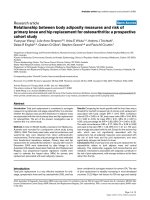
HIV infection aborted in TERT-2 keratinocytesFigure 4
HIV infection aborted in TERT-2 keratinocytes.
TERT-2 cell monolayers were incubated with HIV-1 (IIIb or
BaL), trypsinized and washed. Cells were sub-cultured at 48
h post incubation. At 0.5 to 72 h post incubation, total RNA
was isolated and cDNA was synthesized as described in the
Materials and Methods. HIV
gag
-specific RNA was detected by
SYBR real time PCR. β-actin served as the reference house-
keeping gene. Data are the mean ± standard deviation of
three independent experiments, each performed in triplicate.
Retrovirology 2008, 5:66 />Page 8 of 14
(page number not for citation purposes)
1 to permissive cells in vitro. HIV trans infection in vitro
from primary TE cells to PBMCs showed variation among
tonsil donors. Since TE cells were derived from excised
tonsils obtained with uncharacterized inflammatory
backgrounds, proinflammatory cytokines might be differ-
entially expressed in TE cells. Some cytokines may modu-
late HIV entry (reviewed in [39]), but whether donor-
specific expression patterns affect primary infection is not
known. Clearly, keratinocyte-associated virions remain
infectious and can be transferred to infect co-cultured per-
missive cells.
Oral keratinocytes support the life cycle of HIV-1 step-by-
step until integration. HIV
gag
-specific RNA peaked in
TERT-2 cells after a 6 h incubation with HIV-1, which is
consistent with the internalization of HIV-1 genomic RNA
over time (Fig. 4; [40]). After internalization in TERT-2
cells, HIV-1 begins a replication cycle, which is not com-
pleted. HIV-1 genomic RNA is reverse transcribed into
DNA, which can be inhibited by treatment of the cells
with AZT (Fig. 2A). Among other reverse transcriptase
products, linear HIV DNA was detected in oral keratinoc-
ytes, whereas two-LTR circles, stable forms of unintegrated
HIV DNA, were not seen. This pattern of products is con-
sistent with the low level of HIV-1 integration into TERT-
2 cell genomic DNA.
To clarify the viral life cycle in TERT-2 cells, the presence
of integrated HIV DNA was sought as a product of reverse
transcriptase activity. Integrated HIV DNA was consist-
ently detected in TERT-2 cell nuclei (Fig. 3). After incuba-
tion with HIV-1 in vitro, Liu et al. [20] had previously
reported that oral keratinocytes contain linear HIV-spe-
cific DNA. We noted that contaminating DNA from the
propagating cells is present in the viral inoculum and can
be amplified by nested PCR, giving a false indication of
integration. To avoid this artifact, we isolated HIV-1 DNA
directly from the TERT-2 cell nuclei. As previously
reported in permissive cells [40], integrated HIV DNA is
detected consistently in TERT-2 nuclei and in all keratino-
cyte lines tested (data not shown) as early as 3 h post inoc-
ulation (Fig. 3). Integrated HIV DNA persisted in the
TERT-2 genome after several passages of the cells, but the
signal decayed for linear HIV DNA. To this point, replica-
tion kinetics in oral keratinocytes and permissive cells
[40] were similar. The HIV-1 life cycle in TERT-2 cells was
marked by viral internalization, uncoating, reverse tran-
scriptase and integrase activities.
In response to infection by HIV-1 IIIb or BaL, the rate of
decay of nonintegrated linear HIV-1 DNA in TERT-2 cells
appeared to be too rapid to support substantial gene
expression [40]. Likewise, we were unable to detect the
Infectious HIV-1 harbored by TERT-2 cellsFigure 5
Infectious HIV-1 harbored by TERT-2 cells. TERT-2 monolayers were incubated for 6 h with HIV-1 (IIIb or BaL). TERT-
2 cells were then trypsinized, washed, and maintained in growth media. At the indicated time post inoculation, TERT-2 cells
were co-cultured with (A) PHA-activated PBMCs to test for direct transfer of HIV-1. To learn if infectious HIV-1 is released
from TERT-2 cells, (B) spent media were recovered and used to inoculate PHA-activated PBMCs. After exposure to TERT-2
cells or media, PBMC supernatants were harvested at day 9 and analyzed for p24
gag
production by ELISA. Data shown are the
mean ± standard deviation from three independent experiments, each performed in triplicate.
Retrovirology 2008, 5:66 />Page 9 of 14
(page number not for citation purposes)
specific RNA product U5-U3 RNA. HIV-1 specific mRNAs
appeared at levels that could not be clearly distinguished
from contamination (data not shown). After low-level
integration, therefore, HIV-1 replication aborts.
Many steps in the HIV life cycle may be restricted by
intrinsic cellular factors targeting viral entry, viral uncoat-
ing, viral DNA synthesis, intracellular trafficking of viral
nucleic acids, integration, viral gene expression or viral
packaging [41]. TERT-2 cells clearly restrict HIV replica-
tion after integration when infected with HIV-1. We
sought to determine whether the internalization pathway
used by HIV-1 in TERT-2 cells contributed to the restric-
tion. Therefore we inoculated TERT-2 cells with VSV-G
pseudotyped HIV-NL4-3Δenv-EGFP particles, which
internalizes promiscuously into an endosomal pathway
[42]. When integrated, HIV LTR from the pseudotyped
particles regulates green fluorescence expression in TERT-
2 cells (Fig. 3A and 3B). When the conventional, gp120-
mediated viral entry is circumvented, the VSV-pseudo-
typed HIV-1 particles integrate and new RNA is tran-
scribed. Since EGFP is expressed, viral-specific proteins are
likely to be synthesized. This is in contrast to infection
with the wild-type HIV-1 strains, where new transcripts
are minimally expressed. Hence the CD4- and CCR5-inde-
pendent internalization may represent a major restriction
against HIV-1 replication.
The HIV entry mechanisms in oral keratinocytes and other
epithelial cells are not well understood. Unlike oral kerat-
inocytes, gastrointestinal epithelial cells constitutively
express CCR5 and selectively internalize R5-tropic HIV-1
[43]. Oral keratinocytes from different sources are CD4-
and express different putative receptors and co-receptors
for HIV-1 including galactosylceramide [20] and heparin
sulfate proteoglycans (HSPGs) [37,44-51]. HSPG binds
HIV-1 gp120 [16,47,52-54], which can enter endosomes
[55,56] and enable co-localization of HIV-1 particles with
endosomal markers in TERT-2 cells (Dietrich E. et al, in
preparation). Except for HSPGs, most putative receptors
and co-receptors for HIV-1 are inconsistently expressed
(Table 1) and can vary with the microanatomic location
[37].
Although we saw no evidence of new HIV transcripts or
newly replicated virions, TERT-2 cells clearly harbor infec-
tious HIV-1 virions. Harbored HIV-1 can be effectively
transferred to infect permissive cells including PBMCs for
up to 48 h, but appear to become less infectious during
the interval from 6 to 48 h after inoculation. After 48 h,
TERT-2 cells were ineffective at trans infecting cell-associ-
ated harbored virus (Fig. 5A) and infectious supernatants
(Fig 5B) to activated PBMCs. Since most experiments were
performed after trypsinizing TERT-2 cells to remove extra-
cellular virus, internalized HIV-1 was a harbored infec-
tious reservoir.
HIV uptake and transfer are temperature and microtubule
dependent (Fig. 6), as reported for endothelial cells [16].
With trypsin or colchicine treatment, harbored, internal-
ized particles were distinguished from surface-bound par-
ticles. Both surface-bound and internalized particles are
infectious and effectively trans infect CD4+ cells (Fig. 6).
Cell-associated particles effectively trans infect PBMCs and
MOLT-4 cells. Few infectious viral particles are released
from TERT-2 cells. Optimal HIV transfer from TERT-2 cells
is suggested therefore to involve direct cell-to-cell interac-
tions with PBMCs and other permissive cells.
In the oral mucosa, the transfer of infectious virus to prox-
imal lymphoid cells may be of clinical importance. Proxi-
mal to mucosal stratified squamous keratinocytes,
Langerhans cells and CD4-positive lymphocytes are avail-
able to be trans infected in vivo. Indeed, a recent report
TERT-2 cells trans infect VLPs to MOLT-4/CCR5 cellsFigure 6
TERT-2 cells trans infect VLPs to MOLT-4/CCR5
cells. TERT-2 cell monolayers were incubated for 6 h at
37°C with replication-incompetent HIV-NL4-3 particles
pseudotyped to express VSV-G envelope (VLPs) at a MOI
100. Cells were washed, then co-cultured with MOLT-4/
CCR5 (2 × 10
5
) cells, and EGFP expression in MOLT-4/
CCR5 cells was analyzed at 48 h using flow cytometry. The
percentage of infected MOLT-4/CCR5 cells was quantified.
Some TERT-2 monolayers were either treated with trypsin,
colchicine (500 μM for 30 min), pre-cooled to 4°C, trypsin
and pre-cooled to 4°C, or trypsin and colchicine as described
in the Materials and Methods. Data shown are the mean ±
standard deviation from three independent experiments.
Retrovirology 2008, 5:66 />Page 10 of 14
(page number not for citation purposes)
suggests that Langerin-positive dendritic cells degrade
internalized HIV-1, reducing transfer to CD4+ T cells in
the mucosa [57], while others show that activated CD34-
positive Langerhans cells increase trans infection of per-
missive target cells [58]. Unlike the female genital epithe-
lium [17], oral Langerhans cells (dendritic cells) are not
known to sample antigens or capture HIV-1 at the
mucosal surface. Oral mucosal keratinocytes, therefore,
could contribute to HIV transmission in vivo, however, by
activating and trans infecting Langerhans cells, which can
dock and transfer virus to CD4+ cells, or by transferring
infectious harbored HIV-1 particles to proximal permis-
sive cells.
For the first time, we show that oral keratinocytes become
infected by HIV-1, initiating a defined, truncated viral life
cycle. While infection is non-productive, an intracellular
pool of infectious HIV-1 is harbored for up to 48 h and
fully capable of trans infecting CD4+ permissive cells.
Hence, the oral epithelium may actively disseminate HIV-
1 infection and is more than an inert barrier. Since R5-
tropic HIV-1 is most frequently associated with primary
infections, oral epithelium could function as a selective
"gatekeeper" and exclude X4-tropic virus. When com-
pared, oral keratinocytes from different sources selectively
harbor and transfer HIV-1 in either an X4- or R5-tropic
HIV-1-specific manner (data not shown). TERT-2 cells
consistently harbor all HIV-1 strains tested, while primary
tonsil epithelial cells from some donors did not support
trans infection (Fig. 1A). Since CXCR4+ CCR5- TERT-2
cells (Table 1) appear to harbor R5-tropic HIV-1 BaL more
effectively than IIIb (Fig. 4), infection appears to be inde-
pendent of the co-receptor tropism of the HIV envelope
protein. We have recently shown that the endogenous oral
pathogen, Porphyromonas gingivalis, selectively up-regu-
lates CCR5 on CXCR4+ oral keratinocytes [59]. Up-regu-
lation of CCR5 selectively promotes the harboring and
transfer of R5-tropic HIV-1 from TERT-2 cells to permis-
sive targets [60].
If oral mucosal keratinocytes serve as a clinical focus for
HIV-1 infection, endogenous restriction factors notwith-
standing, novel uptake, harboring and transfer mecha-
nisms may become potential targets for antiviral drugs
and vaccines. Following the initial short period of primary
virus exposure, infectious HIV-1 persists in oral keratinoc-
ytes for several days. The harbored virus could be trans-
ferred to permissive cells and arguably serve to
disseminate infection systemically. In the oral cavity, sali-
vary components have been suggested to reduce the risk of
HIV transmission [61-63]. For example, salivary mucins
agglutinate the virus in vitro and appear to reduce viral
uptake into permissive cells [64]. In the presence of saliva,
however, HIV-1 still internalizes into oral keratinocytes in
vitro and infectious virus can be effectively transferred to
permissive reporter cells (Dietrich et al, 2008 in prepara-
tion). The rate of uptake of infectious HIV-1 into oral
keratinocytes in the presence of saliva appears to occur
more rapidly than complete inactivation of virus. Even in
the presence of saliva, shedding oral epithelial cells may
also serve as an infectious source for HIV transmission
during oral sexual contacts. To protect against mucosal
HIV transmission and dissemination, therefore, mucosal
vaccines and microbicides should target the viral reservoir
in oral keratinocytes.
Conclusion
The oral mucosa is exposed to infectious HIV-1 during
oral-sexual contact and breast-feeding. The surface oral
and oropharyngeal epithelium is a potential site of pri-
mary HIV infection and dissemination even though these
cells do not express the common HIV-1 receptors and co-
receptors found on permissive cells. Using an atypical
uptake mechanism (CD4-independent), oral epithelial
keratinocytes were hypothesized to capture or internalize
infectious HIV-1 and reverse transcribe the RNA HIV-1
genome into DNA, which then integrates into the kerati-
nocyte genome. For the first time, integration, a major fea-
ture of infection, is shown to persist in daughter cells after
the keratinocytes divide. After integration, the life cycle of
the virus aborts and no newly assembled virus particles
are detectable. By using HIV-1 that was engineered to
bypass the usual receptors, we showed that the virus life
cycle is prolonged. Although the life cycle aborts, captured
infectious HIV-1 is harbored for at least 48 h and trans-
ferred to highly permissive peripheral blood mononu-
clear cells, which in vivo could result in systemic CD4+ T
cell infection. While often considered passive bystanders
in HIV-1 infection, mucosal epithelial cells could be
actively providing a route to systemic infection.
Materials and methods
Cells
OKF6/TERT-2 immortalized keratinocytes (TERT-2), pro-
vided by Dr. James G. Rheinwald (Harvard Medical
School, MA) were cultured in Keratinocyte-SFM (Invitro-
gen) supplemented (to final concentrations) with 0.2 ng/
mL recombinant epidermal growth factor (rEGF; Invitro-
gen), 25 μg/mL bovine pituitary extract (BPE), and 0.4
mM CaCl
2
. Tonsil epithelial cells (TE) were isolated from
tissue excised from HIV-seronegative individuals under-
going tonsillectomy at Hennepin County Medical Center,
Minneapolis, MN. Use of surgical waste TE cells in
research was reviewed and approved by the Research and
Development Committee of the Minneapolis VA Medical
Center and the Human Subjects Research Committee of
the Hennepin County Medical Center. The protocol was
determined to be exempt upon full IRB review and no
subject consent was necessary. For culture, tonsillar epi-
thelial cells were prepared by a modified method of Oda
Retrovirology 2008, 5:66 />Page 11 of 14
(page number not for citation purposes)
and Watson [65]. Briefly, tissue was cut and digested at
4°C overnight in 0.2% Dispase grade II (Boehringer Man-
nheim) in MEM supplemented with 10% FBS. The next
day, epithelial sheets were separated from connective tis-
sue, digested using 0.05% Trypsin/0.53 mM EDTA
(GIBCO) at 37°C for 5 min, and dispersed into single cell
suspensions using a pipette. Cells were cultured in kerati-
nocyte-SFM supplemented with 5 ng/mL hEGF, 30 μg/mL
BPE, and 0.06 mM CaCl
2
. For use in the experiments, TE
cells from passage 3 or 4 were seeded at 10
4
cells/cm
2
.
Molt-4/CCR5, ACH2, and TZM-bl cells were provided by
the NIH AIDS Research and Reference Program. MOLT-4/
CCR5 T cells were cultured in RPMI medium 1640 (Invit-
rogen) supplemented with 10% FBS and 1 mg/mL G418
sulfate. TZM-bl, and 293T were cultured in Dulbecco's
Modified Eagle Medium (D-MEM; Invitrogen) containing
10% FBS. Peripheral blood mononuclear cells (PBMCs)
were isolated from buffy coats obtained from 10 healthy
seronegative donors by Ficoll-Histopaque density gradi-
ent centrifugation [66] and cryopreserved in liquid nitro-
gen until use. Source leukocytes from healthy adult
donors were purchased from the Memorial Blood Centers.
The Memorial Blood Centers IRB reviewed and approved
the protocol. As part of the consent process, the blood
donors agreed that their donated blood could be used for
research purposes. When needed, PBMCs (2 × 10
6
cells/
mL) were activated overnight in PBMC media (RPMI1640
medium containing L-glutamine (Mediatech, Inc.), 5%
human interleukin-2 (Roche) with 10% FBS) supple-
mented with 5 μg/ml phytohemagglutinin (PHA-P;
Sigma). After activation, cells were washed to remove
PHA-P and cultured for 3 days in PBMC media before use.
Viruses
HIV-1 strains IIIB (X4-tropic) and BaL (R5-tropic) were
obtained from the NIH AIDS Research and Reference Rea-
gent program. HIV-1 was propagated and TCID
50
of virus
stocks was determined in PHA-activated PBMCs as
described in the Manual for HIV Laboratories, National
Institutes of Health, Division of Acquired Immune Defi-
ciency Syndrome (DAIDS) Virology (Publication NIH-97-
3838).
Virus-like particles
Plasmids encoding non-replicative NL4-3 (pNL4-3-Δenv-
EGFP; Catalog number 11100) and the vesicular stomati-
tis virus G (VSV-G) glycoprotein (pHEF-VSV-G; Catalog
number 4693) were obtained from NIH AIDS Research
and Reference Reagent Program. To generate VLPs, 293T
cells were transiently transfected with pNL4-3-Δenv-EGFP
(10 μg) and pHEF-VSV-G (1 μg), using calcium phosphate
precipitation as described previously [67]. To determine
TCID
50
of VLPs, TZM-bl cells (1 × 10
4
cells/well) were cul-
tured overnight in 96-well tissue culture plates, and then
incubated with six replicates of ten serial dilutions (1:4) of
a VLPs stock in 50 μl growth media per well with the addi-
tion of 10 μg/mL Sequa-brene (Sigma). After 2 h, cells
were washed three times and incubated in 200 μl of
growth media. After 48 h, cells were fixed with 0.05% glu-
taraldehyde for 5 min at room temperature and washed
twice with Dulbecco's phosphate-buffered saline (Medi-
atech, Inc.; DPBS). To detect the expression of β-galactos-
idase, cells were stained with 1 mg/mL X-Gal in 5 mM
KFe
4
(CN
6
) 3H
2
O, 5 mM KFe
3
(CN
6
) 3H
2
O, and 1 mM
MgCl
2
and incubated at 37°C for 2 h. A positive well con-
tained two or more blue cells. Positive- and negative-
stained wells were tabulated and TCID
50
was calculated
using the Reed-Muench TCID
50
calculation [68].
Flow cytometry
TERT-2 or TE cells were washed once with DPBS and incu-
bated with 0.02% (W/V) EDTA for 10 min. Detached cells
were washed twice with DPBS supplemented with 2% FBS
(wash buffer), and resuspended at 5 to 10 × 10
5
cells in
200 μL wash buffer. To identify putative HIV receptors
and co-receptors, cells were incubated at 4°C for 30 min
with 1 μg of anti-CD104, CXCR4, CCR5, galactosylcera-
mide (GalCer), heparin sulfate (HSPGs), DC-SIGN, or
macrophage mannose receptor (Table 1). Similarly, to
characterize the purity of primary tonsil keratinocytes in
culture, antibodies against CD3, CD4, CD11a/LFA1,
CD32, CD64, CD89, and human fibroblast were used
(Table 1). All antibodies were obtained from BD
Pharmingen, except anti-GalCer (Chemicon), anti-
heparin sulfate (Seikagaku) and anti-human fibroblasts
(Sigma). Cells were then washed twice with 1 mL wash
buffer to remove unbound antibody. If needed, cells were
stained with goat anti-mouse IgG or IgM conjugated with
fluorescein isothiocyanate (FITC) (Jackson ImmunoRe-
search Laboratories, West Grove, PA) in 200 μL wash
buffer at 4°C for 30 min to detect primary antibodies. Iso-
type controls and other staining controls were included.
After staining, cells were washed three times with 1 mL
wash buffer, fixed in 200 μL of 2% paraformaldehyde,
and stored at 4°C until analysis using a FACSVantage SE
flow cytometer (BD Biosciences).
HIV infection
To infect with HIV-1, TERT-2 cells were plated in 96-well
tissue culture plates (1.5 × 10
4
cells/well) and grown over-
night in monolayers to 80–90% confluence and infected
at a MOI 0.01 (TCID
50
per seeded cells), for 0.5 to 120 h.
Every 48 h, media were replaced with fresh growth media
to maintain viability of TERT-2 and TE cells. In some
experiments, viruses were heat-inactivated (HV) by incu-
bating in a water bath at 70°C for 3 h and used as a nega-
tive control. At indicated times, HIV-1 was aspirated. To
remove surface-bound HIV-1, some cultures were treated
with 0.05% trypsin/0.53 mM EDTA for 3 min at room
temperature, and then an equal volume of soybean
Retrovirology 2008, 5:66 />Page 12 of 14
(page number not for citation purposes)
trypsin inhibitor (250 μg/mL; Invitrogen) in HBSS was
added. Trypsinization did not appear to disrupt the mon-
olayers, which were washed three times in HBSS and
maintained in growth media. Some cells were sub-cul-
tured for 3 to 8 passages post inoculation. In some exper-
iments, cells were pre-treated with azidothymidine (AZT;
500 μM; Sigma) for 2 h, or colchicine (500 μM; Sigma) for
30 min and then inoculated with HIV-1. Colchicine was
washed from cultures before HIV-1 was added, but AZT
remained with TERT-2 cells during HIV incubation. To
determine if reagent carry over inhibited replication in
permissive cells, colchicine or AZT was incubated with
TERT-2 cells. The treated and untreated TERT-2 cells were
co-cultured with MOLT-4/CCR5 cells and VLPs (see
below). Infectivity of VLPs (EGFP expression) in MOLT-4/
CCR5 cells was similar when co-cultured with TERT-2
cells in the presence or absence of AZT, suggesting that
contamination from TERT-2 cell cultures was insufficient
to inhibit infection in permissive lymphoid cells.
PBMC co-culture assays
At indicated times post inoculation, TERT-2 cells were co-
cultured in triplicate wells of 96-well plates with 2 × 10
5
activated PBMCs to estimate trans infection of cell-associ-
ated HIV. Co-culture was performed in 200 μL of PBMC
medium, which selectively supports viability of the
PBMCs at the expense of the TERT-2 cells (require K-SFM
supplement as above). After co-culture with HIV-infected
TERT-2 cells, PBMC media were replaced (100 μL) on day
4, and supernatants were collected (100 μL) on day 9 by
centrifugation at 330 × g for 5 min. The recovery of p24
gag
was estimated in the PBMC supernatants with the Coulter
HIV-1 p24
gag
Antigen Assay (Beckman Coulter) using the
manufacturer's protocol. To estimate the release of HIV-1
from TERT-2 cells, TERT-2 culture supernatants (50 μL)
were collected and then inoculated into 2 × 10
5
activated
PBMCs at selected times post inoculation. p24
gag
produc-
tion was estimated in PBMCs cultures nine days later as
described above.
Identification of integrated HIV DNA, linear HIV DNA and
two-LTR circles
To identify integrated HIV DNA in TERT-2 cells, contami-
nating DNA from viral inocula (MOI 0.01) derived from
propagating cells was carefully excluded. To partition con-
taminating DNA copies from TERT-2 cell integrated HIV
DNA, TERT-2 cells (9 × 10
4
cells) were grown in 6-well
plates and nuclei were isolated using the Nuclei EZ Prep
Nuclei Isolation kit (Sigma). DNA was then extracted
from the nuclei using the DNeasy kit (Qiagen) and quan-
tified spectrophotometrically. PCR reactions contained
500 ng of TERT-2 DNA, primers and PCR conditions were
as described (Table 2; [25,40]). Integrated HIV DNA was
detected by nested PCR to increase sensitivity and fidelity
[25]. PCR products were identified on 3% agarose gels
stained with ethidium bromide.
Analysis of multiply spliced, singly spliced, unspliced and
U5-U3 HIV-1 RNA
Total RNA was collected from infected TERT-2 cells using
Rneasy Plus Mini kit (Qiagen) and quantified spectropho-
tometrically. To detect viral-specific RNA using real time
RT-PCR, 5 μg of total RNA was reverse transcribed to
cDNA using an Iscript™ cDNA Synthesis Kit (BioRad). In
separate PCRs, the cDNA product (10 μl) was incubated
with primers specific to multiply spliced HIV RNA,
unspliced HIV RNA and U5-U3 RNA. Primer sequences
and PCR conditions were as shown (Table 2; [25]). Glyc-
eraldehyde-3-phosphate dehydrogenase (GAPDH)
sequence was amplified as a control. PCR products were
identified on 3% agarose gels stained with ethidium bro-
mide. The concentration and purity of RNA preparations
was performed using the 2100 Bioanalyzer (Agilent).
Total RNA (500 ng) was reverse transcribed to cDNA using
the Superscript III First Strand Synthesis System. The
cDNA was then diluted 1:5 with RNase/DNase free water
and 1 μl (5 ng) was used as a template in the Platinum
SYBR Green qPCR SuperMix-UDG with ROX (Invitrogen).
Real time PCR was performed on each sample in triplicate
on an ABI7900 HT Real Time PCR machine (Applied Bio-
systems) and data was analyzed using SDS 2.1 software
(Applied Biosystems). All genes were normalized to
expression of human β-actin (SuperArray Bioscience).
Relative expression was quantified using the delta-delta
CT method [69].
VLP infection
To prepare for infection with virus-like particles (VLPs;
pseudovirus), cells were grown overnight on gelatin-
coated cover slips in 24-well plates to approximately 50%
confluence. Cell monolayers were then incubated with
VLPs at a MOI 10 for 6 h with 10 μg/mL Sequa-brene and
then aspirated. At indicated times post inoculation, cells
were fixed in 4% paraformaldehyde at room temperature
for 10 min, washed three times in 1 mL DPBS, and nuclei
were stained with 4', 6-diamidino-2-phenylindole, dihy-
drochloride (DAPI; Molecular Probes). Cells were then
washed three times in DPBS and the glass cover slips were
mounted with Fluoromount G (Southern Biotech). EGFP
expression was visualized with a fluorescence microscope
(Eclipse E800, Nikon) under a 20× objective. Images were
acquired using Spot Insight QE (Diagnostic Instrument,
Inc.) and MetaMorph software (Molecular Devices). To
characterize trans infection, TERT-2 cultures in 24-well tis-
sue culture plates were incubated with VLPs at a MOI of
100 for 6 h in the presence of 10 μg/mL Sequa-brene.
Supernatants were aspirated and TERT-2 cells were co-cul-
tured with MOLT-4/CCR5 (2 × 10
5
) cells. In some experi-
ments, TERT-2 cells were treated with trypsin or colchicine
Retrovirology 2008, 5:66 />Page 13 of 14
(page number not for citation purposes)
as above, or cooled to 4°C (or combinations of treat-
ments) and transfer of VLPs was compared. At 48 h after
co-culture, MOLT-4/CCR5 cells were collected by centrif-
ugation at 330 × g for 5 min, and washed three times in 1
mL DPBS with 2% FBS. Cells were then resuspended in
200 μL of 2% paraformaldehyde, and stored at 4°C.
EGFP-positive cells were analyzed by use of a FACSVan-
tage SE flow cytometer (BD Biosciences).
Competing interests
The authors declare that they have no competing interests.
Authors' contributions
AV contributed to the design of the study, evaluated the
data, drafted the manuscript and performed all of the
experimental procedures except as noted. AA carried out
the SYBR real time PCR assays. KG prepared HIV stocks.
CF performed flow cytometric analyses. RG and all
authors contributed to the critical appraisal of the data. EJ
contributed to the early design of the study. KR and MH
conceived of the study, contributed to the design and
coordination of the experiments, and critically reviewed
and edited the manuscript. All authors read and approved
the final manuscript.
Acknowledgements
These studies were supported by NIH grants-in-aid DE015503 (to MCH),
DE15506 (KFR), HD41361 (ENJ), DE72621 (ENJ), the Veterans Affairs
Research Service, and the Mucosal and Vaccine Research Center. This man-
uscript has been submitted in partial fulfillment of the requirements for the
PhD degree in oral biology by AV.
References
1. Baron S, Poast J, Richardson CJ, Nguyen D, Cloyd M: Oral transmis-
sion of human immunodeficiency virus by infected seminal
fluid and milk: a novel mechanism. J Infect Dis 2000,
181:498-504.
2. Dunn DT, Newell ML, Ades AE, Peckham CS: Risk of human
immunodeficiency virus type 1 transmission through breast-
feeding. Lancet 1992, 340:585-588.
3. Syrjanen S: PL7 Oral viral infections that could be transmitted
oro-genitally. Oral Dis 2006, 12(Suppl 1):2.
4. Schacker T, Collier AC, Hughes J, Shea T, Corey L: Clinical and epi-
demiologic features of primary HIV infection. Ann Intern Med
1996, 125:257-264.
5. Lifson AR, O'Malley PM, Hessol NA, Buchbinder SP, Cannon L,
Rutherford GW: HIV seroconversion in two homosexual men
after receptive oral intercourse with ejaculation: implica-
tions for counseling concerning safe sexual practices. Am J
Public Health 1990, 80(12):1509-1511.
6. Rodriguez-Inigo E, Jimenez E, Bartolome J, Ortiz-Movilla N, Bar-
tolome Villar B, Jose Arrieta J, Manzarbeitia F, Carreno V: Detection
of human immunodeficiency virus type 1 RNA by in situ
hybridization in oral mucosa epithelial cells from anti-HIV-1
positive patients. J Med Virol 2005, 77:17-22.
7. Qureshi MN, Barr CE, Hewlitt I, Boorstein R, Kong F, Bagasra O,
Bobroski LE, Joshi B: Detection of HIV in oral mucosal cells.
Oral Dis 1997, 3(Suppl 1):S73-78.
8. Milush JM, Kosub D, Marthas M, Schmidt K, Scott F, Wozniakowski
A, Brown C, Westmoreland S, Sodora DL: Rapid dissemination of
SIV following oral inoculation. Aids 2004, 18(18):2371-2380.
9. Haase AT: Perils at mucosal front lines for HIV and SIV and
their hosts. Nat Rev Immunol 2005, 5(10):783-792.
10. Ruprecht RM, Baba TW, Liska V, Ray NB, Martin LN, Murphey-Corb
M, Rizvi TA, Bernacky BJ, Keeling ME, McClure HM, Andersen J: Oral
transmission of primate lentiviruses. J Infect Dis 1999,
179(Suppl 3):S408-412.
11. Shaheen F, Collman RG: Co-receptor antagonists as HIV-1
entry inhibitors.
Curr Opin Infect Dis 2004, 17:7-16.
12. Fauci AS: The human immunodeficiency virus: infectivity and
mechanisms of pathogenesis. Science 1988, 239:617-622.
13. Greene WC: The molecular biology of human immunodefi-
ciency virus type 1 infection. N Engl J Med 1991, 324:308-317.
14. Walsh LJ, Ishii T, Savage NW, Gemmell E, Seymour GJ: Immunohis-
tologic analysis of epithelial cell populations in oral lichen
planus. J Oral Pathol Med 1990, 19:177-181.
15. Fantini J, Hammache D, Delezay O, Yahi N, Andre-Barres C, Rico-
Lattes I, Lattes A: Synthetic soluble analogs of galactosylcera-
mide (GalCer) bind to the V3 domain of HIV-1 gp120 and
inhibit HIV-1-induced fusion and entry. J Biol Chem 1997,
272:7245-7252.
16. Bobardt MD, Salmon P, Wang L, Esko JD, Gabuzda D, Fiala M, Trono
D, Schueren B Van der, David G, Gallay PA: Contribution of pro-
teoglycans to human immunodeficiency virus type 1 brain
invasion. J Virol 2004, 78:6567-6584.
17. Bobardt MD, Chatterji U, Selvarajah S, Schueren B Van der, David G,
Kahn B, Gallay PA: Cell-free human immunodeficiency virus
type 1 transcytosis through primary genital epithelial cells. J
Virol 2007, 81:395-405.
18. Pang S, Yu D, An DS, Baldwin GC, Xie Y, Poon B, Chow YH, Park NH,
Chen IS: Human immunodeficiency virus Env-independent
infection of human CD4(-) cells. J Virol 2000, 74:10994-11000.
19. Bomsel M, Alfsen A: Entry of viruses through the epithelial bar-
rier: pathogenic trickery. Nat Rev Mol Cell Biol 2003, 4:57-68.
20. Liu X, Zha J, Chen H, Nishitani J, Camargo P, Cole SW, Zack JA:
Human immunodeficiency virus type 1 infection and replica-
tion in normal human oral keratinocytes. J Virol 2003,
77:3470-3476.
21. Moore JS, Hall SD, Jackson S: Cell-associated HIV-1 infection of
salivary gland epithelial cell lines. Virology 2002,
297:89-97.
22. Moore JS, Rahemtulla F, Kent LW, Hall SD, Ikizler MR, Wright PF,
Nguyen HH, Jackson S: Oral epithelial cells are susceptible to
cell-free and cell-associated HIV-1 infection in vitro. Virology
2003, 313:343-353.
23. Chen J, Reeves L, Sanburn N, Croop J, Williams DA, Cornetta K:
Packaging cell line DNA contamination of vector superna-
tants: implication for laboratory and clinical research. Virol-
ogy 2001, 282:186-197.
24. Asin SN, Fanger MW, Wildt-Perinic D, Ware PL, Wira CR, Howell
AL: Transmission of HIV-1 by primary human uterine epithe-
lial cells and stromal fibroblasts. J Infect Dis 2004, 190:236-245.
25. Brussel A, Sonigo P: Analysis of early human immunodeficiency
virus type 1 DNA synthesis by use of a new sensitive assay for
quantifying integrated provirus. J Virol 2003, 77:10119-10124.
26. Freed EO: HIV-1 and the host cell: an intimate association.
Trends Microbiol 2004, 12:170-177.
27. Gomez C, Hope TJ: The ins and outs of HIV replication. Cell
Microbiol 2005, 7:621-626.
28. Rheinwald JG, Hahn WC, Ramsey MR, Wu JY, Guo Z, Tsao H, De
Luca M, Catricala C, O'Toole KM: A two-stage, p16(INK4A)- and
p53-dependent keratinocyte senescence mechanism that
limits replicative potential independent of telomere status.
Mol Cell Biol 2002, 22:5157-5172.
29. Campo J, Perea MA, del Romero J, Cano J, Hernando V, Bascones A:
Oral transmission of HIV, reality or fiction? An update. Oral
Dis 2006, 12:219-228.
30. Rupniak HT, Rowlatt C, Lane EB, Steele JG, Trejdosiewicz LK, Lask-
iewicz B, Povey S, Hill BT: Characteristics of four new human
cell lines derived from squamous cell carcinomas of the head
and neck. J Natl Cancer Inst 1985, 75(4):621-635.
31. Eagle H: Propagation in a fluid medium of a human epider-
moid carcinoma, strain KB. Proc Soc Exp Biol Med 1955,
89(3):362-364.
32. Andreadis D, Epivatianos A, Poulopoulos A, Nomikos A, Christidis K,
Papazoglou G, Antoniades D, Barbatis C:
Immunohistochemical
detection of the expression of the cell adhesion molecules E-
cadherin, desmoglein-2, beta4-integrin, ICAM-1 and HCAM
(CD44s) in Warthin's tumour of the parotid gland. Oral Oncol
2005, 41:799-805.
33. Folks TM, Clouse KA, Justement J, Rabson A, Duh E, Kehrl JH, Fauci
AS: Tumor necrosis factor alpha induces expression of
Retrovirology 2008, 5:66 />Page 14 of 14
(page number not for citation purposes)
human immunodeficiency virus in a chronically infected T-
cell clone. Proc Natl Acad Sci USA 1989, 86:2365-2368.
34. Kage A, Shoolian E, Rokos K, Ozel M, Nuck R, Reutter W, Kottgen
E, Pauli G: Epithelial uptake and transport of cell-free human
immunodeficiency virus type 1 and gp120-coated micropar-
ticles. J Virol 1998, 72(5):4231-4236.
35. Kotler DP: HIV infection and the gastrointestinal tract. Aids
2005, 19:107-117.
36. Milush JM, Stefano-Cole K, Schmidt K, Durudas A, Pandrea I, Sodora
DL: Mucosal innate immune response associated with a
timely humoral immune response and slower disease pro-
gression after oral transmission of simian immunodeficiency
virus to rhesus macaques. J Virol 2007, 81:6175-6186.
37. Kumar RB, Maher DM, Herzberg MC, Southern PJ: Expression of
HIV receptors, alternate receptors and co-receptors on ton-
sillar epithelium: implications for HIV binding and primary
oral infection. Virol J 2006, 3:25.
38. Moutsopoulos NM, Nares S, Nikitakis N, Rangel Z, Wen J, Munson P,
Sauk J, Wahl SM: Tonsil epithelial factors may influence
oropharyngeal human immunodeficiency virus transmission.
Am J Pathol 2007, 171:571-579.
39. Decrion AZ, Dichamp I, Varin A, Herbein G: HIV and inflamma-
tion. Curr HIV Res 2005, 3:243-259.
40. Brussel A, Sonigo P: Evidence for gene expression by uninte-
grated human immunodeficiency virus type 1 DNA species.
J Virol 2004, 78:11263-11271.
41. Goff SP: Retrovirus restriction factors. Mol Cell 2004,
16:849-859.
42. Aiken C: Pseudotyping human immunodeficiency virus type 1
(HIV-1) by the glycoprotein of vesicular stomatitis virus tar-
gets HIV-1 entry to an endocytic pathway and suppresses
both the requirement for Nef and the sensitivity to
cyclosporin A. J Virol 1997, 71(8):5871-5877.
43. Meng G, Wei X, Wu X, Sellers MT, Decker JM, Moldoveanu Z, Oren-
stein JM, Graham MF, Kappes JC, Mestecky J, Shaw GM, Smith PD:
Primary intestinal epithelial cells selectively transfer R5 HIV-
1 to CCR5+ cells. Nat Med 2002, 8:150-156.
44. Saphire AC, Bobardt MD, Zhang Z, David G, Gallay PA: Syndecans
serve as attachment receptors for human immunodeficiency
virus type 1 on macrophages. J Virol 2001, 75:9187-9200.
45. Saidi H, Magri G, Nasreddine N, Requena M, Belec L: R5- and X4-
HIV-1 use differentially the endometrial epithelial cells HEC-
1A to ensure their own spread: implication for mechanisms
of sexual transmission. Virology 2007, 358:55-68.
46. Patel M, Yanagishita M, Roderiquez G, Bou-Habib DC, Oravecz T,
Hascall VC, Norcross MA: Cell-surface heparan sulfate prote-
oglycan mediates HIV-1 infection of T-cell lines. AIDS Res Hum
Retroviruses 1993, 9:167-174.
47. Vidricaire G, Gauthier S, Tremblay MJ: HIV-1 infection of tro-
phoblasts is independent of gp120/CD4 Interactions but
relies on heparan sulfate proteoglycans. J Infect Dis 2007,
195:1461-1471.
48. Rahemtulla F, Moorer CM, Wille JJ Jr: Biosynthesis of proteogly-
cans by proliferating and differentiating normal human
keratinocytes cultured in serum-free medium. J Cell Physiol
1989, 140:98-106.
49. Tonnaer EL, Hafmans TG, Van Kuppevelt TH, Sanders EA, Verweij PE,
Curfs JH: Involvement of glycosaminoglycans in the attach-
ment of pneumococci to nasopharyngeal epithelial cells.
Microbes Infect 2006, 8:316-322.
50. Larjava H, Hakkinen L, Rahemtulla F: A biochemical analysis of
human periodontal tissue proteoglycans. Biochem J 1992,
284(Pt 1):267-274.
51. Yura Y, Iga H, Kondo Y, Harada K, Tsujimoto H, Yanagawa T, Yoshida
H, Sato M: Heparan sulfate as a mediator of herpes simplex
virus binding to basement membrane. J Invest Dermatol 1992,
98:494-498.
52. Alfsen A, Yu H, Magerus-Chatinet A, Schmitt A, Bomsel M: HIV-1-
infected blood mononuclear cells form an integrin- and
agrin-dependent viral synapse to induce efficient HIV-1 tran-
scytosis across epithelial cell monolayer. Mol Biol Cell 2005,
16:4267-4279.
53. Guibinga GH, Miyanohara A, Esko JD, Friedmann T: Cell surface
heparan sulfate is a receptor for attachment of envelope
protein-free retrovirus-like particles and VSV-G pseudo-
typed MLV-derived retrovirus vectors to target cells. Mol
Ther 2002, 5:538-546.
54. Bugatti A, Urbinati C, Ravelli C, De Clercq E, Liekens S, Rusnati M:
Heparin-mimicking sulfonic acid polymers as multitarget
inhibitors of HIV-1 Tat and gp120 proteins. Antimicrob Agents
Chemother 2007.
55. Wiley RD, Gummuluru S: Immature dendritic cell-derived exo-
somes can mediate HIV-1 trans infection. Proc Natl Acad Sci
USA 2006, 103:738-743.
56. Daecke J, Fackler OT, Dittmar MT, Krausslich HG: Involvement of
clathrin-mediated endocytosis in human immunodeficiency
virus type 1 entry. J Virol 2005, 79:1581-1594.
57. de Witte L, Nabatov A, Pion M, Fluitsma D, de Jong MA, de Gruijl T,
Piguet V, van Kooyk Y, Geijtenbeek TB: Langerin is a natural bar-
rier to HIV-1 transmission by Langerhans cells. Nat Med 2007,
13:367-371.
58. Fahrbach KM, Barry SM, Ayehunie S, Lamore S, Klausner M, Hope TJ:
Activated CD34-derived Langerhans cells mediate transin-
fection with human immunodeficiency virus. J Virol 2007,
81:6858-6868.
59. Giacaman RANA, Ross KF, Herzberg MC: Porphyromonas gingiva-
lis selectively up-regulates the HIV-1 coreceptor CCR5 in
oral keratinocytes. J Immunol 2007, 179:2542-2550.
60. Giacaman RA, Asrani AC, Gebhard KH, Dietrich EA, Vacharaksa A,
Ross KF, Herzberg MC: Porphyromonas gingivalis induces CCR5-
dependent transfer of infectious HIV-1 from oral keratinoc-
ytes to permissive cells. Retrovirology 2008, 5:29.
61. Moore BE, Flaitz CM, Coppenhaver DH, Nichols M, Kalmaz GD,
Bessman JD, Cloyd MW, Lynch DP, Prabhakar BS, Baron S: HIV
recovery from saliva before and after dental treatment:
inhibitors may have critical role in viral inactivation. J Am
Dent Assoc 1993, 124(10):67-74.
62. Kazmi SH, Naglik JR, Sweet SP, Evans RW, O'Shea S, Banatvala JE,
Challacombe SJ: Comparison of human immunodeficiency
virus type 1-specific inhibitory activities in saliva and other
human mucosal fluids. Clin Vaccine Immunol 2006, 13:1111-1118.
63. Skott P, Lucht E, Ehnlund M, Bjorling E: Inhibitory function of
secretory leukocyte proteinase inhibitor (SLPI) in human
saliva is HIV-1 specific and varies with virus tropism. Oral Dis
2002, 8:160-167.
64. Habte HH, Mall AS, de Beer C, Lotz ZE, Kahn D: The role of crude
human saliva and purified salivary MUC5B and MUC7
mucins in the inhibition of Human Immunodeficiency Virus
type 1 in an inhibition assay. Virol J 2006, 3:99.
65. Oda D, Watson E: Human oral epithelial cell culture I.
Improved conditions for reproducible culture in serum-free
medium. In Vitro Cell Dev Biol 1990, 26:589-595.
66. Ulmer AJ, Scholz W, Ernst M, Brandt E, Flad HD: Isolation and sub-
fractionation of human peripheral blood mononuclear cells
(PBMC) by density gradient centrifugation on Percoll. Immu-
nobiology 1984, 166:238-250.
67. Naldini L, Blomer U, Gallay P, Ory D, Mulligan R, Gage FH, Verma IM,
Trono D: In vivo gene delivery and stable transduction of non-
dividing cells by a lentiviral vector. Science 1996, 272:263-267.
68. Matumoto M: A note on some points of calculation method of
LD50 by Reed and Muench. Jpn J Exp Med 1949, 20:175-179.
69. Livak KJ, Schmittgen TD: Analysis of relative gene expression
data using real-time quantitative PCR and the 2(-Delta Delta
C(T)) Method. Methods 2001, 25:402-408.
70. Vives RR, Imberty A, Sattentau QJ, Lortat-Jacob H: Heparan sulfate
targets the HIV-1 envelope glycoprotein gp120 coreceptor
binding site. J Biol Chem 2005, 280:21353-21357.
71. Augustin LA, Fantini J, Mootoo DR: C-Glycoside analogues of
beta-galactosylceramide with a simple ceramide substitute:
synthesis and binding to HIV-1 gp120. Bioorg Med Chem 2006,
14:1182-1188.
72. Sattentau QJ, Dalgleish AG, Weiss RA, Beverley PC: Epitopes of the
CD4 antigen and HIV infection.
Science 1986, 234:1120-1123.
73. Deng H, Liu R, Ellmeier W, Choe S, Unutmaz D, Burkhart M, Di Mar-
zio P, Marmon S, Sutton RE, Hill CM, Davis CB, Peiper SC, Schall TJ,
Littman DR, Landau NR: Identification of a major co-receptor
for primary isolates of HIV-1. Nature 1996, 381:661-666.
74. Asin SN, Wildt-Perinic D, Mason SI, Howell AL, Wira CR, Fanger
MW: Human immunodeficiency virus type 1 infection of
human uterine epithelial cells: viral shedding and cell con-
tact-mediated infectivity. J Infect Dis 2003, 187:1522-1533.