Báo cáo y học: "Highly specific inhibition of leukaemia virus membrane fusion by interaction of peptide antagonists with a conserved region of the coiled coil of envelope" potx
Bạn đang xem bản rút gọn của tài liệu. Xem và tải ngay bản đầy đủ của tài liệu tại đây (2.14 MB, 14 trang )
BioMed Central
Page 1 of 14
(page number not for citation purposes)
Retrovirology
Open Access
Research
Highly specific inhibition of leukaemia virus membrane fusion by
interaction of peptide antagonists with a conserved region of the
coiled coil of envelope
Daniel Lamb
1
, Alexander W Schüttelkopf
2
, Daan MF van Aalten
2
and
David W Brighty*
1
Address:
1
The Biomedical Research Centre, College of Medicine, Ninewells Hospital, The University, Dundee, DD1 9SY, Scotland, UK and
2
The
Division of Biological Chemistry and Drug Discovery, College of Life Sciences, University of Dundee, Dow Street, Dundee, DD1 5EH, Scotland, UK
Email: Daniel Lamb - ; Alexander W Schüttelkopf - ; Daan MF van
Aalten - ; David W Brighty* -
* Corresponding author
Abstract
Background: Human T-cell leukaemia virus (HTLV-1) and bovine leukaemia virus (BLV) entry into
cells is mediated by envelope glycoprotein catalyzed membrane fusion and is achieved by folding of
the transmembrane glycoprotein (TM) from a rod-like pre-hairpin intermediate to a trimer-of-
hairpins. For HTLV-1 and for several virus groups this process is sensitive to inhibition by peptides
that mimic the C-terminal α-helical region of the trimer-of-hairpins.
Results: We now show that amino acids that are conserved between BLV and HTLV-1 TM tend
to map to the hydrophobic groove of the central triple-stranded coiled coil and to the leash and
C-terminal α-helical region (LHR) of the trimer-of-hairpins. Remarkably, despite this conservation,
BLV envelope was profoundly resistant to inhibition by HTLV-1-derived LHR-mimetics.
Conversely, a BLV LHR-mimetic peptide antagonized BLV envelope-mediated membrane fusion but
failed to inhibit HTLV-1-induced fusion. Notably, conserved leucine residues are critical to the
inhibitory activity of the BLV LHR-based peptides. Homology modeling indicated that hydrophobic
residues in the BLV LHR likely make direct contact with a pocket at the membrane-proximal end
of the core coiled-coil and disruption of these interactions severely impaired the activity of the BLV
inhibitor. Finally, the structural predictions assisted the design of a more potent antagonist of BLV
membrane fusion.
Conclusion: A conserved region of the HTLV-1 and BLV coiled coil is a target for peptide
inhibitors of envelope-mediated membrane fusion and HTLV-1 entry. Nevertheless, the LHR-based
inhibitors are highly specific to the virus from which the peptide was derived. We provide a model
structure for the BLV LHR and coiled coil, which will facilitate comparative analysis of leukaemia
virus TM function and may provide information of value in the development of improved,
therapeutically relevant, antagonists of HTLV-1 entry into cells.
Published: 4 August 2008
Retrovirology 2008, 5:70 doi:10.1186/1742-4690-5-70
Received: 14 April 2008
Accepted: 4 August 2008
This article is available from: />© 2008 Lamb et al; licensee BioMed Central Ltd.
This is an Open Access article distributed under the terms of the Creative Commons Attribution License ( />),
which permits unrestricted use, distribution, and reproduction in any medium, provided the original work is properly cited.
Retrovirology 2008, 5:70 />Page 2 of 14
(page number not for citation purposes)
Background
Bovine Leukemia Virus (BLV) and Human T-Cell Leuke-
mia Virus Type-1 (HTLV-1) are closely related deltaretro-
viruses that cause aggressive lymphoproliferative
disorders in a small percentage of infected individuals [1-
3]. In order to efficiently enter cells, both viruses are
dependent on a fusion event between viral and cell mem-
branes. As with other retroviruses, fusion is catalyzed by
the virally encoded Env complex, which is synthesized as
a polyprotein precursor and is subsequently cleaved to
yield the surface glycoprotein (SU) and transmembrane
glycoprotein (TM) subunits. On the surface of the virus or
infected cell, Env is displayed as a trimer, with three SU
subunits linked by disulphide bonds to a spike of three
TM subunits.
The amino-acid sequences of the HTLV-1 and BLV enve-
lope glycoproteins are strikingly similar [4] and, in com-
mon with other oncoretroviruses, share a characteristic
modular structure [4-8]. A receptor-binding domain is
located at the amino-terminal end of SU and is connected
to a C-terminal domain by a proline-rich linker [4,6,9].
The C-terminal domain includes a conserved CXCC
sequence and is required for interactions with TM [10-12].
The modular nature of envelope extends into TM, and it is
here that the homology between retroviruses and phylo-
genetically diverse viral isolates is most apparent. The
functional regions of TM include a hydrophobic fusion
peptide linked to an isoleucine/leucine heptad repeat, a
membrane spanning segment and a cytoplasmic tail of
variable length. These conserved modules identify retrovi-
ral TM proteins as members of a diverse family of virally
expressed class 1 membrane fusion proteins.
Accumulating evidence advocates a conserved mechanism
of retroviral envelope-mediated membrane fusion [13-
15]. SU binds to the cellular receptor, which is accompa-
nied by isomerisation of the disulphide linkages between
SU and TM [11,12], and triggers a conformational change
in TM. The N-terminal hydrophobic fusion peptide of TM
is then inserted into the target cell membrane, while the
C-terminus remains anchored in the viral or host cell
membrane. This transient rod-like conformation, referred
to as a "pre-hairpin" intermediate, is stabilized by the
assembly of a trimeric coiled coil composed of one alpha
helix from each of the three adjacent TM monomers. A
more C-terminal region of the TM ecto-domain, which in
HTLV-1 includes an extended non-helical leash and short
α-helix [16], then folds onto the coiled coil to generate a
six-helix bundle or trimer-of-hairpins [16-19]. These dra-
matic conformational changes draw the opposing mem-
branes together, destabilise the lipid bilayers, promote
lipid mixing and culminate in membrane fusion [13,14].
Despite the sequence homology and conserved modular
structure, there are notable differences in primary
sequence, size, and function of the HTLV-1 and BLV enve-
lope proteins. It is likely that these differences contribute
in a substantial way to the species-specificity, and the dis-
tinctive patterns of tissue tropism and pathogenesis that
are observed for these viruses [2,3]. Consequently, com-
parative analysis of the envelope glycoproteins will pro-
vide significant insight into the determinants of species-
and tissue-specific tropism, the strategies for immune
modulation, and the mechanisms of membrane fusion
that are adopted by these viruses. Information derived
from such studies will aid the development of effective
vaccines and small-molecule inhibitors of viral entry and
cell-to-cell viral transfer.
Significantly, our laboratory [20-22], and others [23],
have demonstrated that synthetic peptides that mimic the
C-terminal non-helical l
eash and α-helical region (LHR)
of HTLV-1 TM are inhibitory to envelope-mediated mem-
brane fusion. Prototypic α-helical TM-mimetic inhibitory
peptides have also been characterized for a number of
highly divergent enveloped viruses, including HIV and
paramyxoviruses [24-27]. The HTLV-derived peptide
binds to the coiled coil of TM and, in a trans-dominant
negative manner, blocks resolution of the pre-hairpin
intermediate to the trimer-of-hairpins, thus impairing the
fusogenic activity of TM. The potency of these inhibitors
makes them attractive leads for antiviral therapeutics.
Although the HTLV-1 peptide inhibitor also blocks viral
entry of the divergent HTLV-2 it is inactive against a vari-
ety of heterologous viral envelope proteins [20,23]. How-
ever, the molecular features that determine the target
specificity, activity, and potency of these peptide inhibi-
tors is only beginning to be understood [20-22]. In this
study, we examine the target specificity and activity of
peptide inhibitors derived from the conserved C-terminal
leash and α-helical region (LHR) of the HTLV-1 and BLV
trans-membrane glycoproteins. We demonstrate that a
synthetic peptide that mimics the BLV LHR is a potent
antagonist of BLV envelope-mediated membrane fusion.
Surprisingly, despite the high level of identity between the
HTLV-1 and BLV derived peptides, the inhibitory activity
of the peptides is limited exclusively to the virus from
which they were derived. While the peptides display
remarkable target specificity, the activity of each peptide is
nevertheless dependent upon the interaction of conserved
amino acid side chains with their respective targets. An
amino acid substitution analysis reveals that several con-
served residues within the BLV LHR play a critical role in
determining peptide potency and identifies a single
amino acid substitution within the BLV peptide that
yields a more potent inhibitor. Finally, based on homol-
ogy with HTLV-1 TM, the inhibition data and amino acid
substitution analysis support a model for the BLV trimer-
of-hairpins.
Retrovirology 2008, 5:70 />Page 3 of 14
(page number not for citation purposes)
Materials and methods
Cells
HeLa and BLV-FLK (a kind gift of Dr Arsène Burny and Dr
Luc Willems; Universitaire des Sciences Agronomiques de
Gembloux, Belgium) cells were maintained in Dulbecco's
modified Eagle medium supplemented with 10% fetal
bovine serum (FBS).
Plasmids
The Plasmid HTE-1 [28] and pRSV-Rev [29] have been
described. The plasmid pCMV-BLVenv-RRE was con-
structed by replacing a fragment of the HIV-1 envelope
open reading frame in pCMVgp160ΔSA [30] with a
genomic fragment spanning the entire BLV envelope. In
brief, pCMVgp160ΔSA was digested with EcoR I, which
cuts the recipient vector after the CMV early promoter but
prior to the initiating ATG of the HIV-1 env sequences. The
vector was subsequently digested with BglII, which
removes the HIV-1 SU region but retains the HIV RRE. A
fragment encompassing the entire BLV envelope open
reading frame between a 5' Xho I site and a 3' BamH I site
(nucleotides 4347–6997 of NC_001414) was ligated into
the vector backbone using an EcoR I-Xho I linker. The
resulting plasmid encodes BLV env including the natural
BLV env stop codon placed upstream of the HIV RRE; the
transcription unit is terminated by the SV40 poly A site
and is expressed from the CMV early promoter.
Peptides
Peptides (Table 1) were synthesized using standard solid-
phase Fmoc chemistry and unless stated otherwise have
acetylated N-termini and amidated C-termini. The pep-
tides were purified by reverse-phase high-pressure liquid
chromatography and verified for purity by MALDI-TOF
mass spectrometry. All peptides were dissolved in dime-
thyl sulfoxide (DMSO), the concentration of peptide
stock solutions was confirmed where possible by absorb-
ance at 280 nm in 6 M guanidine hydrochloride and pep-
tides were used at the final concentrations indicated. For
the peptide P
BLV
-ΔN, peptide concentration was estimated
by Bradford assay at 5 two-fold serial dilutions from a
stock solution using the P
BLV
-ΔC peptide in concentra-
tions verified by absorbance at 280 nm in 6 M guanidine
hydrochloride to plot a standard curve. The HTLV-1-
derived peptides are based on the sequence of HTLV-1
strain CR and conform closely to the consensus sequence
for HTLV-1 and HTLV-2 strains, the BLV peptides conform
to the consensus sequence for most BLV isolates.
Peptide biotinylation
Peptides to be biotinylated were reduced using immobi-
lized Tris [2-carboxyethyl] phosphine (TCEP) reducing
agent (Pierce), and subsequent biotinylation was carried
out with EZ-Link
®
Iodoacetyl-PEO
2
-Biotin (Pierce), in
both cases according to the manufacturer's protocols. The
biotinylation reaction was quenched with cysteine. The
biotinylated peptide was incubated for 30 mins at room
temperature with either streptavidin-agarose (Gibco-BRL)
or amylose-agarose (New England Biolabs) in a spin-col-
umn. Unbound peptide was recovered by centrifugation,
the flow-through was re-applied to the column, and the
incubation and centrifugation was repeated. The flow-
through from the second centrifugation was used in syn-
cytium interference assays; the peptide concentration of
the amylose-agarose flow through was established by UV
spectrometry as described above, and added to tissue cul-
ture medium to produce the final assay concentrations as
indicated. In the case of the flow-through from the
streptavidin-agarose column, volumes equivalent to those
used with the amylose-agarose flow-through were added
to the wells.
Determination of relative peptide solubility
A two-fold serial dilution of peptide in DMSO was per-
formed, and added in duplicate to 96-well microplates.
Filtered PBS was added to give a total volume of 200 μl
and a final DMSO concentration of 1.5 % in all wells. The
plates were incubated at room temperature for 1 hr and
the relative solubility of peptides was established by meas-
uring forward scattered light using a NEPHELOstar laser-
Table 1: Peptides used in this study.
Peptide Amino Acid Position Sequence MW Maximum Solubility (μM)*
P
cr
-400 gp21 400–429 CCFLNITNSHVSILQERPPLENRVLTGWGL 3,411 > 90.00
P
cr
-400 L/A gp21 400–429 A A A A A 3,200 45.00
P
BLV
-391 gp30 391–419 CCFLRIQNDSIIRLGDLQPLSQRVSTDWQ 3,447 > 90.00
P
BLV
-ΔN gp30 400–419 S 2,312 > 90.00
P
BLV
-ΔC gp30 391–410 L 2,317 > 90.00
P
BLV
-L/A gp30 391–419 A A A A 3,236 45.00
P
BLV
-L404/410A gp30 391–419 A A 3,321 > 90.00
P
BLV
-ΔCCF gp30 394–419 L 3,052 11.25
P
BLV
-R403A gp30 391–419 A 3,321 22.50
C34 gp41 627–661 GWMEWDREINNYTSLLIHSLIEESQNQQEKNEQELL 4,418 > 90.00
* Maximum solubility in aqueous solution determined by laser nephelometry.
Retrovirology 2008, 5:70 />Page 4 of 14
(page number not for citation purposes)
based microplate nephelometer (BMG LABTECH). Wells
containing PBS and 1.5 % DMSO only were used as
blanks. Data analysis was carried out using ActivityBase,
and peptides giving readings up to and including 3-fold
higher than the average reading for the DMSO control
were considered to be in solution at the concentrations
specified.
Syncytium Interference Assays
Syncytium interference assays were performed by stand-
ard methods [20,31]. Briefly, HeLa cells for use as effector
cells were transfected with the envelope expression vector
pHTE-1 or with equal amounts of pCMV-BLVenv-RRE and
pRSV-Rev using the Genejuice™ transfection reagent
(Novagen) in accordance with the manufacturer's instruc-
tions. 24 h later, 3 × 10
5
effector cells were added to 7 ×
10
5
untransfected HeLa target cells in six-well dishes
(Nunc). Where appropriate, the co-culture was incubated
in the presence of peptides at the concentrations specified.
To assess the ability of the peptides to inhibit fusion
induced by virally expressed BLV envelope, 2 × 10
5
BLV-
infected FLK cells were used as effectors and added to 8 ×
10
5
uninfected HeLa cells. After incubation at 37°C for 16
h, cells were washed twice with PBS and fixed in PBS + 3%
paraformaldehyde. Assays were performed in triplicate
and the number of syncytia (defined as multinucleated
cells with 4 or more nuclei) from 10 low-power fields
(LPF) per replicate was scored by light microscopy; some
assays were stained using Giemsa. A syncytium formation
value of 100% is defined as the number of syncytia
formed in the absence of peptide but in the presence of
1.5% DMSO. The peptide concentration required to give
50% inhibition (IC
50
) of syncytium formation was calcu-
lated using GraphPad Prism 4.
Results
Amino acid residues conserved between the HTLV-1 and
BLV TM ectodomains map to the interacting surfaces of
the LHR and coiled-coil
Although there are considerable differences in the amino
acid sequence of class-1 fusion proteins from diverse viral
groups there is exceptional conservation of secondary and
tertiary structure. To compare the class-1 fusion proteins
from the related retroviruses BLV and HTLV-1, the pre-
dicted coiled-coil regions of the BLV TM were identified
using the program LearnCoil-VMF [32] and the BLV and
HTLV-1 amino acid sequences were aligned using Clustal-
W [33] (Figure 1A). The alignment revealed that for the
TM 33% of the residues are identical and a further 10% are
conservative substitutions. The homology is particularly
evident in the predicted coiled-coil region incorporating
the heptad repeat and in the LHR of the TM ectodomain
(Figure 1A), the LHR lies distal to a CX
6
CC motif common
to oncoretroviral fusion proteins. The crystal structure of
the HTLV-1 six-helix-bundle has been solved and the
structure spans these regions of homology [16].
Using the crystal structure of the HTLV-1 TM as a tem-
plate, we mapped on the coiled coil and LHR the location
of amino acid residues that are conserved between the
ectodomain of HTLV-1 and BLV TM (Figure 1B). Using
this approach, we observed that for the core coiled-coil
the majority of conserved residues map along the grooves
formed by the interface of each pair of interacting N heli-
ces. Importantly, these grooves act as docking sites for the
LHR as TM folds from the pre-hairpin intermediate to the
trimer-of-hairpins. Moreover, many of the conserved
amino acids of the LHR are located on the face of the LHR
that interacts with the grooves on the coiled coil. By exam-
ining the location of substituted residues on the HTLV-1
TM it becomes clear that where there are amino acid sub-
stitutions on the BLV LHR there are complimentary or
accommodating amino acid changes within the hydro-
phobic grooves of the core coiled coil (Figure 1C). For
example, leucines 413 and 419 in the HTLV-1 LHR are
conserved in BLV, and these leucines interact with eight
coiled coil residues of which seven are identical in BLV
and one is a conservative substitution (Figure 1C). In con-
trast, HTLV-1 LHR residues H409 and R416 interact with
the side chains of six residues of the coiled coil, but H409
and R416 are not conserved in BLV and of the six interact-
ing coiled coil residues four have diverged and only one
residue is semi-conserved (Figure 1C). Overall, the analy-
sis indicates that the majority of the conserved residues
occupy positions that form the interacting surfaces of the
trimer-of-hairpins. In agreement with these observations,
those residues that do not involve the interacting surfaces
of the TM are invariably solvent exposed on the trimer-of-
hairpins and are subject to the highest degree of variation
between the two viruses.
A synthetic peptide, P
cr
-400, which mimics the LHR of the
HTLV-1 TM is a potent inhibitor of envelope-catalysed
membrane fusion [20]. This peptide interacts directly and
specifically with a recombinant coiled coil derived from
HTLV-1 TM and substitution of critical amino acid resi-
dues within the peptide disrupts coiled coil binding and
impairs the biological activity of the peptide [20-22].
These findings are consistent with the view that the pep-
tide blocks membrane fusion by binding to the coiled coil
of fusion-active envelope. As illustrated above, there are
remarkable similarities in the interacting surfaces of the
coiled coil and LHR between HTLV-1 and BLV (Figure 1).
Considering the noted differences, it was not clear if the
HTLV-1-derived synthetic peptide could inhibit mem-
brane fusion mediated by BLV envelope. The HTLV-1 pep-
tide inhibits viral entry by the divergent HTLV-2 but does
not inhibit membrane fusion catalysed by a number of
heterologous viral envelopes including HIV-1, feline
Retrovirology 2008, 5:70 />Page 5 of 14
(page number not for citation purposes)
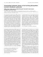
Analysis of the conserved regions of BLV and HTLV-1 TMFigure 1
Analysis of the conserved regions of BLV and HTLV-1 TM. (A) Alignment of the BLV and HTLV-1 TM sequences, the
predicted coiled coil of BLV TM is indicated between the arrow heads; the LHR is in bold; the helical regions of the HTLV-1
TM are boxed; the limits of the HTLV-1 crystal structure are marked by asters; and the membrane spanning region is under-
lined. (B) The HTLV-1 core coiled-coil and, on the right, the leash and α-helical region that is mimicked by the HTLV-1 inhibi-
tory peptide (from PDB 1MG1
). The face of the peptide that interacts with the coiled coil is shown. For the sequence
alignment and structural renderings, residues identical between BLV and HTLV-1 are shown in red, conservative substitutions
are blue, and non-conserved are rendered white. Amino acid coordinates refer to the full-length envelope precursor. (C)
Detail of the predicted interaction of the HTLV-1 LHR-mimetic peptide (ribbon structure) with the surface of the coiled coil
(space filling form) based on the structure of Kobe et al. [16]; shading as above.
Retrovirology 2008, 5:70 />Page 6 of 14
(page number not for citation purposes)
immunodeficiency virus and vesicular stomatitis virus G
protein [20,23] (our unpublished results). Moreover, the
HTLV-1 inhibitory peptide is unusual among C helix-
based fusion inhibitors in that it includes both α-helical
and extended non-helical peptide segments. It was there-
fore uncertain if peptides based on the LHR of BLV would,
like the HTLV-mimetic peptide, display anti-fusogenic
activity. We therefore compared the fusogenic activity of
HTLV-1 and BLV envelope and examined the sensitivity of
BLV envelope to inhibition by peptide inhibitors.
A robust BLV Env-mediated membrane fusion assay
Preliminary experiments with a variety of BLV envelope
expression constructs produced only low levels of BLV
envelope expression and little fusogenic activity in syncy-
tium formation assays (data not shown); this may, in part,
be due to the nuclear retention of the envelope transcripts
as observed for HIV-1 and HTLV-1. Therefore, we devel-
oped an envelope expression vector whereby BLV env was
inserted downstream of the strong cytomegalovirus
(CMV) early promoter, and immediately upstream of the
human immunodeficiency virus Rev-response element
(RRE). The RRE forms a region of extensive secondary
structure in the mRNA that is recognized by Rev and the
resulting ribonucleoprotein complex is subsequently
exported out of the nucleus. The BLV envelope expression
construct was examined for envelope-induced membrane
fusion in syncytium formation assays. Briefly, HeLa cells
were either transfected with pCMV-BLVenv-RRE or pRSV-
Rev individually, or cotransfected with equal amounts of
both vectors. These cells were then used as effector cells to
induce syncytia when co-cultured with non-transfected
cells. Neither vector induced syncytium formation when
transfected alone, but cotransfection of effector cells with
pCMV-BLVenv-RRE and pRSV-Rev resulted in the wide-
spread formation of large syncytia (Figure 2). Further-
more, BLV envelope expressed in this system produced
levels of syncytia that were comparable to that of HTLV-1
envelope expressed from pHTE-1 and consequently this
approach was used to express BLV envelope for these stud-
ies.
Inhibition of envelope-mediated membrane fusion by
LHR-mimetic peptides is limited to the parental virus
To compare the inhibitory properties and specificity of
LHR-based synthetic peptides from HTLV-1 and BLV a
peptide based on the LHR of BLV was generated. The syn-
thetic peptide designated P
BLV
-391 includes residues
Cys391 to Gln419 of BLV Env and spans a region that is
equivalent to the HTLV-1 LHR-derived peptide P
cr
-400
(Table 1). To aid comparison with TM, we refer to the res-
idues of each peptide using the co-ordinates for the full-
length envelope precursor (thus for the BLV-derived pep-
tide residue 1 is referred to as Cys391). The BLV and
HTLV-1 peptides share 45 % identity (Figure 1A, B), but it
should be noted that only a fragment of the HTLV-1 LHR
that is mimicked by P
cr
-400 is resolved in the available
HTLV-1 TM crystal structure (Table 1, Figure 1) [20].
Both HTLV-1 and BLV envelope induced widespread syn-
cytium formation in cultures incubated in the absence of
peptide inhibitors or in the presence of inactive control
peptides (Figure 3A, B). However, in keeping with previ-
ous studies [20-22], HTLV envelope-mediated syncytium
formation was robustly blocked in a dose-dependent
manner by P
cr
-400 with an IC
50
of 0.28 ± 0.01 μM (Figure
3A). However, despite the marked conservation of amino
acid sequence between the LHRs and coiled coils of HTLV-
1 and BLV, P
cr
-400 failed to inhibit membrane fusion
induced by BLV envelope even at concentrations up to 15
μM (Figure 3B) and above (data not shown). Also, like the
inactive control peptides, the BLV LHR-mimetic peptide at
concentrations up to 20 μM (Figure 3A) and above (data
not shown) failed to inhibit membrane fusion induced by
HTLV-1 envelope. By contrast, the peptide P
BLV
-391 spe-
cifically antagonized BLV envelope-mediated membrane
fusion (Figure 3B) with a calculated IC
50
of 3.49 ± 0.03
μM; control peptides including C34 and P
cr
-400 L/A did
not interfere with BLV Env-induced membrane fusion
(Figure 3B). In addition, P
BLV
-391 robustly antagonized
membrane fusion induced by virally expressed envelope
as shown by the inhibition of syncytium formation
between chronically BLV infected FLK cells and target cells
(Figure 3C); whereas, the HTLV-1 peptide inhibitor did
not block BLV-induced membrane fusion. Thus, it appears
that the inhibitory properties of the LHR-mimetic pep-
tides are highly specific to the virus from which they were
derived.
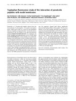
BLV Env-induced syncytiaFigure 2
BLV Env-induced syncytia. Mock transfected HeLa cells
(Mock) or HeLa cells transfected with pRSV-Rev alone (rev),
pCMV-BLVenv-RRE alone (env), or both pRSV-Rev and
pCMV-BLVenv-RRE (rev + env) were co-cultured with target
untransfected HeLa cells. Cells were stained with Giemsa
and typical syncytia profiles are shown.
Retrovirology 2008, 5:70 />Page 7 of 14
(page number not for citation purposes)
The C- and N-terminal regions of P
BLV
-391 are necessary
but not individually sufficient to block membrane fusion
Our group recently demonstrated that truncations at the
N- or C-terminal end of P
cr
-400 abolished fusion-inhibi-
tory function [29]. To test whether or not the N- and C-ter-
minal leash regions are required for the activity of P
BLV
-
391, we synthesized two peptides, P
BLV
-ΔN and P
BLV
-ΔC,
which lack nine amino acid residues at the N-terminus or
C-terminus respectively (Table 1). The peptides retain an
eleven-residue overlap, and have solubility profiles com-
parable to the parental peptide P
BLV
-391 (Table 1). Unlike
the parental peptide, the peptide derivatives P
BLV
-ΔN and
P
BLV
-ΔC lacked detectable inhibitory activity in syncytium
interference assays (Figure 4A). These data illustrate that
amino acid residues contained within the regions Cys391
to Asp399, and Ser411 to Gln419, are critical to the activ-
ity of the mimetic peptide, and that both the amino-termi-
nal and C-terminal regions are necessary but not sufficient
for antagonism of membrane fusion. Importantly, the
data also demonstrate that the central 11-residue region of
the BLV peptide, equivalent to Ser400-Leu410 and
homologous to the short C-terminal α-helix of the HTLV-
1 trimer-of-hairpins is not sufficient for inhibition of syn-
cytium formation.
Moreover, the BLV peptide was remarkably intolerant of
even relatively small deletions. For example, a peptide,
P
BLV
-ΔCCF, in which only 3 amino acids were deleted
from the N-terminus exhibited dramatically reduced abil-
ity to inhibit membrane fusion (Figure 4B). The P
BLV
-
ΔCCF peptide blocked syncytium formation by only 30%
at 20 μM (Figure 4B), compared to > 95% for the parental
peptide, and even at a concentration of 30 μM peptide
P
BLV
-ΔCCF achieved only 40% inhibition (data not
shown). These results can be explained only in part by the
decrease in peptide solubility at concentrations above 11
μM that is associated with the loss of the three N-terminal
amino acid residues (Table 1). At peptide concentrations
below 11 μM, P
BLV
-ΔCCF is soluble under the conditions
used in the syncytium interference assays and yet fails to
inhibit membrane fusion (Figure 4B). It should be noted
that disulphide formation between the peptide and enve-
lope is not required for inhibitory activity, as reduction of
P
BLV
-391 and subsequent modification of the cysteine res-
idues with the sulfhydryl reactive agent Iodoacetyl-PEO
2
-
Biotin failed to disrupt the inhibitory properties of the
peptide (Figure 4C). Moreover, the activity of the bioti-
nylated peptide was indistinguishable from that of the
unmodified P
BLV
-391, indicating that potential dimeriza-
tion of the peptide through inter-molecular disulphide
bonding does not influence peptide potency (Figure 4C).
The first 3 amino acids of the BLV peptide, which includes
the two cysteine residues and an adjacent phenylalanine,
are conserved between HTLV-1 and BLV. Given the data
obtained for the BLV peptide it is surprising to note that
Figure 3
The specificity of peptide inhibitors of Envelope-
mediated membrane fusion is limited to the parental
virus. HeLa cells expressing HTLV-1 (A) or BLV (B) enve-
lope were used as effector cells and co-cultured with
untransfected HeLa cells. Cells were incubated in the pres-
ence of the peptides P
cr
-400, P
BLV
-391, P
cr
-400 L/A a non-
functional derivative of P
cr
-400 [20], or the control HIV C
helix mimetic peptide C34 [51]. (C) Syncytia formation
between BLV infected FLK cells and non-infected HeLa cells.
Syncytia were counted in 10 low-power light microscope
fields. Data points show the mean ± SD of triplicate assays.
Retrovirology 2008, 5:70 />Page 8 of 14
(page number not for citation purposes)
substitution of the cysteines with alanine did not affect
the activity of the HTLV-1 inhibitor P
cr
-400 [22]. Thus it
seems that, at least for the BLV peptide, the first 3 amino
acids aid peptide solubility and contribute in an impor-
tant but, as yet, ill-defined way to the binding or orienta-
tion of the peptide within the target-binding site on TM.
Two conserved leucines are essential for the inhibitory
activity of P
BLV
-391
Leucine residues in P
cr
-400 play a key functional role in
peptide activity [20]. The crystal structure of the HTLV-1
TM [16] reveals that within the LHR several leucine and
isoleucine residues reach down into deep pockets within
the groove of the coiled coil. It appears that the LHR-
derived peptide P
cr
-400 makes similar contacts with the
coiled coil and that these contacts are necessary for stable
binding of the peptide to the coiled coil and thus are crit-
ical to the inhibitory activity of the peptide [22]. Intrigu-
ingly, some but not all of these leucine and isoleucine
residues are conserved between the LHRs of HTLV-1 and
BLV. We therefore sought to determine the importance of
these conserved residues to the inhibitory properties of
the BLV LHR-mimetic peptide. Two peptides were synthe-
sized, P
BLV
-L/A in which all leucines were substituted with
alanine, and P
BLV
-L404/410A in which the Leu404 and
Leu410 of BLV envelope were replaced by alanine (Table
1) these particular leucines are equivalent to the well-con-
served Leu413 and Leu419 of HTLV-1 isolates. Syncytium
interference assays revealed that compared to the parental
peptide (P
BLV
-391) the alanine-substituted peptides were
Deletions or substitutions of specific amino acids in P
BLV
-391 have a detrimental effect on inhibitory activityFigure 4
Deletions or substitutions of specific amino acids in P
BLV
-391 have a detrimental effect on inhibitory activity.
Syncytium interference assays using BLV envelope-expressing HeLa cells as effectors. (A) The inhibitory properties of P
BLV
-391,
P
BLV
-ΔN, P
BLV
-ΔC and the P
cr
-400 control were examined. (B) The activity of P
BLV
-391, the derivative P
BLV
-ΔCCF, and the con-
trol peptide P
cr
-400 were compared. (C) The activity of P
BLV
-391 was compared to Bio-P
BLV
-391
Ar
a biotinylated peptide recov-
ered from the flow-through of an amylose column (see methods), Bio-P
BLV
-391
Sd
the same peptide depleted over a streptavidin
column (volumes of column buffer equal to those required to give the specified concentrations of Bio-P
BLV
-391
Ar
were used),
and the control peptide C34. (D) The inhibitory properties of P
BLV
-391, P
BLV
-L/A, P
BLV
-L404/410A and the control P
cr
-400
were compared. Syncytia were counted in 10 low-power light microscope fields. Data points show the mean ± SD of triplicate
assays.
Retrovirology 2008, 5:70 />Page 9 of 14
(page number not for citation purposes)
severely compromised in their ability to inhibit mem-
brane fusion (Figure 4D); in particular, P
BLV
-L/A did not
exhibit any discernible inhibition up to 20 μM (Figure
4D) or above (data not shown). Hence, the leucine resi-
dues are important to peptide function. Moreover,
although P
BLV
-L404/410A was just as soluble as the paren-
tal peptide (Table 1), P
BLV
-L404/410A also failed to dis-
play any fusion-blocking activity up to 20 μM (Figure 4D);
indicating that the leucines equivalent to BLV envelope
residues 404 and 410 are particularly important to the
inhibitory properties of the LHR-mimetic peptide.
A model for the BLV trimer-of-hairpins
Our analysis reveals that for the ectodomain of the TM the
majority of the amino acid residues that are conserved
between HTLV-1 and BLV map to the interacting surfaces
of the trimer-of-hairpins. Moreover, a BLV homologue of
the HTLV-1 LHR-derived peptide inhibitor also exhibits
robust but highly specific inhibitory activity against BLV-
induced membrane fusion. Significantly, conserved leu-
cine residues are critical to the inhibitory activity of both
peptides. Encouraged by these results and to gain greater
insight into the mechanism of fusion and the likely con-
tacts made by P
BLV
-391 with the coiled coil, we con-
structed a homology model of the BLV trimer-of-hairpins
that is based on the crystal structure of the HTLV-1 TM
(Figure 1B) [16].
Having identified the predicted BLV coiled-coil (Figure
1A), the Clustal-W alignment of the TM ectodomain
sequences of BLV and HTLV-1 (Figure 1A) permitted the
substitution of the BLV residues onto the HTLV-1-derived
scaffold, consisting of the complete trimer of N-helices
and a single LHR. The geometry of the crude model was
improved by simulated annealing and energy minimisa-
tion in explicit solvent with the GROMACS (Groningen
Machine for Chemical Simulations) package using the
GROMOS96 43a1 force field [34]. It should be noted that,
compared to the HTLV-1 trimer of hairpins, there are two
additional residues in the predicted BLV chain-reversal
region at positions 380 and 381 of BLV envelope. Since
these residues are within a flexible loop there is insuffi-
cient information to model these residues with any degree
of accuracy therefore these residues are omitted in the cur-
rent model. Nonetheless, the restraint provided by the
disulphide bond between Cys384 and Cys391 coupled
with a high level of sequence conservation within the hep-
tad repeat region and within the LHR suggests that the
model is likely to be a reasonably accurate representation
of the interaction between the LHR and the coiled coil.
The model for the BLV coiled coil and LHR is presented in
Figure 5A.
Consistent with the sequence alignment and the structure
of the HTLV-1 TM ectodomain (Figure 1), the BLV TM
model indicates that Leu394 and Ile396 likely project into
a hydrophobic pocket at the membrane-distal end of the
core coiled-coil (Figure 5B). It also implies that Ile401,
Leu404 and Leu407, which all lie on the same side of the
putative α-helix of the LHR, are oriented such that they
project into the groove of the coiled coil. Notably, Leu410
is predicted to make a significant contact with a deep
pocket situated towards the membrane-proximal end of
the core coiled-coil. Therefore, the BLV coiled coil and
LHR model is highly consistent with the experimental
data and provides a molecular explanation for the loss of
activity associated with substitutions in the BLV LHR-
derived peptide.
Substituting an arginine residue for an alanine in P
BLV
-391
results in a more potent peptide inhibitor
The accumulated experimental data correlate well with
the structural model, implying that predications based on
the BLV trimer-of-hairpins model are likely to be inform-
ative. The homology model of the BLV TM ectodomain
(Figure 6) suggests that Arg403, a residue within the pre-
dicted α-helix of the LHR and mimicked by P
BLV
-391 pep-
tide, may be electrostatically unfavourable for efficient
binding of the C-terminal LHR into the groove of the core
coiled-coil. We predicted that removing this unfavourable
charge interaction would improve the binding of the pep-
tide to the BLV coiled coil and thereby improve the inhib-
itory activity of the peptide. We therefore synthesized a
peptide, P
BLV
-R403A, which incorporated an alanine resi-
due in place of the arginine equivalent to Arg403 of Env
(Table 1). As anticipated, substitution of the arginine res-
idue resulted in a modest but highly consistent and signif-
icant (p < 0.0001, Student's t-test) improvement in
peptide potency when compared to P
BLV
-391. The peptide
P
BLV
-R403A is more than twice as potent as P
BLV
-391 in
syncytium interference assays, with a calculated IC
50
of
1.56 ± 0.05 μM compared to 3.49 μM ± 0.03 μM for P
BLV
-
391 (Figure 6). The data show that a single amino-acid
substitution in the predicted short α-helix of the LHR-
mimetic peptide increases the ability of the peptide to
block membrane fusion and provides further support for
the utility of the model of the BLV TM core.
Discussion
Experimental evidence points towards a remarkably con-
served mechanism by which virally encoded envelope
glycoproteins catalyse membrane fusion and facilitate
delivery of the viral core into the target cell [13,14]. The
structures of several class 1 fusion proteins reveal a char-
acteristic "trimer-of-hairpins" motif believed to represent
a late or post-fusion conformation [16-19,35-37]. Investi-
gating the way in which envelope proteins fold from a
rod-like, pre-hairpin intermediate into the trimer-of-hair-
pins to pull the viral and cellular membranes together is
important not only for our understanding of viral entry
Retrovirology 2008, 5:70 />Page 10 of 14
(page number not for citation purposes)
but also for the development of therapeutically relevant
inhibitors of this process.
The protein sequences of the TM ectodomains of BLV and
HTLV-1 display a striking level of conservation. By scruti-
nizing the position of conserved residues in the context of
the HTLV-1 six-helix-bundle structure, we have found that
the majority of the conserved residues map to the interact-
ing surfaces of the LHR and core coiled-coil. It is interest-
ing to note that there are several non-conserved residues
within the LHR of each virus; significantly, these modifi-
cations are mirrored by compensating substitutions
within the specific area of the core coiled-coil with which
the variant residue interacts (Figure 1C) and conse-
quently, the association with the coiled coil is main-
tained. It appears that in order to support variation and
speciation but to maintain biological function comple-
mentary regions of the fusion proteins have evolved in
parallel. The greatest functional constraint and therefore
most highly conserved regions map along the interacting
surfaces of the trimer-of-hairpins. Conversely, regions of
the TM that are likely exposed to the aqueous environ-
ment both during and after fusion exhibit considerable
divergence and display relatively few amino acids in com-
mon. Such changes may reflect strong selective pressures
exerted on the virus, perhaps due to the need for particular
regions of the TM to interact functionally with the rela-
tively divergent surface glycoproteins of the respective
viruses. Alternatively, the selective pressure may be due to
the differing immunological environments of the respec-
tive hosts. It is worth noting, that the TM and the trimer-
of-hairpins of HTLV-1 are immunogenic [38,39], that
antibodies targeting TM often recognise non-neutralizing
conformational epitopes [39,40], and that trimer-of-hair-
pin structures are frequently displayed on the surface of
infected cells [40]. Whether or not these features of the TM
contribute to the pathogenesis or immune evasion of leu-
kaemia viruses remains to be determined.
The HTLV-1-derived LHR-based peptide is able to inhibit
membrane fusion mediated by the divergent envelope of
HTLV-2 and, given the level of conservation between the
HTLV-1 and BLV TM ectodomain, we anticipated that the
HTLV-1-derived peptide P
cr
-400 would also inhibit the
fusogenic activity of BLV envelope. Surprisingly, although
P
cr
-400 is an extremely effective inhibitor of HTLV-1-
Homology model of the BLV core coiled-coil and the interacting LHRFigure 5
Homology model of the BLV core coiled-coil and the interacting LHR. The protein sequence of BLV TM was mod-
elled onto the HTLV-1 TM ectodomain structure (PDB ID 1MG1
). (A) The predicted BLV core coiled-coil is shown as a space-
filling model in grey with the LHR in green. (B) Detail of the coiled coil in blue, grey and red, with the C-terminal section mim-
icked by P
BLV
-391 shown as a green ribbon, the predicted position of relevant side chains are shown as sticks. The membrane
proximal region is uppermost. The arrowhead marks the position of Leu404.
Retrovirology 2008, 5:70 />Page 11 of 14
(page number not for citation purposes)
mediated fusion, the peptide had no detectable activity in
BLV syncytium interference assays. Moreover, the BLV
LHR-based peptide P
BLV
-391 does not inhibit HTLV-1
envelope-catalysed syncytium formation. Sequence align-
ment and homology modelling (Figures 1 and 5) indicate
that within the first eight residues only two residues differ
between the HTLV-1 and BLV peptides and these residues
are likely to be solvent exposed and unable to contribute
to the interaction with the core coiled-coil. The residues
that determine the specificity of inhibition are therefore
located within or overlapping the short α-helix or C-ter-
minal leash segments of the peptide. In terms of peptide
function, it is clear that the putative α-helix within the
central region of these peptides is important for inhibitory
activity. Nonetheless, both the N- and to the C-terminal
leash residues contribute to the inhibitory properties of
the peptide as deletion of these regions severely attenuates
inhibitory activity. The structure of residues C-terminal of
Asn421 in the HTLV-1 TM (equivalent to Gln412 of BLV
Env) has not been resolved [16]. Consequently, it is not
yet possible to account in molecular terms for the con-
served interactions beyond this point. However, our data
highlight a number of features that play a key role in the
biological activity of the BLV-derived peptide. The first
three N-terminal amino acid residues appear to be critical
to activity. Given the orientation of the phenylalanine res-
idue in the BLV TM model and the equivalent Phe402 in
the crystal structure of HTLV-1 TM, it is unlikely that this
side chain directly contributes to the interaction with the
coiled coil. Consistent with this view, Maerz et al. [41]
have demonstrated that Phe402 likely plays a structural
role in pre-fusogenic envelope and is required for enve-
lope processing, but likely becomes solvent exposed dur-
ing assembly of the fusion-associated trimer-of-hairpins
structure [41]. Furthermore, although disulphide bonding
regulates TM function [11,12] and association with the SU
subunit [10], the adjacent cysteines at the N-terminus of
P
cr
-400 are not required for disulphide formation, for
binding to the coiled-coil, or for inhibitory activity [22].
Similarly, modification of the adjacent cysteine residues
in the BLV-derived peptide reveals that disulphide forma-
tion is not required for coiled coil binding or inhibition of
membrane fusion. The apparent requirement for the
cysteine residues for functional activity of the BLV-derived
peptide may reflect an intrinsic difference between BLV
and HTLV-1 peptide target interactions. Currently, our
preferred view is that the N-terminus of the BLV peptide
aids alignment of the adjacent peptide sequences relative
to the target-binding site on the coiled coil.
A recurring theme in the interaction of the C-terminal
helix of the trimer-of-hairpins with the coiled coil of viral
fusion proteins is the interaction of non-polar side chains
with deep pockets on the coiled coil [16-18,35,36,42].
The model for the BLV trimer-of-hairpins suggests that
this is also the case for BLV and this interpretation is sup-
ported by the peptide inhibition data. The model suggests
that a series of leucine residues, which include L404, L407
and L410, make contact with the coiled coil. Moreover,
the inhibitory activity of P
BLV
-391 is completely abrogated
following substitution of all the leucine residues with
alanine. Similar results have been observed for the P
cr
-400
inhibitor of HTLV-1 [20]. In particular, two leucines,
Leu413 and Leu419, are important for the inhibitory
activity of P
cr
-400 [22]. Leucine 413 is situated within the
short α-helix, whereas Leu419 is situated within the C-ter-
minal leash-like domain. Significantly, both of these leu-
cine residues are conserved in BLV, at positions 404 and
410 respectively, and the model for the BLV trimer-of-
hairpins suggests that they are located in areas of similar
structure. Importantly, substitution of these residues in
P
BLV
-391 results in a non-functional peptide. This is a sig-
nificantly more dramatic outcome than is observed for
specific substitutions at each of these residues in P
cr
-400
[22] and suggests that disruption of both of the potential
contacts made with the coiled coil has a profound cumu-
lative effect on loss of peptide activity. Given that these
leucines are critical to the inhibitory properties of the
LHR-mimetic we suspect, and are currently testing the
view, that within envelope such substitutions would
severely impair envelope-mediated membrane fusion.
Our data also reveal that P
BLV
-391 is significantly less
potent against BLV than the comparable peptide (P
cr
-400)
Substitution of a single arginine residue with alanine yields an improved inhibitorFigure 6
Substitution of a single arginine residue with alanine
yields an improved inhibitor. The syncytium inhibition
activity of the peptides P
cr
-400, P
BLV
-391 and the derivative
peptide P
BLV
-R403A was examined. The percentage syncy-
tium inhibition following co-incubation of cells with the pep-
tides is shown. Syncytia were counted in 10 low-power light
microscope fields. Data points show the mean ± SD of tripli-
cate assays. The asters show the data points for which the p
values were calculated (see main text).
Retrovirology 2008, 5:70 />Page 12 of 14
(page number not for citation purposes)
against HTLV-1. The structure and model of the HTLV-1
and BLV TM suggests a plausible explanation for this
observation in that, relative to the HTLV-derived peptide,
the BLV peptide displays a smaller surface area available
for interaction with the core coiled-coil. In addition, non-
conserved residues within the HTLV-1 peptide may con-
tribute disproportionately to the stability of the interac-
tion between the HTLV-1 peptide and the core coiled-coil.
The model and accumulated data also underscore the
importance of a deep pocket that is situated towards the
membrane-proximal end of the trimer-of-hairpins and is
conserved between leukaemia viruses. The peptide inhib-
itors engage this pocket and this interaction appears to
contribute substantially to the stability of peptide associa-
tion with the coiled coil and is required for optimal inhib-
itory activity. The data provides further validation of the
BLV coiled coil and LHR model and reveals that conserved
hydrophobic amino acid side-chains within the helical
and non-helical regions mediate interaction of the pep-
tide inhibitors with their target.
An intriguing finding of this study is that, directed by anal-
ysis of the model structure, an improved inhibitor of BLV
envelope-mediated membrane fusion can be generated by
the substitution of a single amino acid residue, Arg403,
with alanine. A similar observation has been made for the
Ile412 residue of the HTLV-1 fusion inhibitors [22]. Inter-
estingly, the relative location of these beneficial substitu-
tions is conserved: the BLV residue Arg403 and the HTLV-
1 residue Ile412 are immediately N-terminal of an impor-
tant coiled-coil contact mediated by a conserved leucine
residue. It is likely that the substitutions relieve a steric
and/or electrostatic clash between the peptides and the
relevant viral core coiled-coil, and thereby allow the adja-
cent leucine residue to dock more effectively with the
coiled coil. For BLV, the clash between Arg403 and the
coiled coil is highlighted in the model of the trimer-of-
hairpins (Figure 5B), and this structure is validated by the
collected experimental data. Surprisingly, the data derived
from the peptide inhibitors identifies a conserved posi-
tion at which a residue impedes assembly of the trimer-of-
hairpins. It appears that during evolution two related but
diverging viruses have maintained non-optimal residues
within the LHR and that the LHR has not been selected for
the best possible fit with the coiled coil. It seems strange
not only that such clashes occur, but that they occur in
ostensibly the same place. Perhaps, these non-optimal res-
idues act to modulate the fusogenic activity of the TM. It
is worth noting that highly fusogenic or readily activated
fusion proteins have been described for a number of
viruses and these proteins display an array of mutations or
deletions, implying that fusogenic activity is modulated
by multiple regions of envelope [43-46]. Of course, it is
also possible that the non-optimal residues for LHR asso-
ciation with the coiled coil modulate envelope activity at
an earlier pre-fusogenic stage of envelope assembly. Stud-
ies are currently underway to test these ideas. Importantly,
the ability to remove residues that hinder LHR:coiled coil
interaction provides an opportunity to design peptides
with "super-binding" characteristics and thereby pave the
way towards more drug-like HTLV-1 entry inhibitors.
BLV is prevalent among cattle throughout many regions of
the world [3]. The combined effect of decreased milk pro-
duction, mortality due to lymphoma, reduced productive
lifespan and increased susceptibility of infected cattle to
opportunistic pathogens has significant economic ramifi-
cations [3]. Our data indicate that the core coiled-coil of
gp30 is exposed at least transiently during the fusion proc-
ess and is accessible to a small inhibitory peptide and that
inhibitory peptides will be of significant utillity in the
analysis of BLV entry into cells. Moreover, it will be inter-
esting to determine if the BLV coiled coil is also accessible
to neutralising antibodies and whether coiled-coil-based
immunogens could be of value as components of a subu-
nit vaccine to prevent BLV transmission between animals.
Although retroviral TM displays significant resistance to
neutralisation by coiled-coil-specific antibodies [47,40]
recent efforts indicate that such hurdles can be success-
fully overcome [48]. Moreover, attenuated BLV strains
provide long-term protection against experimental BLV
infection of cattle [49]; and an HTLV-1 envelope-derived
subunit vaccine candidate provides significant protection
against virus challenge in primate models [50]. The accu-
mulating evidence therefore suggests that a subunit vac-
cine based on viral envelope may be an achievable
objective for prophylactic treatment against leukaemia
virus infections.
Our data further define a membrane-proximal region of
TM that is conserved between BLV and HTLV-1, which has
potential as an anti-HTLV-1 drug target. This study dem-
onstrates that comparative analysis of BLV and HTLV-1
induced membrane fusion will provide significant insight
into envelope function and ultimately will be of value in
the quest for compounds that block HTLV-1 entry into
cells.
Competing interests
The authors declare that they have no competing interests.
Authors' contributions
DL performed the experiments and helped to draft the
manuscript, AS provided technical expertise in molecular
modeling, DvA provided assistance and technical exper-
tise in structural analysis, DWB designed the experiments
and wrote the manuscript. All authors read and approved
the final manuscript.
Retrovirology 2008, 5:70 />Page 13 of 14
(page number not for citation purposes)
Acknowledgements
We thank Dr Arsène Burny and Dr Luc Willems for kindly supplying rea-
gents. The Leukaemia Research Fund generously supported this work
through a project grant (LRF-354) to D.W.B. D.L. is the recipient of a Med-
ical Research Council studentship. D.v.A. is supported by a Wellcome
Trust Senior Research Fellowship. We thank Clare Connolly, Dr Daniella
Zheleva and Cyclacel Pharmaceuticals, Inc. for assistance with laser neph-
elometry.
References
1. Anonymous: Human T-cell Lymphotropic viruses. In Human
Immunodeficiency viruses and human T-cell lymphotropic viruses Volume
67. Group IW: IARC Monographs; 1996:261-390.
2. Cann AJ, Chen ISY: Human T-cell Leukaemia virus types I and
II. In Fields Virology Volume 2. 3rd edition. Philadelphia: Lippincott-
Raven; 1996:1849-1880. [Fields BN, Knipe, D.M., Howley, P.M.,
Channock, R.M., Melnick, J.L., Monath, T.P., Roizman, B. and Straus,
S.E. (Series Editor)
3. Gillet N, Florins A, Boxus M, Burteau C, Nigro A, Vandermeers F,
Balon H, Bouzar AB, Defoiche J, Burny A, et al.: Mechanisms of
leukemogenesis induced by bovine leukemia virus: prospects
for novel anti-retroviral therapies in human. 2007, 4:16.
4. Johnston ER, Albritton LM, Radke K: Envelope proteins contain-
ing single amino acid substitutions support a structural
model of the receptor-binding domain of bovine leukemia
virus surface protein. J Virol 2002, 76:10861-10872.
5. Gallaher WR, Ball JM, Garry RF, Martin-Amedee AM, Montelaro RC:
A general model for the surface glycoproteins of HIV and
other retroviruses. AIDS Res Hum Retroviruses 1995, 11:191-202.
6. Kim FJ, Seiliez I, Denesvre C, Lavillette D, Cosset FL, Sitbon M: Def-
inition of an amino-terminal domain of the human T-cell
leukemia virus type 1 envelope surface unit that extends the
fusogenic range of an ecotropic murine leukemia virus. J Biol
Chem 2000, 275:23417-23420.
7. Gatot JS, Callebaut I, Van Lint C, Demonte D, Kerkhofs P, Portetelle
D, Burny A, Willems L, Kettmann R: Bovine leukemia virus SU
protein interacts with zinc, and mutations within two inter-
acting regions differently affect viral fusion and infectivity in
vivo. J Virol 2002, 76:7956-7967.
8. Kim FJ, Battini JL, Manel N, Sitbon M: Emergence of vertebrate
retroviruses and envelope capture. Virology 2004, 318:183-191.
9. Kim FJ, Manel N, Garrido EN, Valle C, Sitbon M, Battini JL: HTLV-1
and -2 envelope SU subdomains and critical determinants in
receptor binding. Retrovirology 2004, 1:41.
10. Johnston ER, Radke K: The SU and TM envelope protein subu-
nits of bovine leukemia virus are linked by disulfide bonds,
both in cells and in virions. J Virol 2000, 74:2930-2935.
11. Wallin M, Ekstrom M, Garoff H: Isomerization of the intersubu-
nit disulphide-bond in Env controls retrovirus fusion. Embo J
2004, 23:54-65.
12. Wallin M, Ekstrom M, Garoff H: The fusion-controlling disulfide
bond isomerase in retrovirus Env is triggered by protein
destabilization. J Virol 2005, 79:1678-1685.
13. Weissenhorn W, Dessen A, Calder LJ, Harrison SC, Skehel JJ, Wiley
DC: Structural basis for membrane fusion by enveloped
viruses. Mol Membr Biol 1999, 16:3-9.
14. Eckert DM, Kim PS: Mechanisms of viral membrane fusion and
its inhibition. Annu Rev Biochem 2001, 70:777-810.
15. Park HE, Gruenke JA, White JM: Leash in the groove mechanism
of membrane fusion. Nat Struct Biol 2003, 10:1048-1053.
16. Kobe B, Center RJ, Kemp BE, Poumbourios P: Crystal structure of
human T cell leukemia virus type 1 gp21 ectodomain crystal-
lized as a maltose-binding protein chimera reveals structural
evolution of retroviral transmembrane proteins. Proc Natl
Acad Sci USA 1999, 96:4319-4324.
17. Chan DC, Fass D, Berger JM, Kim PS: Core structure of gp41
from the HIV envelope glycoprotein. Cell 1997, 89:263-273.
18. Malashkevich VN, Chan DC, Chutkowski CT, Kim PS: Crystal
structure of the simian immunodeficiency virus (SIV) gp41
core: conserved helical interactions underlie the broad inhib-
itory activity of gp41 peptides. Proc Natl Acad Sci USA 1998,
95:9134-9139.
19. Malashkevich VN, Singh M, Kim PS: The trimer-of-hairpins motif
in membrane fusion: Visna virus. Proc Natl Acad Sci USA 2001,
98:8502-8506.
20. Pinon JD, Kelly SM, Price NC, Flanagan JU, Brighty DW: An antiviral
peptide targets a coiled-coil domain of the human T-cell
leukemia virus envelope glycoprotein. J Virol
2003,
77:3281-3290.
21. Mirsaliotis A, Nurkiyanova K, Lamb D, Kuo CW, Brighty DW: An
antibody that blocks human T-cell leukemia virus type 1 six-
helix-bundle formation in vitro identified by a novel assay for
inhibitors of envelope function. J Gen Virol 2007, 88:660-669.
22. Mirsaliotis A, Lamb D, Brighty DW: Non-helical Leash and α-hel-
ical Structures Determine the Potency of a Peptide Antago-
nist of Human T Cell Leukaemia Virus Entry. J Virol 2008,
82:4965-4973.
23. Sagara Y, Inoue Y, Shiraki H, Jinno A, Hoshino H, Maeda Y: Identifi-
cation and mapping of functional domains on human T-cell
lymphotropic virus type 1 envelope proteins by using syn-
thetic peptides. J Virol 1996, 70:1564-1569.
24. Wild C, Oas T, McDanal C, Bolognesi D, Matthews T: A synthetic
peptide inhibitor of human immunodeficiency virus replica-
tion: correlation between solution structure and viral inhibi-
tion. Proc Natl Acad Sci USA 1992, 89:10537-10541.
25. Wild C, Greenwell T, Matthews T: A synthetic peptide from
HIV-1 gp41 is a potent inhibitor of virus-mediated cell-cell
fusion. AIDS Res Hum Retroviruses 1993, 9:1051-1053.
26. Lambert DM, Barney S, Lambert AL, Guthrie K, Medinas R, Davis DE,
Bucy T, Erickson J, Merutka G, Petteway SR Jr: Peptides from con-
served regions of paramyxovirus fusion (F) proteins are
potent inhibitors of viral fusion. Proc Natl Acad Sci USA 1996,
93:2186-2191.
27. Rapaport D, Ovadia M, Shai Y: A synthetic peptide correspond-
ing to a conserved heptad repeat domain is a potent inhibi-
tor of Sendai virus-cell fusion: an emerging similarity with
functional domains of other viruses. Embo J 1995,
14:5524-5531.
28. Dokhelar MC, Pickford H, Sodroski J, Haseltine WA: HTLV-I
p27rex regulates gag and env protein expression. J Acquir
Immune Defic Syndr 1989, 2:431-440.
29. Hope TJ, McDonald D, Huang XJ, Low J, Parslow TG: Mutational
analysis of the human immunodeficiency virus type 1 Rev
transactivator: essential residues near the amino terminus. J
Virol 1990, 64:5360-5366.
30. Churchill MJ, Moore JL, Rosenberg M, Brighty DW: The rev-
responsive element negatively regulates human immunode-
ficiency virus type 1 env mRNA expression in primate cells.
J Virol 1996, 70:5786-5790.
31. Jassal SR, Lairmore MD, Leigh-Brown AJ, Brighty DW: Soluble
recombinant HTLV-1 surface glycoprotein competitively
inhibits syncytia formation and viral infection of cells. Virus
Res 2001, 78:17-34.
32. Singh M, Berger B, Kim PS: LearnCoil-VMF: computational evi-
dence for coiled-coil-like motifs in many viral membrane-
fusion proteins. J Mol Biol 1999, 290:1031-1041.
33. Thompson JD, Higgins DG, Gibson TJ: CLUSTAL W: improving
the sensitivity of progressive multiple sequence alignment
through sequence weighting, position-specific gap penalties
and weight matrix choice. Nucleic Acids Res 1994, 22:4673-4680.
34. Lindahl EaH B, Spoel D van der: Gromacs 3.0: a package for
molecular simulation and trajectory analysis. J Mol Mod 2001,
7:306-317.
35. Weissenhorn W, Carfi A, Lee KH, Skehel JJ, Wiley DC: Crystal
structure of the Ebola virus membrane fusion subunit, GP2,
from the envelope glycoprotein ectodomain. Mol Cell 1998,
2:605-616.
36. Malashkevich VN, Schneider BJ, McNally ML, Milhollen MA, Pang JX,
Kim PS: Core structure of the envelope glycoprotein GP2
from Ebola virus at 1.9-A resolution. Proc Natl Acad Sci USA
1999, 96:2662-2667.
37. Melikyan GB, Markosyan RM, Hemmati H, Delmedico MK, Lambert
DM, Cohen FS: Evidence that the transition of HIV-1 gp41 into
a six-helix bundle, not the bundle configuration, induces
membrane fusion. J Cell Biol 2000, 151:413-423.
38. Hadlock KG, Rowe J, Perkins S, Bradshaw P, Song GY, Cheng C, Yang
J, Gascon R, Halmos J, Rehman SM, et al.: Neutralizing human
monoclonal antibodies to conformational epitopes of human
Publish with BioMed Central and every
scientist can read your work free of charge
"BioMed Central will be the most significant development for
disseminating the results of biomedical research in our lifetime."
Sir Paul Nurse, Cancer Research UK
Your research papers will be:
available free of charge to the entire biomedical community
peer reviewed and published immediately upon acceptance
cited in PubMed and archived on PubMed Central
yours — you keep the copyright
Submit your manuscript here:
/>BioMedcentral
Retrovirology 2008, 5:70 />Page 14 of 14
(page number not for citation purposes)
T-cell lymphotropic virus type 1 and 2 gp46. J Virol 1997,
71:5828-5840.
39. Mirsaliotis A, Nurkiyanova K, Lamb D, Woof JM, Brighty DW: Con-
formation-specific Antibodies targeting the trimer-of-hair-
pins motif of the HTLV-1 Trans-membrane glycoprotein
recognise viral envelope but fail to neutralise viral entry. J
Virol 2007.
40. Mirsaliotis A, Nurkiyanova K, Lamb D, Kuo CW, Brighty DW:
Resistance to neutralization by antibodies targeting the
coiled coil of fusion-active envelope is a common feature of
retroviruses. J Biol Chem 2007, 282:36724-36735.
41. Maerz AL, Center RJ, Kemp BE, Kobe B, Poumbourios P: Functional
implications of the human T-lymphotropic virus type 1
transmembrane glycoprotein helical hairpin structure. J Virol
2000, 74:6614-6621.
42. Chen J, Skehel JJ, Wiley DC: N- and C-terminal residues com-
bine in the fusion-pH influenza hemagglutinin HA(2) subunit
to form an N cap that terminates the triple-stranded coiled
coil. Proc Natl Acad Sci USA 1999, 96:8967-8972.
43. Kim FJ, Manel N, Boublik Y, Battini JL, Sitbon M: Human T-cell
leukemia virus type 1 envelope-mediated syncytium forma-
tion can be activated in resistant Mammalian cell lines by a
carboxy-terminal truncation of the envelope cytoplasmic
domain. J Virol 2003, 77:963-969.
44. Poon B, Chen IS: Identification of a domain within the human
T-cell leukemia virus type 2 envelope required for syncytium
induction and replication. J Virol 1998, 72:1959-1966.
45. Platt EJ, Kuhmann SE, Rose PP, Kabat D: Adaptive mutations in
the V3 loop of gp120 enhance fusogenicity of human immu-
nodeficiency virus type 1 and enable use of a CCR5 corecep-
tor that lacks the amino-terminal sulfated region. J Virol 2001,
75:12266-12278.
46. Saha K, Yan H, Nelson JA, Zerhouni-Layachi B: Infection of human
and non-human cells by a highly fusogenic primary CD4-
independent HIV-1 isolate with a truncated envelope cyto-
plasmic tail. Virology 2005, 337:30-44.
47. Zwick MB, Saphire EO, Burton DR: gp41: HIV's shy protein. Nat
Med
2004, 10:133-134.
48. Miller MD, Geleziunas R, Bianchi E, Lennard S, Hrin R, Zhang H, Lu
M, An Z, Ingallinella P, Finotto M, et al.: A human monoclonal anti-
body neutralizes diverse HIV-1 isolates by binding a critical
gp41 epitope. Proc Natl Acad Sci USA 2005, 102:14759-14764.
49. Kerkhofs P, Gatot JS, Knapen K, Mammerickx M, Burny A, Portetelle
D, Willems L, Kettmann R: Long-term protection against bovine
leukaemia virus replication in cattle and sheep. J Gen Virol
2000, 81:957-963.
50. Ibuki K, Funahashi SI, Yamamoto H, Nakamura M, Igarashi T, Miura T,
Ido E, Hayami M, Shida H: Long-term persistence of protective
immunity in cynomolgus monkeys immunized with a recom-
binant vaccinia virus expressing the human T cell leukaemia
virus type I envelope gene. J Gen Virol 1997, 78:147-152.
51. Chan DC, Chutkowski CT, Kim PS: Evidence that a prominent
cavity in the coiled coil of HIV type 1 gp41 is an attractive
drug target. Proc Natl Acad Sci USA 1998, 95:15613-15617.