Báo cáo y học: " Induction of galectin-1 expression by HTLV-I Tax and its impact on HTLV-I infectivity" ppt
Bạn đang xem bản rút gọn của tài liệu. Xem và tải ngay bản đầy đủ của tài liệu tại đây (1.3 MB, 15 trang )
BioMed Central
Page 1 of 15
(page number not for citation purposes)
Retrovirology
Open Access
Research
Induction of galectin-1 expression by HTLV-I Tax and its impact on
HTLV-I infectivity
Sonia Gauthier
1
, Isabelle Pelletier
1
, Michel Ouellet
1
, Amandine Vargas
2
,
Michel J Tremblay
1
, Sachiko Sato
1
and Benoit Barbeau*
2
Address:
1
Research Center in Infectious Diseases, CHUL Research Center, 2705 boul. Laurier; Ste-Foy, Québec, G1V 4G2, Canada and
2
Université
du Québec à Montréal, Département des sciences biologiques, 2080 St-Urbain, Montréal, Québec, H2X 3X8, Canada
Email: Sonia Gauthier - ; Isabelle Pelletier - ;
Michel Ouellet - ; Amandine Vargas - ;
Michel J Tremblay - ; Sachiko Sato - ;
Benoit Barbeau* -
* Corresponding author
Abstract
Background: Cell-free Human T-cell Leukemia Virus type I (HTLV-I) virions are poorly infectious
and cell-to-cell contact is often required to achieve infection. Other factors might thus importantly
contribute in increasing infection by HTLV-I. Galectin-1 is a galactoside-binding lectin which is
secreted by activated T lymphocytes. Several functions have been attributed to this protein
including its capacity to increase cell-to-cell adhesion. Based on previous studies, we postulated that
this protein could also accentuate HTLV-I infection.
Results: Herein, we demonstrate that galectin-1 expression and release are higher in HTLV-I-
infected T cells in comparison to uninfected T cells. Furthermore, galectin-1 expression was
activated in various cell lines expressing the wild type viral Tax protein while this induction was
minimal upon expression of NF-κB activation-defective TaxM22. Cotransfection of these Tax
expression vectors with galectin-1 promoter-driven luciferase constructs confirmed that Tax
upregulated galectin-1 promoter activity. However, a NF-κB-independent mechanism was strongly
favoured in this induction of galectin-1 expression as no activation of the promoter was apparent
in Jurkat cells treated with known NF-κB activators. Using HTLV-I envelope pseudotyped HIV-1
virions, galectin-1 was shown to increase infectivity. In addition, a co-culture assay with HTLV-I-
infected cells also indicated an increase in cell fusion upon addition of galectin-1. This effect was not
mediated by factors present in the supernatant of the HTLV-I-infected cells.
Conclusion: These data suggest that HTLV-I Tax increases galectin-1 expression and that this
modulation could play an important role in HTLV-I infection by stabilizing both cell-to-cell and
virus-cell interactions.
Background
Human T-cell Leukemia Virus type I (HTLV-I) is the etio-
logical agent of adult T cell leukemia (ATL) and HTLV-I-
associated myelopathy/tropical spastic paraparesis
(HAM/TSP) [1-3]. It has been estimated that 20 million
individuals are infected worldwide [4]. The in vivo target
cells are mature CD4+CD45RO T lymphocytes and CD8+
T lymphocytes [5], although other cell types have been
Published: 25 November 2008
Retrovirology 2008, 5:105 doi:10.1186/1742-4690-5-105
Received: 16 June 2008
Accepted: 25 November 2008
This article is available from: />© 2008 Gauthier et al; licensee BioMed Central Ltd.
This is an Open Access article distributed under the terms of the Creative Commons Attribution License ( />),
which permits unrestricted use, distribution, and reproduction in any medium, provided the original work is properly cited.
Retrovirology 2008, 5:105 />Page 2 of 15
(page number not for citation purposes)
suggested to be potential target including lung epithelial
cells, as recently demonstrated [6]. HTLV-I is transmitted
between individuals by the transfer of infected lym-
phocytes and is thought to require repeated contacts as
only one out of 1 × 10
5
to 1 × 10
6
viruses is infectious [7-
9]. During viral transmission, a contact is established
between an uninfected and an infected T cell by the inter-
action of the gp46 viral protein with its cellular receptor
subsequently followed by the polarization of the infected
cell cytoskeleton at the site of cell-to-cell contact and the
accumulation of viruses at the cell junction [7]. GLUT-1
has been reported to be part of this receptor and to be
involved in the first step of viral entry, although its exact
role is still ill-defined [10,11]. Although the cellular
ICAM-1 protein has been established as a potential
inducer of microtubule reorganization, the viral Tax pro-
tein has also been shown to be active in this process
[12,13].
Tax is the viral transactivator of HTLV-I allowing transcrip-
tion through the three Tax-responsive elements (TRE1)
present in the U3 region of the Long Terminal Repeat
(LTR) [14-16]. This viral protein also promotes transcrip-
tion of many cellular genes. To activate transcription, Tax
does not bind directly to the different cellular and viral
promoters but forms complexes with transcription fac-
tors, such as the cAMP Response Element Binding tran-
scription factor (CREB). In uninfected cells, CREB
phosphorylation leads to its interaction with CBP (CREB-
binding protein) and the recruitment of the transcrip-
tional machinery to CRE elements. In HTLV-I infected
cells, Tax binds simultaneously to CBP and CREB and
recruits the complex to viral TRE1 allowing constitutive
LTR-dependent transcription [17]. Several studies have
also provided detailed analysis on the mechanism of Tax-
mediated activation of NF-κB by its association to IKK and
upstream kinases [18]. Modulation of cellular genes by
Tax has been extensively studied and has been shown to
involve various transcription factors. In a previous study,
using high-density gene arrays, 763 genes were shown to
have differential gene expression profiles in HTLV-I-trans-
formed and immortalized cell lines compared to periph-
eral blood mononuclear cells (PBMCs) [19]. One of the
genes from which the expression was upregulated corre-
sponded to the mammalian soluble β-galactoside-bind-
ing lectin, galectin-1 (LGALS1).
Galectins are a phylogenetically conserved family of pro-
teins, present from invertebrates to mammals [20-22].
This family is constituted of at least 14 different galectins,
most of which have an affinity for β-galactoside contain-
ing glycoconjugates, such as lactosamine residues [20,23].
The galectin family is further subdivided into three sub-
families: the prototype, the tandem repeat and the chi-
mera groups [20]. Galectin-1 is a member of the prototype
subfamily. While galectin-1 is primarily synthesized as a
monomer that has one carbohydrate recognition domain
(CRD), it also forms a dimer, which thus has the capacity
to bind to two different β-galactoside-containing ligands.
Galectin-1 is present in the cytoplasm of many cell types
but can also be secreted [24-26]. Indeed, although nascent
galectin-1 does not contain any signal sequence or hydro-
phobic domain necessary for usage of the secretory path-
way, it has been well established that certain type of cells,
such as activated T cells and thymus epithelial cells,
secrete this lectin through a leaderless secretion pathway
without compromising membrane integrity [22,24-28].
The expression of the galectin-1 gene is modulated during
cellular differentiation and transformation [22,29]. Its
expression is controlled by DNA methylation [30,31],
known to restrict the access of transcription factors to
binding sites [32]. The +1/+30 region of the galectin-1
gene is well preserved between different species [33] and
the upstream (-57/-31) and downstream elements (+10/
+57) of the initiation site account for the majority of the
basal promoter activity [34]. However, little information
is available on the transcription factor(s) involved in the
modulation of the expression of this gene.
Being a dimer, galectin-1 could mediate cell-cell or cell-
pathogen interactions. Indeed, our recent report suggests
that galectin-1 stabilizes HIV-1 binding to its target, acti-
vating CD4+ T lymphocytes and therefore promoting
HIV-1 infectivity [35,36]. Since an early report has sug-
gested that HTLV-I-infected cells express galectin-1 [19]
and HTLV-I infection requires cell-cell contact for several
cell types, we investigated the pattern of expression of
galectin-1 in infected cells and its possible impact on
HTLV-I transmission. Our data show that Tax significantly
induces transcription from the galectin-1 promoter in an
NF-κB-, SRF- and CREB-independent manner. In fact, cell
lines chronically infected by HTLV-I release more galectin-
1 when compared to non-infected T cell lines. Further-
more, soluble galectin-1 increases HTLV-I cellular infec-
tion by HTLV-I gp46-pseudotyped HIV-1 virions. In
addition, our data suggest that soluble galectin-1
enhances HTLV-I-mediated cell fusion between chroni-
cally infected cells and uninfected cells.
Methods
Cell culture and reagents
The following HTLV-I-infected cell lines were used in this
study: C8166-45 [37], C91-PL [38], MJ [39], MT2 [40]
and S1T [41]. The non-infected T cell lines, A2.01 [42],
CEM-T4 [42], HSB-2 [43], Jurkat (clone E6.1) [44], Molt-
4 [45], PM1 [46] and SupT1 [47] were also used. A2.01,
CEM-T4, C8166-45, C91-PL, HSB-2, Molt-4, MT2 and
PM1 were provided by the NIH AIDS Repository Reagent
Program (Germantown, MD), while MJ and Jurkat E6.1
cells were provided by the American Type Culture Collec-
Retrovirology 2008, 5:105 />Page 3 of 15
(page number not for citation purposes)
tion (ATCC) (Manassas, CA) and the S1T cell line was
obtained from Dr. D. Branch (University of Toronto,
Toronto, Canada). The 293T cell line [48] derives from
human embryonic kidney cells and was obtained from
the ATCC. PBMCs were isolated from healthy donors
using Ficoll-Hypaque density gradient centrifugation.
PBMCs were stimulated for 72 h with PHA-L (1 μg/ml)
(Sigma-Aldrich, Oakville, Canada) and IL-2 (30 U/ml)
and subsequently maintained in the presence of IL-2. All
cell lines were maintained in complete medium (RPMI-
1640 or DMEM) supplemented with 10% foetal bovine
serum (Wisent, St-Jean-Baptiste de Rouville, Canada), L-
glutamine (2 mM), penicillin (100 U/ml) and streptomy-
cin (100 μg/ml) (Wisent, St-Jean-Baptiste de Rouville,
Canada). The following reagent was obtained through the
AIDS Research and Reference Reagent Program, Division
AIDS, NIAID, NIH: Human rIL-2 from Dr. Maurice
Gately, Hoffmann-La Roche Inc [49].
Plasmids
Expression vectors for wild-type and mutant Tax proteins
(i.e. Tax 703, Tax Δ3 and Tax M22) were obtained from
Dr. K. Matsumoto (Osaka Red Cross Blood Center, Osaka,
Japan) and cloned into phβPr.1neo under the control of
the β-actin promoter [50]. The K30 proviral DNA was
obtained from the NIH AIDS Repository Reagent Pro-
gram. The pHTLV-Luc vector (kindly provided by Dr. W.C.
Greene, University of California of San Francisco; San
Francisco, CA) contains the luciferase gene under the con-
trol of HTLV-I LTR. The pNF-κB-Luc and pSRE-Luc luci-
ferase expression vectors were purchased from Clontech
(Mountain View CA). The pNL4.3Luc+Env-Vpr+ vector
(kindly provided by Dr. N.R. Landau; The Salk Institute
for Biological Studies, La Jolla, CA) encodes a complete
HIV-1 genome in which the envelope gene has been inac-
tivated and the luciferase gene inserted in the region cod-
ing for the Nef viral protein. The pSV HTLV-I env vector
(kindly provided by Dr. R. Sutton, Baylor College of Med-
icine, Houston, TX) harbours the HTLV-I gp46 cDNA
under the control of the SV40 promoter. The pActin-LacZ
vector contains the β-galactosidase gene under the control
of the actin promoter. The pLTRX-Luc construct was
kindly provided by O. Schwartz (Unité d'oncologie virale,
Institut Pasteur, Paris, France) and contains the HIV-1 LTR
from the HIV-1 LAI strain positioned upstream of the luci-
ferase reporter gene [51].
Construction of the human galectin-1 promoter vector
A PCR-based approach was used to insert the luciferase
gene under the control of the galectin-1 promoter.
Genomic DNA was isolated from 293T cells with the
QIAamp DNA Blood Mini Kit (QIAGEN, Mississauga,
Canada). Two fragments of the galectin-1 promoter
region (0.5 kb or 1.2 kb) were amplified from 200 ng of
genomic DNA by PCR with the forward primers gal-0.5 kb
(5'-GTTAAGTCAGTGGCCCTCTGCAG-3') or gal-1.2 kb
(5'-CAGAGGAGATGTTAAGAGAGCAGAC-3') and the
reverse primer gal-as1 (5'-CGCACCAGCTGTCAGAA-
GACTCC-3'). PCR amplifications were then performed in
the presence of 0.2 mM dNTPs, 1 μM of each primer, 1 U
of Vent polymerase (New England Biolab, Pickering, Can-
ada) through 35 cycles (denaturing at 95°C for 1 min,
annealing at 63°C for 1 min and polymerizing at 72°C for
1 min). The PCR products were purified with the
QIAquick PCR purification kit (Qiagen, Mississauga, Can-
ada) and ligated into the pBluescript SK (pBSK) vector in
SmaI. Positive clones were sequenced and compared to
the human galectin-1 promoter sequence (Genbank
Accession no [Z83844.5
]). The 0.5 kb and 1.2 kb galectin-
1 promoter fragments were cut out of pBSK with SacI and
NdeI enzymes and ligated into pGL3-Basic (Promega;
Neapean, Canada) digested by SacI and SmaI.
Preparation of galectin-1
Recombinant human galectin-1 was purified as previously
described [35]. Purified galectin-1 was passed through
Detoxi-gel endotoxin-removing gels (Pierce; Rockford,
IL). The activity of galectin-1 to bind to glycan and to
cross-link neighbouring cells was weekly tested by per-
forming a hemagglutination assay with concentrations
ranging from 1 to 4 μM.
RT-PCR
Total RNA from A2.01, HSB-2, Jurkat (clone E6.1), Molt-
4, CEM-T4, PM1, Sup T1, C8166-45, C91-PL, MJ, MT2
and S1T cell lines or from transfected 293T cells was
extracted with the TRIzol reagent (Invitrogen; Burlington,
Canada). Extracted RNA (5 μg) was then reverse tran-
scripted with the M-MLV reverse transcriptase (1 U) (Inv-
itrogen; Burlington, Canada) and oligo dT primers. Next,
PCR amplification was performed on the resulting cDNA
with primers act-s (5'-CGTGACATTAAGGAGAAGCT-
GTGC-3') and act-as (5'-TCTAGGAGGAGCAATGATCTT-
GAT-3') for β-actin mRNA; gal-s (5'-
GACTCAATCATGGCTTGTGGTCTG-3') and gal-as (5'-
GCTGATTTCAGTCAAAGGCCACAC-3') for galectin-1
mRNA; or tax-s (5'-ATGGCCCACTTCCCAGGGTTT-
GGAC-3') and tax-as (5'-TCAGACTTCTGTTTCGAG-
GAAATG-3') for Tax mRNA. PCR amplifications were
performed in the presence of 0.2 mM dNTPs, 1 μM of each
primer, 1 U Vent polymerase and 30 amplification cycles
(denaturation at 95°C for 1 min, annealing at 55°C for
galectin, 58°C for β-actin and 65°C for Tax for 1 min and
polymerization at 72°C for 1 min). The PCR products
were then migrated on a 1.5% agarose gel.
Real-time RT-PCR
RNA was first isolated from 293T transfected cells, by the
RNeasy
®
Plus mini Kit (Qiagen, Mississauga, ON, Canada)
according to the manufacturer's instructions. Real-time
Retrovirology 2008, 5:105 />Page 4 of 15
(page number not for citation purposes)
RT-PCR reactions were then performed in the presence of
each specific primer. Briefly, RNA (5 μg) was reverse tran-
scripted with the M-MLV reverse transcriptase (1 U) (Inv-
itrogen) and oligo dT primers. PCR reactions were then
initiated in a final volume of 10 μl containing 1 μl of
cDNA, 0.5 μM of each primer, and 1× reaction mix,
including Taq DNA polymerase, the reaction buffer, and
SYBR green (SYBR
®
Premix Ex Taq™ Perfect Real Time,
Fisher Scientific Canada, Montréal, Canada). All primer
sequences were generated using the Light Cycler Probe
Design Software 2.0 (Roche, Basel, Switzerland) and
checked for specificity using GenBank Blast analysis. The
galectin-1 primers were the following: 5'-GACTCAATCAT-
GGCTTGTGGTCTG-3' (reverse) and 5'-GCTGATTTCAGT-
CAAAGGCCACAC-3' (forward). In all PCR reactions,
negative controls consisting of a RT-like reaction step with
no added reverse transcriptase in addition to a blank sam-
ple were carried out and showed no PCR amplification
(data not shown). Thermal cycling for quantification of
both transcripts was initiated with a denaturation step of
95°C for 10 seconds, followed by 50 cycles (denaturation
at 94°C for 3 seconds, 57°C for annealing during 15 sec-
onds, and elongation at 72°C for 12 seconds). Amplifica-
tion of the human HPRT-1 (Hypoxanthine
Phosphoribosyl Transferase 1) cDNA with forward and
reverse primers (5'-AAGCTTGCGACCTTGACC-3' and 5'-
GACCAGTCAACAGGGGACATAA-3', respectively) was
used as a reference gene for normalisation. To verify the
amplification of each single product with its suitable
melting temperature, and to provide an accurate quantifi-
cation with the Rel Quant Software, dissociation curves
were run for all reactions and amplified products were vis-
ualized by electrophoresis on a 1.5% agarose gel.
Transient transfections
Jurkat, CEM-T4 and SupT1 cells (1 × 10
7
) were transiently
transfected by electroporation as previously described
[52]. Briefly, cells were electroporated with 15–20 μg of
DNA in complete medium containing 10 μg/ml DEAE-
DEXTRAN in a 0.4 cm electroporation cuvette with the
Bio-Rad Gene Pulser II system (250 V, 950 μF). In trans-
fection experiments assessing NF-κB activation, 24 hours
after transfection, cells were either untreated or treated
with PMA (20 ng/ml) or TNF-α (10 ng/ml) (Sigma-
Aldrich, St-Louis MO) for a period of 8 hours. For the Sup
T1 cell line, DMSO was also added at a final concentration
of 1.25%. For certain experiments, extracted RNA were
analysed by RT-PCR, while luciferase activity was evalu-
ated in other transfection experiments as previously
described [53]. In these latter experiments, β-galactosi-
dase activity was also measured through the Galacto-
Light™ commercial kit (Applied Biosystems, Bedford, MA)
according to the manufacturer's protocol. Experiments
were conducted in triplicates and both luciferase and β-
galactosidase activities are represented as the average
value +/- standard deviation. Transfection of 293T cells
with the various Tax expression vectors (40 μg) were per-
formed as previously described [54].
Quantification of extracellular galectin-1 levels
A2.01, HSB-2, Jurkat (clone E6.1), Molt-4, PM1, CEM-T4,
SupT1, C8166-45, C91-PL, MJ, MT2 and S1T cell lines
were seeded at 5 × 10
5
cells/ml, and incubated for 48
hours. The supernatants were passed through a 0.22 μm
filter, and lysed with a 5× disruption buffer (PBS 1×,
0.05% Tween-20, 2.5% Triton X-100 and 1% Trypan
blue). Galectin-1 concentration was determined by an in
house ELISA assay specific for galectin-1.
Virus production and infection assay
HIV-1-based viruses pseudotyped with the HTLV-I enve-
lope protein were prepared as previously described [54].
Briefly, 293T cells were cotransfected with 13 μg of the
envelope-defective luciferase-expressing HIV-1 proviral
clone pNL4.3L+E-Vpr+ and 26 μg of pSV HTLV-I env by
calcium phosphate coprecipitation. The cells were washed
with PBS 1× 16 hours after transfection and incubated
another 24 hours. Supernatants were then filtered
through a 0.22 μm-pore-size filter to remove cells and cel-
lular debris. Viral preparations were stored at -85°C until
needed. Virus particles were titrated through the use of a
sandwich ELISA specific for the HIV-1 p24 capsid protein
[55]. Pseudotyped virions were subsequently used in
infection experiments of Jurkat and PBMCs. Cells were
initially incubated with various concentrations of galec-
tin-1 (ranging from 0 to 4 μM) for 30 minutes in the
absence or presence of 50 mM lactose and then infected
with luciferase-encoding HTLV-I env-pseudotyped viruses
(10 ng of p24 per 1 × 10
5
cells) for 48 hours at 37°C
before lysis. In certain experiments, 24 hours after trans-
fection, TNF-α was added at a concentration of 10 ng/ml.
Luciferase activity was next measured as previously
described [53]. Experiments were conducted in triplicates
and luciferase activity represents the average value +/-
standard deviation.
Co-culture assays
Jurkat cells were transfected with pHTLV-Luc by electropo-
ration as described above. HTLV-I-infected C91-PL cells (1
× 10
5
) were then added to an equal number of transfected
Jurkat cells in a flat-bottom 96-well plate. Galectin-1 was
added in various concentrations (ranging from 0 to 4 μM)
in the absence or presence of 50 mM lactose for 24 hours
at 37°C before lysis and quantification of luciferase activ-
ity. As a control, transfected cells were similarly incubated
with supernatant of C91-PL cells harvested after a 24 hour
incubation at a concentration of 1 × 10
6
cells/ml and fil-
tered through a 0.22 μM filter. Values are expressed as the
average luciferase activity +/- standard deviation calcu-
lated from triplicates.
Retrovirology 2008, 5:105 />Page 5 of 15
(page number not for citation purposes)
Statistical analyses
Statistical analyses were carried out according to the meth-
ods outlined in Zar (1984) [56]. Homoscedasticity were
determined using F
max
. When homoscedasticity assump-
tions were met, means were compared using Student's t
test, or a single factor ANOVA followed by Dunnett's mul-
tiple comparisons when more that two means were con-
sidered. When homoscedasticity assumptions were not
met, means were compared using a Kruskal-Wallis single
factor ANOVA followed by Dunnett's multiple compari-
sons when more than two means were considered. P val-
ues of less than 0.05 were deemed statistically significant,
whereas p values lower than 0.01 were considered highly
significant. Computations were carried out using Graph-
Pad PRISM version 3.03 statistical software.
Results
Galectin-1 is more strongly expressed in HTLV-I-infected T
cells than in non-infected T cells
Previous studies have suggested that expression of various
genes are positively modulated in HTLV-I-infected cells
[19,57]. In order to determine whether galectin-1 expres-
sion is indeed altered in HTLV-I-infected cells, RT-PCR
experiments were performed to compare the level of
galectin-1 gene expression between non infected human T
cells and HTLV-I-infected human T cells. Sequence-spe-
cific primers were derived from two different exons to
insure that amplified products were derived from cDNA
and not contaminating genomic DNA. As presented in
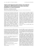
Figure 1, results showed that galectin-1 was expressed in
all HTLV-I-infected cell lines studied in contrast to non-
infected T cell lines in which galectin-1 mRNA expression
was either undetectable or slightly expressed. These results
hence suggested a possible association between HTLV-I
infection of T cells and increased expression of galectin-1.
Tax induces galectin-1 expression
As some of the tested HTLV-I-infected cells have been
reported to only express the viral Tax protein, we then
looked if Tax expression indeed could modulate galectin
mRNA levels. 293T cells were transfected with either a vec-
tor containing a complete HTLV-I proviral genome (i.e.
K30), or expression vectors coding for Tax WT or Tax
mutants defective in their ability to activate transcription
factors NF-κB, SRF and/or CREB. Galectin-1 expression
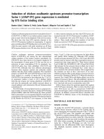
was then analyzed by RT-PCR. As shown in Figure 2A,
transfection of the K30 proviral DNA led to an induction
in the expression of galectin-1. In addition, comparable
induced levels of galectin-1 mRNA were observed in 293T
cells expressing wild-type Tax and both Tax mutants defec-
tive for CREB and SRF activation (Tax 703 and Tax Δ3). In
contrast, cells that were transfected with the Tax M22
(deficient in NF-κB activation) expression vector did not
demonstrate a significant difference in galectin-1 mRNA
levels when compared to cells transfected with the control
vector (Figure 2A). As RT-PCR experiments further show
that cells expressed similar levels of Tax, this difference in
upregulation of galectin-1 mRNA level was not due to dif-
ferences in the expression level of the different Tax pro-
teins in transfected 293T cells. In order to confirm these
results, RNA from 293T cells transfected with the various
Tax expression vectors were quantitatively analysed for
galectin-1 expression by real-time RT-PCR. Results pre-
sented in Figure 2B again revealed an important decrease
in Tax M22-mediated activation of galectin-1 expression
while other Tax mutants demonstrated a comparable
upregulation to the one measured with wild-type Tax.
Next, RT-PCR analyses were performed in a more repre-
sentative context, i.e T cell lines. Hence, the wild-type Tax
expression vector was transfected in CEM-T4 and SupT1 T
cell lines and analysed by RT-PCR for galectin-1 expres-
sion. As denoted in Figure 2C, Tax expression indeed
increased the expression of galectin-1 in both T cell lines.
As the data suggest that HTLV-I Tax induces the expression
of galectin-1 in non-T and T cell lines, it is likely that Tax
plays a role in the modulation of galectin-1 mRNA levels
in HTLV-I-infected cell lines.
Tax induces transcription from the galectin-1 promoter
To determine whether the effect of Tax on galectin-1 expres-
sion resulted from direct activation of transcription from the
galectin-1 promoter, two different luciferase-encoding vec-
tors driven by the human galectin-1 promoter were con-
structed. Two fragments of 0.5 kbp and 1.2 kbp containing
the transcription initiation site deduced from sequence
homology with the mouse galectin-1 gene were derived from
the human galectin-1 promoter region. Both fragments were
cloned upstream of the luciferase reporter gene of the pGL3-
Basic vector. Before determining the effect of Tax on these
constructs, the Tax M22 expression vector was first tested in
the context of Jurkat cells to see if it was specifically deficient
in activating NF-κB (Figure 3A). These results indeed con-
firmed previous studies in Jurkat cells: Tax M22 was only
defective in activating NF-κB unlike Tax 703, which was
comparable to wild-type Tax for NF-κB activation but greatly
affected in its capacity to activate both SRF and CREB (the lat-
ter being tested with the HTLV-I LTR-driven reporter con-
struct mainly responsive to CREB activation). As Tax M22
was behaving as expected in the Jurkat T cell line, the two
galectin-1 promoter constructs were next cotransfected with
Tax WT or Tax M22 expression vectors along with pActin-
LacZ into CEM-T4, Jurkat E6.1 and SupT1 T cell lines and
promoter activity was then evaluated by luciferase activity
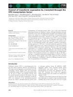
after normalisation (Figure 3B, C). When compared to cells
transfected with the control vector, the 0.5 kb galectin-1 pro-
moter construct demonstrated an increase of 10- to 15-fold
following expression of Tax WT while Tax M22 expression
led to a modest 2 to 4-fold induction (Figure 3B). For the 1.2
Retrovirology 2008, 5:105 />Page 6 of 15
(page number not for citation purposes)
kb galectin-1 promoter construct, expression of TaxWT led to
a 10- to 35-fold increase in promoter activity compared to 2
to 6 fold activation when the TaxM22 expression vector was
transfected (Figure 3C). These results suggested that the viral
protein Tax upregulates transcription from the galectin-1
promoter region, which likely accounts for the observed
increase in galectin-1 mRNA levels in both HTLV-I-infected
cells and cells transfected with the Tax expression vector.
Lower induction of the galectin-1 promoter by TaxM22,
which is deficient for NF-κB activation, raised the possi-
bility that this transcription factor was crucial for Tax-
mediated increase in galectin-1 expression. However, Jur-
kat cells transfected with the 1.2 kb galectin-1 promoter
construct did not show higher luciferase activity upon
stimulation with two known potent NF-κB activating
agents, PMA and TNF-α, thereby strongly suggesting that
NF-κB was not involved in the modulation of galectin-1
promoter activity by Tax (Figure 3D). As no known NF-
κB-binding sites have been identified from galectin-1 pro-
moter sequence analyses, these results strongly hint on the
involvement of a Tax-activated transcription factor differ-
ent from NF-κB in galectin-1 expression.
Galectin-1 is more abundant in the supernatant of HTLV-I
chronically infected T cell lines than in the supernatant of
non-infected cells
As we have demonstrated that HTLV-I-infected cell lines
express higher levels of galectin-1 mRNA, we next studied
whether these cells produced more extracellular galectin-
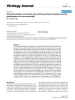
1. Figure 4 indeed shows that HTLV-I-infected T cell lines
released 13 to 50 times higher levels of extracellular galec-
tin-1 than the average level produced by uninfected T cell
lines. Interestingly, the S1T T cell line demonstrated the
lowest level of extracellular galectin-1 and is known to
poorly express Tax.
Together, the data suggest that mRNA and secretion of
galectin-1 were both upregulated in cells chronically
infected with HTLV-I.
Galectin-1 increases the infectivity of pseudotyped viruses
As galectin-1 can stabilize cell-to-cell and cell-virus inter-
actions by cross-linking different entities, we studied
whether extracellular galectin-1 could facilitate HTLV-I
infection. To initiate this study, Jurkat E6.1 cells were first
infected with luciferase-expressing HIV virions pseudo-
typed with the HTLV-I gp46 envelope in the presence of
various concentrations of purified galectin-1 (0–4 μM) for
48 hours; luciferase activity was then measured. The use of
HTLV-I gp46-pseudotyped virions that can express luci-
ferase allows us to detect a single round of infection and
although different from wild-type HTLV-I virions, it
should be representative of the type of interactions and
fusogenic activities of gp46 occurring on the surface of
HTLV-I virions upon infection. Infection of Jurkat E6.1
cells by the pseudotyped virions was increased by 1.6 fold
in the presence of 2 μM of galectin-1, an increase which
was statistically significant (F = 6.764, p = 0.0138) (Figure
5A). Lactose, an inhibitor of galectin-1, inhibited this
Comparative analysis of galectin-1 expression in different uninfected T cell lines and HTLV-I chronically-infected cell linesFigure 1
Comparative analysis of galectin-1 expression in different uninfected T cell lines and HTLV-I chronically-
infected cell lines. Galectin-1 mRNA levels were measured by RT-PCR analyses on total RNA isolated from non-infected
(A2.01, CEM-T4, HSB-2, JurkatE6.1, Molt-4, PM1, and Sup T1) and chronically HTLV-I-infected cells (C8166-45, C91-PL, MJ,
MT2 and S1T). PCR products were separated by electrophoresis on 1.5% agarose gels. Expression of β-actin mRNA served as
an internal control for normalization.
A
2
.
0
1
C
E
M
-
T
4
H
S
B
-
2
J
u
r
k
a
t
M
o
l
t
-
4
S
u
p
T
1
100pb marker
C
8
1
6
6
-
4
5
C
9
1
-
P
L
M
J
M
T
2
S
1
T
P
M
1
Galectin-1
β-Actin
Uninfected Infected
Retrovirology 2008, 5:105 />Page 7 of 15
(page number not for citation purposes)
galectin-1-promoting effect on HTLV-I infectivity, suggest-
ing that the carbohydrate binding activity of this protein
is involved in this increase. In order to increase the luci-
ferase signal, infection of Jurkat cells were also conducted
in the presence of the LTR activating agent TNF-α. Results
depicted in Figure 5B again demonstrated a highly signif-
icant (t = 5, p = 0.0069) positive effect of galectin-1 on
infectivity of gp46-pseudotyped virions.
A more physiological model was also used to study the
impact of soluble galectin-1 on infection by HTLV-I pseu-
dotyped virus. PBMCs isolated from a healthy donor were
stimulated with IL-2 and PHA-L for 72 hours and, after
washing, were then similarly treated upon infection by the
HTLV-I gp46-pseudotyped virions. The infection of
PBMCs by pseudotyped virions was increased by 1.8 fold
in the presence of 4 μM of galectin-1 (Figure 5C). The pos-
itive modulation on virus infection was determined to be
statistically significant (F = 4.364, p = 0.0425).
To eliminate the possibility that galectin-1 was positively
modulating LTR activity of the integrated proviral DNA of
our gp46-pseudotyped virions, Jurkat cells were trans-
fected with a vector containing the luciferase reporter gene
under the control of the HIV-1 LTR, after which different
concentrations of galectin-1 (0–4 μM) was added. Meas-
urement of luciferase activity demonstrated that the pres-
ence of galectin-1 had no impact on the transcription
levels dependent on the HIV-1 LTR (data not shown).
Analysis of galectin-1 expression in WT and mutant Tax-expressing cellsFigure 2
Analysis of galectin-1 expression in WT and mutant Tax-expressing cells. A,B. 293T cells were transfected with 40
μg of the control vector phβPr.1neo, Tax expression vectors (Tax 703, TaxΔ3, Tax M22, and Tax WT) or full-length proviral
DNA K30 clone. RT-PCR analyses for galectin-1, Tax and β-actin RNA levels (A) and real-time RT-PCR for galectin-1 RNA
levels (B) were conducted on RNA from each transfected conditions. The activated transcription factors for each Tax expres-
sion vectors are indicated below panel A. C. CEM-T4 and Sup T1 cell lines were transfected with 20 μg of the control vector
pHβPr.1neo or Tax WT expression vector. Total RNA was analyzed by RT-PCR for galectin-1 and β-actin RNA levels. PCR
products were separated by electrophoresis on 1.5% agarose gels.
A
Galectin-1
Tax
β-Actin
p
h
β
P
r
.
1
n
e
o
T
a
x
7
0
3
T
a
x
3
T
a
x
M
2
2
T
a
x
W
T
K
3
0
+++
SRF
+++-+/
CREB
++-++-
NF-κB
K30
Tax
WT
Tax
M22
Tax
3
Tax
703
phβPr.1
neo
p
h
β
P
r
.
1
n
e
o
T
a
x
WT
p
h
β
P
r
.
1
n
e
o
T
a
x
WT
β-Actin
Galectin-1
CEM-T4
Sup T1
C
0
0,05
0,1
0,15
0,2
0,25
phΒ Tax703 Tax3 TaxM22 TaxWT
Relative Galectin-1 mRNA expression
B
Retrovirology 2008, 5:105 />Page 8 of 15
(page number not for citation purposes)
Hence, these results show that extracellular galectin-1
increases infection of a T cell line and PBMCs by free
HTLV-I gp46-pseudotyped viruses and that this increase
relies on the binding of cell/virus surface carbohydrates by
the galectin-1 CRD.
Effect of galectin-1 on gp46-mediated cell fusion in a co-
culture assay
To study whether galectin-1 can possibly facilitate cell
fusion events, a co-culture system allowing a quantitative
evaluation of cell fusion by luciferase assay was used [58].
This cell line model provided another useful system to
assess the gp46-mediated fusion and was thus used to fur-
ther confirm the results obtained with the gp46-pseudo-
typed virions. Our results had previously strongly
suggested that this induction of luciferase activity could
not be attributed to HTLV-I infection following cell-to-cell
contact, but was rather involving cytoplasmic exchange
likely mediated by the fusogenic capacity of gp46. Briefly,
Jurkat E6.1 cells were transfected with pHTLV-Luc con-
taining the HTLV-I LTR upstream of the luciferase gene
and were subsequently co-cultured with the HTLV-I-
infected cell line, C91-PL. Cytoplasmic exchange can then
be estimated by assessing luciferase activity as Tax present
Activation of the galectin-1 promoter by Tax expression in transfected T cell linesFigure 3
Activation of the galectin-1 promoter by Tax expression in transfected T cell lines. A. Jurkat cells were transfected
with either pNF-κB-Luc, pHTLV-Luc or pSRE-Luc (7.5 μg) along with pHβPr.1neo (control vector) or expression vectors for
Tax WT, Tax M22 or Tax 703 (7.5 μg) and pActin-LacZ (5 μg). B,C. Jurkat, CEM-T4 and Sup T1 T cell lines were co-trans-
fected with pHβPr.1neo (control vector) or expression vectors for Tax WT or Tax M22 (7.5 μg), the galectin-1 promoter
reporter constructs pGL3-gal-1 0.5 kb (B) or pGL3-gal-1 1.2 kb (C) (7.5 μg) and pActin-LacZ (5 μg). D. Jurkat cells were
transfected with pNF-κB-Luc or pGL3-gal-1 1.2 kb (15 μg). After transfection (24 hours), cells were either left untreated or
stimulated with PMA or TNF-α for 8 hours. Luciferase and β-galactosidase activities were determined 48 hours after transfec-
tion as described in Materials and Methods. In panels A, B and C, luciferase activity was normalized on the basis of the β-galac-
tosidase activity. The results represent the mean of three independent transfections +/- standard deviations (*p < 0.05; **p <
0.01).
B
0
50
100
150
200
250
300
350
CEM-T4 Jurkat E6.1 Sup T1
Normalized luciferase activity (RLU)
phβPr.1neo Tax M22
Tax WT
**
*
**
**
**
C
0
500
1000
1500
2000
2500
3000
**
**
**
**
**
CEM-T4 Jurkat E6.1 Sup T1
phβPr.1neo Tax M22
Tax WT
Normalized luciferase activity (RLU)
0,1
1
10
100
1000
10000
NF-κ
κκ
κB-Luc
HTLV-Luc SRE-Luc
Normalized luciferase activity (Log RLU
)
phβPr.1neo
Tax WT
Tax M22
Tax 703
A
D
0
10
20
30
40
50
60
Untreated
PMA
TNF-
α
αα
α
Luciferase activity (RLU)
NF-κ
κκ
κB-Luc
pGL3-gal-1 1.2 kb
Retrovirology 2008, 5:105 />Page 9 of 15
(page number not for citation purposes)
in infected C91-PL cells should, upon cellular fusion, acti-
vate HTLV-I LTR activity in transfected Jurkat cells. This
assay was thus tested in the presence of different amounts
of galectin-1 (0–4 μM) for 24 hours, after which luciferase
activity was measured. A dose-dependent (and statistically
significant at 4 μM; F = 4.192, p = 0.0466) increase in luci-
ferase activity mediated by galectin-1 was noted (Figure
6A). Again, this induction was lactose-sensitive. Of note,
a small but non-significant effect of lactose was apparent
in co-cultured cells which were not treated with galectin-
1, suggesting a possible impact of endogenous galectin-1
in cell fusion affecting luciferase activity. As a control,
supernatant from C91-PL cells incubated in the presence
of transfected Jurkat cells did not lead to any significant
increase in luciferase activity either in the absence or pres-
ence of galectin-1, thereby ruling out the effect of extracel-
lular factors acting on HTLV-I LTR activity (Figure 6B). In
addition, although we cannot rule out a contribution in
this signal from infection events by HTLV-I particles on
Jurkat cells, which would similarly induce luciferase
expression, previous experiments have suggested that the
first 24-hour time course preferentially involves HTLV-I-
driven syncytium formation in the modulation of luci-
ferase assay [58].
These results show that soluble galectin-1 can also
increase cytoplasmic cell exchange likely occurring
though gp46-dependent cell fusion events between an
HTLV-I-infected cells and uninfected T cells, again being
inhibited by the addition of lactose.
Discussion
HTLV-I is a poorly infectious virus and, in this regard, the
presence of various molecules that facilitate infection may
Comparative analysis of extracellular galectin-1 levels between uninfected and HTLV-I-chronically-infected cell linesFigure 4
Comparative analysis of extracellular galectin-1 levels between uninfected and chronically HTLV-I-infected
cell lines. A2.01, CEM-T4, HSB-2, Jurkat E6.1, Molt-4, PM1, Sup T1, C8166-45, C91-PL, MJ, MT2 and S1T cell lines were cul-
tured for 48 hours starting at a concentration of 5 × 10
5
cells/ml. The supernatants were then collected, passed through a 0.22
μm filter and analysed for galectin-1 secretion by a galectin-1-specific ELISA as described in Materials and Methods.
Non-infected
Infected
0
600
1200
1800
2400
3000
3600
4200
A2.01
CEM-T4
HSB.2
Jurkat
MOLT.4
PM1
Sup T1
C8166-45
C9L-PL
MJ
MT2
S1T
Galectin-1 (picoM)
4800
Retrovirology 2008, 5:105 />Page 10 of 15
(page number not for citation purposes)
be important for viral transmission. Several studies have
been conducted on the implication of adhesion mole-
cules incorporated by retroviruses (especially for HIV-1)
and their positive impact on viral replication [59]. Similar
studies have revealed that cell surface adhesion molecules
could affect the infection and syncytium formation
related to HTLV-I [8,13,60-63]. In addition, certain stud-
ies have also indicated that soluble factors were also pos-
sible modulators of the HTLV-I infection process [64,65].
Galectins are a family of proteins involved in cell adhe-
sion but few studies have been conducted on their possi-
ble involvement in viral infection [66]. In the present
study, we have focused on galectin-1, mainly because of
its capacity to mediate cell-to-cell contact but also because
this protein is expressed by activated T cells and cells from
lymphoid tissue, a major site of infection by HTLV-I.
In this study, we have demonstrated that galectin-1 is
more strongly expressed and secreted in chronically
HTLV-I-infected T cell lines compared to uninfected T
cells. These results agree with the study of Pise-Masison
and colleagues, which showed through DNA microarray
experiments that galectin-1 gene expression is upregulated
in HTLV-I-transformed and immortalized cell lines [19].
Furthermore, we have demonstrated that the viral Tax pro-
tein could be involved in the upregulation of galectin-1
expression. Generally, Tax directly activates gene tran-
scription by the activation of CREB, NF-κB and/or SRF
transcription factor [67]. Using Tax mutants and known
Soluble galectin-1 positively impacts on the infection of T cell line and PBMCs by HTLV-I-envelope-pseudotyped virusesFigure 5
Soluble galectin-1 positively impacts on the infection of T cell line and PBMCs by HTLV-I-envelope-pseudo-
typed viruses. Jurkat cells (A, B) or PBMCs (C) (1 × 10
5
cells) were infected with 10 ng (p24) of HTLV-I envelope-pseudo-
typed HIV-1 viruses in the presence of different concentrations of purified galectin-1 (0–4 μM), with or without lactose (50
mM). B, Jurkat cells were also treated with TNF-α (10 ng/ml). Luciferase activities were measured 48 hours post-infection. The
results represent three independent infections and are expressed as the mean luciferase activity value +/- standard deviation
(*p < 0.05; **p < 0.01).
A
NL4.3L+E- /
pSV HTLV-I env
0
2
4
6
8
10
12
14
16
18
Luciferase activity (RLU)
PBS
Lactose (50mM)
2μM
1μM0μM0μM
Galectin-1
+++
++
-
+++
++
+
Jurkat E6.1
*
2μM
0μMGalectin-1
0
50
100
150
200
250
300
350
400
450
500
Luciferase activity (RLU)
PBS
Lactose (50mM)
**
B
-
+
+
4μM2μM1μM0μM0μM
Galectin-1
++
+
+++++
NL4.3L+E- /
pSV HTLV-I env
++
+
+++++++
PBMCs
0
1
2
3
4
5
6
Luciferase activity (RLU)
PBS
Lactose (50mM)
*
C
+
Retrovirology 2008, 5:105 />Page 11 of 15
(page number not for citation purposes)
Soluble galectin-1 increases the extent of HTLV-I LTR activation in co-culture assayFigure 6
Soluble galectin-1 increases the extent of HTLV-I LTR activation in co-culture assay. Jurkat cells were transfected
with 15 μg of pHTLV-Luc and cultured for 24 hours. The transfected cells were then incubated for an additional 24 hour with
an equal number of HTLV-I-infected C91-PL cells (A) or supernatant of C91-PL cells (B) in the presence of galectin-1 added at
various concentrations with or without lactose (50 mM). The cells were lysed 24 hours after co-culture and luciferase activities
were measured. The results represent three independent co-culture assays and are expressed as the mean luciferase activity
value +/- standard deviations (*p < 0.05; **p < 0.01).
0
10
20
30
40
50
60
70
80
Luciferase activity (RLU)
PBS
Lactose (50mM)
*
0
1
2
3
4
5
6
Luciferase activity (RLU)
4μM2μM1μM0μM0μM
Galectin-1
++++++++
C91-PL
++++++++++
Jurkat E6.1/
pHTLV-Luc
4μM2μM1μM0μM0μM
Galectin-1
++++-
C91-PL sup.
+++++
Jurkat E6.1/
pHTLV-Luc
A
B
Retrovirology 2008, 5:105 />Page 12 of 15
(page number not for citation purposes)
NF-κB activators, we have shown that CREB, SRF and NF-
κB are not involved in Tax-induced galectin-1 expression.
This was surprising given that the Tax mutant TaxM22,
which is deficient for the activation of NF-κB, was less effi-
cient in activating galectin-1 expression when compared
to wild-type Tax. However, no typical NF-κB binding con-
sensus sequences have been identified in the galectin-1
promoter region tested in this study. In this regard, it
should be noted that Tax M22 has been demonstrated to
be deficient for the activation of another transcription fac-
tor namely NFAT [68,69]. In addition, as the galectin-1
promoter has eight Sp1-potential sites in its 1.2 kbp
region, six of which are shared with the 0.5 kbp region,
Sp1 might be such a potential transcription factor. Indeed,
it has been shown that Tax can interact with Sp1 and the
resulting complex is important for the transactivation of
PTHrP P3 and GATA3 promoters [70,71]. Alternatively,
Tax may indirectly induce galectin-1 expression by main-
taining a chronic activation of infected cells. In HTLV-I-
infected cells, the constitutive expression of the LTR
allows a weak expression of Tax. Since activated T lym-
phocytes express galectin-1, it may be possible that this
chronic activation of HTLV-I-infected cells by Tax indi-
rectly induces galectin-1 expression. Finally, although our
results argue for an implication of Tax, it should be stated
that other HTLV-I proteins might also participate in the
modulation of galectin-1 expression in infected cells as
elevated galectin-1 mRNA levels were detected in the S1T
cell line, which poorly expresses Tax.
As cell-free virus has very low infectivity, it is assumed that
HTLV-I infection is mediated by the interaction between
non-infected and infected cells, although recent evidence
has demonstrated that cell-free virus can infect isolated
dendritic cells [72-74]. We have previously demonstrated
that galectin-1 increases HIV-1 infectivity through stabiliz-
ing the virus adsorption step [35]. In addition, galectin-1
may mediate cell-cell interaction as galectin-1 can cross-
link cells. Using pseudotyped HIV-1 virions, which har-
bour HTLV-I gp46 envelope protein and express the luci-
ferase reporter gene, we showed that, in both Jurkat and
PBMCs, virion infection was significantly promoted in the
presence of galectin-1 in a glycan binding-dependent
manner, suggesting that galectin-1 increases the infectivity
of HTLV-I virus particles in this system. We also used a
quantitative system which mimics the mechanism involv-
ing gp46-mediated fusion in a cell-to-cell fashion. Again,
galectin-1 increased this HTLV-I-induced gp46-mediated
cell fusion showing that galectin-1 might also stabilize the
interaction between infected and non-infected cells. Of
note, no extracellular factors seemed to act upon galectin-
1-mediated induction of HTLV-I LTR activity in this co-
culture system as judged by results obtained with C91-PL
supernatant. However, at this point, it cannot be dis-
missed that other possible gp46-independent cytoplasmic
exchange modulated by galectin-1 might be taking place
and lead to this upregulation of luciferase activity. Further
experiments will be needed to assess this issue.
Although galectin-1 concentrations in our infection exper-
iments were higher than the measured levels from the
supernatant of infected cells, in vivo conditions are likely
to differ from our cell culture settings. Previous studies
have demonstrated that lymphoid organ tissues are sites
where an important number of infected T lymphocytes are
located [75-77]. Given that these tissues represent a more
confined space, the concentration of galectin-1 secreted
by surrounding infected cells may represent a more appro-
priate environment and increase the concentration of
galectin-1 to effective levels. Indeed, we have previously
reported that the tonsil tissue contains 10 to 20 μM galec-
tin-1 [35]. Moreover, the transmission of HTLV-I to target
cells has been shown to require the formation of a virolog-
ical synapse following a cell-cell contact [7]. This synapse
is formed by the binding of host molecules between the
HTLV-I-infected T cells and the uninfected T lymphocytes,
thereby facilitating virus transmission. Galectin-1 may be
concentrated in the vicinity of this virological synapse and
more favourably act upon infection. A final issue which
needs to be taken into consideration in the current study
relates to breast feeding, an important route for HTLV-I
transmission. Lactose is an important constituent of
breast milk and therefore could be suggested to hinder the
action of galectin-1 during this route of HTLV-I transmis-
sion. Although one might then argue that galectin-1 is less
important for this mode of transmission, it remains to be
determined whether high lactose concentrations are also
present at sites where initial HTLV-I infection does occur
following HTLV-I transmission during breast feeding and
where galectin-1 could modulate HTLV-I binding.
Conclusion
In summary, our study demonstrates a bidirectional inter-
action between HTLV-I and galectin-1. The data demon-
strated that expression of galectin-1 was increased in
chronically HTLV-I-infected cells and that this modula-
tion of galectin-1 expression was largely attributed to the
viral transactivator Tax in NF-κB- and CREB-independent
manners. In addition, this study showed that HTLV-I-
infected cells secrete galectin-1 at a higher level than the
uninfected cells and that extracellular galectin-1 facilitates
HTLV-I infection and promotes higher levels of gp46-
dependent cell fusion. Given that our previous studies
had demonstrated that HIV-1 is also enhanced in its infec-
tivity by galectin-1 [35,36], other retroviruses (or even
other enveloped viruses) could potentially be more infec-
tious in the presence of galectin-1. Further studies will be
needed to assess the possible universal action of this β-
galactoside-binding protein on the replicative cycle of var-
ious pathogens.
Retrovirology 2008, 5:105 />Page 13 of 15
(page number not for citation purposes)
Competing interests
The authors declare that they have no competing interests.
Authors' contributions
SG carried all RT-PCR analyses, transfection experiments
and infection and syncytium formation assay and has
drafted the manuscript. IP has conducted the ELISA assay.
MO has participated in the design of the study. AV has
performed the real-time RT-PCR experiments and has
helped in drafting the manuscript. MJT and SS have
helped in drafting and finalizing the manuscript and have
provided important input on the design of the study. BB
conceived the study, participated in its coordination and
helped in drafting and finalizing the manuscript.
Acknowledgements
We thank Ms. Sylvie Méthot for editorial assistance. This work was per-
formed by SG in partial fulfillment of a M.Sc. degree in the Microbiology-
Immunology Program at Laval University. MJT. is the recipient of the Can-
ada Research Chair in Human Immuno-Retrovirology (Tier 1 level) and SS
has been awarded a Scholarship Award (Senior level) from the Fonds de la
Recherche en Santé du Québec (FRSQ). BB holds a Canada Research Chair
in Human Retrovirology (Tier 2 level).
References
1. Watanabe T: HTLV-1-associated diseases. Int J Hematol 1997,
66:257-278.
2. Zaninovic V: On the etiology of tropical spastic paraparesis
and human T-cell lymphotropic virus-I-associated myelopa-
thy. Int J Infect Dis 1999, 3:168-176.
3. Manns A, Hisada M, La Grenade L: Human T-lymphotropic virus
type I infection. Lancet 1999, 353:1951-1958.
4. Derse D, Heidecker G, Mitchell M, Hill S, Lloyd P, Princler G: Infec-
tious transmission and replication of human T-cell leukemia
virus 1. Front Biosci 2004, 9:2495-2499.
5. Richardson JH, Edwards AJ, Cruickshank JK, Rudge P, Dalgleish AG:
In vivo cellular tropism of human T-cell leukemia virus type
1. J Virol 1990, 64:5682-5687.
6. Teruya H, Tomita M, Senba M, Ishikawa C, Tamayose M, Miyazato A,
Yara S, Tanaka Y, Iwakura Y, Fujita J, et al.: Human T-cell leukemia
virus type I infects human lung epithelial cells and induces
gene expression of cytokines, chemokines and cell adhesion
molecules. Retrovirology 2008, 5:86.
7. Igakura T, Stinchcombe JC, Goon PK, Taylor GP, Weber JN, Griffiths
GM, Tanaka Y, Osame M, Bangham CR: Spread of HTLV-I
between lymphocytes by virus-induced polarization of the
cytoskeleton. Science 2003, 299:1713-1716.
8. Fan N, Gavalchin J, Paul B, Wells KH, Lane MJ, Poiesz BJ: Infection
of peripheral blood mononuclear cells and cell lines by cell-
free human T-cell lymphoma/leukemia virus type I. J Clin
Microbiol 1992, 30:905-910.
9. Derse D, Hill SA, Lloyd PA, Chung H, Morse BA: Examining human
T-lymphotropic virus type 1 infection and replication by cell-
free infection with recombinant virus vectors. J Virol 2001,
75:8461-8468.
10. Manel N, Taylor N, Kinet S, Kim FJ, Swainson L, Lavanya M, Battini JL,
Sitbon M: HTLV envelopes and their receptor GLUT1, the
ubiquitous glucose transporter: a new vision on HTLV infec-
tion? Front Biosci
2004, 9:3218-3241.
11. Kinet S, Swainson L, Lavanya M, Mongellaz C, Montel-Hagen A,
Craveiro M, Manel N, Battini JL, Sitbon M, Taylor N: Isolated recep-
tor binding domains of HTLV-1 and HTLV-2 envelopes bind
Glut-1 on activated CD4+ and CD8+ T cells. Retrovirology 2007,
4:31.
12. Nejmeddine M, Barnard AL, Tanaka Y, Taylor GP, Bangham CR:
Human T-lymphotropic virus, type 1, tax protein triggers
microtubule reorientation in the virological synapse. J Biol
Chem 2005, 280:29653-29660.
13. Barnard AL, Igakura T, Tanaka Y, Taylor GP, Bangham CR: Engage-
ment of specific T-cell surface molecules regulates cytoskel-
etal polarization in HTLV-1-infected lymphocytes. Blood
2005, 106:988-995.
14. Fujisawa J, Seiki M, Sato M, Yoshida M: A transcriptional enhancer
sequence of HTLV-I is responsible for trans-activation medi-
ated by p40 chi HTLV-I. EMBO J 1986, 5:713-718.
15. Paskalis H, Felber BK, Pavlakis GN: Cis-acting sequences respon-
sible for the transcriptional activation of human T-cell leuke-
mia virus type I constitute a conditional enhancer. Proc Natl
Acad Sci USA 1986, 83:6558-6562.
16. Shimotohno K, Takano M, Teruuchi T, Miwa M: Requirement of
multiple copies of a 21-nucleotide sequence in the U3
regions of human T-cell leukemia virus type I and type II long
terminal repeats for trans-acting activation of transcription.
Proc Natl Acad Sci USA 1986, 83:8112-8116.
17. Goren I, Semmes OJ, Jeang KT, Moelling K: The amino terminus
of Tax is required for interaction with the cyclic AMP
response element binding protein. J Virol 1995, 69:5806-5811.
18. Munoz E, Israel A: Activation of NF-kappa B by the Tax protein
of HTLV-1. Immunobiology 1995, 193:128-136.
19. Pise-Masison CA, Radonovich M, Mahieux R, Chatterjee P, Whiteford
C, Duvall J, Guillerm C, Gessain A, Brady JN: Transcription profile
of cells infected with human T-cell leukemia virus type I
compared with activated lymphocytes. Cancer Res
2002,
62:3562-3571.
20. Hirabayashi J, Kasai K: The family of metazoan metal-independ-
ent beta-galactoside-binding lectins: structure, function and
molecular evolution. Glycobiology 1993, 3:297-304.
21. Leffler H, Carlsson S, Hedlund M, Qian Y, Poirier F: Introduction to
galectins. Glycoconj J 2004, 19:433-440.
22. Toscano MA, Ilarregui JM, Bianco GA, Campagna L, Croci DO, Salat-
ino M, Rabinovich GA: Dissecting the pathophysiologic role of
endogenous lectins: glycan-binding proteins with cytokine-
like activity? Cytokine Growth Factor Rev 2007, 18:57-71.
23. Hirabayashi J, Hashidate T, Arata Y, Nishi N, Nakamura T, Hirashima
M, Urashima T, Oka T, Futai M, Muller WE, et al.: Oligosaccharide
specificity of galectins: a search by frontal affinity chroma-
tography. Biochim Biophys Acta 2002, 1572:232-254.
24. Hughes RC: Secretion of the galectin family of mammalian
carbohydrate-binding proteins. Biochim Biophys Acta 1999,
1473:172-185.
25. Nickel W: The mystery of nonclassical protein secretion. A
current view on cargo proteins and potential export routes.
Eur J Biochem 2003, 270:2109-2119.
26. Nickel W: Unconventional secretory routes: direct protein
export across the plasma membrane of mammalian cells.
Traffic 2005, 6:607-614.
27. Baum LG, Pang M, Perillo NL, Wu T, Delegeane A, Uittenbogaart CH,
Fukuda M, Seilhamer JJ: Human thymic epithelial cells express
an endogenous lectin, galectin-1, which binds to core 2 O-
glycans on thymocytes and T lymphoblastoid cells. J Exp Med
1995, 181:877-887.
28. Blaser C, Kaufmann M, Muller C, Zimmermann C, Wells V, Mallucci
L, Pircher H: Beta-galactoside-binding protein secreted by
activated T cells inhibits antigen-induced proliferation of T
cells. Eur J Immunol 1998, 28:2311-2319.
29. Chiariotti L, Salvatore P, Frunzio R, Bruni CB: Galectin genes: reg-
ulation of expression.
Glycoconj J 2004, 19:441-449.
30. Salvatore P, Benvenuto G, Caporaso M, Bruni CB, Chiariotti L: High
resolution methylation analysis of the galectin-1 gene pro-
moter region in expressing and nonexpressing tissues. FEBS
Lett 1998, 421:152-158.
31. Salvatore P, Benvenuto G, Pero R, Lembo F, Bruni CB, Chiariotti L:
Galectin-1 gene expression and methylation state in human
T leukemia cell lines. Int J Oncol 2000, 17:1015-1018.
32. Klose RJ, Bird AP: Genomic DNA methylation: the mark and
its mediators. Trends Biochem Sci 2006, 31:89-97.
33. Salvatore P, Contursi C, Benvenuto G, Bruni CB, Chiariotti L: Char-
acterization and functional dissection of the galectin-1 gene
promoter. FEBS Lett 1995, 373:159-163.
34. Kondoh N, Hada A, Ryo A, Shuda M, Arai M, Matsubara O, Kimura F,
Wakatsuki T, Yamamoto M: Activation of Galectin-1 gene in
human hepatocellular carcinoma involves methylation-sen-
sitive complex formations at the transcriptional upstream
and downstream elements. Int J Oncol 2003, 23:1575-1583.
Retrovirology 2008, 5:105 />Page 14 of 15
(page number not for citation purposes)
35. Ouellet M, Mercier S, Pelletier I, Bounou S, Roy J, Hirabayashi J, Sato
S, Tremblay MJ: Galectin-1 acts as a soluble host factor that
promotes HIV-1 infectivity through stabilization of virus
attachment to host cells. J Immunol 2005, 174:4120-4126.
36. Mercier S, St-Pierre C, Pelletier I, Ouellet M, Tremblay MJ, Sato S:
Galectin-1 promotes HIV-1 infectivity in macrophages
through stabilization of viral adsorption. Virology 2008,
371:121-9.
37. Salahuddin SZ, Markham PD, Wong-Staal F, Franchini G, Kalyanara-
man VS, Gallo RC: Restricted expression of human T-cell
leukemia – lymphoma virus (HTLV) in transformed human
umbilical cord blood lymphocytes. Virology 1983, 129:51-64.
38. Popovic M, Sarngadharan MG, Read E, Gallo RC: Detection, isola-
tion, and continuous production of cytopathic retroviruses
(HTLV-III) from patients with AIDS and pre-AIDS. Science
1984, 224:497-500.
39. Popovic M, Lange-Wantzin G, Sarin PS, Mann D, Gallo RC: Transfor-
mation of human umbilical cord blood T cells by human T-
cell leukemia/lymphoma virus. Proc Natl Acad Sci USA 1983,
80:5402-5406.
40. Harada S, Koyanagi Y, Yamamoto N: Infection of HTLV-III/LAV
in HTLV-I-carrying cells MT-2 and MT-4 and application in a
plaque assay. Science 1985, 229:563-566.
41. Mills GB, Arima N, May C, Hill M, Schmandt R, Li J, Miyamoto NG,
Greene WC: Neither the LCK nor the FYN kinases are oblig-
atory for IL-2-mediated signal transduction in HTLV-I-
infected human T cells. Int Immunol 1992, 4:1233-1243.
42. Folks T, Powell DM, Lightfoote MM, Benn S, Martin MA, Fauci AS:
Induction of HTLV-III/LAV from a nonvirus-producing T-cell
line: implications for latency. Science 1986, 231:600-602.
43. Hara J, Benedict SH, Champagne E, Mak TW, Minden M, Gelfand EW:
Comparison of T cell receptor alpha, beta, and gamma gene
rearrangement and expression in T cell acute lymphoblastic
leukemia. J Clin Invest 1988, 81:989-996.
44. Weiss A, Imboden J, Shoback D, Stobo J: Role of T3 surface mol-
ecules in human T-cell activation: T3-dependent activation
results in an increase in cytoplasmic free calcium. Proc Natl
Acad Sci USA 1984, 81:
4169-4173.
45. Kikukawa R, Koyanagi Y, Harada S, Kobayashi N, Hatanaka M,
Yamamoto N: Differential susceptibility to the acquired
immunodeficiency syndrome retrovirus in cloned cells of
human leukemic T-cell line Molt-4. J Virol 1986, 57:1159-1162.
46. Lusso P, Cocchi F, Balotta C, Markham PD, Louie A, Farci P, Pal R,
Gallo RC, Reitz MS Jr: Growth of macrophage-tropic and pri-
mary human immunodeficiency virus type 1 (HIV-1) isolates
in a unique CD4+ T-cell clone (PM1): failure to downregulate
CD4 and to interfere with cell-line-tropic HIV-1. J Virol 1995,
69:3712-3720.
47. Smith SD, Shatsky M, Cohen PS, Warnke R, Link MP, Glader BE:
Monoclonal antibody and enzymatic profiles of human
malignant T-lymphoid cells and derived cell lines. Cancer Res
1984, 44:5657-5660.
48. Pear WS, Nolan GP, Scott ML, Baltimore D: Production of high-
titer helper-free retroviruses by transient transfection. Proc
Natl Acad Sci USA 1993, 90:8392-8396.
49. Lahm HW, Stein S: Characterization of recombinant human
interleukin-2 with micromethods. J Chromatogr 1985,
326:357-361.
50. Matsumoto K, Shibata H, Fujisawa JI, Inoue H, Hakura A, Tsukahara
T, Fujii M: Human T-cell leukemia virus type 1 Tax protein
transforms rat fibroblasts via two distinct pathways. J Virol
1997, 71:4445-4451.
51. Schwartz O, Virelizier JL, Montagnier L, Hazan U: A microtransfec-
tion method using the luciferase-encoding reporter gene for
the assay of human immunodeficiency virus LTR promoter
activity. Gene 1990, 88:197-205.
52. Lemieux AM, Pare ME, Audet B, Legault E, Lefort S, Boucher N, Lan-
dry S, van Opijnen T, Berkhout B, Naghavi MH, et al.: T-cell activa-
tion leads to poor activation of the HIV-1 clade E long
terminal repeat and weak association of nuclear factor-kap-
paB and NFAT with its enhancer region. J Biol Chem 2004,
279:52949-52960.
53. Barbeau B, Bernier R, Dumais N, Briand G, Olivier M, Faure R, Posner
BI, Tremblay M: Activation of HIV-1 long terminal repeat tran-
scription and virus replication via NF-kappaB-dependent and
-independent pathways by potent phosphotyrosine phos-
phatase inhibitors, the peroxovanadium compounds. J Biol
Chem
1997, 272:12968-12977.
54. Fortin JF, Cantin R, Lamontagne G, Tremblay M: Host-derived
ICAM-1 glycoproteins incorporated on human immunodefi-
ciency virus type 1 are biologically active and enhance viral
infectivity. J Virol 1997, 71:3588-3596.
55. Bounou S, Leclerc JE, Tremblay MJ: Presence of host ICAM-1 in
laboratory and clinical strains of human immunodeficiency
virus type 1 increases virus infectivity and CD4(+)-T-cell
depletion in human lymphoid tissue, a major site of replica-
tion in vivo. J Virol 2002, 76:1004-1014.
56. Zar JH: Biostatistical Analysis Prentice Hall, Englewoods Cliffs, NJ; 1984.
57. Harhaj EW, Good L, Xiao G, Sun SC: Gene expression profiles in
HTLV-I-immortalized T cells: deregulated expression of
genes involved in apoptosis regulation. Oncogene 1999,
18:1341-1349.
58. Paré ME, Gauthier S, Landry S, Sun J, Legault E, Leclerc D, Tanaka Y,
Marriott SJ, Tremblay MJ, Barbeau B: A new sensitive and quanti-
tative HTLV-I-mediated cell fusion assay in T cells. Virology
2005, 338:309-322.
59. Cantin R, Methot S, Tremblay MJ: Plunder and stowaways: incor-
poration of cellular proteins by enveloped viruses. J Virol 2005,
79:6577-6587.
60. Hildreth JE, Subramanium A, Hampton RA: Human T-cell lympho-
tropic virus type 1 (HTLV-1)-induced syncytium formation
mediated by vascular cell adhesion molecule-1: evidence for
involvement of cell adhesion molecules in HTLV-1 biology. J
Virol 1997, 71:1173-1180.
61. Valentin H, Lemasson I, Hamaia S, Casse H, Konig S, Devaux C, Gaz-
zolo L: Transcriptional activation of the vascular cell adhesion
molecule-1 gene in T lymphocytes expressing human T-cell
leukemia virus type 1 Tax protein. J Virol 1997, 71:8522-8530.
62. Ceccaldi PE, Delebecque F, Prevost MC, Moris A, Abastado JP, Ges-
sain A, Schwartz O, Ozden S: DC-SIGN facilitates fusion of den-
dritic cells with human T-cell leukemia virus type 1-infected
cells. J Virol 2006, 80:4771-4780.
63. Daenke S, McCracken SA, Booth S: Human T-cell leukaemia/
lymphoma virus type 1 syncytium formation is regulated in
a cell-specific manner by ICAM-1, ICAM-3 and VCAM-1 and
can be inhibited by antibodies to integrin beta2 or beta7. J
Gen Virol 1999, 80(Pt 6):1429-1436.
64. Moriuchi M, Moriuchi H: Seminal fluid enhances replication of
human T-cell leukemia virus type 1: implications for sexual
transmission. J Virol 2004, 78:12709-12711.
65. Moriuchi M, Inoue H, Moriuchi H: Reciprocal interactions
between human T-lymphotropic virus type 1 and prostaglan-
dins: implications for viral transmission. J Virol 2001,
75:192-198.
66. Hsu DK, Hammes SR, Kuwabara I, Greene WC, Liu FT: Human T
lymphotropic virus-I infection of human T lymphocytes
induces expression of the beta-galactoside-binding lectin,
galectin-3. Am J Pathol 1996, 148:1661-1670.
67. Shimizu T, Kawakita S, Li QH, Fukuhara S, Fujisawa J: Human T-cell
leukemia virus type 1 Tax protein stimulates the interferon-
responsive enhancer element via NF-kappaB activity. FEBS
Lett 2003, 539:73-77.
68. Good L, Maggirwar SB, Sun SC: Activation of the IL-2 gene pro-
moter by HTLV-I tax involves induction of NF-AT com-
plexes bound to the CD28-responsive element. EMBO J 1996,
15:3744-3750.
69. Rivera I, Harhaj EW, Sun SC: Involvement of NF-AT in type I
human T-cell leukemia virus Tax-mediated Fas ligand pro-
moter transactivation. J Biol Chem 1998, 273:22382-22388.
70. Richard V, Nadella MV, Green PL, Lairmore MD, Feuer G, Foley JG,
Rosol TJ: Transcriptional regulation of parathyroid hormone-
related protein promoter P3 by ETS-1 in adult T-cell leuke-
mia/lymphoma. Leukemia 2005, 19:1175-1183.
71. Gilli SC, Salles TS, Saad ST: Regulation of the GATA3 promoter
by human T-cell lymphotropic virus type I Tax protein. J Cell
Biochem 2004, 93:
1178-1187.
72. Popovic M, Sarin PS, Robert-Gurroff M, Kalyanaraman VS, Mann D,
Minowada J, Gallo RC: Isolation and transmission of human ret-
rovirus (human t-cell leukemia virus). Science 1983,
219:856-859.
73. Yamamoto N, Okada M, Koyanagi Y, Kannagi M, Hinuma Y: Trans-
formation of human leukocytes by cocultivation with an
Publish with BioMed Central and every
scientist can read your work free of charge
"BioMed Central will be the most significant development for
disseminating the results of biomedical research in our lifetime."
Sir Paul Nurse, Cancer Research UK
Your research papers will be:
available free of charge to the entire biomedical community
peer reviewed and published immediately upon acceptance
cited in PubMed and archived on PubMed Central
yours — you keep the copyright
Submit your manuscript here:
/>BioMedcentral
Retrovirology 2008, 5:105 />Page 15 of 15
(page number not for citation purposes)
adult T cell leukemia virus producer cell line. Science 1982,
217:737-739.
74. Jones KS, Petrow-Sadowski C, Huang YK, Bertolette DC, Ruscetti
FW: Cell-free HTLV-1 infects dendritic cells leading to trans-
mission and transformation of CD4(+) T cells. Nat Med 2008,
14:429-36.
75. Takenouchi N, Matsuoka E, Moritoyo T, Nagai M, Katsuta K, Hasui K,
Ueno K, Eizuru Y, Usuku K, Osame M, et al.: Molecular pathologic
analysis of the tonsil in HTLV-I-infected individuals. J Acquir
Immune Defic Syndr 1999, 22:200-207.
76. Tanaka M, Sun B, Fang J, Nitta T, Yoshida T, Kohtoh S, Kikukawa H,
Hanai S, Uchida K, Miwa M: Human T-cell leukemia virus type 1
(HTLV-1) infection of mice: proliferation of cell clones with
integrated HTLV-1 provirus in lymphoid organs. J Virol 2001,
75:4420-4423.
77. Kazanji M: HTLV type 1 infection in squirrel monkeys (Saimiri
sciureus): a promising animal model for HTLV type 1 human
infection. AIDS Res Hum Retroviruses 2000, 16:1741-1746.