Báo cáo y học: " Human Immunodeficiency Virus type-1 reverse transcriptase exists as post-translationally modified forms in virions and cells" pdf
Bạn đang xem bản rút gọn của tài liệu. Xem và tải ngay bản đầy đủ của tài liệu tại đây (650.14 KB, 12 trang )
BioMed Central
Page 1 of 12
(page number not for citation purposes)
Retrovirology
Open Access
Research
Human Immunodeficiency Virus type-1 reverse transcriptase exists
as post-translationally modified forms in virions and cells
Adam J Davis
1
, Jillian M Carr*
1,2
, Christopher J Bagley
3
, Jason Powell
4
,
David Warrilow
5
, David Harrich
5,6
, Christopher J Burrell
1,2
and Peng Li
1
Address:
1
Infectious Diseases Laboratories, SA Pathology, Adelaide 5000, Australia,
2
School of Molecular and Biomedical Science, University of
Adelaide, Adelaide 5005, Australia,
3
Adelaide Proteomics Centre, University of Adelaide, Adelaide 5005, Australia,
4
Division of Human
Immunology, SA Pathology, Adelaide 5000, Australia,
5
Division of Infectious Disease, Queensland Institute of Medical Research, Brisbane 4029,
Australia and
6
Griffith Medical Research College, a joint program of Griffith University and the Queensland Institute of Medical Research,
Queensland 4029, Australia
Email: Adam J Davis - ; Jillian M Carr* - ;
Christopher J Bagley - ; Jason Powell - ;
David Warrilow - ; David Harrich - ;
Christopher J Burrell - ; Peng Li -
* Corresponding author
Abstract
Background: HIV-1 reverse transcriptase (RT) is a heterodimer composed of p66 and p51
subunits and is responsible for reverse transcription of the viral RNA genome into DNA. RT can
be post-translationally modified in vitro which may be an important mechanism for regulating RT
activity. Here we report detection of different p66 and p51 RT isoforms by 2D gel electrophoresis
in virions and infected cells.
Results: Major isoforms of the p66 and p51 RT subunits were observed, with pI's of 8.44 and 8.31
respectively (p66
8.44
and p51
8.31
). The same major isoforms were present in virions, virus-infected
cell lysates and intracellular reverse transcription complexes (RTCs), and their presence in RTCs
suggested that these are likely to be the forms that function in reverse transcription. Several minor
RT isoforms were also observed. The observed pIs of the RT isoforms differed from the pI of
theoretical unmodified RT (p66
8.53
and p51
8.60
), suggesting that most of the RT protein in virions
and cells is post-translationally modified. The modifications of p66
8.44
and p51
8.31
differed from each
other indicating selective modification of the different RT subunits. The susceptibility of RT
isoforms to phosphatase treatment suggested that some of these modifications were due to
phosphorylation. Dephosphorylation, however, had no effect on in vitro RT activity associated with
virions, infected cells or RTCs suggesting that the phospho-isoforms do not make a major
contribution to RT activity in an in vitro assay.
Conclusion: The same major isoform of p66 and p51 RT is found in virions, infected cells and
RTC's and both of these subunits are post-translationally modified. This post-translational
modification of RT may be important for the function of RT inside the cell.
Published: 18 December 2008
Retrovirology 2008, 5:115 doi:10.1186/1742-4690-5-115
Received: 1 August 2008
Accepted: 18 December 2008
This article is available from: />© 2008 Davis et al; licensee BioMed Central Ltd.
This is an Open Access article distributed under the terms of the Creative Commons Attribution License ( />),
which permits unrestricted use, distribution, and reproduction in any medium, provided the original work is properly cited.
Retrovirology 2008, 5:115 />Page 2 of 12
(page number not for citation purposes)
Background
The human immunodeficiency virus type 1 (HIV) reverse
transcriptase (RT) enzyme catalyses reverse transcription
of the viral RNA genome into double-stranded DNA in
infected cells, a crucial early step in the virus life-cycle. RT
is encoded by the Pol open reading frame, and is trans-
lated as a Gag-Pol protein precursor that is subsequently
proteolysed by viral protease (PR) into 66 kDa (p66) and
51 kDa (p51) subunits with active RT formed as a het-
erodimer of p66 and p51 [1-3]. The p51 subunit shares
the same N-terminal sequence but lacks the C-terminal
140 amino acids of p66. The subunits are functionally dif-
ferent: p66 possesses RNA-dependent and DNA-depend-
ent DNA polymerase and RNase H activity, and p51
provides essential structural and conformational stability
[4-7].
Reverse transcription of the viral RNA genome initially
leads to synthesis of a 181 nt single-stranded, negative-
sense DNA product called minus-strong stop DNA (-
ssDNA) (reviewed in [8]). This first intermediate of
reverse transcription is detected at low levels in a small
proportion of intact virions [9-11] and although isolated
intact HIV core structures can perform reverse transcrip-
tion [12], following the entry of virions into cells, synthe-
sis of -ssDNA and subsequent intermediate products of
reverse transcription increases dramatically [13]. The -
ssDNA subsequently hybridises to the 3' terminus of the
viral genome (first strand transfer) allowing negative
strand DNA synthesis to continue [14]. Plus strand DNA
synthesis is initiated and following a second strand trans-
fer, double-stranded viral DNA is completed. The kinetics
of HIV reverse transcription during virus replication has
been analysed in several studies [13-17], including a syn-
chronous one-step cell-cell HIV infection model used in
our laboratory which shows distinct time delays in the
appearance of -ssDNA (1.5 hr post infection; pi), first
strand transfer (2 hr pi) and second strand transfer DNA
products (2.5 hr pi) [18]. The presence of these time
delays during reverse transcription has suggested that
recruitment or modification of cellular and viral factors
and/or conformational changes in RT may be required for
specific steps of the reverse transcription process [18].
Protein phosphorylation is known to regulate the enzy-
matic activity of a number of proteins including polymer-
ases. Phosphorylation of RNA polymerase II (RNAPII) is
essential for transition from the initiation to elongation
phase of transcription [19], while de-phosphorylation of
RNAPII is required for re-forming a competent RNAPII
initiation complex [20]. Similarly, the HIV polymerase (or
RT) may be regulated by phosphorylation. HIV RT can be
phosphorylated in vitro by a number of kinases including
auto-activated protein kinase (AK), myelin basic protein
kinase (MBPK), cytosolic protamine kinase (CPK), casein
kinase II (CKII) and protein kinase C (PKC) [21]. Further-
more, CKII-mediated phosphorylation of RT stimulates
polymerase and RNase H activity in vitro [22] and recom-
binant HIV RT can be phosphorylated in insect cells [21].
Kinase-specific consensus sequences in HIV RT have also
been found to be highly conserved within HIV subtypes
[23,24]. Together, these results suggest that the RT process
is activated during early infection, that RT is a substrate for
phosphorylation and that phosphorylation may affect RT
activity. We therefore investigated whether HIV RT under-
went post-translational modification, specifically phos-
phorylation, during the progression of a normal HIV
infection.
We report that RT p66 and p51 exist in virions and during
HIV infection of cells as a number of protein isoforms,
some of which are phosphorylated. The majority of RT is
post-translationally modified and the major RT isoforms
are present in HIV RTCs, suggesting that these isoforms
play a biological function in the reverse transcription
process inside the cell.
Results
Validation of pI measurements
We firstly verified that our 2D gel electrophoresis system
could accurately measure small changes in pI by deter-
mining the theoretical and experimental pIs of recom-
binant histidine tagged (His)-RT and GAPDH. The
theoretical pIs for unmodified recombinant His-p66, and
His-p51 from the HIV LAI strain, RT
LAI
were calculated to
be 8.53 and 8.60 respectively (Table 1). These calculated
pIs were greater than 2 pH units above the pKa of His and
thus the His-tag would reduce the pI of either protein by
only 0.002 pH units, as estimated by ExPASy Compute,
and produce a negligible shift in our 2D gel electrophore-
sis system. The theoretical pI's for RT
HXB2
and recom-
binant RT
LAI
were the same (Table 1). The theoretical pI of
GAPDH, used as an internal standard, was calculated to be
8.52. Additionally, we calculated the expected changes in
pI for p66, p51 and GAPDH due to post-translational
modification by phosphorylation or deamidation (Table
1). Other post-translational modifications such as acetyla-
tion could occur and would similarly induce an acidic
shift in protein pI.
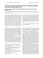
We determined the experimental pIs of purified recom-
binant RT
LAI
and GAPDH using 2D gel electrophoresis. RT
was detected using western blot and GAPDH by Coomas-
sie staining. A number of isoforms consistent in size with
p66 or p51 were detected (Figure 1) with the major iso-
forms present having pIs of 8.13 and 8.33, respectively.
The pIs of the most basic isoforms, p66
8.38
and p51
8.44
(Table 2), were lower than the theoretical pI values of
unmodified p66
8.53
and p51
8.6
(Table 1), consistent with
deamidation of a single asparagine residue calculated to
Retrovirology 2008, 5:115 />Page 3 of 12
(page number not for citation purposes)
change the pI by -0.17 and -0.19 pI units respectively
(Table 1). The pI difference between p66
8.38
and the major
p66
8.13
(-0.25 pI units) was consistent with a second
deamidation predicted to affect the pI by -0.23 pI units
(Tables 1 and 2). 2D gel electrophoresis analysis of
GAPDH detected three isoforms by Coomassie staining
(Figure 1). The major and most basic GAPDH isoform had
an observed pI of 8.50 corresponding to the theoretical pI
of unmodified GAPDH (8.52). The more negatively
charged GAPDH isoforms had pI values -0.37 and -0.87 pI
units lower than GAPDH
8.52
, consistent with singly and
doubly deamidated forms of GAPDH with theoretical pI
differences of -0.27 and -0.70 respectively (Table 1). These
results are consistent with deamidation of both recom-
binant RT and GAPDH and demonstrate that changes in
pI associated with post-translational modifications can be
accurately measured using our 2D gel electrophoresis for-
mat.
Table 1: Theoretical pIs of unmodified and modified RT containing phosphorylation or deamidations of 6His-tagged recombinant
RT
LAI
(rRT) [37], RT
HXB2
(Swiss-Prot: P04585), and GAPDH [42].
Theoretical isoelectric point (pI)
Protein Unmodified No. of Phosphorylation groups Deamidations
12312
rRT
LAI
p66 8.53 8.16 7.60 7.19 8.36 8.13
rRT
LAI
p51 8.60 8.17 7.44 7.02 8.41 8.13
RT
HXB2
p66 8.53 8.19 7.55 7.09 8.36 8.12
RT
HXB2
p51 8.60 8.21 7.56 7.07 8.43 8.18
GAPDH 8.52 7.54 7.0 6.71 8.25 7.82
2D gel electrophoresis analysis of recombinant RT identifies protein isoformsFigure 1
2D gel electrophoresis analysis of recombinant RT identifies protein isoforms. Recombinant RT
LAI
+ GAPDH pro-
tein (3 μg each) was solubilised in 2D gel electrophoresis buffer, focussed on a pH 7–11 non-linear, 11 cm Immobiline DryStrip
gel then resolved on a 10% acrylamide SDS-PAGE gel followed by transfer to PVDF membranes. RT was detected by Western
blot using an anti-RT antibody (upper panel) and GAPDH detected by Coomassie stain (lower panel). RT isoforms are desig-
nated by black arrows and calculated pI indicated. Position of triangles (Δ) denote the reference marks used for calculation of
pI.
Retrovirology 2008, 5:115 />Page 4 of 12
(page number not for citation purposes)
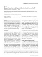
HIV RT exists as multiple isoforms
To examine RT in purified HIV virus, HIV
HXB2
virions were
pelleted through 25% sucrose and then solubilised in 2D
sample buffer. An aliquot was analysed by 1D SDS-PAGE
and western blot for RT. As expected, two distinct bands
corresponding to p66 and p51 were detected (Figure 2A).
The remaining sample was then analysed by 2D gel elec-
trophoresis. Three distinct isoforms of p66 and p51 were
identified (Table 2). A summary of the reproducibly
detected isoforms and potential post-translational modi-
fications is presented in Table 3. The isoforms of virion
p66
8.44
and p51
8.31
were most abundant and reproducibly
seen (Figure 2B). Densitometric quantitation of images
showed that these isoforms represented 85–90% of vir-
ion-associated RT (data not shown). The pIs of both of
these major isoforms differed from that predicted for
unmodified p66
8.53
and p51
8.60
. The virion p51 isoforms
showed a similar pI profile to the isoforms detected in
recombinant RT, with the virion p51
8.31
and p51
8.41
iso-
forms similar to the recombinant p51
8.33
and p51
8.44
iso-
forms (Table 1). The minor RT isoforms suggest multiple
modifications of p66 and p51 in HIV virions. The pI val-
ues for p51
8.41
and p51
8.15
closely correspond to the theo-
retical pI's for RT
HXB2
p51 deamidation (p51
8.43
, p51
8.18
,
Table 1).
We next assessed the presence of these RT isoforms in
other biological situations: in (i) virus producer cells (Fig-
ure 3A), (ii) intracellularly following HIV infection (Fig-
ure 3B) and (iii) in HIV RTC's (Figure 3C–E). H3B cells are
chronically HIV infected cells that produce infectious
virus and although they contain forms of HIV RT that are
active in vitro, RT is not active inside the cell and newly
synthesised HIV DNA is not formed until stimulation by
mixing with uninfected recipient cells [2]. H3B cells thus
represent a system to analyse changes in RT that occur co-
incident with intracellular stimulation of reverse tran-
scription and additionally offers the advantage of a syn-
chronous and highly efficient infection model compared
with a cell-free infection [13]. This allows high sensitivity
in detecting RT protein. To analyse the RT in H3B pro-
ducer cells we mixed H3B cells with uninfected Hut-78
cells and immediately lysed cells prior to the opportunity
for interaction, stimulation of RT or infection. Proteins
were then immunoprecipitated and subjected to 2D gel
electrophoresis. p51
8.41
, p51
8.31
, p51
8.15
and p51
7.91
and
p66
8.57
, p66
8.44
, p66
8.40
, p66
8.28
isoforms were seen, repre-
senting RT present in H3B cells (Figure 3A). The two most
abundant p66
8.44
and p51
8.31
isoforms had pI values iden-
Table 2: Observed pI of 6His-tagged recombinant RT
LAI
(rRT),
and HIV-1 virion RT
HXB2
p66 and p51 isoforms. Isoform in bold is
the major isoform observed.
Protein Observed isoelectric point (pI)
rRT p66 8.38 8.13 7.94 7.75
rRT p51 8.44 8.33 8.00 7.80
virion RT p66 8.44 8.40 8.28
virion RT p51 8.41 8.31 8.15
RT isoforms are present in purified HIV virionsFigure 2
RT isoforms are present in purified HIV virions. Viral
particles from H3B cells were pelleted through 25% sucrose,
solubilised in 2D gel electrophoresis buffer and an aliquot
resolved by 1D SDS-PAGE (A). The remaining sample was
spiked with 3 μg of GAPDH protein, focussed on a pH 7–11
non-linear, 11 cm Immobiline DryStrip gel and then resolved
by SDS-PAGE followed by transfer to PVDF membranes (B).
RT was detected by Western blot using an anti-RT antibody.
RT isoforms (B) are designated by black arrows and the cal-
culated pI and expected position of p66 and p51 indicated.
Table 3: Summary of the routinely observed isoforms of RT
HXB2
.
Isoform pI Modification
p66 8.44 unknown
8.40 unknown
8.28 phosphorylation + basic addition
8.57 unmodified
p51 8.41
a
phosphorylation + basic addition or
b
deamidation
8.31
a
phosphorylation + basic addition
8.15
b
2 deamidations
7.91 2 phosphates + basic addition
a
= de-phosphorylation observed in a one experiment only.
b
= based on theoretical pI (see table 1)
Major isoforms are highlighted in bold.
Retrovirology 2008, 5:115 />Page 5 of 12
(page number not for citation purposes)
tical to the two most abundant isoforms detected in viri-
ons (Figure 2B). Similar to that seen in virions,
quantitation of western images indicated that these iso-
forms represented 76 ± 12 and 79 ± 2% of the p51 and
p66 RT protein, respectively. New minor RT isoforms, not
seen in virions were observed (p66
8.57
and p51
7.91
) which
for p66
8.57
closely corresponds to the theoretical pI of
unmodified p66
8.53
. Minor differences in the p66 and p51
profiles were observed between these and the subse-
quently described experiments which are likely attributa-
ble to variation in HIV infection, immunoprecipitation
efficiency, and sensitivity of western blot detection and
spots that were variably observed are indicated on the fig-
ures with a white arrow. A higher molecular weight RT
immunoreactive species was sometimes observed (eg Fig-
ure 3A, 3D) which likely represents unprocessed Gag-Pol
arising from the H3B producer cells.
We next analysed RT present after HIV infected H3B cells
were mixed with uninfected Hut-78 cells at 37°C to allow
virus entry and replication. The same two major p66
8.44
and p51
8.31
isoforms were again observed (Figure 3B).
However, the relative proportions of the major and minor
isoforms differed, with the minor isoforms becoming
more prominent and the major p66
8.44
and p51
8.31
iso-
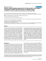
Figure 3
The same major RT isoforms are present in virus producer cells, newly infected cells and HIV RTCsFigure 3
The same major RT isoforms are present in virus
producer cells, newly infected cells and HIV RTCs.
H3B and Hut-78 cells were co-cultured for the indicated
time period then lysed. For panels A and B, lysates were
immunoprecipitated using heat-inactivated AIDS patient sera
cross-linked to protein A sepharose beads and washed. In
panels A, B and D, E samples were subjected to 2D gel elec-
trophoresis on a pH 7–11 non-linear, 11 cm Immobiline
DryStrip gel along with 3 μg of GAPDH protein. Proteins
were resolved by SDS-PAGE and transferred to PVDF mem-
branes. RT was detected by Western blot using an anti-RT
antibody and RT isoforms are designated by a black arrow (n
= 2 for each panel). Minor differences in the p66 and p51
profiles were observed between experiments and spots not
routinely observed are indicated by a white arrow. (A) H3B
virus producer cells. H3B and Hut-78 cells were co-cultured
and lysed immediately. (B) Infected cell lysates. H3B and Hut-
78 cells were co-cultured and lysed at 40 min post-cell mix-
ing. (C-E) HIV RTC's. Lysates were subjected to 15–30%
sucrose velocity gradient sedimentation. Fractions (1 ml)
were collected from the top of the gradient and viral -ssDNA
analysed by real time PCR (C). The remainder of two
selected fractions; (D) from the top of the gradient (fraction
1) and (E) co-incident with the known sedimentation of
RTCs (fraction 5), were TCA precipitated and subjected to
2D gel electrophoresis, as for panels A and B, above. Experi-
ments were replicated, at least n = 2, for each presented bio-
logical situation.
Retrovirology 2008, 5:115 />Page 6 of 12
(page number not for citation purposes)
forms representing only 64 ± 11 and 60 ± 9% of the p51
and p66 RT protein, respectively. Similar minor isoforms
were present in these cells undergoing active reverse tran-
scription compared with those detected in chronically
infected virus producer H3B cells.
After viral entry some RT remains part of a nucleoprotein
complex termed the reverse transcription complex (RTC)
but the majority of virion associated RT dissociates from
the RTC [25]. We next assessed if specific isoforms of RT
were associated with RTCs following HIV infection. Infec-
tions were initiated by cell-cell mixing as previously, and
after 120 min, cell lysates were prepared and subjected to
sucrose velocity gradient sedimentation. This sedimenta-
tion technique was chosen since we have previously
observed that it yields good separation of free protein
(fraction 1) and any remaining unactivated RT in pre-exis-
iting complexes from H3B cells (fraction 7) from RTCs
(fraction 5) [2,26], the latter which we can monitor by vir-
tue of the presence of newly synthesised reverse transcrip-
tion products. HIV reverse transcription products showed
a peak in gradient fraction 5 (1.08 g/ml sucrose; Figure
3C) consistent with the previously characterised sedimen-
tation rate of RTCs as defined by the presence of newly
synthesised DNA, RT activity and HIV integrase protein
[26]. Sucrose gradient fractions were then subjected to 2D
gel electrophoresis and western blot for RT, as above. Frac-
tion 1 from the top of the gradient and containing free
protein showed RT isoforms with migration characteris-
tics consistent with p66
8.57
, p66
8.44
and p51
8.41
, p51
8.31
,
p51
8.15
and p51
7.91
, with the major isoforms p66
8.44
and
p51
8.31
(Figure 3D) as seen previously (Figure 2, 3A, 3B).
However, in fraction 5 containing RTCs, only isoforms
with migration characteristics consistent with p66
8.44
and
p51
8.31
could be detected (Figure 3E). Although this does
not exclude the presence of other less abundant RT iso-
forms in RTCs that were not detected due to the much
lower levels of RT protein present, our results confirm that
the major isoforms of p66
8.44
and p51
8.31
RT, seen in the
virion and in infected cells, are associated with active
RTCs and thus are the likely to be biologically relevant RT
isoforms.
Newly HIV infected cells contain phosphorylated isoforms
of RT
As one of the most important forms of protein modifica-
tion is phosphorylation, we analysed the susceptibility of
RT isoforms to phosphatase treatment prior to 2D gel elec-
trophoresis. Validation of the efficiency of de-phosphor-
ylation in our in vitro reactions was demonstrated by
treating phosphorylated recombinant beta common (βc)
chain of the GM-CSF receptor with phosphatase and con-
firming the loss of reactivity with anti-phospho-Ser-585βc
polyclonal antibody by Western blot (data not shown)
[27]. Next, HIV infection was initiated by mixing of H3B
and Hut-78 cells and after 40 min the cells were lysed and
viral proteins immunoprecipitated. Precipitated proteins
were divided equally and treated with or without calf
intestinal alkaline phosphatase (CIAP). The RT proteins
were then analysed by 2D gel electrophoresis and detected
by Western blot. The sample without phosphatase treat-
ment showed a profile of p66 and p51 isoforms (Figure
4A) of calculated pI equivalent to p66
8.57
, p66
8.44
, p66
8.40
,
p66
8.28
, and p51
8.41
, p51
8.31
, p51
8.15
and p51
7.91
as seen
previously (Figure 2 and 3). Some additional minor p66
and p51 isoforms were also observed, again highlighting
the experimental variation in the minor RT isoforms.
Removal of phosphate groups should increase protein pI
if phosphorylation is present. Phosphatase treatment
clearly altered the observed p66 and p51 isoforms (Figure
4B). The minor p66 isoforms, (p66
8.28
and p66
8.16
) were
greatly diminished or abolished by phosphatase treat-
ment and this was reproducibly observed in replicate
experiments, suggesting that these isoforms are phospho-
rylated. p66
8.16
differed by -0.37 pI units from the theoret-
ical pI of unmodified p66
8.53
, consistent with the -0.34 pI
unit change associated with addition of a single phos-
phate group. This p66
8.16
phosphorylated isoform was not
routinely detected in all experiments. p66
8.28
differed by -
0.25 pI units from unmodified p66, suggesting that while
p66
8.28
is phosphorylated it also possesses additional
modifications which make it more basic. p51
7.91
was also
consistently reduced by phosphatase treatment and dif-
fered by -0.69 pI units compared with unmodified p51,
corresponding to a predicted addition of two phosphate
groups and additional basic modification. Although most
of p51 RT was relatively phosphatase resistant (Figure 4B)
in one experiment phosphatase treatment reduced the lev-
els of both p51
8.41
and p51
8.31
(data not shown). We have
previously observed variation in de-phosphorylation and
that total de-phosphorylation of ovalbumin is time-
dependent; indicating slow removal of certain phosphate
groups (CJ Bagley, unpublished results). Thus the variable
susceptibly of some RT isoforms to de-phosphorylation
may reflect reduced activity or restricted accessibility of
the phosphatase enzyme to some phosphate groups
present in the RT protein and thus we believe that p51
8.41
and p51
8.31
are most likely phosphorylated. Together the
pI value and susceptibility to phosphatase treatment indi-
cate that the RT isoforms p66
8.28
, p66
8.16
and p51
7.91
and
potentially p51
8.41
and p51
8.31
are phospho-RT isoforms.
To analyse the significance of phosphorylated RT iso-
forms, cell lysates and virions were treated with or without
phosphatase and RT activity was then assessed by in vitro
exogenous RT activity assay (Figure 5). Since phosphatase
itself could theoretically dephosphorylate dNTP's and
influence the in vitro RT activity assay, we first validated
measurement of RT activity in the presence of phos-
Retrovirology 2008, 5:115 />Page 7 of 12
(page number not for citation purposes)
Phosphatase treatment alters the RT isoforms detectedFigure 4
Phosphatase treatment alters the RT isoforms detected. H3B and Hut-78 cells were mixed and incubated at 37°C for
40 mins, cells were then lysed and virus protein immunoprecipitated using heat-inactivated AIDS patient antibody cross-linked
to protein A sepharose beads. Immunoprecipitates were incubated without (A) or with (B) calf intestinal alkaline phosphatase
(CIAP), proteins pelleted, washed and subjected to 2D gel electrophoresis on a pH 7–11 non-linear, 11 cm Immobiline DryS-
trip gel along with 3 μg of GAPDH protein, and then resolved by SDS-PAGE. RT was detected by Western blot using an anti-
RT antibody. RT isoforms are designated by a black arrow and spots not routinely observed are indicated by a white arrow.
Experiments were replicated (n = 3).
Retrovirology 2008, 5:115 />Page 8 of 12
(page number not for citation purposes)
phatase and phosphatase buffering conditions. Incuba-
tion of recombinant M-MuLV RT in an in vitro RT activity
assay in the presence of CIAP buffer alone or with CIAP
enzyme had no effect on the quantitation of RT activity
(Figure 5A). We next analysed the effect of phosphatase
treatment on RT activity present in HIV virions, cell lysates
and RTCs. RTCs were isolated by sucrose density gradient
sedimentation, since this technique is best suited for con-
centrating particles into a more tightly sedimenting band
than the velocity gradients used in Figure 3. Fractions 7–
8, sedimenting at the previously defined density for RTCs
[26] and containing newly synthesised reverse transcrip-
tion products (Figure 5B,) were immunoprecipitated and
subjected to dephosphorylation with CIAP, along with
virions and cell lysates. Dephosphorylation reactions
were performed as previously, which we know success-
fully dephosphorylates the βc chain of the GM-CSF recep-
tor [27] and some isoforms of HIV RT (Figure 4).
Dephosphorylation had no effect on the ability of RT
found in virions, inside newly infected cells or associated
with RTCs to perform in vitro reverse transcription (Figure
5C). Additionally, other sources of phosphatase; Antarctic
phosphatase and lambda phosphatase similarly had no
effect on RT activity of virions (data not shown), suggest-
ing that phosphorylation makes limited contribution to
the inherent activity of naturally occurring RT when meas-
ured in an in vitro assay.
Discussion
Previous literature has suggested that RT may be subjected
to post-translational modification, such as phosphoryla-
tion and it is well known that the process of reverse tran-
scription is substantially activated upon cell infection. We
thus hypothesised that this activation of RT may be related
to its post-translational modification, particularly phos-
phorylation. In this study we have shown by 2D gel elec-
trophoresis that modified RT forms are the major RT
protein present in virions, newly infected cells and RTC's.
The same predominant RT isoforms with pI's of p66
8.44
and p51
8.31
were seen in purified virions, intracellularly
and associated with RTC's, and this suggests that these are
the major biologically active RT form. The possibility that
Phosphatase treatment does not affect in vitro RT activityFigure 5
Phosphatase treatment does not affect in vitro RT activity. (A) Recombinant M-MuLV RT was assayed directly (RT1 =
500 milliUnits [mU], RT2 = 100 mU, RT3 = 20 mU, RT4 = 4 mU) or was incubated for 60 mins at 37°C in PBS or CIAP buffer
+/- CIAP enzyme prior to exogenous RT activity assay, overnight at 37°C using DIG-UTP and colourimetric detection of incor-
porated DIG. (B) H3B and Hut-78 cells were co-cultured and lysed at 40 min post-cell mixing and lysates were subjected to 0–
60% linear sucrose equilibrium gradient sedimentation. Fractions (1 ml) were collected from the top of the gradient and viral
Gag DNA analysed by real time PCR. Fractions 7–8, containing HIV DNA and sedimenting at 1.19–1.25 g/ml sucrose was
immunoprecipitated with AIDS patient sera and represented the RTCs used subsequently in the in vitro RT activity assay. (C)
Samples from virions, cell lysates and RTCs were incubated for 60 mins at 37°C in PBS or CIAP buffer +/- CIAP enzyme prior
to exogenous RT activity assay, overnight at 37°C using DIG-UTP and colourimetric detection of incorporated DIG. Results
were normalized against the RT activity observed in the absence of CIAP and represents data from 3 independent dephospho-
rylation and RT activity assays and from 2 independent RTC preparations.
Retrovirology 2008, 5:115 />Page 9 of 12
(page number not for citation purposes)
these represented an excess of inactive molecules present
together with smaller levels of a modified active form, was
considered unlikely since these forms predominated in
semi-purified RTCs that are known to be supporting active
reverse transcription. The major RT isoforms observed
corresponded to an undefined post-translational modifi-
cation for p66
8.44
, and potentially phosphorylation plus
an undefined basic modification for p51
8.31
. The major
p51
8.31
isoform had lower pI than the major p66
8.44
iso-
form, contrary to that seen for recombinant RT (p66
8.13
and p51
8.33
) and the theoretical pI of the unmodified
p51
8.60
or p66
8.53
. Additionally, susceptibility of p51
8.31
to
phosphatase treatment in one experiment suggested that
p51
8.31
may be phosphorylated, while p66
8.44
was phos-
phatase resistant in all instances. Thus the major p51
8.31
isoform contains modifications that are different from
those in the major p66
8.44
isoform. This observed differen-
tial modification of p51 compared to p66 may be the
result of (i) modification of a single p66 molecule of the
RT homodimer that is then selectively targeted for cleav-
age giving rise to p51 and a mature RT heterodimer or (ii)
selective modification of the p51 in the heterodimer post
p66 cleavage. This differential modification of p51 and
p66 may be important for selective regulation of RT enzy-
matic functions via p66 post-translational modifications
or alterations to RT structure/conformation via post-trans-
lational modifications of p51. The identification of these
RT isoforms is novel. Previous studies have identified at
least two isoforms of MA and CA [28-30] in HIV virions
by 2D gel electrophoresis analysis followed by silver stain
or western blot, but these studies have not identified iso-
forms of RT, possibly due to lower levels of RT or the use
of isoelectric focussing strips of insufficient resolving
power for the pI range of RT [30,31]. The RT isoforms we
observed changed little between virus producer cells, viri-
ons and newly infected cells, although the minor RT iso-
forms became more abundant following infection.
Some of the RT isoforms detected were phosphorylated, as
suggested by their pI value and their susceptibility to
dephosphorylation. Phosphorylation is known to modu-
late the activity of many proteins that interact with nucleic
acids, including HIV proteins Tat, and Rev [32,33] and
RNAPII [19,34]. Indeed phosphorylation of HIV RT in
vitro led to increased polymerase and RNase H activities
[21,22,35]. Similarly the phosphorylated forms of RT that
we have identified may lead to p66/p51 heterodimers
with different physical characteristics, activities or func-
tionality and hence may play an important role in regulat-
ing reverse transcription in newly infected cells. Our
results, however, show that dephosphorylation of RT
from virions, cells lysates or RTCs had no effect on in vitro
RT activity. This is not surprising given our results show-
ing that the major isoforms that would be present in sam-
ples from virions, infected cells and RTCs are p66
8.44
and
p51
8.31
that are not phosphorylated, and were phos-
phatase resistant in 2/3 experiments, respectively. Thus,
naturally occurring phospho-RT isoforms are not a major
contributor to RT activity, as measured in vitro, but could
still be important for RT activity in the complex milieu of
the infected cell, or may play a role in important structural
interactions required for stability, movement and activity
of the RTC intracellularly. Conclusive analysis of the roles
of phosphorylation at specific sites in the RT enzyme
remain to be determined by mutagenesis of potential RT
phosphorylation sites and analysis of subsequent 2D gel
electrophoresis profiles. However, at present this kind of
analysis is hampered by the reduced sensitivity for detec-
tion of RT following infection with cell-free virus and 2D
gel analysis, as would be necessitated in these experi-
ments.
In conclusion, we describe for the first time the presence
of modified p66 and p51 RT isoforms and report that the
same major p51
8.31
and p66
8.44
isoforms are present in
HIV virions, newly infected cells and active RTCs and thus
are likely to be the forms playing a significant role in the
reverse transcription process. The major p51
8.31
and
p66
8.44
isoforms are modified differently, demonstrating
selective modification of the RT subunits and although
some RT isoforms are phosphorylated, phospho-isoforms
of RT are not a major contributor to the inherent activity
of RT, as measured in an in vitro activity assay. A better
understanding of the post-translational modifications,
the cellular enzymes involved and how these specifically
influence RT activity inside the cell will be essential in elu-
cidating the mechanisms for control of reverse transcrip-
tion in newly infected cells.
Methods
Cells, virus and recombinant RT
H3B cells are a laboratory clone of H9 cells persistently
infected with the HTLV-IIIB (HXB2) strain of HIV-1 [13].
Virus particles were isolated from clarified H3B cell cul-
ture medium by filtration (Sartorius, 0.22 μm filter), con-
centration (100,000 MwCO centrifugal filter, Millipore)
and pelleting through 25% (w/v) sucrose at 86,500 g, 4°C
for 1.5 hr (Beckman Optima™ TLX Ultracentrifuge).
Recombinant RT (p6HRT; hexahistidine-tagged p66/p51
heterodimer, Dr. Nicolas Sluis-Cremer, University of Pitts-
burgh and derived from p6HRT-PROT [36]) was from the
LAI sequence of HIV-1 [37] and produced by expression
in M15 Escherichia coli and purified as described previ-
ously [38]. Purified recombinant RT was generously pro-
vided by Dr. Gilda Tachedjian, Burnet Institute,
Melbourne, Australia.
Cell-to-cell infection and lysis
H3B cells were mixed with Hut-78 cells at a ratio of 1:4
and incubated for 3 hr at 23°C to produce a temperature-
Retrovirology 2008, 5:115 />Page 10 of 12
(page number not for citation purposes)
arrested stage of infection [39]. Cells were then shifted to
37°C to allow infection to proceed. To extract protein, 1 ×
10
8
cells were washed twice in ice-cold PBS and lysed by
rotating at 4°C for 1 hr in 1 ml lysis buffer (5 mM Tris-HCl
pH 7.4, 50 mM KCl, 0.05 mM spermine, 0.125 mM sper-
midine, 2 mM DTT, protease inhibitors [20 μg/ml aprot-
onin, complete mini protease inhibitor tablet (Roche), 2
mM PMSF), phosphatase inhibitors (2 mM NaF, 10 mM
sodium pyrophosphate, 2 mM sodium orthovanadate],
and 0.2% (v/v) Triton X-100). The cell lysate was clarified
twice by centrifugation at 17,000 g/4°C for 30 min before
immunoprecipitation.
Immunoprecipitation of viral protein from infected cell
lysate
Sera from four HIV-1 positive patients were pooled and
heat-inactivated (AIDS patient sera (APS)) and incubated
with protein A sepharose CL-4B beads (Pharmacia) at
4°C, rotating for 16 hr. Antibody was cross-linked to pro-
tein A using 5 mg/ml dimethyl pimelimidate (DMP)
(Pierce) as described previously [40]. To immunoprecipi-
tate viral proteins, cell lysates were incubated with APS-
protein A sepharose CL-4B for 16 hr rotating at 4°C. The
beads were then pelleted by low-speed centrifugation and
washed in ice-cold water three times then proteins eluted
directly into 2D gel electrophoresis buffer (see below).
Fractionation of HIV reverse transcription complexes
HIV RTCs were fractionated on sucrose gradients as
described previously [26,41]. Briefly, infections were initi-
ated by mixing of H3B and Hut-78 cells, as described
above. At 120 min post mixing cells were harvested,
washed, lysed in buffer containing 0.1% (v/v) Triton X-
100 and subjected to 15–30% sucrose velocity gradient
sedimentation or 0–60% sucrose equilibrium density gra-
dient sedimentation. 1 ml fractions were collected from
the top of the gradient and 1/10
th
of each fraction was ana-
lysed for HIV reverse transcription products by real time
PCR. The remainder of the velocity gradient fractions were
TCA precipitated and 85 μg of the total protein from each
fraction was subjected to 2D gel electrophoresis, as below.
2D gel electrophoresis and Western blot analysis of protein
Samples were solubilised directly in 2D buffer (7 M urea,
2 M thiourea, 2% (w/v) CHAPS, and 0.5% pH 7–11 NL
carrier ampholytes) and spiked with 3 μg glyceraldehyde-
3-phosphate dehydrogenase (GAPDH, from rabbit mus-
cle, Sigma) and 65 mM DTT. Samples (100 μl) were
loaded, by anodic cup loading, onto a pH 7–11 non-lin-
ear, 11 cm Immobiline DryStrip (GE Healthcare) gel
which had been hydrated in 2D sample buffer containing
1.2% (v/v) 2-hydroxethyldisulfide. Gels were run in a
step-wise voltage gradient: 0–300 V/2 hr; 300–500 V/2 hr;
500–1000 V/2 hr; 1000–4000 V/5 hr followed by 4000 V/
3 hr and then maintained at 500 V. Total volt hours (V/hr)
ranged between 25–30,000 V/h. Focused proteins from
individual gel strips were then separated by SDS-PAGE,
using a 10% or 12% gel with a 29:1 acrylamide:bis-acryla-
mide ratio, alongside BenchMark™ prestained protein
markers (Invitrogen), before transferring to PVDF transfer
membrane (Hybond™-P; GE Healthcare). Membranes
were blocked for 1 hr in TBST (50 mM Tris pH 7.4, 135
mM NaCl, 0.1% (v/v) Tween-20) containing 5% (w/v)
skim-milk powder before incubating with rabbit anti-RT
antibody (1:5000 dilution), (NIH AIDS Research and Ref-
erence Reagent Program, Dr. Stuart Le Grice, Division of
AIDS, NIAID, NIH). Bound antibody was detected using
horseradish-peroxidase-conjugated goat anti-rabbit IgG
secondary antibody, and visualised using Super Signal
West Dura Extended Duration Substrate (Pierce) and
Kodak BioMax film (Integrated Sciences). To determine
the relative proportion of p66 and p51 isoforms, protein
spots in were quantitated by volume integration (Image-
quant v3.3, Molecular Dynamics) and expressed as a per-
cent of the total intensity of signal for RT p66 or p51.
Phosphatase treatment of viral proteins
Viral proteins were immunoprecipitated from infected
cell lysates with APS conjugated protein A sepharose CL-
4B beads as described above, virions were prepared by
PEG precipitation of high titre virus supernatant, and
RTCs were prepared by equilibrium gradient sedimenta-
tion, as above. One half of each sample was treated with
40 units of calf intestinal alkaline phosphatase (CIAP;
Promega) in CIAP buffer; (50 mM Tris, pH 9.3, 1 mM
MgCl
2
0.1 mM ZnCl
2
and 1 mM spermidine and protease
inhibitors (20 ug/ml aprotonin, complete mini protease
inhibitor tablet [Roche], 2 mM PMSF). The other half was
resuspended in CIAP buffer, protease and phosphatase
inhibitors (2 mM PMSF, 2 mM NaF, 10 mM sodium pyro-
phosphate, 2 mM sodium orthovanadate). Reactions were
incubated 37°C for 1.5 hr. For subsequent 2D gel analy-
sis, bead bound samples from cell lysates were pelleted,
washed in ice-cold water three times and the bound virus
protein was eluted in 2D gel electrophoresis sample
buffer. For subsequent RT activity assay, reactions were
used directly, without further processing.
RT activity assay
RT activity was quantitated in vitro using an exogenous
activity assay. Briefly, microtitre plates (Covalink, Nunc)
were coated with poly-A (Roche) then incubated with RT
mix containing the test sample with 4.2 μM Digoxigenin
(DIG)-UTP (Roche Diagnostics) and 2.5 μg/ml Oligo
dT
12–18
(GE Healthcare) in 8.4 μM dTTP, 25 mM KCl, 6.25
mM MgCl
2
, 62.5 mM Tris, pH 7.8, 1.25 mM DTT, 0.1%
(v/v) Triton X-100, overnight at 37°C. Polymerised DIG-
UTP was detected with anti-DIG-HRP conjugate (Roche
Diagnostics, at 1/2500 dilution), reacted with 3,3',5,5'-
tetramethylbenzidine (TMB substrate, Sigma) and quanti-
Retrovirology 2008, 5:115 />Page 11 of 12
(page number not for citation purposes)
tated by measurement of OD at 490 nm. Recombinant
Moloney Murine leukemia virus (M-MuLV, New England
Biolabs) was used as a comparative standard.
Estimation of protein isoelectric point
The distance migrated along the IEF strip from the loading
point (anodic, pH 7 end) was measured as a percentage of
the total gel-strip length (11 cm) and the pI calculated
from an idealised pH 7–11 non-linear migration reference
graph (GE Healthcare). For internal calibration, GAPDH
was spiked into individual viral protein samples before
focusing and small puncture holes made in the PVDF
membrane were used to align the Coomassie-stained and
the Western blot images. Theoretical pI values for
unmodified HIV
HXB2
p66 and p51 (Swiss-Prot: P04585),
recombinant hexahistidine-tagged p66 and p51 proteins,
and GAPDH [42], with one or more phosphate or deami-
dation modifications, in 8 M urea, were calculated using
pKa values as used by the ExPASy Compute pI/Mw tool
/> with the assump-
tion that the pKa values of the protein's phosphate groups
were 2.1 and 7.2.
Competing interests
The authors declare that they have no competing interests.
Authors' contributions
JMC performed isolation and analysis of RTCs, the
dephosphorylation and RT activity experiments, contrib-
uted to interpretation of results and was the primary man-
uscript author, AJD was the main research worker and
performed the 2D gel analysis experiments and pI calcula-
tions, CJBagley assisted in interpretation of all 2D gel elec-
trophoresis and pI calculations, JP contributed in the
design of CIAP experiments, DW, DH, CJBurrell and PL
contributed to the design of the study. All authors read
and approved the final manuscript.
Acknowledgements
We would like to thank Adrian Purins for maintenance of cell culture
stocks, Megan Retallick for 2D gel electrophoresis, John Karlis and Carl
Coolen for technical assistance. We also acknowledge Dr. Gilda Tachedjian
for generously providing the purified recombinant RT protein. This work
and AJD was supported by an Australian NHMRC project grant. JMC was
supported by the Australian Centre for HIV and Hepatitis Research.
References
1. Chattopadhyay D, Evans DB, Deibel MR Jr, Vosters AF, Eckenrode
FM, Einspahr HM, Hui JO, Tomasselli AG, Zurcher-Neely HA, Hein-
rikson RL, et al.: Purification and characterization of het-
erodimeric human immunodeficiency virus type 1 (HIV-1)
reverse transcriptase produced by in vitro processing of p66
with recombinant HIV-1 protease. J Biol Chem 1992,
267:14227-14232.
2. Li P, Stephenson AJ, Brennan PA, Karageorgos L, Kok T, Kuiper LJ,
Swift J, Burrell CJ: Initiation of reverse transcription during
cell-to-cell transmission of human immunodeficiency virus
infection uses pre-existing reverse transcriptase. J Gen Virol
1994, 75(Pt 8):1917-1926.
3. di Marzo Veronese F, Copeland TD, DeVico AL, Rahman R, Oroszlan
S, Gallo RC, Sarngadharan MG: Characterization of highly immu-
nogenic p66/p51 as the reverse transcriptase of HTLV-III/
LAV. Science 1986, 231:1289-1291.
4. Tachedjian G, Aronson HE, de los Santos M, Seehra J, McCoy JM, Goff
SP: Role of residues in the tryptophan repeat motif for HIV-1
reverse transcriptase dimerization. J Mol Biol 2003,
326:381-396.
5. Hostomsky Z, Hostomska Z, Fu TB, Taylor J: Reverse tran-
scriptase of human immunodeficiency virus type 1: function-
ality of subunits of the heterodimer in DNA synthesis. J Virol
1992, 66:3179-3182.
6. Hizi A, McGill C, Hughes SH: Expression of soluble, enzymati-
cally active, human immunodeficiency virus reverse tran-
scriptase in Escherichia coli and analysis of mutants. Proc Natl
Acad Sci USA 1988, 85:1218-1222.
7. Mulky A, Sarafianos SG, Arnold E, Wu X, Kappes JC: Subunit-spe-
cific analysis of the human immunodeficiency virus type 1
reverse transcriptase in vivo. J Virol 2004, 78:7089-7096.
8. Harrich D, Hooker B: Mechanistic aspects of HIV-1 reverse
transcription initiation. Rev Med Virol 2002, 12:31-45.
9. Zhang H, Pomerantz RJ, Dornadula G, Sun Y: Human immunode-
ficiency virus type 1 Vif protein is an integral component of
an mRNP complex of viral RNA and could be involved in the
viral RNA folding and packaging process. J Virol 2000,
74:8252-8261.
10. Huang M, Martin MA:
Incorporation of Pr160(gag-pol) into
virus particles requires the presence of both the major
homology region and adjacent C-terminal capsid sequences
within the Gag-Pol polyprotein. J Virol 1997, 71:4472-4478.
11. Arts EJ, Mak J, Kleiman L, Wainberg MA: Mature reverse tran-
scriptase (p66/p51) is responsible for low levels of viral DNA
found in human immunodeficiency virus type 1 (HIV-1).
Leukemia 1994, 8(Suppl 1):S175-178.
12. Warrilow D, Stenzel D, Harrich D: Isolated HIV-1 core is active
for reverse transcription. Retrovirology 2007, 4:77.
13. Li P, Burrell CJ: Synthesis of human immunodeficiency virus
DNA in a cell-to-cell transmission model. AIDS Res Hum Retro-
viruses 1992, 8:253-259.
14. Li P, Stephenson AJ, Kuiper LJ, Burrell CJ: Double-stranded
strong-stop DNA and the second template switch in human
immunodeficiency virus (HIV) DNA synthesis. Virology 1993,
194:82-88.
15. Kim SY, Byrn R, Groopman J, Baltimore D: Temporal aspects of
DNA and RNA synthesis during human immunodeficiency
virus infection: evidence for differential gene expression. J
Virol 1989, 63:3708-3713.
16. Barbosa P, Charneau P, Dumey N, Clavel F: Kinetic analysis of
HIV-1 early replicative steps in a coculture system. AIDS Res
Hum Retroviruses 1994, 10:53-59.
17. Vandegraaff N, Kumar R, Burrell CJ, Li P: Kinetics of human
immunodeficiency virus type 1 (HIV) DNA integration in
acutely infected cells as determined using a novel assay for
detection of integrated HIV DNA. J Virol 2001, 75:11253-11260.
18. Karageorgos L, Li P, Burrell CJ: Stepwise analysis of reverse tran-
scription in a cell-to-cell human immunodeficiency virus
infection model: kinetics and implications. J Gen Virol 1995,
76(Pt 7):1675-1686.
19. Payne JM, Laybourn PJ, Dahmus ME: The transition of RNA
polymerase II from initiation to elongation is associated with
phosphorylation of the carboxyl-terminal domain of subunit
IIa. J Biol Chem 1989, 264:19621-19629.
20. Archambault J, Chambers RS, Kobor MS, Ho Y, Cartier M, Bolotin D,
Andrews B, Kane CM, Greenblatt J: An essential component of a
C-terminal domain phosphatase that interacts with tran-
scription factor IIF in Saccharomyces cerevisiae. Proc Natl
Acad Sci USA 1997, 94:14300-14305.
21. Idriss H, Kawa S, Damuni Z, Thompson EB, Wilson SH: HIV-1
reverse transcriptase is phosphorylated in vitro and in a cel-
lular system. Int J Biochem Cell Biol 1999, 31:1443-1452.
22. Harada S, Haneda E, Maekawa T, Morikawa Y, Funayama S, Nagata N,
Ohtsuki K: Casein kinase II (CK-II)-mediated stimulation of
HIV-1 reverse transcriptase activity and characterization of
selective inhibitors in vitro. Biol Pharm Bull 1999, 22:1122-1126.
23. Gordon M, De Oliveira T, Bishop K, Coovadia HM, Madurai L, Engel-
brecht S, Janse van Rensburg E, Mosam A, Smith A, Cassol S: Molec-
Publish with BioMed Central and every
scientist can read your work free of charge
"BioMed Central will be the most significant development for
disseminating the results of biomedical research in our lifetime."
Sir Paul Nurse, Cancer Research UK
Your research papers will be:
available free of charge to the entire biomedical community
peer reviewed and published immediately upon acceptance
cited in PubMed and archived on PubMed Central
yours — you keep the copyright
Submit your manuscript here:
/>BioMedcentral
Retrovirology 2008, 5:115 />Page 12 of 12
(page number not for citation purposes)
ular characteristics of human immunodeficiency virus type 1
subtype C viruses from KwaZulu-Natal, South Africa: impli-
cations for vaccine and antiretroviral control strategies. J
Virol 2003, 77:2587-2599.
24. Ojesina AI, Sankale JL, Odaibo G, Langevin S, Meloni ST, Sarr AD,
Olaleye D, Kanki PJ: Subtype-specific patterns in HIV Type 1
reverse transcriptase and protease in Oyo State, Nigeria:
implications for drug resistance and host response. AIDS Res
Hum Retroviruses 2006, 22:770-779.
25. Fassati A, Goff SP: Characterization of intracellular reverse
transcription complexes of human immunodeficiency virus
type 1. J Virol 2001, 75:3626-3635.
26. Carr JM, Davis AJ, Coolen C, Cheney K, Burrell CJ, Li P: Vif-defi-
cient HIV reverse transcription complexes (RTCs) are sub-
ject to structural changes and mutation of RTC-associated
reverse transcription products. Virology 2006, 351:80-91.
27. Guthridge MA, Stomski FC, Barry EF, Winnall W, Woodcock JM,
McClure BJ, Dottore M, Berndt MC, Lopez AF: Site-specific serine
phosphorylation of the IL-3 receptor is required for hemo-
poietic cell survival. Mol Cell 2000, 6:99-108.
28. Fouchier RA, Simon JH, Jaffe AB, Malim MH: Human immunodefi-
ciency virus type 1 Vif does not influence expression or virion
incorporation of gag-, pol-, and env-encoded proteins. J Virol
1996, 70:8263-8269.
29. Fuchigami T, Misumi S, Takamune N, Takahashi I, Takama M, Shoji S:
Acid-labile formylation of amino terminal proline of human
immunodeficiency virus type 1 p24(gag) was found by pro-
teomics using two-dimensional gel electrophoresis and
matrix-assisted laser desorption/ionization-time-of-flight
mass spectrometry. Biochem Biophys Res Commun 2002,
293:1107-1113.
30. Misumi S, Fuchigami T, Takamune N, Takahashi I, Takama M, Shoji S:
Three isoforms of cyclophilin A associated with human
immunodeficiency virus type 1 were found by proteomics by
using two-dimensional gel electrophoresis and matrix-
assisted laser desorption ionization-time of flight mass spec-
trometry. J Virol 2002, 76:10000-10008.
31. Gaddis NC, Chertova E, Sheehy AM, Henderson LE, Malim MH:
Comprehensive investigation of the molecular defect in vif-
deficient human immunodeficiency virus type 1 virions. J Virol
2003, 77:5810-5820.
32. Holmes AM: In vitro phosphorylation of human immunodefi-
ciency virus type 1 Tat protein by protein kinase C: evidence
for the phosphorylation of amino acid residue serine-46. Arch
Biochem Biophys 1996, 335:8-12.
33. Fouts DE, True HL, Cengel KA, Celander DW: Site-specific phos-
phorylation of the human immunodeficiency virus type-1
Rev protein accelerates formation of an efficient RNA-bind-
ing conformation. Biochemistry 1997, 36:13256-13262.
34. Parada CA, Roeder RG: Enhanced processivity of RNA
polymerase II triggered by Tat-induced phosphorylation of
its carboxy-terminal domain. Nature 1996, 384:375-378.
35. Lazaro JB, Boretto J, Selmi B, Capony JP, Canard B: Phosphoryla-
tion of AZT-resistant human immunodeficiency virus type 1
reverse transcriptase by casein kinase II in vitro: effects on
inhibitor sensitivity. Biochem Biophys Res Commun 2000,
275:26-32.
36. Le Grice SF, Gruninger-Leitch F: Rapid purification of
homodimer and heterodimer HIV-1 reverse transcriptase
by metal chelate affinity chromatography. Eur J Biochem 1990,
187:307-314.
37. Shi C, Mellors JW: A recombinant retroviral system for rapid
in vivo analysis of human immunodeficiency virus type 1 sus-
ceptibility to reverse transcriptase inhibitors. Antimicrob
Agents Chemother 1997, 41:2781-2785.
38. Tachedjian G, Moore KL, Goff SP, Sluis-Cremer N: Efavirenz
enhances the proteolytic processing of an HIV-1 pol polypro-
tein precursor and reverse transcriptase homodimer forma-
tion. FEBS Lett 2005, 579:379-384.
39. Mkrtchyan SR, Markosyan RM, Eadon MT, Moore JP, Melikyan GB,
Cohen FS: Ternary complex formation of human immunode-
ficiency virus type 1 Env, CD4, and chemokine receptor cap-
tured as an intermediate of membrane fusion. J Virol 2005,
79:11161-11169.
40. Schneider C, Newman RA, Sutherland DR, Asser U, Greaves MF: A
one-step purification of membrane proteins using a high effi-
ciency immunomatrix. J Biol Chem 1982, 257:10766-10769.
41. Karageorgos L, Li P, Burrell C: Characterization of HIV replica-
tion complexes early after cell-to-cell infection.
AIDS Res Hum
Retroviruses 1993, 9:817-823.
42. Cowan-Jacob SW, Kaufmann M, Anselmo AN, Stark W, Grutter MG:
Structure of rabbit-muscle glyceraldehyde-3-phosphate
dehydrogenase. Acta Crystallogr D Biol Crystallogr 2003,
59:2218-2227.