Báo cáo y học: "Dynamic features of the selective pressure on the human immunodeficiency virus type 1 (HIV-1) gp120 CD4-binding site in a group of long term non progressor (LTNP) subjects" pptx
Bạn đang xem bản rút gọn của tài liệu. Xem và tải ngay bản đầy đủ của tài liệu tại đây (557.47 KB, 15 trang )
BioMed Central
Page 1 of 15
(page number not for citation purposes)
Retrovirology
Open Access
Research
Dynamic features of the selective pressure on the human
immunodeficiency virus type 1 (HIV-1) gp120 CD4-binding site in a
group of long term non progressor (LTNP) subjects
Filippo Canducci*
1
, Maria Chiara Marinozzi
1
, Michela Sampaolo
1
,
Stefano Berrè
1
, Patrizia Bagnarelli
2
, Massimo Degano
3
, Giulia Gallotta
4
,
Benedetta Mazzi
5
, Philippe Lemey
6
, Roberto Burioni
1
and
Massimo Clementi
1
Address:
1
Laboratorio di Microbiologia e Virologa, Università Vita-Salute San Raffaele, Milan, Italy,
2
Istituto di Microbiologia, Università
Politecnica delle Marche, Ancona, Italy,
3
Unità di Biocristallografia, Istituto Scientifico San Raffaele, Milan, Italy,
4
Dipartimento di Malattie
Infettive, Università Vita-Salute San Raffaele, Milan, Italy,
5
Laboratorio di Ematologia Molecolare, Istituto Scientifico San Raffaele, Milan, Italy and
6
Rega Institute, Katholieke Universiteit Leuven, Leuven, Belgium
Email: Filippo Canducci* - ; Maria Chiara Marinozzi - ;
Michela Sampaolo - ; Stefano Berrè - ; Patrizia Bagnarelli - ;
Massimo Degano - ; Giulia Gallotta - ; Benedetta Mazzi - ;
Philippe Lemey - ; Roberto Burioni - ; Massimo Clementi -
* Corresponding author
Abstract
The characteristics of intra-host human immunodeficiency virus type 1 (HIV-1) env evolution were
evaluated in untreated HIV-1-infected subjects with different patterns of disease progression,
including 2 normal progressor [NP], and 5 Long term non-progressor [LTNP] patients. High-
resolution phylogenetic analysis of the C2-C5 env gene sequences of the replicating HIV-1 was
performed in sequential samples collected over a 3–5 year period; overall, 301 HIV-1 genomic RNA
sequences were amplified from plasma samples, cloned, sequenced and analyzed. Firstly, the
evolutionary rate was calculated separately in the 3 codon positions. In all LTNPs, the 3
rd
codon
mutation rate was equal or even lower than that observed at the 1
st
and 2
nd
positions (p = 0.016),
thus suggesting strong ongoing positive selection. A Bayesian approach and a maximum-likelihood
(ML) method were used to estimate the rate of virus evolution within each subject and to detect
positively selected sites respectively. A great number of N-linked glycosylation sites under positive
selection were identified in both NP and LTNP subjects. Viral sequences from 4 of the 5 LTNPs
showed extensive positive selective pressure on the CD4-binding site (CD4bs). In addition,
localized pressure in the area of the IgG-b12 epitope, a broad neutralizing human monoclonal
antibody targeting the CD4bs, was documented in one LTNP subject, using a graphic colour grade
3-dimensional visualization. Overall, the data shown here documenting high selective pressure on
the HIV-1 CD4bs of a group of LTNP subjects offers important insights for planning novel strategies
for the immune control of HIV-1 infection.
Published: 15 January 2009
Retrovirology 2009, 6:4 doi:10.1186/1742-4690-6-4
Received: 6 October 2008
Accepted: 15 January 2009
This article is available from: />© 2009 Canducci et al; licensee BioMed Central Ltd.
This is an Open Access article distributed under the terms of the Creative Commons Attribution License ( />),
which permits unrestricted use, distribution, and reproduction in any medium, provided the original work is properly cited.
Retrovirology 2009, 6:4 />Page 2 of 15
(page number not for citation purposes)
Background
Virus-host relationships in human immunodeficiency
type 1 virus (HIV-1) infection are characterized by a great
complexity. The virus is strictly dependent on the host cell
for replication, but it is constantly exposed to the immune
response of the infected host. Although the innate and
adaptive immune responses restrict HIV-1 replication
after primary infection [1-3], efficient control of virus rep-
lication and consequent stable levels of CD4+ T-cells are
observed only in a minority of patients designated long-
term non progressors (LTNPs). In LTNPs virus replication
is limited, suggesting that HIV-1 variants are less fit than
those detectable in normal or rapid progressors in this
subgroup of infected persons [4] Since in the absence of
anti-retroviral therapy (ART), the HIV-1 replication capac-
ity (RC) is largely related to the efficiency of viral entry
[5,6]-, the selective pressure exerted either by CTL or neu-
tralizing antibodies can account for particular evolution-
ary patterns in the env gene in LTNPs [7-10].
HIV-1 evades the immune response of the host using dif-
ferent mechanisms, including steric occlusion, conforma-
tional masking of critical parts of the protein, and
insertions or deletions in variable loops [2,11]. Addition-
ally, the vast majority of antibodies directed against the
viral envelope recognize non-neutralizing epitopes of the
glycoprotein monomers, thus probably being ineffectual
against the trimeric functional complex [6,12]. Further-
more, a shifting "glycan shield" has been shown to protect
the virus from neutralization by monoclonal antibodies
[13-16]. Finally, many envelope surface elements are
believed to serve as a decoy for the host immune system,
being largely tolerant to variation with no effect on virus
RC [17]. However, conserved env regions have been
described and they are generally associated with func-
tional properties, including virus binding to receptors and
co-receptors. In particular, the CD4 binding-site (CD4bs)
is believed to be a highly conserved region exposed to the
solvent for ligand binding [18] In LTNPs, control of virus
replication seems to correlate with the presence of anti-
bodies against this critical domain, and sera from these
patients show broad cross-neutralizing responses against
primary HIV-1 isolates, mainly due to antibodies against
this epitope [19-22].
In the past few years, a growing body of studies has inves-
tigated the HIV-1 env gene evolution in order to evaluate
its role during the natural course of infection [19,23-27],
and to identify the crucial characteristics of active and pas-
sive immunization strategies [15,18,20,28-30] Positively
selected sites have frequently been observed within the
C2-V5 region of the viral surface glycoprotein in samples
from recently and chronically infected patients
[1,9,10,23,24,26,27,31,32]. In the present study, a high-
resolution phylogenetic analysis of partial env gene nucle-
otide sequences (C2-C5 region) was performed using
samples collected over a period of 3–5 years from 7 HIV-
1 infected, untreated, asymptomatic patients with differ-
ent patterns of disease progression. The aim of this study
was to identify conformational epitopes and sites of the
viral protein surface with specific patterns of virus evolu-
tion in LTNPs.
Results
HIV-1 evolutionary rate in normal progressors and in long-
term non progressor patients
Virus evolutionary rate (substitutions/site/year) within
each patient was estimated separately for the first + second
(μ
1st+2nd
) and third codon position (μ
3rd
) separately (Fig-
ure 1). The average viral mutation rate among all patients
was estimated to be around 2.34E-02 mutations/site/year.
In patients A, B (normal progressors; NP), the average
mutation rate (
μ
) was significantly higher at the third
position compared to that of the first and second posi-
tions (μ3
rd
compared to μ
1st+2nd
). In all LTNPs, the third
codon mutation rate was estimated to be lower or almost
equal to that inferred for the other codon positions (μ3
rd
compared to μ
1st+2nd
). This difference was found to be sta-
tistically significant when LTNP and NP results were com-
pared with the Student t-Test (p = 0,016).
Maximum likelihood analysis of positive selection on non
recombinant data sets
We compared the fit of two sets of nested site-specific
models to the data (including a neutral model that is
restricted to purifying selection and an alternative model
that also allows for positive selection): Model 1a vs.
Model 2a and Model 7 vs. Model 8. To assess whether
allowing codons to evolve under positive selection gives a
significantly better fit to the data, the log likelihood values
obtained for each pair of nested models were compared
using the Likelihood Ratio Test (LRT) (Additional file 1).
In all cases Model 2a and Model 8 were significantly
favoured over Model 2a and Model 7 respectively (P <
0.001), and the empirical Bayes approach identified sev-
eral positively selected sites.
Site specific dN/dS values for each patient and the entropy
value for each position along the sequence were calcu-
lated (data not shown). Subsequently, a color-grade 3-
dimensional visualization of the dN/dS score (the poste-
rior mean value derived from the Empirical Bayes
approach using Model M8) was generated (Figure 2 and
3). Using Model 8, the following numbers of sites with a
dN/dS ratio higher than 1 were observed: patient A: 24;
patient B: 33, patient C: 53; patient D: 45; patient E: 45;
patient F: 81 patient G: 52. The following number of sites
with dN/dS > 2 were observed: patient A: 15; patient B: 23,
patient C: 27; patient D: 36; patient E: 33; patient F: 56
patient G: 34. The following numbers of sites with a dN/
Retrovirology 2009, 6:4 />Page 3 of 15
(page number not for citation purposes)
dS ratio higher than 3 were observed: patient A: 13;
patient B: 0, patient C: 19; patient D: 25; patient E: 23;
patient F: 42; patient G: 17.
The following number of sites with a posterior probability
of being under positive selection > 95% and > 99%,
respectively, were identified: patient A: 6 and 4; patient B:
7 and 1; patient C: 8 and 3; patient D: 10 and 7; patient E:
9 and 5; patient F: 23 and 11; patient G: 8 and 2. Selective
constraints appear to act along all the proteic sequence in
all patients. In all patients, positively selected sites
appeared to be unevenly distributed. In particular the
majority of sites were located in C3 and in V4, where
many N-linked glycosylation sites are known to be
present and used to protect from antibody mediated neu-
tralization [30].
To examine the molecular footprint of deleterious muta-
tional load on within-host evolution, and its putative
impact on the identification on positively selected sites,
we tested for differences in selective pressure among inter-
nal and external branches in each patient. dN/dS esti-
mates were almost always higher on external branches
compared to internal branches, but only for three patients
this was statistically supported by the LRT model compar-
ison (see Additional file 2). When the internal-external
differences were tested on the data combined for all
patients, however, a higher dN/dS on external branches
(0.46 for internal vs 0.78 for external) was strongly sup-
ported by the LRT (< 0.001). This analysis confirms that
external branches are subject to deleterious load, which
might result in an elevated dN/dS ratio for these branches
[33]. When we inferred the sites under selection only for
the internal branches using the Fixed Effects Likelihood
(FEL), several of the sites identified using the previous
models were confirmed to be under positive selection
(Figure 4).
For the 5 patients for which the HLA typing was obtained
(see below), the majority of positively selected sites were
localized outside the known HLA class I linear epitopes
except for patients B, C, and E, where residues immedi-
ately next to or belonging to an HLA-A11 epitope were
identified (position 339 to 350). In particular, in patient
B and E residues 344Q (that is also exposed on the sur-
face) and 346A and position 339N in patient C was
inferred to be under positive selection.
3-dimesional analysis of the dN/dS score
A 3-dimensional visualization of the posterior mean dN/
dS value was generated using a color grade scale. Both on
the CD4 binding site and on the outer domain of the mol-
ecule the majority of sites appeared as under purifying
selection (Figures 2, 3 and 5, light blue areas), especially
in patients C, D, and E. In many cases, amino acids that
were identified as under positive selection along the
Site specific mutation rateFigure 1
Site specific mutation rate. Virus mutation rate (mutations/site/year) within each patient. For each patient the mutation
rate for each codon position was estimated.
A B C D E F G
0
5.0×10
-3
1.0×10
-2
1.5×10
-2
2.0×10
-2
2.5×10
-2
3.0×10
-2
3.5×10
-2
4.0×10
-2
4.5×10
-2
5.0×10
-2
5.5×10
-2
6.0×10
-2
6.5×10
-2
7.0×10
-2
7.5×10
-2
8.0×10
-2
8.5×10
-2
Codon site 1&2
Codon site 3
Patients
mutations/site/year
Retrovirology 2009, 6:4 />Page 4 of 15
(page number not for citation purposes)
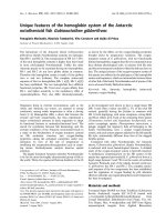
dN/dS score visualization on the surface of gp120 (the 'silent' face of the molecule)Figure 2
dN/dS score visualization on the surface of gp120 (the 'silent' face of the molecule). Visualization of the dN/dS
score (the posterior mean value derived from the Empirical Bayes approach using Model M8) onto the molecular surface of
gp120 (pdb code 2B4C) using a color grade scale. Sites with no data or with a dN/dS score < 0.002 are depicted in white, sites
with a dN/dS score between 0.002 and 0.15 are in light blue, sites between 0.15 and 1 are in light brown, sites with a dN/dS
score between 1 and 2 are yellow, sites with a dN/dS score between 2 and 3 are orange, sites with a dN/dS score > 3 are red
on the surface. A gp120 molecule was added in the upper left quadrants to localize CD4 and/or IgGb12 contact residues and
the C3 alpha helix. Residues that are involved only in CD4 binding are depicted in blue, residues involved in IgGb12 binding are
depicted in yellow, residues that interact both with CD4 and IgGb12 are displayed in green colour (modified from Zhou et al,
2007). The alpha helix present in the C3 region is shown in magenta.
Retrovirology 2009, 6:4 />Page 5 of 15
(page number not for citation purposes)
dN/dS score visualization on the surface of gp120 (the internal portion and the CD4 binding region)Figure 3
dN/dS score visualization on the surface of gp120 (the internal portion and the CD4 binding region). Visualiza-
tion of the dN/dS score (the posterior mean value derived from the Empirical Bayes approach using Model M8) onto the
molecular surface of gp120 (pdb code 2B4C) using a color grade scale. Sites with no data or with a dN/dS score < 0.002 are
depicted in white, sites with a dN/dS score between 0.002 and 0.15 are in light blue, sites between 0.15 and 1 are in light
brown, sites with a dN/dS score between 1 and 2 are yellow, sites with a dN/dS score between 2 and 3 are orange, sites with
a dN/dS score > 3 are red on the surface. A gp120 molecule was added in the upper left quadrants to localize CD4 and/or
IgGb12 contact residues and the C3 alpha helix. Residues that are involved only in CD4 binding are depicted in blue, residues
involved in IgGb12 binding are depicted in yellow, residues that interact both with CD4 and IgGb12 are displayed in green col-
our (modified from Zhou et al, 2007). The alpha helix present in the C3 region is shown in magenta.
Retrovirology 2009, 6:4 />Page 6 of 15
(page number not for citation purposes)
Positively selected sites identified along internal branchesFigure 4
Positively selected sites identified along internal branches. Amino acid (aa) positions are indicated according to HXB2
sequence.
aa
position
aa
position
aa
position
Aa
position
S -> I V -> S N-> D T -> S
I -> V
283
V -> I
279
D -> N
394
S -> T
283
S -> V G -> E A-> V S -> L
S -> P
321
E -> G
281
V -> I L -> Q
291
A -> S K -> T I -> R
410
L -> E
E -> S T -> E R-> G T -> S
392
E -> T T -> A G-> E S -> G
N -> S K -> R E-> G
461
G -> N
S -> P K -> E
335
I -> G M -> T
460
S -> D
336
K -> Q R-> E T -> E
E -> N R -> K
350
E -> R
462
M -> L
D -> N K -> E V-> A R -> K
pt.
A
462
R -> K
354
R -> G A-> V
pt.
F
500
K -> R
A -> V
360
V -> I
279 D -> N
360
V -> F S-> N 360 V -> A
K -> G E -> K N-> K D -> N
354
K -> N K -> T N-> S D -> K
R -> N T -> S
362
N -> T K -> R
444
N -> D S -> K D-> Y
396
K -> N
K -> N K -> E Y-> N N -> V
D -> K
399
K -> N N-> G
398
V -> D
D -> N V -> E D-> V N -> G
460
N -> T
pt.
D
404
E -> R
397
V -> F
401
G -> W
pt.
B
496 V -> I 405 G -> K N -> D
I -> L D -> I
N -> H
454
L -> I
pt.
G
406
I -> N
H -> D I -> N
339
D -> N
I -> K
360 V -> I
460
I -> T
N -> Y R-> T
H -> Y
461
R -> E
H -> P T-> E
396
N -> K T-> N
I -> T
462
N -> D
T -> N E-> N
399
I -> V
N-> S
R -> K S-> N
K -> E E-> G
405
K -> N
E-> Q
D -> E E-> K
pt.
C
463
E -> G
pt.
E
463
G -> E
Retrovirology 2009, 6:4 />Page 7 of 15
(page number not for citation purposes)
gp120 linear sequence, defined clusters on the surface,
suggesting their role in conformational epitopes pre-
sented on exposed antigenic areas. In all patients a high
level of variation was observed in the C3 region, where an
α-helix (position 335 to 350) is located and exposed on
one side to the solvent and can be recognized by humoral
immune defences. On the outer domain of gp120, many
clusters were identified in all patients, but with a different
distribution. A conformational epitope was identified in
patient D, which was defined by Lys337, Ser334, Ala336,
Asn339, Asn340 and Gln344. In patient F, a linear epitope
in the C3 region that is exposed on the surface was identi-
fied and formed by Lys362, Glu363, Ser364 and Ser365.
Another wide site of positive selection appeared to be
formed by Glu269, Asn289, Ser291, Lys337, Gln340,
Lys343, Gln344, and located on the outer surface. In
patient G, the exposed surface harboured only two resi-
dues under positive selection: Ile371 and Gly471, which
cluster together on the 3-D structure.
All patients had positively selected sites in the V3 region,
specifically patient F (5 sites with a dN/dS > 1 located both
on the tip and at its base). In all patients, no sites were
identified among known CD4 induced epitopes.
Analysis of the CD4 binding site
Positively selected sites were identified in the CD4 bind-
ing region in patients C, D, E and F, but not in patients A
and B, where almost all positively selected sites were
located on the outer surface or on the α-helix in the C3
region. In all patients except patient B, Thr283, located in
the CD4 binding region (though not directly in contact
with it), was inferred to be under positive selection. In
patients C and D, distinct sites were under positive selec-
tion in this area. Arg476 in patient C, and Thr283 and
Asp368 in patient D, were under positive selection and
potentially involved in direct receptor binding. A more
clearly delimited constraint seems to act on patients E, F
and G. In particular, a conformational epitope appeared
to be present in patient E and G and formed by Thr278,
Asp279 and Ala 281. In patient F, a complex and large
area located partially within the CD4 binding site and in
a usually highly conserved region immediately next to it
was observed to be under positive selection. This region
includes Ala281, Trp427, Glu460, Ser461, Glu462 and
Leu452 and Leu453. When the IgGb12 heavy chain CDRs
structures were superimposed on patient G-derived gp120
3-dimentional visualization, a high number of positively
selected sites identified in this patient coincided with res-
idues recognized by this broad neutralizing antibody on
the gp120 surface [34].
Identification of rare mutations
When the amino acid entropy of positively selected sites
was studied, the majority of substitutions observed for all
patients were between residues present in that same posi-
tion with a high frequency in the 500 database sequence
alignment. Nevertheless, in some patients, rare substitu-
tions seem to have been selected, including E269D,
N339H, N339D, N340D, N340K, T341A, N343Q,
N343E, A346F, A346Y, T394A, T394I, R476K, R476M.
Amino acid frequencies in those positions in the 500
sequence database alignment and how these sites evolved
during the observation period are shown in Table 1.
dN/dS score visualization on the surface of gp120 (a close-up view of the interaction site between gp120 of patient F and the IgGb12 heavy chain (pdb code NY7))Figure 5
dN/dS score visualization on the surface of gp120 (a
close-up view of the interaction site between gp120
of patient F and the IgGb12 heavy chain (pdb code
NY7)). Visualization of the dN/dS score (the posterior mean
value derived from the Empirical Bayes approach using Model
M8) onto the molecular surface of gp120 (pdb code 2B4C)
using a color grade scale. Sites with no data or with a dN/dS
score < 0.002 are depicted in white, sites with a dN/dS score
between 0.002 and 0.15 are in light blue, sites between 0.15
and 1 are in light brown, sites with a dN/dS score between 1
and 2 are yellow, sites with a dN/dS score between 2 and 3
are orange, sites with a dN/dS score > 3 are red on the sur-
face. Residues that are involved only in CD4 binding are
depicted in blue, residues involved in IgGb12 binding are
depicted in yellow, residues that interact both with CD4 and
IgGb12 are displayed in green colour (modified from Zhou et
al, 2007). The alpha helix present in the C3 region is shown
in magenta. The carbon atoms of CDR1, CDR2 and CDR3
are coloured white, green and cyan respectively. The amino
acid residues are shown as sticks. Of note, the binding region
of the broadly neutralizing antibody overlaps the positively
selected sites in the patient G derived structure.
Retrovirology 2009, 6:4 />Page 8 of 15
(page number not for citation purposes)
HLA typing
A low- or high-resolution HLA typing was also performed
for patient A to E. HLA typing was not possible for patients
F and G. Results of HLA typing are shown in Additional
file 3.
Discussion
In the present study, a high-resolution phylogenetic anal-
ysis of the gp120 envelope glycoprotein evolution was
performed in HIV-1 infected patients with a different pat-
tern of disease progression. All patients under study had
never been treated for HIV-1 infection, leaving the host
immune system as the only selective force acting on virus
evolution and quasispecies selection. Firstly, an analysis
was performed to identify putative recombinants. Recom-
bination may occur frequently in vivo in HIV-1 evolution,
and artificial chimeric sequences due to PCR crossovers
can significantly affect phylogenetic analysis. The PHI test
based on the refined incompatibility score was used to
overcome this bias with our data set [35]. When recom-
binant sequences were excluded (about 15%, see materi-
als and methods) from the analysis, the number of sites
with a dN/dS value > 1 was reduced in some of the
patients. Nevertheless, the number of positively selected
sites identified with a Bayesian posterior probability >
0.95 in our datasets was not significantly affected. The
best fitting model of evolution was chosen in the phyloge-
netic reconstruction, and maximum likelihood methods
were used to fit codon models of evolution for all
patients, to identify positively selected sites, and Bayesian
inference was used to estimate virus evolutionary rates. In
addition, an HLA typing and a color-grade 3-dimensional
visualization of the dN/dS score were used.
Finally, since external branches are subject to substitu-
tions as well as mutational load, which involves random
mutations and therefore potentially many nonsynony-
mous substitutions, we inferred the sites under selection
for the internal branches only, using the Fixed Effects Like-
lihood (FEL) approach [36]. This analysis infers dN and
dS for each site and also tests whether dN = dS or not for
the sites [36]. All the sites identified with the FEL
approach were also identified with the previous methods,
further confirming the possibility of identifying sites
showing diversifying selection when sequential time
points are considered even using cloned sequences. A
multiple-step analysis was in fact necessary in the present
study to address correctly the evolution of a large portion
of the HIV-1 env gene, since a high background is expected
when the dN/dS score/site is performed in highly variable
viral populations under continuous positive selection. In
these cases, only sites with high dN/dS ratio and con-
firmed by Bayesian posterior probability should be taken
into consideration [32,37,38]
In order to highlight the effect of positive selection on
virus evolution, the evolutionary rate was calculated sepa-
rately in the three codon positions. In the third codon
position, mutations are silent in about 70% of all possibly
occurring nucleotide changes, and if no selective con-
straints act on the virus, evolution occurs at a faster rate
compared to the first and second codon positions. In all
LTNPs, the third codon mutation rate is equal to or lower
than that compared to the averaged 1
st
and 2
nd
position (p
= 0.016), thus being compatible with positive selection
[39-41].
The impact of HLA-associated selection pressure on viral
evolution has recently been demonstrated at the popula-
tion level [42-50]. No HLA B57 associated positively
selected sites were identified in our patients, but a poten-
tial HLA A11 associated epitope was present in patients B,
C, and E. Within this epitope, the position 346 exhibited
a high dN/dS ratio in all three patients.
Although positive selection was evident in the replicating
virus from all subjects, differences were observed between
NPs and LTNPs. In subjects A and B (NPs) selective con-
straints are less intense, in terms of dN/dS score calculated
even for the highly selected hotspots (Figure 2 and 3), and
are limited to the external surface of the crystal and to the
α-helix in the C3 region. These sites and the V3 loop
appear to be targets for the immune response in all
patients, with a single exception (patient A). This observa-
tion is apparently in contrast with the results obtained by
other studies, where the C3 alpha helix was observed to be
under positive selection for clade C envelopes and only
modestly for clade B [27,51]. Although we cannot exclude
that differences in the intensity of the immune response
against different HIV-1 subtypes exist at these levels, the
previous analyses were based on cross-sectional C-clade
and B-clade sequence datasets downloaded from HIV-1
databases, thus not reflecting the intra-patient evolution-
ary dynamics and the heterogeneity of host immune
responses during the different phases of HIV-1 infection
(or the different patterns of disease progression observed).
Other studies analyzed the sequence evolution in infected
individuals and showed that the C3 region, including the
externally accessible residues, is under strong positive
selection both in clade B [24-26] and in HIV-1 subtype C
infections [23]. These results may be of particular interest
since this antigenic portion of the gp120 molecule has
been considered in the development of candidate vaccines
[52-56]
Many N-linked glycosylation sites were identified to be
under positive selection and exposed on the surface in the
group of LTNPs and in the 2 NP subjects. In particular
N442, R444 and S446, N295, N332, N340, N339 were
identified as being potentially involved in the glycan
Retrovirology 2009, 6:4 />Page 9 of 15
(page number not for citation purposes)
Table 1: Evolution of positively selected amino acids that were rarely found in the 500 sequences database.
AA
pos
Frequency
in the
database
AB C D E F G
I II III I II III I II III I II III I II III I II III I II III
269 Glu 76.8%
Asp 8.1%
EE E D
15/15
D
15/15
D
10/13
EE D
19/19
D 9/14 D
9/10
E
3/13
E
5/14
E
1/10
339 Asn 78.5%
Asp 4.3%
His 0.5%
NN N
8/13
N
14/15
N
8/14
VNKKNN
D
4/13
H
1/15
H
6/14
H
1/13
340 Lys 32.5%
Asn 31.3%
Asp 7.2%
E D
16/16
D
3/15
N
10/10
D KNNN
N
12/15
341 Thr 91.4%
Ala 5.7%
TT T T
13/15
T
5/12
T
1/3
TT T
A
2/15
A
7/12
A
12/13
343 Asn 2.4%
lys 25.9%
Gln 40.5%
Glu 9.6%
KK N
2/13
N
14/15
N
5/14
K
15/15
K
7/12
E
3/13
K
12/12
K
6/8
K
5/14
G
13/13
E
14/19
K
14/14
K
19/19
K
11/11
E
2/7
K
11/13
K
1/15
K
5/14
E
4/12
K
10/13
R
1/8
Q
8/14
R
5/19
Q
2/10
R
4/14
T
1/12
E
1/8
R
1/14
346 Val 37.6%
Ala 26.8%
Ser11.2%
Phe 0.2%
Tyr 0%
VA
5/16
V
15/15
V
10/10
V
4/13
V
15/15
V
10/14
VV
3/12
V
6/8
V
6/14
VV
19/19
V
7/11
A
9/9
V
11/16
A
4/13
A
4/14
F
5/12
F
2/8
Y
8/14
A
4/11
A 4/12
Retrovirology 2009, 6:4 />Page 10 of 15
(page number not for citation purposes)
394 Thr 81.3%
Iso 3.1%
Ala 1.7%
TT T
4/13
T
13/15
T
6/14
TTTT
I 9/13 I 1/15 I 8/14
A 1/15
476 Arg 81.3%
Lys 18.4%
Met 0%
K R R 11/13 R 3/15 R 8/14 R M RR
K 2/13 K 12/15 K 6/14
Their frequency in the sequence database and their proportion (number of clones with the mutation/number of clones sequenced) in the viral quasispecies at each time point (I, II, and III) are shown.
Table 1: Evolution of positively selected amino acids that were rarely found in the 500 sequences database. (Continued)
Retrovirology 2009, 6:4 />Page 11 of 15
(page number not for citation purposes)
shield that protects the virus against host defences [57].
Interestingly, it has been demonstrated that the neutraliz-
ing activity of a human monoclonal antibody, designated
as mAb 2G12, is associated with the presence of glycosyla-
tion sites at these positions, including 295, 332 and 339
[58-60]. IgG 2G12-like antibodies have previously been
detected in LTNP patients by competitive ELISA experi-
ments with high levels in sera associated with the broad
neutralizing activity [19]. This observation is in perfect
agreement with our data, suggesting that antibodies that
bind close to the 2G12 binding site exist in some patients
and exert selective pressure on the viral surface.
It has recently been observed that cross-neutralizing activ-
ity characterizing a small subset of LTNPs is associated
with antibodies recognizing the CD4bs [22]. However,
only a few broadly neutralizing human monoclonal anti-
bodies have been isolated at present; among them, only
the IgGb12 (directed against the CD-4bs) and mAb 2G12
(recognizing oligomannose residues) target the gp120
[58,61,62]. Notably, 4 out of the 5 LTNP patients exhibit
strong selective constraints at the level of the CD4bs. In
patient F in particular, an IgGb12 epitope-like area is
under strong positive selection (Figure 5). These data doc-
ument that this epitope can be modified in vivo in
response to specific selective pressure. Further analyses are
necessary to clarify if mutations in this region may alter
the viral RC, thus being able to delay disease progression.
Conclusion
The present study describes the dynamic evolution of the
HIV-1 env gene in a subset of LTNP subjects and docu-
ments that the CD4bs is under strong selective pressure in
the replicating virus of a group of LTNPs and evolves dur-
ing the course of the disease. These data may be of interest
not only for the understanding of the complex HIV-1-host
relationships, but also for planning new immune-based
strategies against HIV-1 infection.
Methods
Patients and sequences
Seven HIV-positive patients, never treated for HIV infec-
tion, were selected on the basis of the slope of their CD4+-
T-cell counts and the level of HIV-1 viremia (Table 2).
Two of them (subjects A and B) were showing a typical
progression of HIV infection (TP), with a gradual decline
of CD4+ T cells over time (loss of circulating CD4+ T cells
per year: subject A, 87; and subject B, 153). The patients
designated C, D, E, F, and G were LTNPs with CD4+-T-cell
constantly higher than 500 per ml. They were showing the
following mean variation/year of circulating CD4+lym-
phocytes: -31, -24, -2, -10, and +12 respectively). Lengths
of infection and sampling dates are shown in Table 2.
Plasma specimens were concentrated by centrifugation at
23,600 × g for 1 h at 4°C and RNA was extracted by using
a QIAamp viral RNA mini kit (Qiagen, Valencia, CA). The
following outer primers were used in the nested PCR
amplification reaction: V31 (nucleotides 6939 to 6966 in
the pNL4-3 numbering system) and V52 (nucleotides
7803 to 7778). The internal primers were: V32 (nucle-
otides 7367 to 7340), and V41 (nucleotides 7304 to
7326). The reverse transcription of HIV-1 RNA present in
plasma was performed with primer V52 (25 pmol) and
200 U of SuperScript II RNase H-RT (Bethesda Research
Laboratories, Gaithersburg, Md.) at 37°C for 60 min in a
final volume of 20 μl in the presence of 3.0 mM MgCl
2
, 75
mM KCl, 50 mM Tris (pH 8.3), 10 mM dithiothreitol, 0.5
mM concentrations of each deoxynucleosidetriphosphate
(dNTP), and 20 U of recombinant RNasin RNase inhibi-
tor (Promega Corp., Madison, Wis.). An amount of cDNA
equivalent to 50–1000 copies of template (as evaluated by
HIV-1 RNA copies/ml) was used for PCR amplification.
An Expand High Fidelity PCR system (Roche Diagnostic
Corporation, Indianapolis, IN) in 1× Expand PCR buffer
containing 1.5 mM MgCl
2
, 0.2 mM of each deoxynucleo-
side triphosphate, and 0.2 μM of V31 and V52 primers
was used for the first round. The following cycling condi-
tions were applied: 94°C for 2 min followed by 35 cycles
of 94°C for 15 s, 55°C for 30 s, and 68°C for 4 min, with
a final extension of 68°C for 10 min. Second-round PCR
was performed using 2 μl of the first-round PCR product
and primers V32 and V41 under the same conditions used
for the first-round PCR.
Only one sample at a time was processed, and clinical
samples were amplified in triplicate. Before molecular
cloning, a 10-μl aliquot of the amplified product was run
on a 10% polyacrylamide gel electrophoresis to screen for
the appropriate-sized band (ca. 865 bp); the remaining 90
μl was resolved by electrophoresis on a 1.5% low-melting-
point agarose gel (SeaPlaque; FMC BioProducts, Rock-
land, Maine) in TAE buffer (Tris-acetate, 1 mM EDTA).
The DNA fragment was excised from the gel, purified by
the QIAquick DNA Clean-Up system (Qiagen GmbH,
Hiden, Germany) and cloned into pGEM-T vector
(Promega) according to the manufacturer's instructions.
After PCR colony screening, 8 to 20 positive clones were
selected and the insert was sequenced with primers V31
and V52 on an automatic sequencer (ABI Prism 3100,
Appliedbiosystems, Foster City, CA).
For patient A, 15 clones per time point were studied; for
patient B 16,15 and 10 clones respectively; for patient C,
13, 15, and 24 clones; for patient D, 15,12 and 13; for
patient E, 12, 8, and 14 clones, for patient F, 13, 19, and
14 clones; and for patient G, 19, 15, and 9 clones.
Retrovirology 2009, 6:4 />Page 12 of 15
(page number not for citation purposes)
Because some analyses could potentially be affected by
recombinats, we tested for recombination using the PHI
test implemented in the SplitsTree package version 4.8.
Significance of the PHI statistic for the presence of recom-
bination is assessed with the normal approximation of a
permutation test where, under the null hypothesis of no
recombination, sites along the alignment are randomly
permuted to obtain the null distribution of PHI: p < 0.05
indicate significant presence of recombination. About
15% of sequences for each time point/patient were dis-
carded after this analysis.
GenBank accession numbers of sequences are EU329847
– EU330175.
High resolution phylogenetic analysis & graphical 3-
dimensional visualization
Overall, 278 non recombinant genomic-HIV-1 RNA
sequences were aligned collectively and individually for
each patient, using amino acidic sequences as template for
nucleotide alignment by using DAMBE http://
dambe.bio.uottawa.ca/software.asp and manually cor-
rected with BioEdit />Table 2: Immunologic and virologic parameters of patients that were selected for the study.
Patient Timepoint (years from infection) HIVRNA copies/ml of plasma CD4+ T cell counts/mm
3
A I (9) 28000 561
II(11) 32360 379
III(13) 127200 337
B I (8.5) 25000 374
II(9.5) 65000 255
III(10.5) 40000 259
C I(10) 99 1144
II(11) 100 944
III(12) 549 776
D I(9.5) 2918 993
II(10.5) 3318 735
III(12) 6850 640
E I(11.5) 1200 795
II(12.5) 2000 720
III(13.2) 3034 527
F I(8) 2100 645
II(10) 1200 657
III(13) 6393 625
G I(10) 560 864
II(13) 106 926
III(14) 152 750
Retrovirology 2009, 6:4 />Page 13 of 15
(page number not for citation purposes)
bioedit.html. A Neighbor-Joining (NJ) tree was recon-
structed using the best fitting evolutionary model as eval-
uated with MODELTEST v3.6 (Posada, D., and K.A.
Crandall, 2001) was generated.
To obtain a maximum-likelihood tree topology, a local
rearrangement search with the maximum-likelihood
method was conducted by starting from the topology of
the NJ tree, as implemented in PAUP* http://
paup.csit.fsu.edu. The ratio of transitions to transversions,
and the gamma distribution of rate variation among sites
were estimated from the data. To evaluate if intra-patient
virus evolution showed patterns of positive selection, a
ML method was applied by using CODEML implemented
in the PAML package />and the substitution rate at individual codon position was
also estimated for each patient using the TipDate model as
implemented in BEAST />.
The CODEML program fits various models of codon evo-
lution to sequence data related by a phylogenetic tree,
which allow to test for varying selection pressures at indi-
vidual codon sites. The models of codon evolution differ
in their distribution of dN/dS values among codons. Two
couples of nested models were employed: M1a vs M2a
and M7 vs M8. M1a (neutral/purifying model) allows
only two categories of dN/dS across codons and the dN/
dS ratio is constrained to be > 0 and < 1 in one category
and equal to 1 in the other. Hence, M1a only accommo-
dates neutral evolution. M2a adds an extra class of codons
to account for positive selection (i.e., a class of codons
with dN/dS > 1). M7 (neutral model) assumes a beta dis-
tribution of dN/dS between 0 and 1 with 10 categories to
discretize the distribution. M8 adds an extra class of
codons with dN/dS > 1 [63]. The likelihoods of the mod-
els were than compared using the likelihood ratio test. To
allow further definition of HIV-1 env positively selected
sites within each patient, all the RNA-sequences amplified
and cloned from the same subject, were analysed by using
an empirical Bayes approach [64]. The posterior mean
dN/dS value per site was calculated and a Bayesian
approach was used to identify codons undergoing positive
selection with a posterior probability of > 95% or > 99%,
using CODEML. To better identify conformational
epitopes and sites on the protein surface with possibly dis-
tinct roles on disease progression, and distinct patterns of
virus evolution driven by host-selective constraints along
the C2-V5 region, a graphic colour-grade 3-dimensional
visualization the dN/dS score (ratio between non-synon-
ymous/synonymous mutations per site) was generated
using PyMol
and the
structure of a V3-containing gp120 core [65].
Moreover, to better understand the impact of deleterious
mutational load in within-host HIV evolution and its
impact on identifying positively selected sites, we per-
formed a ML analysis of varying selection pressures
among lineages. In this analysis, we compared model M0
(all branches have the same dN/dS) with an alternative
model that allows a different dN/dS for internal and exter-
nal branches.
Sites under selection along internal branches of recon-
structed phylogenetic trees, were inferred using the Fixed
Effects Likelihood (FEL) approach implemented in Hy-
Phy
.
To evaluate the HIV-1 general site-specific inter-exchange-
ability (site-specific aminoacidic entropy) a collection of
500 aligned env sequences from the Stanford Database
was downloaded and analysed with BioEdit accessory
applications. When positional homology was not main-
tained due to the high genetic variability, that site in the
alignment was not considered in the analyses.
Competing interests
The authors declare that they have no competing interests.
Authors' contributions
FC conceived and coordinated the study and wrote the
manuscript; FC and PL did the analyses. MCM, MS, SB
and PB carried out the amplification and sequencing. MD
did the 3D color-grade mapping on the gp120 structure.
GG followed up patients; BM performed the HLA typing;
RB, MC discussed the data and reviewed the manuscript.
All authors read and approved the final manuscript.
Additional material
Additional file 1
Supplementary Table One. Likelihood ratio statistics (2
Δ
l) for com-
parision of different models of codon evolution.
Click here for file
[ />4690-6-4-S1.doc]
Additional file 2
Supplementary Table Two. Analysis of selective pressure among inter-
nal and external branches in each patient.
Click here for file
[ />4690-6-4-S2.doc]
Additional file 3
Supplementary Table Three. Low- or high-resolution HLA typing.
Click here for file
[ />4690-6-4-S3.doc]
Retrovirology 2009, 6:4 />Page 14 of 15
(page number not for citation purposes)
Acknowledgements
Parts of these results were presented in abstract form at the 2007 Key-
stone Symposia on HIV Vaccines [Whistler, British Columbia], and at the
2006 Meeting on Retroviruses [Cold Spring Harbor, New York]. This work
was partially supported by grants of the "AIDS Project" of the Italian Istituto
Superiore di Sanità to MC and RB; PL was supported by a postdoctoral fel-
lowship from the Fund for Scientific Research (FWO) Flanders.
References
1. Richman DD, Little SJ, Smith DM, Wrin T, Petropoulos C, Wong JK:
HIV Evolution and Escape. Trans Am Clin Climatol Assoc 2004,
115:289-303.
2. Frost SD, Wrin T, Smith DM, Kosakovsky Pond SL, Liu Y, Paxinos E,
Chappey C, Galovich J, Beauchaine J, Petropoulos CJ, et al.: Neutral-
izing antibody responses drive the evolution of human
immunodeficiency virus type 1 envelope during recent HIV
infection. Proc Natl Acad Sci USA 2005, 102(51):18514-18519.
3. Clementi M, Canducci F, Bagnarelli P, Menzo S: Intra-host evolu-
tion of human immunodeficiency virus type 1 and viral fit-
ness. New Microbiol 2004, 27(2 Suppl 1):41-44.
4. Quinones-Mateu ME, Ball SC, Marozsan AJ, Torre VS, Albright JL,
Vanham G, van Der Groen G, Colebunders RL, Arts EJ: A dual
infection/competition assay shows a correlation between ex
vivo human immunodeficiency virus type 1 fitness and dis-
ease progression. J Virol 2000, 74(19):9222-9233.
5. Arts EJ, Quinones-Mateu ME: Sorting out the complexities of
HIV-1 fitness. Aids 2003, 17(5):780-781.
6. Rangel HR, Weber J, Chakraborty B, Gutierrez A, Marotta ML, Mirza
M, Kiser P, Martinez MA, Este JA, Quinones-Mateu ME: Role of the
human immunodeficiency virus type 1 envelope gene in viral
fitness. J Virol 2003, 77(16):9069-9073.
7. Bagnarelli P, Vecchi M, Burighel N, Bellanova D, Menzo S, Clementi
M, De Rossi A: Genotypic and phenotypic correlates of the
HIV Type 1 env gene evolution in infected children with dis-
cordant response to antiretroviral therapy. AIDS Res Hum Ret-
roviruses 2004, 20(12):1306-1313.
8. Menzo S, Sampaolesi R, Vicenzi E, Santagostino E, Liuzzi G, Chirianni
A, Piazza M, Cohen OJ, Bagnarelli P, Clementi M: Rare mutations
in a domain crucial for V3-loop structure prevail in replicat-
ing HIV from long-term non-progressors. Aids 1998,
12(9):985-997.
9. Frost SD, Liu Y, Pond SL, Chappey C, Wrin T, Petropoulos CJ, Little
SJ, Richman DD: Characterization of human immunodefi-
ciency virus type 1 (HIV-1) envelope variation and neutraliz-
ing antibody responses during transmission of HIV-1 subtype
B. J Virol 2005, 79(10):6523-6527.
10. Riddle TM, Shire NJ, Sherman MS, Franco KF, Sheppard HW, Nelson
JA: Sequential turnover of human immunodeficiency virus
type 1 env throughout the course of infection. J Virol 2006,
80(21):10591-10599.
11. Srivastava IK, Ulmer JB, Barnett SW: Role of neutralizing antibod-
ies in protective immunity against HIV. Hum Vaccin 2005,
1(2):45-60.
12. Moore PL, Crooks ET, Porter L, Zhu P, Cayanan CS, Grise H, Corc-
oran P, Zwick MB, Franti M, Morris L, et al.: Nature of nonfunc-
tional envelope proteins on the surface of human
immunodeficiency virus type 1. J Virol 2006, 80(5):2515-2528.
13. Lee WR, Syu WJ, Du B, Matsuda M, Tan S, Wolf A, Essex M, Lee TH:
Nonrandom distribution of gp120 N-linked glycosylation
sites important for infectivity of human immunodeficiency
virus type 1. Proc Natl Acad Sci USA 1992, 89(6):2213-2217.
14. Wei X, Decker JM, Wang S, Hui H, Kappes JC, Wu X, Salazar-
Gonzalez JF, Salazar MG, Kilby JM, Saag MS, et al.: Antibody neutral-
ization and escape by HIV-1. Nature 2003, 422(6929):307-312.
15. Balzarini J: Targeting the glycans of gp120: a novel approach
aimed at the Achilles heel of HIV. Lancet Infect Dis 2005,
5(11):726-731.
16. Poon AF, Lewis FI, Pond SL, Frost SD: Evolutionary interactions
between N-linked glycosylation sites in the HIV-1 envelope.
PLoS Comput Biol 2007, 3(1):e11.
17. Burton DR, Desrosiers RC, Doms RW, Koff WC, Kwong PD, Moore
JP, Nabel GJ, Sodroski J, Wilson IA, Wyatt RT: HIV vaccine design
and the neutralizing antibody problem. Nat Immunol 2004,
5(3):233-236.
18. Zolla-Pazner S: Identifying epitopes of HIV-1 that induce pro-
tective antibodies. Nat Rev Immunol 2004, 4(3):199-210.
19. Braibant M, Brunet S, Costagliola D, Rouzioux C, Agut H, Katinger H,
Autran B, Barin F: Antibodies to conserved epitopes of the
HIV-1 envelope in sera from long-term non-progressors:
prevalence and association with neutralizing activity. Aids
2006, 20(15):1923-1930.
20. Eggink D, Melchers M, Sanders RW: Antibodies to HIV-1: aiming
at the right target.
Trends Microbiol 2007, 15(7):291-294.
21. Dhillon AK, Donners H, Pantophlet R, Johnson WE, Decker JM, Shaw
GM, Lee FH, Richman DD, Doms RW, Vanham G, et al.: Dissecting
the neutralizing antibody specificities of broadly neutralizing
sera from human immunodeficiency virus type 1-infected
donors. J Virol 2007, 81(12):6548-6562.
22. Li Y, Migueles SA, Welcher B, Svehla K, Phogat A, Louder MK, Wu X,
Shaw GM, Connors M, Wyatt RT, et al.: Broad HIV-1 neutraliza-
tion mediated by CD4-binding site antibodies. Nat Med 2007,
13(9):1032-1034.
23. Moore PL, Gray ES, Choge IA, Ranchobe N, Mlisana K, Abdool Karim
SS, Williamson C, Morris L: The c3-v4 region is a major target
of autologous neutralizing antibodies in human immunodefi-
ciency virus type 1 subtype C infection. J Virol 2008,
82(4):1860-1869.
24. Kupfer B, Sing T, Schuffler P, Hall R, Kurz R, McKeown A, Schneweis
KE, Eberl W, Oldenburg J, Brackmann HH, et al.: Fifteen years of
env C2V3C3 evolution in six individuals infected clonally
with human immunodeficiency virus type 1. J Med Virol 2007,
79(11):1629-1639.
25. Bunnik EM, Pisas L, van Nuenen AC, Schuitemaker H: Autologous
neutralizing humoral immunity and evolution of the viral
envelope in the course of subtype B human immunodefi-
ciency virus type 1 infection. J Virol 2008, 82(16):7932-7941.
26. Joos B, Fischer M, Schweizer A, Kuster H, Boni J, Wong JK, Weber R,
Trkola A, Gunthard HF: Positive in vivo selection of the HIV-1
envelope protein gp120 occurs at surface-exposed regions. J
Infect Dis 2007, 196(2):313-320.
27. Choisy M, Woelk CH, Guegan JF, Robertson DL: Comparative
study of adaptive molecular evolution in different human
immunodeficiency virus groups and subtypes. J Virol 2004,
78(4):1962-1970.
28. Burton DR, Stanfield RL, Wilson IA: Antibody vs. HIV in a clash
of evolutionary titans. Proc Natl Acad Sci USA 2005,
102(42):14943-14948.
29. Gaschen B, Taylor J, Yusim K, Foley B, Gao F, Lang D, Novitsky V,
Haynes B, Hahn BH, Bhattacharya T, et al.: Diversity considera-
tions in HIV-1 vaccine selection. Science
2002,
296(5577):2354-2360.
30. Scanlan CN, Offer J, Zitzmann N, Dwek RA: Exploiting the defen-
sive sugars of HIV-1 for drug and vaccine design. Nature 2007,
446(7139):1038-1045.
31. Leal E, Janini M, Diaz RS: Selective pressures of human immun-
odeficiency virus type 1 (HIV-1) during pediatric infection.
Infect Genet Evol 2007, 7(6):694-707.
32. de Oliveira T, Salemi M, Gordon M, Vandamme AM, van Rensburg EJ,
Engelbrecht S, Coovadia HM, Cassol S: Mapping sites of positive
selection and amino acid diversification in the HIV genome:
an alternative approach to vaccine design? Genetics 2004,
167(3):1047-1058.
33. Pybus OG, Rambaut A, Belshaw R, Freckleton RP, Drummond AJ,
Holmes EC: Phylogenetic evidence for deleterious mutation
load in RNA viruses and its contribution to viral evolution.
Mol Biol Evol 2007, 24(3):845-852.
34. Zhou T, Xu L, Dey B, Hessell AJ, Van Ryk D, Xiang SH, Yang X, Zhang
MY, Zwick MB, Arthos J, et al.: Structural definition of a con-
served neutralization epitope on HIV-1 gp120. Nature 2007,
445(7129):732-737.
35. Salemi M, Burkhardt BR, Gray RR, Ghaffari G, Sleasman JW, Goode-
now MM: Phylodynamics of HIV-1 in Lymphoid and Non-Lym-
phoid Tissues Reveals a Central Role for the Thymus in
Emergence of CXCR4-Using Quasispecies. PLoS ONE 2007,
2(9):e950.
36. Pond SL, Frost SD, Grossman Z, Gravenor MB, Richman DD, Brown
AJ: Adaptation to different human populations by HIV-1
revealed by codon-based analyses. PLoS Comput Biol 2006,
2(6):e62.
Publish with BioMed Central and every
scientist can read your work free of charge
"BioMed Central will be the most significant development for
disseminating the results of biomedical research in our lifetime."
Sir Paul Nurse, Cancer Research UK
Your research papers will be:
available free of charge to the entire biomedical community
peer reviewed and published immediately upon acceptance
cited in PubMed and archived on PubMed Central
yours — you keep the copyright
Submit your manuscript here:
/>BioMedcentral
Retrovirology 2009, 6:4 />Page 15 of 15
(page number not for citation purposes)
37. Yamaguchi-Kabata Y, Gojobori T: Reevaluation of amino acid
variability of the human immunodeficiency virus type 1
gp120 envelope glycoprotein and prediction of new discon-
tinuous epitopes. J Virol 2000, 74(9):4335-4350.
38. Sheridan I, Pybus OG, Holmes EC, Klenerman P: High-resolution
phylogenetic analysis of hepatitis C virus adaptation and its
relationship to disease progression. J Virol 2004,
78(7):3447-3454.
39. Bazykin GA, Dushoff J, Levin SA, Kondrashov AS: Bursts of nonsyn-
onymous substitutions in HIV-1 evolution reveal instances of
positive selection at conservative protein sites. Proc Natl Acad
Sci USA 2006, 103(51):19396-19401.
40. Lemey P, Rambaut A, Pybus OG: HIV evolutionary dynamics
within and among hosts. AIDS Rev 2006, 8(3):125-140.
41. Lemey P, Kosakovsky Pond SL, Drummond AJ, Pybus OG, Shapiro B,
Barroso H, Taveira N, Rambaut A: Synonymous substitution
rates predict HIV disease progression as a result of underly-
ing replication dynamics. PLoS Comput Biol 2007, 3(2):e29.
42. McMichael A, Klenerman P: HIV/AIDS. HLA leaves its footprints
on HIV. Science 2002, 296(5572):1410-1411.
43. Carrington M, O'Brien SJ: The influence of HLA genotype on
AIDS. Annu Rev Med 2003, 54:535-551.
44. Peyerl FW, Bazick HS, Newberg MH, Barouch DH, Sodroski J, Letvin
NL: Fitness costs limit viral escape from cytotoxic T lym-
phocytes at a structurally constrained epitope. J Virol 2004,
78(24):13901-13910.
45. Fellay J, Shianna KV, Ge D, Colombo S, Ledergerber B, Weale M,
Zhang K, Gumbs C, Castagna A, Cossarizza A, et al.: A whole-
genome association study of major determinants for host
control of HIV-1. Science 2007, 317(5840):944-947.
46. Moore CB, John M, James IR, Christiansen FT, Witt CS, Mallal SA:
Evidence of HIV-1 adaptation to HLA-restricted immune
responses at a population level. Science 2002,
296(5572):
1439-1443.
47. Mallal S, Nolan D, Witt C, Masel G, Martin AM, Moore C, Sayer D,
Castley A, Mamotte C, Maxwell D, et al.: Association between
presence of HLA-B* HLA-DR7, and HLA-DQ3 and hyper-
sensitivity to HIV-1 reverse-transcriptase inhibitor abacavir.
Lancet 5701, 359(9308):727-732.
48. Bailey JR, Zhang H, Wegweiser BW, Yang HC, Herrera L, Ahonkhai
A, Williams TM, Siliciano RF, Blankson JN: Evolution of HIV-1 in an
HLA-B*57-positive patient during virologic escape. J Infect Dis
2007, 196(1):50-55.
49. Dong T, Stewart-Jones G, Chen N, Easterbrook P, Xu X, Papagno L,
Appay V, Weekes M, Conlon C, Spina C, et al.: HIV-specific cyto-
toxic T cells from long-term survivors select a unique T cell
receptor. J Exp Med 2004, 200(12):1547-1557.
50. Wang S, Sun Y, Zhai S, Zhuang Y, Zhao S, Kang W, Li X, Huang D, Yu
XG, Walker BD, et al.: Identification of HLA-A11-restricted
HIV-1-specific cytotoxic T-lymphocyte epitopes in China.
Curr HIV Res 2007, 5(1):119-128.
51. Gnanakaran S, Lang D, Daniels M, Bhattacharya T, Derdeyn CA, Kor-
ber B: Clade-specific differences between human immunode-
ficiency virus type 1 clades B and C: diversity and
correlations in C3-V4 regions of gp120. J Virol 2007,
81(9):4886-4891.
52. Pinter A: Roles of HIV-1 Env variable regions in viral neutrali-
zation and vaccine development. Curr HIV Res 2007,
5(6):542-553.
53. Pantophlet R, Burton DR: GP120: target for neutralizing HIV-1
antibodies. Annu Rev Immunol 2006, 24:739-769.
54. Vaine M, Wang S, Crooks ET, Jiang P, Montefiori DC, Binley J, Lu S:
Improved induction of antibodies against key neutralizing
epitopes by human immunodeficiency virus type 1 gp120
DNA prime-protein boost vaccination compared to gp120
protein-only vaccination. J Virol 2008, 82(15):7369-7378.
55. Karlsson Hedestam GB, Fouchier RA, Phogat S, Burton DR, Sodroski
J, Wyatt RT: The challenges of eliciting neutralizing antibodies
to HIV-1 and to influenza virus. Nat Rev Microbiol 2008,
6(2):143-155.
56. McKnight A, Aasa-Chapman MM: Clade specific neutralising vac-
cines for HIV: an appropriate target? Curr HIV Res 2007,
5(6):554-560.
57. Dacheux L, Moreau A, Ataman-Onal Y, Biron F, Verrier B, Barin F:
Evolutionary dynamics of the glycan shield of the human
immunodeficiency virus envelope during natural infection
and implications for exposure of the 2G12 epitope. J Virol
2004, 78(22):12625-12637.
58. Mehandru S, Vcelar B, Wrin T, Stiegler G, Joos B, Mohri H, Boden D,
Galovich J, Tenner-Racz K, Racz P, et al.: Adjunctive passive
immunotherapy in human immunodeficiency virus type 1-
infected individuals treated with antiviral therapy during
acute and early infection. J Virol 2007, 81(20):11016-11031.
59. Manrique A, Rusert P, Joos B, Fischer M, Kuster H, Leemann C, Nie-
derost B, Weber R, Stiegler G, Katinger H, et al.: In vivo and in vitro
escape from neutralizing antibodies 2G12, 2F5, and 4E10. J
Virol 2007, 81(16):8793-8808.
60. Scanlan CN, Pantophlet R, Wormald MR, Ollmann Saphire E, Stanfield
R, Wilson IA, Katinger H, Dwek RA, Rudd PM, Burton DR: The
broadly neutralizing anti-human immunodeficiency virus
type 1 antibody 2G12 recognizes a cluster of alpha1 >2 man-
nose residues on the outer face of gp120. J Virol 2002,
76(14):7306-7321.
61. Trkola A, Kuster H, Rusert P, Joos B, Fischer M, Leemann C, Man-
rique A, Huber M, Rehr M, Oxenius A, et al.: Delay of HIV-1
rebound after cessation of antiretroviral therapy through
passive transfer of human neutralizing antibodies. Nat Med
2005, 11(6):615-622.
62. Nakowitsch S, Quendler H, Fekete H, Kunert R, Katinger H, Stiegler
G: HIV-1 mutants escaping neutralization by the human anti-
bodies 2F5, 2G12, and 4E10: in vitro experiments versus clin-
ical studies. Aids 2005, 19(17):1957-1966.
63. Yang Z: Maximum likelihood analysis of adaptive evolution in
HIV-1 gp120 env gene. Pac Symp Biocomput 2001:226-237.
64. Yang Z, Wong WS, Nielsen R: Bayes empirical bayes inference
of amino acid sites under positive selection. Mol Biol Evol 2005,
22(4):1107-1118.
65. Huang CC, Tang M, Zhang MY, Majeed S, Montabana E, Stanfield RL,
Dimitrov DS, Korber B, Sodroski J, Wilson IA,
et al.: Structure of a
V3-containing HIV-1 gp120 core. Science 2005,
310(5750):1025-1028.