Báo cáo y học: "Complete suppression of viral gene expression is associated with the onset and progression of lymphoid malignancy: observations in Bovine Leukemia Virus-infected sheep" pdf
Bạn đang xem bản rút gọn của tài liệu. Xem và tải ngay bản đầy đủ của tài liệu tại đây (486.13 KB, 9 trang )
BioMed Central
Page 1 of 9
(page number not for citation purposes)
Retrovirology
Open Access
Short report
Complete suppression of viral gene expression is associated with
the onset and progression of lymphoid malignancy: observations in
Bovine Leukemia Virus-infected sheep
Makram Merimi
1
, Pavel Klener
1,2
, Maud Szynal
1
, Yvette Cleuter
1
,
Claude Bagnis
3
, Pierre Kerkhofs
4
, Arsène Burny
1
, Philippe Martiat
1
and
Anne Van den Broeke*
1
Address:
1
Laboratory of Experimental Hematology, Institut Jules Bordet, Université Libre de Bruxelles (ULB), 1000 Brussels, Belgium,
2
Institute of
Pathological Physiology, Charles University, Prague, Czech Republic,
3
Etablissement Français du Sang, 13009 Marseille, France and
4
CERVA-
CODA, 1180 Uccle, Belgium
Email: Makram Merimi - ; Pavel Klener - ; Maud Szynal - ;
Yvette Cleuter - ; Claude Bagnis - ; Pierre Kerkhofs - ;
Arsène Burny - ; Philippe Martiat - ; Anne Van den Broeke* -
* Corresponding author
Abstract
Background: During malignant progression, tumor cells need to acquire novel characteristics that lead
to uncontrolled growth and reduced immunogenicity. In the Bovine Leukemia Virus-induced ovine
leukemia model, silencing of viral gene expression has been proposed as a mechanism leading to immune
evasion. However, whether proviral expression in tumors is completely suppressed in vivo was not
conclusively demonstrated. Therefore, we studied viral expression in two selected experimentally-
infected sheep, the virus or the disease of which had features that made it possible to distinguish tumor
cells from their nontransformed counterparts.
Results: In the first animal, we observed the emergence of a genetically modified provirus simultaneously
with leukemia onset. We found a Tax-mutated (Tax
K303
) replication-deficient provirus in the malignant B-
cell clone while functional provirus (Tax
E303
) had been consistently monitored over the 17-month
aleukemic period. In the second case, both non-transformed and transformed BLV-infected cells were
present at the same time, but at distinct sites. While there was potentially-active provirus in the non-
leukemic blood B-cell population, as demonstrated by ex-vivo culture and injection into naïve sheep, virus
expression was completely suppressed in the malignant B-cells isolated from the lymphoid tumors despite
the absence of genetic alterations in the proviral genome. These observations suggest that silencing of viral
genes, including the oncoprotein Tax, is associated with tumor onset.
Conclusion: Our findings suggest that silencing is critical for tumor progression and identify two distinct
mechanisms-genetic and epigenetic-involved in the complete suppression of virus and Tax expression. We
demonstrate that, in contrast to systems that require sustained oncogene expression, the major viral
transforming protein Tax can be turned-off without reversing the transformed phenotype. We propose
that suppression of viral gene expression is a contributory factor in the impairment of immune surveillance
and the uncontrolled proliferation of the BLV-infected tumor cell.
Published: 23 July 2007
Retrovirology 2007, 4:51 doi:10.1186/1742-4690-4-51
Received: 7 March 2007
Accepted: 23 July 2007
This article is available from: />© 2007 Merimi et al; licensee BioMed Central Ltd.
This is an Open Access article distributed under the terms of the Creative Commons Attribution License ( />),
which permits unrestricted use, distribution, and reproduction in any medium, provided the original work is properly cited.
Retrovirology 2007, 4:51 />Page 2 of 9
(page number not for citation purposes)
Background
It is widely accepted that the majority of cancers if not all
result from a combination of multiple cellular events
leading to malignancy after a prolonged period of clinical
latency. Alterations in the cell itself however may not be
sufficient to drive full transformation and evidence has
emerged that the immune system is playing a critical role
in the control of cancer progression. Although the propen-
sity of tumor cells to evade immune attack is well docu-
mented [1-3], there is little direct experimental evidence
suggesting a correlation between immune evasion
through virus- or oncogene-silencing and the onset of
overt leukemia.
Sheep are particularly interesting as a large animal model
for studying certain aspects of cancer biology. Compared
to murine tumor models, information gained from large
animal outbred populations such as sheep can be
expected to be more informative about human malignan-
cies [4]. Furthermore, sheep develop B-cell leukemia and
lymphoma after experimental transmission of BLV, a virus
belonging to the deltaretrovirus family, which encom-
passes HTLV-1 and -2 and STLVs [5-7]. Finally, in contrast
to most rodent leukemia models in which a short mean
latency precedes the aggressive acute phase, the ovine
BLV-associated leukemia effectively recreates the temporal
events that occur during the initiation and progression of
chronic leukemia such as ATL and B-CLL in human.
In the model of BLV-induced leukemia and lymphoid
tumors, viral infection and tumor progression can be
monitored over time following injection with either
naked proviral DNA or virus-producing cells [8,9]. BLV-
infected sheep consistently develop tumors after a 6-
month to 4-year period of latency. The pre-leukemic
phase of infection includes the expansion of infected sur-
face immunoglobulin M-positive (sIgM
+
) B-cells with
proviral insertion at multiple sites, whereas a unique inte-
gration site represents the molecular signature of the
malignant B-cell clone found in each individual after the
onset of overt leukemia/lymphoma. Unlike simple retro-
viruses, which induce tumors by expressing viral products
or by proviral insertional mutagenesis, complex oncoret-
roviruses such as HTLV-1 and BLV induce tumors using
mechanisms which involve Tax, the viral transactivator.
Tax deregulates signal transduction pathways, acts
through the transcriptional modification of host genes
and interactions with cellular proteins which create a cel-
lular environment favoring aneuploidy and DNA damage
[10-13]. Although Tax is an essential contributor to the
oncogenic potential of both viruses, there is compelling
evidence that expression of Tax is not sufficient for trans-
formation. Furthermore, the presence of deletions and
mutations in tumor-associated proviral sequences,
including tax, suggests that neither virus nor Tax expres-
sion are required for the maintenance of the transformed
phenotype [8,14,15].
BLV and HTLV-1 infection are both characterized by low
or undetectable viral expression in vivo but cells isolated
from an infected individual during the pre-malignant
phase spontaneously express viral proteins in vitro
[16,17]. However, in B-cell tumors isolated from BLV-
infected sheep and cell lines that were derived from these
tumors, we previously observed the presence of a silent
provirus [8,15,18]. We raised the hypothesis that silencing
of viral genes might be a strategy to circumvent effective
immune attack. Because in BLV-infected sheep from ear-
lier studies, the malignant cells were not easily distin-
guishable from their non-transformed infected
counterparts, we studied viral expression in two selected
BLV-infected individuals the virus or the disease of which
had features that made it possible to separate tumor cells
from non malignant cells. We found a correlation
between the complete suppression of provirus expression
and tumor onset, providing experimental evidence that
virus and Tax silencing are critical if not mandatory for
progression to overt malignancy.
Results
Sheep S2531: a case illustrating tumor-associated virus
silencing by a genetic mechanism
Sheep S2531 was injected with PBMCs isolated from S19,
a sheep that had been inoculated in a previous study with
YR2
LTaxSN
, a BLV-infected tumor B-cell line carrying both a
silent Tax
K303
-mutated transactivation-deficient BLV pro-
virus and a MoMuLV-derived retroviral vector expressing
a functional Tax protein [8]. In S2531, antibodies to p24,
the BLV capsid protein, were detected two weeks post-
inoculation and persisted over time, suggesting that pro-
ductive infection with a functional wild-type virus was
taking place. Sequence analysis of the BLV provirus inte-
grated in PBMCs isolated from S2531 demonstrated the
presence of a replication-competent provirus character-
ized by a wild-type tax sequence (Fig. 1A), identical to that
initially identified in the S19 PBMCs used in the inocu-
lum. At position 303 of the Tax protein (309 aa), we iden-
tified a glutamic acid (E) resulting from a A
8149
to G
8149
transition which was shown to originate from homolo-
gous recombination between the transduced LTaxSN vec-
tor-derived wild-type tax (Tax
E303
) and the YR2-derived
mutated tax sequence (Tax
K303
), consistent with our ear-
lier studies of BLV-infected animals from the cohort to
which S19 belongs [8]. In S2531, the Tax
E303
replication-
competent provirus was identified throughout the 17-
month aleukemic period, characterized by normal WBC
counts and a polyclonal integration pattern of the provi-
rus, the hallmark of a non-transformed BLV-infected B-
cell population (Fig. 1A, Proviral integration, EcoRI).
S2531 developed a fatal B-cell leukemia as well as lym-
Retrovirology 2007, 4:51 />Page 3 of 9
(page number not for citation purposes)
phoma eighteen months post-infection. This acute phase
was characterized by the development of localized B-lym-
phoid tumors, as well as increasing WBC counts up to
68,900/mm
3
, a significantly increased virus load resulting
from the proliferation of the malignant B-cell clone (Fig.
1A, Viral load Sac I) and a monoclonal integration pattern
of the provirus in both the leukemic PBMCs and the lym-
phoid tumors. Sequence analysis revealed that, in contrast
to the observations with PBMCs isolated at the aleukemic
stage, the provirus identified in the malignant B-cell clone
was a Tax
K303
-mutated replication-deficient provirus car-
rying an A at position 8149 (Fig. 1A, red arrows).
Expression vectors for Tax
2531
were then constructed by
exchanging the wild-type tax sequence in pSGTax with the
PCR-amplified tax DNA from either pre-leukemic (posi-
tion 8149 = G) or leukemic (position 8149 = A) S2531
samples respectively. HeLa cells were co-transfected with
each pSGTax
2531
construct together with the pLTRLuc
reporter plasmid containing the firefly luciferase gene
under the control of the BLV promoter as previously
described [19]. Luciferase activities examined 42 hours
post-transfection of pSGTax
2531
constructs from samples
17-months post-inoculation were not significantly differ-
ent from background levels generated by the control vec-
tor pSGc, confirming the transactivation-deficient
phenotype associated with the genetic change observed in
the tumor-derived proviral tax. As expected, constructs
expressing tax sequences isolated from earlier samples,
before the onset of leukemia, were consistently positive
(Fig. 1A,B). Furthermore, two naïve sheep injected with
the cloned S2531 proviral DNA isolated from leukemic
cells failed to seroconvert and BLV-specific PCR was con-
sistently negative, conclusively demonstrating that the
tumor-associated S2531 provirus was non functional
(data not shown). Thus, in S2531, while functional provi-
rus had been consistently monitored over the 17-month
aleukemic period, we exclusively found the transactiva-
tion-deficient provirus in both the peripheral lymphoid
tumors and the blood isolated after progression to the
acute leukemic phase. Finally, we examined whether the
silent replication-deficient provirus might have been
present as a minor form in the inoculum used to infect
S2531. Therefore, we subcloned the PCR-amplified tax
products obtained with DNA extracted from S19 PBMCs
in the pCRScript
®
-SK(+) vector system (Stratagene) and
sequenced multiple tax clones. Among a total of twenty
sequenced clones we found two clones the sequence of
which corresponded to the mutated tax (Tax
K303
), suggest-
ing that besides wild-type replication-competent provirus
(Tax
E303
) a minor population of replication-deficient pro-
virus was present in the cells that served to infect S2531
(data not shown). Although it remains to be understood
how and where a transactivation-deficient provirus was
able to persist in S2531 before eventually giving rise to a
transformed B-cell, our data show that while functional
provirus was the major replicative form present over the
pre-malignant stage, a transactivation-deficient provirus
was selected after progression to acute leukemia. This in
vivo follow-up strongly suggests that switching off Tax and
virus expression is associated with the onset of full-blown
malignancy.
Sheep S267: a case illustrating tumor-associated virus
silencing by an epigenetic mechanism
Although a proportion of the proviruses isolated from
BLV-induced tumors carry genetic alterations including
mutations and deletions, the vast majority of proviruses
found in ovine tumors display a wild-type sequence. To
determine whether silencing is unique to genetically-
modified proviruses and thus rather an exception, or
whether expression of structurally-intact proviruses found
in tumor cells is also suppressed and thus the rule, we
studied a second case, sheep S267, selected from an exper-
imental cohort previously inoculated with cloned full-
length wild-type proviral DNA [9]. While the majority of
sheep from previous studies by others and our group
developed both leukemia and lymphoma as a result of
BLV infection, sheep S267 developed multiple peripheral
lymphoid tumors (called lymphoma hereafter) in the
absence of leukemia. Provirus was present in circulating
B-cells, but WBC counts remained at a normal level
(11,450 per mm
3
at the time of autopsy, 29 months post-
infection). In sheep S267, it was thus possible to separate
the infected non-transformed (blood) and infected trans-
formed (lymphoma) B-cells. Each individual lymphoma
(L267) consisted of an identical clonal population of
transformed sIgM
+
B-cells carrying a single monoclonally-
integrated BLV provirus, whereas the PBMCs (BL267)
exhibited a non-transformed population characterized by
random polyclonal provirus integration (Fig. 2A,B). The
freshly-isolated lymphoma cells L267-1, -2, -3 and the B-
cell cultures CL267-1, -2, -3 derived from these cells, dis-
played the same monoclonal integration pattern, suggest-
ing that the cell lines were representative of the parental
tumors (Fig. 2C). Whereas the lymphoma-derived CL267-
1, -2, -3 cell lines were established from fresh L267-1, -2
and -3 cells in the absence of cytokines, culture of BL267
cells in similar conditions did not result in the outgrowth
of transformed B-cells. Because cytokine-independent
growth is a characteristic of B-cell transformation [12],
our data strongly suggest that the blood-derived BLV-
infected cells from S267 were not transformed.
B-cells freshly isolated from non-leukemic BLV-infected
sheep spontaneously express viral proteins including Tax,
whereas it is expected, if our hypothesis is correct, that
tumor cells and the cell lines derived from these tumors
harbor a silent provirus [8,15]. Using RT-PCR, we could
not detect transcriptional activity in either the freshly iso-
Retrovirology 2007, 4:51 />Page 4 of 9
(page number not for citation purposes)
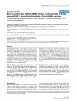
Follow-up of sheep S2531: silencing occurssimultaneously with the onset of leukemiaFigure 1
Follow-up of sheep S2531: silencing occurssimultaneously with the onset of leukemia. (A) Blood samples were col-
lected from S2531 at regular time intervals over a 18-month period from the time of inoculation to the leukemic stage and
examined for several parameters. WBC counts per mm
3
are indicated. Provirus load and integration were examined by South-
ern blot hybridization of SacI- and EcoRI-digests respectively, showing increasing provirus load and the progression from poly-
clonal to monoclonal integration as leukemia develops. The nucleotide sequence of the 3' end of the proviral tax DNA is
illustrated by a polyacrylamide gel autoradiography of dideoxynucleotide sequenced PCR-amplified DNA. Boxes highlight
nucleotides at positions 8149, 8150 and 8151 of the BLV sequence [29]. Arrows indicate the nucleotide identified at position
8149: a G at pre-leukemic stages (yellow arrow); a G to A transition at the time of the first documented WBC increase (17-
month post-infection, red arrow). The resulting amino acid at position 303 of the corresponding Tax proteins is shown below.
The transactivation potential of the putative S2531 proviral Tax proteins were examined in a luciferase reporter assay follow-
ing co-transfection of HeLa cells with the pSGTax
2531
expression vectors containing tax sequences cloned from S2531 PBMCs
collected at different times post-infection and the reporter plasmid pLTR-Luc as detailed in B. "+" indicates a luciferase activity
equivalent to that resulting from transfection with the wild-type pSGTax; "-" indicates the background level activity similar to
that obtained when the empty expression vector pSG5 is co-transfected with pLTR-Luc. (B) Luciferase assay reflecting the
transactivation potential of a selection of four S2531-derived tax sequences. Each pSGTax
2531
construct containing the different
S2531-derived tax sequences downstream of the CMV promoter was used in HeLa co-transfection with pLTR-Luc which
expresses the firefly luciferase under the control of the BLV-LTR promoter. Luciferase activities were measured in cell lysates
42 h posttransfection and were normalized to protein concentrations as previously described [19]. Results are represented as
histograms indicating basal luciferase activities (arbitrary units). pSGTax
2531–6
and pSGTax
2531–14
contain sequences amplified
from PBMCs isolated during the aleukemic stage, 6 and 14 months post-inoculation respectively; pSGTax
2531–18
contains tax
sequences from leukemic PBMC isolated 18 months post-inoculation, and the pSGTax
2531-tum
construct resulted from the
insertion of lymphoma-derived tax sequences collected 18 months post-infection. pSGc is the empty control vector. Values
represent the means of the results of triplicate samples. The results from a representative experiment of four independent
experiments are shown.
Retrovirology 2007, 4:51 />Page 5 of 9
(page number not for citation purposes)
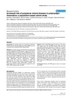
Sheep S267: non-transformed blood-derived B-cells carry a potentially active provirus while virus and Tax expression are com-pletely suppressed in the the co-existing malignant lymphoma B-cellsFigure 2
Sheep S267: non-transformed blood-derived B-cells carry a potentially active provirus while virus and Tax
expression are completely suppressed in the the co-existing malignant lymphoma B-cells. (A) Diagram of the BLV
L267 provirus and major transcripts. The two LTRs and the gag, pro, pol, env, tax, and rex genes are represented. Vertical
arrows indicate restriction sites in the L267 provirus: S, SacI; E, EcoRI. The position and direction of the PCR primers are indi-
cated on the provirus map. The horizontal bar indicates the 8.4 kb-long region that was used as probe. Double lines represent
the sequenced regions. The genomic, env, and tax/rex transcripts are represented below. Alternatively spliced RNAs are not
shown. The translation products of the singly- and doubly-spliced transcripts and the positions of the RT-PCR primers are indi-
cated. (B) Southern blot analysis following hybridization with a full-length BLV probe of SacI-digested DNA isolated from blood
(BL267) and lymphoma (L267-1, -2 and -3) cells collected from S267 twenty nine months post-infection. SacI is indicative of the
proviral load (upper row). Southern blot analysis of EcoRI-digested DNA indicates the presence of a single monoclonally-inte-
grated provirus for all three lymphoma (L267) whereas the blood-derived BL267 cells display a polyclonal integration pattern
(middle and lower panels). EcoRI-cleaved DNA generates two virus-host junction fragments for each integrated L267 provirus
as illustrated in the diagram. Shown here in each lane are the fragments containing the 5' flanking genomic region. (C) Southern
blot analysis of EcoRI-digested DNA isolated from the lymphoma (L267-1, -2, -3) and the cell lines derived from each of these
lymphoma (CL267-1, -2, -3) cultured for four weeks. (D) RT-PCR analysis of RNA isolated from lymphoma-derived cell lines
(CL267), 24 h-cultured blood-derived lymphocytes (BL267-24 h), fresh lymphoma (L267) and freshly isolated blood-derived
lymphocytes (BL267). EnvA/Tax2 primers for the detection of the doubly-spliced tax/rex RNA were used. In the controls YR2
and YR2
LTaxSN
, provirus is silent and active respectively. (E) PCR analysis using BLV tax-specific primer pair Tax1/Tax2 of DNA
isolated from sheep inoculated with the various S267-isolated B-cell populations: six sheep were inoculated using either cul-
tured (CL267) or fresh (L267) transformed B-cells, two sheep were injected with nontransformed PBMCs (BL267).
BL267
L267-1
L267-2
L267-3
CL267-1
CL267-2
CL267-3
Sheep injected with:
PCR
BL267
L267-1
L267-2
L267-3
BL267-
24h
CL267-1
CL267-2
CL267-3
YR2
YR2
LTaxSN
L267-1
L267-2
L267-3
CL267-1
CL267-2
CL267-3
integration
RT-PCR
BL267
L267-1
L267-2
L267-3
viral load
integration
BL267
L267-1
L267-2
L267-3
BL267
L267-1
25
integration
10 DNA input (µg)
A.
C.
D.
E.
Tax
1
tax
LTR LTR
genomic RNA
env
tax/rex
TAX
S S S S
S
Tax
2
U
3
AAA
AAA
AAA
full-length BLV probe
Env
A
Tax
2
E
REX
ENV
rexenv
pol
gag
pro
seq seqseq
U
3
B.
Retrovirology 2007, 4:51 />Page 6 of 9
(page number not for citation purposes)
lated L267 lymphoma or the established CL267 trans-
formed B-cell lines, whereas the blood-derived BL267
cells exhibited BLV-specific transcription (Fig. 2D).
Importantly, the in vivo injection of naïve sheep with
either fresh L267 lymphoma cells or lymphoma-derived
CL267 cell lines did not result in productive infection,
whereas injection of freshly-isolated BL267 cells, the
blood-derived non-leukemic population, readily induced
seroconversion to BLV-p24 as well as a detectable virus
(Fig. 2E). Thus, while there is potentially-active provirus
in the non-transformed blood-derived B-cells, provirus
expression is silenced in the tumor B-cells as demon-
strated by its incapacity to generate infection in vivo. Direct
sequencing of selected regions of both the lymphoma-
and blood-derived S267 proviruses including tax, the pol/
env region required for tax/rex transcript expression as well
as the complete 5'LTR (Fig. 2A) indicated identical
sequences matching the injected wild-type proviral DNA
[9,20-23]. Although it is possible that mutations in other
regions might contribute to proviral extinction, our data
suggest that tumor-associated silencing in S267 results
from molecular mechanisms that are not linked to genetic
changes. Interestingly, a sheep that had been infected with
BL267 cells developed leukemia 25 month post-inocula-
tion, characterized by 166,000 WBC/mm3 and a distinct
provirus integration pattern as compared to that found in
L267. Again, in the malignant clone of this animal, the
BLV provirus was silent. A summary of these data is illus-
trated in Table 1. Overall, our observations in S267 rein-
force the hypothesis that virus silencing plays a pivotal
role in the establishment of a fully-transformed pheno-
type. Furthermore, these findings suggest that besides
genetic changes, epigenetic mechanisms such as DNA
methylation and chromatin modifications might be
involved in tumor-associated virus latency.
Discussion
Using the BLV-associated ovine model of leukemia and
based on the observations in two experimental sheep, we
provide evidence for the role of virus and oncogene silenc-
ing as an important step in the onset of lymphoid malig-
nancy. In the first animal, S2531, we identified a
correlation between the genetic modification of the provi-
ral structure and the emergence of leukemia. We found a
Tax-mutated (Tax
K303
) replication-deficient provirus inte-
grated into the genome of the malignant B-cell clone
while recombinant functional provirus (Tax
E303
) had
been consistently monitored over the aleukemic period.
Although sequencing of individual tax clones identified
the presence of a replication-deficient proviral form in the
inoculum, our data provide no clues as to how this provi-
rus might persist in the infected host. It will be important
to sort out from our future studies whether the Tax
K303
defective provirus found at the time of leukemia develop-
ment in S2531 was already present in the pre-tumoral
clone early after infection. A study is ongoing to answer
this question, based on a BLV-specific inverse PCR tech-
nique for the detection of tumor-specific integration sites
developed by Moules et al. [24]. Using this method, BLV-
positive pre-malignant clones are detectable as early as
two weeks after virus exposure. Whatever the mechanism
responsible for this genetic modification, our observa-
tions suggest that switching off expression of Tax, the
essential contributor to the oncogenic potential of BLV, is
linked with the onset of acute leukemia. We propose that
in this particular case, the mechanism by which the
immune system destroys developing malignancies is
evaded by the malignant cell by reducing its intrinsic
immunogenicity, possibly through recombination-medi-
ated virus silencing. In the second case, S267, both non-
transformed and transformed BLV-infected cells were
present at the same time, but at clearly distinct sites. While
there was potentially-active provirus in the non-leukemic
blood B-cell population, as demonstrated by ex-vivo cul-
ture and injection into naïve recipients, virus expression
was completely suppressed in the malignant B-cells iso-
lated from the lymphoid tumors despite the absence of
genetic alterations in the proviral genome. This independ-
ent observation reinforces our previous conclusion and
suggests that besides genetic alterations, epigenetic mech-
anisms might be involved in tumor-associated silencing.
Altogether, our findings strongly support the hypothesis
that switching-off viral gene expression, including Tax, the
essential contributor to the oncogenic potential of BLV, is
critical, if not mandatory, for progression to overt malig-
nancy.
Sheep infected by BLV mount a strong immune response
to viral antigens. Active killing of infected cells might play
a decisive role in limiting BLV gene expression, but seems
unable to prevent – or perhaps paradoxically favors – the
development of a malignant clone harboring a silent pro-
virus. It is tempting to assign our observations to the fail-
ure of the immune system to eliminate the infected cell
given the absence of proper expression of immunogenic
proteins, in this case Tax. Tax is the major target of CTLs
in HTLV-associated disease [25], and we found significant
levels of Tax-specific CTLs in BLV-infected sheep (Van den
Broeke, unpublished results). The lack of immunogenicity
Table 1: Characterization of PBMC- and lymphoma-derived B-
cells isolated from sheep S267
Cells isolated from: Blood Lymphoma
provirus integration polyclonal monoclonal
cytokine-independent growth/capacity to
derive cell lines
-+
viral expression + -
provirus sequence wild-type wild-type
in vivo infectious potential + -
Retrovirology 2007, 4:51 />Page 7 of 9
(page number not for citation purposes)
of naturally occurring tumors is often understood in terms
of a suboptimal condition in the tumor microenviron-
ment to generate protective immunity, regulatory T-cell
activity, dendritic cell dysfunction, production of suppres-
sive factors such as IL-10, or changes in the pattern of anti-
gen expression [1,3,26], but so far there was no example
of complete suppression of tumor antigen expression,
especially if this antigen is the major transforming pro-
tein.
The demonstration in S2531 of a link between the inter-
ruption of the long clinical latency and the complete sup-
pression of viral expression suggests that silencing is a late
event in the multi-step process leading to the uncon-
trolled growth of a transformed B-cell clone and the onset
of the fatal acute stage of the disease. Early after infection,
cells that do not express viral proteins might have a sur-
vival advantage because they escape CTLs, but such cells
will not outgrow the cells that express virus because of the
absence of functional Tax protein capable of transactivat-
ing the host cell pathways responsible for enhanced B-cell
proliferation. However, if virus silencing occurs when the
cell has undergone sufficient events to reach a point of no
return, impairment of immune surveillance might allow
the uncontrolled proliferation of this fully-transformed B-
cell clone. Whatever the mechanism – genetic or epige-
netic – it is critical for achieving complete silencing of all
viral genes. Cellular changes that have occurred during the
process of leukemogenesis are such that even the Tax
oncoprotein can be turned off without reversing the trans-
formed phenotype. Loss of Tax and virus expression has
been extensively documented in HTLV-1-associated dis-
ease and both genetic and epigenetic silencing mecha-
nisms have been described [13,27,28]. This study in sheep
contributes to the further understanding of tumor-associ-
ated silencing. In particular, the analysis of sequential
samples of the same individual from pre-tumoral to overt
leukemia and the documentation of the timing of the Tax
expression reduction are unique. Our findings are in
strong contrast with observations in other viral-associated
malignancies including HPV-, EBV-, and HBV-associated
cancers, as well as tumors mediated by simple oncornavi-
ruses that all require sustained oncogene or transforming
gene expression. This observation also raises a major con-
cern for the application of effective anti-tumor immuno-
therapy. CTLs to the oncogenic protein might be effective
when elicited during the chronic pre-leukemic stage, but
would be irrelevant for eliminating malignant cells that
do not longer express the initially-immunogenic target
antigen after tumor progression.
Methods
Animals and animal samples
All sheep were housed at the Centre de Recherches Vétéri-
naires et Agrochimiques (Brussels, Belgium). Experimen-
tal procedures were approved by the Comité d'Ethique
Médicale de la Faculté de Médecine ULB and were con-
ducted in accordance with national and institutional
guidelines for animal care and use. S2531 was inoculated
intradermally with 10
7
PBMCs isolated from a BLV-
infected animal (S19) described earlier [8]. S267 was
injected with naked proviral DNA of an infectious BLV
variant (pBLVX3C) [9], isogenic to the full-length wild-
type 344 provirus used for in vivo infection of sheep [9,20-
23]. Blood was collected in EDTA-containing tubes and
PBMCs were isolated using standard Ficoll-Hypaque sep-
aration. S267 lymphoid tumors were collected at
necropsy, minced through a nylon mesh cell strainer (Bec-
ton-Dickinson) to obtain single-cell suspensions. Sheep
used for injection with S267-derived cell populations
were inoculated with 2 × 10
7
BL267, L267, or CL267
respectively. Anti-p24 antibody titers and viral load were
determined as previously described [8].
Cell cultures
PBMCs and single cell suspensions isolated from BLV-
infected sheep were cultured at a concentration of 10
6
cells/ml in OPTIMEM medium (Invitrogen) supple-
mented with 10% FCS, 1 mM sodium pyruvate, 2 mM
glutamine, non-essential amino acids and 100 µg/ml kan-
amycin as previously described [8].
Southern blot, PCR, RT-PCR and sequence analysis
Genomic DNA was prepared and analyzed by Southern
blot and PCR analysis as previously described [8]. The
nylon-bound Sac I or EcoRI-digested genomic DNAs were
hybridized with a
32
P-labeled BLV full-length proviral
DNA probe (Fig. 2A). Primers for PCR were as follow
(nucleotide positions according to Sagata [29]: Tax1
[7321–7340]: 5'-GATGCCTGGTGCCCCCTCTG-3', Tax2
[7604–7623]: 5'-ACCGTCGCTAGAGGCCGAGG-3', U3
[8599–8618]:5'-GCCAGACGCCCTTGGAGCGC-3'. Tax1-
Tax2 and Tax1-U3 were paired together for proviral DNA
detection and sequencing respectively. For RT-PCR exper-
iments, total RNA was extracted using the Tripure reagent
according to the manufacturer's protocol (Roche). 1 µg of
RNA was reverse transcribed and amplified using the Titan
RT-PCR system according to the protocol supplied by the
manufacturer (Roche). Primers EnvA [4766–4787]: 5'-
TCCTGGCTACTAACCCCCCCGT-3', and Tax2 were used
for the detection of the 2.1 kb doubly-spliced tax/rex
mRNA as previously described [8], generating a fragment
of 482 bp (Fig. 2A). For provirus sequencing, amplifica-
tion of selected regions was performed using the Pfu
proofreading DNA polymerase (Stratagene) and the puri-
fied products were sequenced using the Thermosequenase
radiolabeled terminator cycle sequencing method (GE
Healthcare Biosciences).
Retrovirology 2007, 4:51 />Page 8 of 9
(page number not for citation purposes)
Constructs and luciferase assays
DNA extracted from PBMCs isolated from S2531 at differ-
ent times post-infection was amplified using primers
Tax1/U3. Eco RI-restricted products were inserted into
pSGTax [30] for exchange with the wild-type sequence.
Each pSGTax
2531
construct was used in HeLa co-transfec-
tion with pLTR-Luc, and luciferase activities were meas-
ured as described [19]. pSGTax contains the wild-type tax
downstream of the CMV promoter; pLTR-Luc expresses
the firefly luciferase under the control of the BLV-LTR pro-
moter.
Proviral DNA from S2531 leukemic cells was cloned by
insertion of EcoRI-restricted genomic DNA into the
Lambda Dash
®
II vector (Stratagene) according to the
manufacturer and used to evaluate the infectious poten-
tial in sheep.
Abbreviations
ATL: Adult T-cell Leukemia; B-CLL: B-cell Chronic Lym-
phocytic Leukemia; BLV: Bovine Leukemia Virus; EBV:
Epstein-Bar Virus; HBV: Hepatitis-B Virus; HPV: Human
Papilloma Virus; HTLV-1: Human T-lymphotropic Virus-
1; MoMuLV: Moloney Murine Leukemia Virus; PBMCs:
Peripheral Blood Mononuclear Cells; STLV: Simian T-lym-
photropic Virus; WBC: White Blood Cell.
Competing interests
The author(s) declare that they have no competing inter-
ests.
Authors' contributions
MM and PK set up the experiments, carried out most of
the experimental work, and participated to the writing of
the manuscript, MS participated in the transfection and
luciferase assays, YC performed the cloning and sequenc-
ing experiments, PK was responsible for the follow-up of
the animals, CB participated in the experimental design
and analysis of retroviral vector-associated recombination
events, AB and PM helped with the interpretation of the
results and corrected the manuscript, AVDB was the prin-
cipal designer of the study, coordinated its realization and
the writing of the manuscript. All authors read and
approved the final manuscript.
Acknowledgements
This work was supported by the Fonds National de la Recherche Scienti-
fique (F.N.R.S.), the Medic Foundation, the International Brachet Founda-
tion, the Fondation Bekales, les Amis de l'Institut Bordet (Y.C.), and Télévie
Grants to M.M. and M.S.
We thank Jean-Marie Londes for skilful help with the animals.
References
1. Kim R, Emi M, Tanabe K, Arihiro K: Tumor-driven evolution of
immunosuppressive networks during malignant progres-
sion. Cancer Res 2006, 66:5527-5536.
2. Marincola FM, Jaffee EM, Hicklin DJ, Ferrone S: Escape of human
solid tumors from T-cell recognition: molecular mechanisms
and functional significance. Adv Immunol 2000, 74:181-273.
3. Pinzon-Charry A, Maxwell T, Lopez JA: Dendritic cell dysfunction
in cancer: a mechanism for immunosuppression. Immunol Cell
Biol 2005, 83:451-461.
4. Hein WR, Griebel PJ: A road less travelled: large animal models
in immunological research. Nat Rev Immunol 2003, 3:79-84.
5. Burny A Willems,L.,Callebaut,I.,Adam,E.,Cludts,I, Dequiedt,F.,Droog-
mans,L.,Grimonpont,C.,Kerkhofs,P.,Mammerickx,M.,Porte-
telle,D.,Van den Broeke,A.,and Kettman,R.: Bovine Leukemia
Virus: biology and mode of transformation. In: Viruses and Can-
cer Minson, A C , Neil, J C and McRae, M A (eds), Cambridge University
Press, Cambridge 1994:313-334.
6. Willems L, Burny A, Collete D, Dangoisse O, Dequiedt F, Gatot JS,
Kerkhofs P, Lefebvre L, Merezak C, Peremans T, Portetelle D, Twiz-
ere JC, Kettmann R: Genetic determinants of bovine leukemia
virus pathogenesis. AIDS Res Hum Retroviruses 2000,
16:1787-1795.
7. Gillet N, Florins A, Boxus M, Burteau C, Nigro A, Vandermeers F,
Balon H, Bouzar AB, Defoiche J, Burny A, Reichert M, Kettmann R,
Willems L: Mechanisms of leukemogenesis induced by bovine
leukemia virus: prospects for novel anti-retroviral therapies
in human. Retrovirology 2007, 4:18.
8. Van den Broeke A, Bagnis C, Ciesiolka M, Cleuter Y, Gelderblom H,
Kerkhofs P, Griebel P, Mannoni P, Burny A: In vivo rescue of a
silent tax-deficient bovine leukemia virus from a tumor-
derived ovine B-cell line by recombination with a retrovirally
transduced wild-type tax gene. J Virol 1999, 73:1054-1065.
9. Willems L, Kettmann R, Dequiedt F, Portetelle D, Voneche V, Cornil
I, Kerkhofs P, Burny A, Mammerickx M: In vivo infection of sheep
by bovine leukemia virus mutants. J Virol 1993, 67:4078-4085.
10. Klener P, Szynal M, Cleuter Y, Merimi M, Duvillier H, Lallemand F,
Bagnis C, Griebel P, Sotiriou C, Burny A, Martiat P, Van Den BA:
Insights into gene expression changes impacting B-cell trans-
formation: cross-species microarray analysis of bovine leuke-
mia virus tax-responsive genes in ovine B cells. J Virol 2006,
80:1922-1938.
11. Ng PW, Iha H, Iwanaga Y, Bittner M, Chen Y, Jiang Y, Gooden G,
Trent JM, Meltzer P, Jeang KT, Zeichner SL: Genome-wide expres-
sion changes induced by HTLV-1 Tax: evidence for MLK-3
mixed lineage kinase involvement in Tax-mediated NF-kap-
paB activation. Oncogene 2001, 20:4484-4496.
12. Szynal M, Cleuter Y, Beskorwayne T, Bagnis C, Van LC, Kerkhofs P,
Burny A, Martiat P, Griebel P, Van Den BA: Disruption of B-cell
homeostatic control mediated by the BLV-Tax oncoprotein:
association with the upregulation of Bcl-2 and signaling
through NF-kappaB. Oncogene 2003, 22:4531-4542.
13. Matsuoka M, Jeang KT: Human T-cell leukaemia virus type 1
(HTLV-1) infectivity and cellular transformation. Nat Rev Can-
cer 2007, 7:270-280.
14. Takeda S, Maeda M, Morikawa S, Taniguchi Y, Yasunaga J, Nosaka K,
Tanaka Y, Matsuoka M: Genetic and epigenetic inactivation of
tax gene in adult T-cell leukemia cells. Int J Cancer 2004,
109:559-567.
15. Van den Broeke A, Cleuter Y, Chen G, Portetelle D, Mammerickx M,
Zagury D, Fouchard M, Coulombel L, Kettmann R, Burny A: Even
transcriptionally competent proviruses are silent in bovine
leukemia virus-induced sheep tumor cells. Proc Natl Acad Sci U
S A 1988, 85:9263-9267.
16. Hanon E, Asquith RE, Taylor GP, Tanaka Y, Weber JN, Bangham CR:
High frequency of viral protein expression in human T cell
lymphotropic virus type 1-infected peripheral blood mono-
nuclear cells. AIDS Res Hum Retroviruses 2000, 16:1711-1715.
17. Powers MA, Radke K: Activation of bovine leukemia virus tran-
scription in lymphocytes from infected sheep: rapid transi-
tion through early to late gene expression. J Virol 1992,
66:4769-4777.
18. Van den Broeke A Cleuter,Y.,Droogmans,L.,Burny,A.and Kettman,R.:
Isolation and culture of B lymphoblastoid cell lines from
Bovine Leukemia Virus-induced tumors. In:"Immunology meth-
ods manual", In vitro experimental immunology in sheep, Yvan Lefkovits
(ed), Academic Press 1997:2127-2132.
19. Calomme C, Dekoninck A, Nizet S, Adam E, Nguyen TL, Van Den BA,
Willems L, Kettmann R, Burny A, Van LC: Overlapping CRE and E
box motifs in the enhancer sequences of the bovine leukemia
Publish with BioMed Central and every
scientist can read your work free of charge
"BioMed Central will be the most significant development for
disseminating the results of biomedical research in our lifetime."
Sir Paul Nurse, Cancer Research UK
Your research papers will be:
available free of charge to the entire biomedical community
peer reviewed and published immediately upon acceptance
cited in PubMed and archived on PubMed Central
yours — you keep the copyright
Submit your manuscript here:
/>BioMedcentral
Retrovirology 2007, 4:51 />Page 9 of 9
(page number not for citation purposes)
virus 5' long terminal repeat are critical for basal and
acetylation-dependent transcriptional activity of the viral
promoter: implications for viral latency. J Virol 2004,
78:13848-13864.
20. Rice NR, Stephens RM, Burny A, Gilden RV: The gag and pol genes
of bovine leukemia virus: nucleotide sequence and analysis.
Virology 1985, 142:357-377.
21. Rice NR, Stephens RM, Couez D, Deschamps J, Kettmann R, Burny
A, Gilden RV: The nucleotide sequence of the env gene and
post-env region of bovine leukemia virus. Virology 1984,
138:82-93.
22. Willems L, Portetelle D, Kerkhofs P, Chen G, Burny A, Mammerickx
M, Kettmann R: In vivo transfection of bovine leukemia provi-
rus into sheep. Virology 1992, 189:775-777.
23. Willems L, Thienpont E, Kerkhofs P, Burny A, Mammerickx M, Kett-
mann R: Bovine leukemia virus, an animal model for the study
of intrastrain variability. J Virol 1993, 67:1086-1089.
24. Moules V, Pomier C, Sibon D, Gabet AS, Reichert M, Kerkhofs P, Wil-
lems L, Mortreux F, Wattel E: Fate of premalignant clones dur-
ing the asymptomatic phase preceding lymphoid
malignancy. Cancer Res 2005, 65:1234-1243.
25. Bangham CR, Osame M: Cellular immune response to HTLV-1.
Oncogene 2005, 24:6035-6046.
26. Khazaie K, von BH: The impact of CD4+CD25+ Treg on tumor
specific CD8+ T cell cytotoxicity and cancer. Semin Cancer Biol
2006, 16:124-136.
27. Taniguchi Y, Nosaka K, Yasunaga J, Maeda M, Mueller N, Okayama A,
Matsuoka M: Silencing of human T-cell leukemia virus type I
gene transcription by epigenetic mechanisms. Retrovirology
2005, 2:64.
28. Kamoi K, Yamamoto K, Misawa A, Miyake A, Ishida T, Tanaka Y,
Mochizuki M, Watanabe T: SUV39H1 interacts with HTLV-1
Tax and abrogates Tax transactivation of HTLV-1 LTR. Ret-
rovirology 2006, 3:5.
29. Sagata N, Yasunaga T, Tsuzuku-Kawamura J, Ohishi K, Ogawa Y,
Ikawa Y: Complete nucleotide sequence of the genome of
bovine leukemia virus: its evolutionary relationship to other
retroviruses. Proc Natl Acad Sci U S A 1985, 82:677-681.
30. Willems L, Heremans H, Chen G, Portetelle D, Billiau A, Burny A,
Kettmann R: Cooperation between bovine leukaemia virus
transactivator protein and Ha-ras oncogene product in cel-
lular transformation. EMBO J 1990, 9:1577-1581.