Báo cáo y học: " HIV-1 sequence evolution in vivo after superinfection with three viral strains" ppt
Bạn đang xem bản rút gọn của tài liệu. Xem và tải ngay bản đầy đủ của tài liệu tại đây (526.41 KB, 14 trang )
BioMed Central
Page 1 of 14
(page number not for citation purposes)
Retrovirology
Open Access
Research
HIV-1 sequence evolution in vivo after superinfection with three
viral strains
Karolina Kozaczynska
1
, Marion Cornelissen
1
, Peter Reiss
2
, Fokla Zorgdrager
1
and Antoinette C van der Kuyl*
1
Address:
1
Laboratory of Experimental Virology, Department of Medical Microbiology, Centre for Infection and Immunity Amsterdam (CINIMA),
Academic Medical Centre of the University of Amsterdam, Meibergdreef 15, 1105 AZ Amsterdam, The Netherlands and
2
Department of Internal
Medicine, Division of Infectious Diseases, Tropical Medicine and AIDS, Academic Medical Centre of the University of Amsterdam, Meibergdreef
15, 1105 AZ Amsterdam, The Netherlands
Email: Karolina Kozaczynska - ; Marion Cornelissen - ; Peter Reiss - ;
Fokla Zorgdrager - ; Antoinette C van der Kuyl* -
* Corresponding author
Abstract
With millions of people infected worldwide, the evolution of HIV-1 in vivo has been the subject of
much research. Although recombinant viruses were detected early in the epidemic, evidence that
HIV-1 dual infections really occurred came much later. Dual infected patients, consisting of
coinfected (second infection before seroconversion) and superinfected (second infection after
seroconversion) individuals, opened up a new area of HIV-1 evolution studies. Here, we describe
the in-depth analysis of HIV-1 over time in a patient twice superinfected with HIV-1, first with a
subtype B (B2) strain and then with CRF01_AE after initial infection with a subtype B (B1) strain.
The nucleotide evolution of gag and env-V3 of the three strains followed a similar pattern: a very
low substitution rate in the first 2–3 years of infection, with an increase in synonymous
substitutions thereafter. Convergent evolution at the protein level was rare: only a single amino
acid in a gag p24 epitope showed convergence in the subtype B strains. Reversal of CTL-epitope
mutations were also rare, and did not converge. Recombinant viruses were observed between the
two subtype B strains. Luciferase-assays suggested that the CRF01_AE long terminal repeat (LTR)
constituted the strongest promoter, but this was not reflected in the plasma viral load. Specific real-
time PCR assays based upon the env gene showed that strain B2 and CRF01_AE RNA was present
in equal amounts, while levels of strain B1 were 100-fold lower.
All three strains were detected in seminal plasma, suggesting that simultaneous transmission is
possible.
Background
The overall rate of evolution of human immunodeficiency
virus type 1(HIV-1) is the highest documented for viruses
to date. Several mechanisms contribute to this phenome-
non, amongst them the high error rate of the viral reverse
transcriptase (RT), which lacks an 3'→5'exonuclease
proofreading capacity, the short generation time, and the
high rate of recombination between viral genomes.
Recombination is facilitated by the average presence of
three to four proviral genomes in the infected cell [1],
Published: 23 August 2007
Retrovirology 2007, 4:59 doi:10.1186/1742-4690-4-59
Received: 22 June 2007
Accepted: 23 August 2007
This article is available from: />© 2007 Kozaczynska et al; licensee BioMed Central Ltd.
This is an Open Access article distributed under the terms of the Creative Commons Attribution License ( />),
which permits unrestricted use, distribution, and reproduction in any medium, provided the original work is properly cited.
Retrovirology 2007, 4:59 />Page 2 of 14
(page number not for citation purposes)
combined with the template-switching ability of the viral
RT [2]. Recombinant genomes are most easily spotted
when different subtypes of HIV-1 are involved, but as
recombination is typical in HIV replication, recombinant
viruses are present in any infected persons. The rate of evo-
lution, e.g. the rate of nucleotide substitution and recom-
bination, of HIV-1 as governed by the viral RT is supposed
to be more or less constant. However, selection factors,
such as host immune pressure and the use of antiviral
drugs influence the viral quasi-species so that there can be
rapid outgrowth of only a limited number of viral
genomes. The outcome of these evolution and selection
processes is such that viruses at the end of the infection
(AIDS stage) are clearly related, but distinct from the
quasi-species that was present during the acute infection
and from the viruses seen during the chronic phase of the
infection. HIV-1 variation over time has been studied
extensively in patients infected with single strains (e.g. see
[3,4]). It has been suggested that HIV-1 evolution follows
a similar pattern in most patients, whereby a period of lin-
ear increase in divergence and diversity is replaced by a
stabilization of diversity, and finally by an evolutionary
slowdown late in infection, accompanied by the appear-
ance of CXCR4 using viruses [3,4]. Due to the availability
of effective anti-viral treatment, the later stages of viral
evolution are nowadays more difficult to study in vivo.
Studies on HIV-1 evolution, mainly focussing on recom-
bination events, in dually infected patients [5-11] and in
patients coinfected with three HIV-1 strains [12,13] have
also been performed. However, most studies suffer from a
lack of samples (insufficient follow-up), and/or of a pre-
cise timing of the infections. Therefore, a more detailed
description of how different HIV-1 strains present in the
same host influence each other, except for the occurrence
of recombination, is not available yet. We described ear-
lier a Dutch patient who was twice superinfected with
HIV-1 at identified time points; once with a subtype B
virus, and once with CRF01_AE after initial infection with
a subtype B strain [14]. Here we present an extensive fol-
low-up of the HIV-1 quasi-species in this patient after tri-
ple infection, both in blood and in seminal plasma. The
influence of infection with a second or third strain upon
the evolution of the other strains was investigated in the
gag and env genes, as well as was the frequency of conver-
gent evolution. Biological clones were generated to esti-
mate the occurrence of recombination. Virus production
of the distinct strains in blood and seminal plasma was
measured to see if, and to what extent, replication of the
three strains continues or whether there is outgrowth of a
single virus species. Continuous expression of all three
strains was observed. LTR-luciferase experiments sug-
gested that the CRF01_AE LTR has substantially higher
promoter activity than the LTR's of both subtype B strains
from this patient. This increased promoter activity was not
reflected in plasma viral load differences, where strain B2
and CRF01_AE had similar copy numbers, while the
strain B1 viral load was substantially lower.
Methods
Patient samples and HLA-typing
Patient H01-10366 is infected with three HIV-1 strains
(in, or shortly before 2001 with subtype B (strain B1), in
autumn 2002 with subtype B (strain B2), and in summer
2003 with CRF01_AE [14]). The patient was first demon-
strated to be HIV-1-seropositive in March 2001 at the
Municipal Health Service anonymous testing facility in
Amsterdam, and referred for follow up to the Academic
Medical Centre of the University of Amsterdam. Blood
plasma samples were thereafter obtained at regular hospi-
tal visits of the patient. At a few time-points, PBMC's were
collected using the BD Vacutainer
®
CPT™ system (Becton
Dickinson, Plymouth, UK). Semen samples were collected
at the same visits, and centrifuged for 20 minutes at 600 g
to collect the seminal plasma used in the experiments.
HLA-typing of patient H01-10366 was routinely per-
formed at Sanquin Diagnostiek (Amsterdam, The Nether-
lands) and the following results were obtained: HLA class
I: A3, A32(19), B8, B62(15), and Cw3 (Cw4–8 were not
tested); HLA class II: DRB1*12, DRB1*13, DRB3* posi-
tive, DQB1*03 and DQB1*06.
Plasma viral load
Blood plasma HIV-1 RNA was measured using the VER-
SANT HIV-1 RNA 3.0 assay (bDNA) (Bayer Diagnostics
Division, Tarrytown, NY), which has a detection level of
50 copies/ml. The HIV-1 viral load of the seminal plasma
was determined with an in-house real-time PCR assay,
with primers located in the HIV-1 pol gene. Primer/probe
sequences were: upstream primer 5'TGC ATT YAC CAT-
ACC TAG T 3', downstream primer 5'ATT GCT GGT GAT
CCT TTC CA 3', and probe 5'AAA CAA TGA GAC ACC
AGG GAT TAG ATA 3'. The detection limit of this assay
was 5 HIV-1 RNA copies per reaction.
Viral strain-specific PCR assays
Although the PCR primers used in this study are able to
amplify both HIV-1 subtype B and CRF01_AE, the effi-
ciency with which the strains are detected in a mixed sam-
ple differs. Therefore, three additional strain-specific
nested primer sets located at approximately the same posi-
tions in the env gene were developed to detect the B1, B2
and AE strains more accurately (for primer sequences see
Table 1). Reverse transcriptase (RT) reactions were per-
formed with AMV RT (Roche Applied Science, Indianapo-
lis, IN) and the 3'outer primer.
Viral strain-specific real-time PCR assays
To measure the viral copy number of each of the three
strains independently in a single sample, three additional
Retrovirology 2007, 4:59 />Page 3 of 14
(page number not for citation purposes)
real-time PCR assays were developed. Primers and probes
for the three strains, B1, B2, and CRF01_AE, were located
at approximately the same positions in the V3 region of
the HIV-1 env gene (for primer and probe sequences see
Table 1). No cross-reaction was found between each spe-
cific primer and probe set with the other strains of patient
H01-10366. The detection limits of the assays were 10
HIV-1 RNA copies per reaction.
Generation of biological clones
Freshly phytohemagglutinin (bioTRADING Benelux,
Mijdrecht, The Netherlands), glutamax and interleukin-2
(Proleukine) stimulated peripheral blood mononuclear
cells (PBMC's), obtained from four healthy (HIV-1 nega-
tive) human donors, were combined and cultured in
RPMI 1640 medium (Invitrogen Corporation, Carlsbad,
CA) supplemented with antibiotics, L-glutamine and 15%
heat-inactivated foetal calf serum for 3 days. CD8+ T cells
were depleted after 2 days using the Dynabeads M-450
CD8 kit (Invitrogen Corporation, Carlsbad, CA). Differ-
ent concentrations (10
4
, 2.5 × 10
4
, 4 × 10
4
6 × 10
4
cells/
well) of PBMC's from the HIV-1 infected patient were coc-
ultivated with 1 × 10
6
CD4+ T cells in the same medium
in 96-wells plates for 21 and 28 days, respectively. Each 7
days culture supernatants were tested for the presence of
p24 with an in-house antigen capture enzyme-linked
immunosorbent assay (ELISA). At the same time, to prop-
agate the culture, one-third of the cell culture was trans-
ferred to new 96-wells plates and fresh PHA, Il-2
stimulated CD4+ cells were added. Viruses were consid-
ered to be clonal if less than one-third of the microcul-
tures are positive at a given cell number (Poisson
distribution). HIV-1 clones were expanded and cultured
[15]. After 7 days the clones were harvested. PBMC's and
supernatant were cryopreserved at -150°C [16].
RT-PCR of gag and env
A 804 nucleotide HIV-1 gag gene fragment, encompassing
the complete p17 gene and the first part of p24, and a 264
nucleotide V3 sequence of the HIV-1 envelope gene were
amplified by RT-PCR as described [17,18]. To amplify the
whole of gag-p17 the 5'primers described by Cornelissen
et al [17] were replaced with outer primer 5'GAC GCA
GGA CTC GGC TTG CTG A 3', and nested primer 5'TCC
TTC TAG CCT CCG CTA GTC AA 3' (the original 5'outer
primer). Primers used are able to amplify both subtype B
and CRF01_AE.
PCR amplification of vpr and vpu
The complete vpr and vpu genes of the biological clones
were amplified and completely sequenced as described
[19].
Table 1: Primer and probe sequences
Primers Primer sequence 5'-3'
V3 evolution B1 virus
5'tripleB1_1 GAA AAT TTC ACA GAC AAT GCT 1
st
PCR
3'tripleB1_rt TTA ATT TTG TAA CTA TCA GTT C 1
st
PCR
5'tripleB1_2 TAA TAG TAC AGC TGA ATG CAT Nested PCR
3'tripleB1_3 AGT GTT ATT CCA TTT TGT TAA Nested PCR
V3 evolution B2 virus
5'tripleB2_1 GAC AAT TTC ACA GAC AAT AAG 1
st
PCR
3'tripleB2_rt TTA ATT TTT CAA CTG TCT GAT T 1
st
PCR
5'tripleB2_2 TAA TAG TAC AGC TGA AGA CAG Nested PCR
3'tripleB2_3 AGC ATT ACC CCA TTC TAC TCC Nested PCR
V3 evolution AE virus
5'tripleAE_1 GAA AAT CTC ACA GAT AAT ACC 1
st
PCR
3'tripleAE_rt AGT GCT CTT TTA ATT TTT CAG 1
st
PCR
5'tripleAE_2 CAT AAT AGT GCA CCT TAA TAA Nested PCR
3'tripleAE_3 CCA TTT TGT TCT ATT AAT CTC Nested PCR
Taqman strain specific assay B1
5'B1/B2triple-taqman TTA ATT GTA CAA GAC CCA GCA ACA
3'B1triple-taqman AAG GTT ACA ATG TGC TTG CCT TA
B1triple-probe2rev TCT CCT ATT ATT TCT CCT GTT GCA T 5'label 6-FAM
Taqman strain specific assay B2
5'B1/B2triple-taqman TTA ATT GTA CAA GAC CCA GCA ACA
3'B2triple-taqman ACT AAT GTT ACA ATG TGC CTT T
B2triple-probe TAA AAA ATG CTT TCC CTG GTC CCA TA 5'label 6-FAM
Taqman strain specific assay AE
5'AEtriple-taqman TCA ATT GTA CCA GAC CCT CTA AC
3'AEtriple-taqman TTG TTC TAT TAA TCT CAC AAT A
AEtriple-probe TAT AGA ATA CTT GTC CTG GTC CCA TA 5'label 6-FAM
Retrovirology 2007, 4:59 />Page 4 of 14
(page number not for citation purposes)
Cloning and sequencing
HIV-1 gag and V3 fragments were cloned with the TOPO
TA cloning kit (Invitrogen, Carlsbad, CA, USA), and
sequenced with the BigDye Terminator cycle sequencing
kit (Applied Biosystems, Foster City, CA, USA). Electro-
phoresis and data collection are performed on an ABI
PRISM 3100 genetic analyser (also from Applied Biosys-
tems). The number of clones (n) for each virus strain per
time point varied from n = 4 till n = 54, with an average of
10 clones per virus strain per time point. For 2002 three
consecutive time points were sequenced and pooled in
the analysis.
For the biological clones multiple primer sets were used to
generate overlapping fragments that were directly
sequenced as described above.
Nucleotide distance calculation and phylogenetic analysis
Sequences were aligned with and without reference HIV-1
gag, vpr,vpu and env-V3 sequences [20] using ClustalW
available in BioEdit Sequence Alignment Editor version
7.0.1 [21]. Recombination events between the B1 and B2
strains in gene fragments from the biological clones were
identified from the nucleotide alignments. Nucleotide
distances were estimated with the Tamura-Nei [22] dis-
tance with the gamma model. This model corrects for
multiple hits, and takes into account the different rates of
substitution between nucleotides and the inequality of
nucleotide frequencies. The nucleotide composition of
the HIV genome is quite different from other species,
being A-rich and C-poor. The gamma shape parameter α
for HIV-1 gag (α = 0.25) and env-V3 (α = 0.38) was taken
from Leitner et al. [23].
Neighbour-Joining (NJ) trees based upon Tamura-Nei dis-
tances were constructed with the MEGA 3.1 software pack-
age [24], and 1000 bootstrap replicates were analysed.
Bootstrap values ≥ 80 were considered significant. Addi-
tional phylogenetic analyses were done with the parallel
version of MrBayes 3.1 [25], modified so that the program
now uses the sprng library [26] to generate independent
streams of random numbers. MrBayes3.1 was run at the
SARA High Performance Computing Facilities [27]. Here,
posterior probability values ≥ 0.8 were considered signifi-
cant.
LTR-constructs and luciferase-assays
The LTR region of the viral genome (from the biological
clones or from plasma for subtype AE) was amplified by
nested reverse transcription (RT)-PCR, with primer sets
described earlier [28]. PCR products were cloned into
pCRII-TOPO (Invitrogen Corporation, Carlsbad, CA)
using the BfrI-site. Four clones from each strain; B1, B2,
AE, X and B (LAI), were sequenced as described above.
Subtype X is a novel HIV-1 subtype distantly related to
subtype K, discovered recently in a single patient [29]. For
strain B2, two sizes of LTR fragments were discovered of
which the longer one contained a duplication of 23 nucle-
otides and was named B2_L(ong), while the shorter LTR
was designated B2_S(hort). Sequences were aligned and
transcription factor binding sites were identified with
TFSEARCH [30] and Alibaba 2.1 [28,31,32].
A representative clone for each subtype was selected for
subcloning into pBlue3'LTR, which is a Bluescript KS(+)
plasmid containing a XhoI-BglI LAI 3'LTR fragment. Then,
constructs were digested with BseAI and BfrI and the frag-
ment (position -147 to +63 of the viral genome) was
cloned into pBlue3'LTR-luc as described previously [28].
The cervix carcinoma cell line C33A was used in all luci-
ferase experiments. Cells were grown in 2-cm
2
wells to
60%–70% confluency as described earlier [28,33] and
transfected by the calcium phosphate method [34]. Mix-
tures contained 100 ng of different LTR-luciferase con-
structs (B1, B2_S, B2_L, AE, X and B(LAI)), 0.5 ng of pRL-
CMV plasmid (Promega, Madison, WI) expressing Renilla
luciferase as an internal control for transfection efficiency
[33], and pBluescript in such a concentration that the total
amount of DNA would always be 1000 ng. To test the acti-
vation of the promoters by tat, constructs were titrated
with different concentrations of a tat-expressing plasmid
(pTAT). Cells were cultured for two days and lysed in Pas-
sive Lysis Buffer (Promega, Madison, WI). Firefly and
Renilla luciferase activities were determined with the dual-
luciferase reporter assay (Promega, Madison, WI) as
described previously [33]. The activity of different con-
structs was calculated as the ratio of the firefly and Renilla
luciferase activities, and corrected for between-session var-
iation [35].
Results
Detection of the three viral strains in blood and seminal
plasma
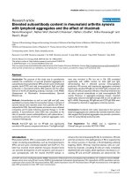
To verify the presence of the three viral strains over time
in both blood and seminal plasma, three specific nested
PCR primer sets were developed in the env-V3 region. Fig.
1 shows the overall viral load in blood and seminal
plasma (panel A) and the detection of strains B1, B2 and
CRF01_ AE in seminal plasma (panel B). In blood plasma,
all three env fragments were detected by PCR amplifica-
tion at all time-points, in line with the relatively high viral
load (result not shown). In seminal plasma, however, the
viral load was much lower and was sometimes even below
the detection limit. In line with this, not all strains could
be detected at every occasion (Fig. 1B), but over the course
of the 1.5 years analysed here the patient was able to trans-
mit any strain at some point. At all time points except one,
at least two strains were simultaneously present. Interest-
Retrovirology 2007, 4:59 />Page 5 of 14
(page number not for citation purposes)
ingly, the env gene of the first infecting virus B1 was
detected the least in seminal plasma.
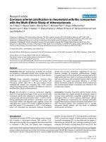
To determine the contribution of each viral strain to the
total blood plasma viral load, three strain-specific real-
time PCR assays were used to amplify a fragment of env-
V3 of strains B1, B2 and CRF01_AE in sequential plasma
samples of patient H01-10366 (Fig. 2). As expected,
sequences of strain B2 were not detected until the B2
superinfection moment, and the CRF01_AE sequences
were similarly not detected until the CRF01_AE superin-
fection moment. At the latter time-point, the very high
plasma viral load was mainly due to the newly infecting
virus CRF01_AE. From the later time points, when three
viruses are present in blood plasma, env-V3 sequences
from strain B2 and CRF01_AE are present in more or less
equal amounts (± 30.000 copies/ml), while strain B1 env-
V3 sequences form a minority (less than 300 copies/ml).
CD4+ cell counts are stable after superinfection with
strain B2, but rapidly decrease after the second superinfec-
tion with CRF01_AE (Fig. 2).
Detection of the three viral strains over timeFigure 1
Detection of the three viral strains over time. (A) HIV-1 viral load in blood and seminal plasma. (B) Strain-specific RT-
PCR detection of HIV-1 subtypes B (strains 1 and 2) and CRF01_AE in seminal plasma.
HIV-1 load plasma versus semen
1.00E+01
1.00E+02
1.00E+03
1.00E+04
1.00E+05
J
a
n
-
0
4
A
p
r
-
0
4
J
u
l
-
0
4
O
c
t
-
0
4
J
a
n
-
0
5
A
p
r
-
0
5
sampling date
viral load copies/mL
plasma
semen
detection level
J
a
n
‘
0
4
B1
B2
AE
M
a
y
’
0
4
F
e
b
’
0
5
J
u
n
’
0
5
N
o
v
‘
0
4
b
l
A
B
Retrovirology 2007, 4:59 />Page 6 of 14
(page number not for citation purposes)
Analysis of biological clones
To assess the occurrence of recombination between the
three strains, a total of 20 biological clones were generated
corresponding to three time points (time point 1 = Janu-
ary 2004, time point 2 = February 2004, time point 3 =
November 2004, approximately 6, 7, and 15 months after
triple infection). Of these 20 clones, three were com-
pletely sequenced, while the structure of the other 17 was
roughly analysed by amplifying and sequencing LTR, gag,
vpr, vpu, and env-V3 fragments (Table 2). Two clones,
2301#12 and 2602#1, of which the former was com-
pletely sequenced, appeared to contain a complete strain
B2 virus, while the other 18 clones were all recombinants
between B1 and B2 virus sequences. No full-length B1 or
CRF01_AE viruses found, nor were any CRF01_AE
sequence fragments detected in the clones. Recombina-
tion between the strain B1/B2 viruses was found at differ-
ent sites; the analysis of 10 clones suggested that
recombination occurred between the gag and the vpr
genes, in one virus recombination occurred in the gag
gene (between p17/p24), and in another virus recombina-
tion was found in the vpr gene. Six clones showed a more
complex pattern of recombination, with multiple crosso-
ver sites being present (Table 2). In general in the recom-
binants, the genome composition was such that the 5' end
of the virus originated from strain B1, while the 3'half of
the viruses corresponded to strain B2, except of course for
the 3'LTR, which belonged to B1 again.
Of the completely sequenced virus clones, the genomic
structure is shown in Fig. 3. Clone 2301#5 was found to
be almost completely composed of strain B1 sequences,
except for a small part in the middle of the genome
encompassing the vif and vpr genes, which originated
from strain B2. Clone 2301#12 contained a complete
strain B2 virus. Clone 2301#14 was a more complex
recombinant virus where recombination did occur once
between the pol and vif genes, and again between the env
and nef genes.
Because no CRF01_AE sequences were found amongst the
biological clones, the presence of CRF01_AE DNA in the
PBMC samples used in the biological cloning procedure
was analysed with PCR primers specific for CRF01_AE.
Indeed, CRF01_AE env-V3 sequences were present in the
preparations (not shown); suggesting the deficiency of the
clones is not explained by the absence of viral DNA.
Evolution of gag: nucleotide distances
Having three distinguishable virus strains in one patient is
a great opportunity to learn whether or not the nucleotide
evolution of a single virus is influenced by the presence of
other virus strains. Therefore we amplified, cloned and
sequenced gag gene fragments from consecutive time
points for the B1, B2 and AE viral strains using generic
PCR-primers, and calculated their overall diversity (=
nucleotide distance) per year in both blood and seminal
plasma. Mean nucleotide distances for the gag gene frag-
ments are summarized in Table 3. From this table it is
clear that nucleotide variation in blood plasma follows a
Table 2: Genomic organization of 20 biological clones
Date Clone LTR Gag Vpr Vpu V3
env
23.01.04 2301#1 B1 B1 B2 B2 B2
2301#2 B1 B1 B2 B2 B2
2301#3 B1 B1 B2 B2 B2
2301#4 B2 B1 B2 B2 B2
2301#5 B1 B1 B2 B1 B1
2301#6 B1 B1 B2 B2 B2
2301#7 B1 B1 B2 B1 B2
2301#8 B1 B1 B2 B2 B2
2301#9 B1 B1 B2 B2 B2
2301#10 B1 B1 B2 B2 B2
2301#11 B1 B1/B2 B2 B2 B2
2301#12 B2 B2 B2 B2 B2
2301#13 B1 B1 B1/B2 B2 B2
2301#14 B1 B1 B2 B2 B2
2301#15 B1 B1 B2 B2 B2
26.02.04 2602#1 B2 B2 B2 B2 B2
2602#2 B1 B1 B2 B1/B2 B1
03.11.04 0311#1 B1 B1 B2 B2 B2
0311#2 B1 B1 B2 B1/B2 B1
0311#3 B1 B1 B2 B2 B1
Viral load of the three HIV-1 strains in blood plasmaFigure 2
Viral load of the three HIV-1 strains in blood plasma.
Real-time PCR analysis with specific primers and probes
located in env-V3 was performed on sequential blood plasma
samples of patient H01-10366. The plasma viral loads meas-
ured by real-time, strain specific PCR is shown, as well as the
overall plasma viral load determined with the VERSANT
HIV-1 RNA 3.0 assay, which is based upon the pol gene.
CD4+ cell counts (× 10
9
) measured at the same time points
are also shown.
1.E+00
1.E+01
1.E+02
1.E+03
1.E+04
1.E+05
1.E+06
1.E+07
01-01 01-02 01-03 01-04 01-05
date
HIV-1 RNA (copies/ml)
0
0.2
0.4
0.6
0.8
1
CD4+ cell counts
Viral lo ad plasma
Viral load B1-ENV
Viral load B2-ENV
Viral load AE-ENV
CD4+ cell counts
detection limit
Retrovirology 2007, 4:59 />Page 7 of 14
(page number not for citation purposes)
similar pattern for viral strains B1 and B2, although strain
B2 has a relatively high amount of synonymous variation
in the year of initial infection (2002). In approximately
the first 2–3 years of infection, overall nucleotide varia-
tion is low for both strains. After this period (in 2004 for
B1, and in 2005 for B2), mean nucleotide differences start
to rise. This rise is almost completely accounted for by an
increase in synonymous substitutions. The amount of
non-synonymous substitutions does not differ signifi-
cantly over the years in both strains. A phylogenetic NJ
tree based upon gag sequences is shown in Fig. 4. From
this tree, the low level of evolution of HIV-1 gag in this
patient is also obvious from the short branch lengths. A
similar phylogenetic tree was obtained with a Bayesian
approach.
Mean nucleotide distances in seminal plasma are lower
than in blood plasma for virus strains B1 and B2. This is
probably correlated with the low HIV-1 copy number in
seminal plasma compared with the blood compartment
(Fig. 1). Phylogenetic analysis of blood plasma and semi-
nal plasma derived HIV-1 gag sequences suggest that there
are no semen specific sequences and that compartmental-
ization does not occur in the seminal compartment (Fig.
4). Seminal plasma gag sequences cluster together with
blood plasma sequences from the corresponding time
points for both strains. This sampling time-related cluster-
ing was also seen for the gag-sequences obtained from the
biological clones (not shown).
Evolution of env-V3: nucleotide distances
Mean nucleotide distances per year of the env-V3 region of
the viral genome of strains B1, B2, and CRF01_AE are
shown in Table 4. Overall nucleotide distances in blood
plasma slowly rise over the years for all three strains. This
rise is mostly accounted for by an increase in synonymous
substitutions, while non-synonymous nucleotide dis-
tances are more or less constant throughout the period
investigated. Mean nucleotide distances were also calcu-
Table 3: Mean nucleotide distances within the gag gene over time in blood and seminal plasma
B1 strain B2 strain Subtype AE
Year Tamura-
Nei
a
Syn
b
Nonsyn
c
Tamura-
Nei
a
Syn
b
Nonsyn
c
Tamura-
Nei
a
Syn
b
Nonsyn
c
2001 blood 0.008 ±
0.001
0.011 ±
0.004
0.006 ±
0.001
2002 blood 0.007 ±
0.001
0.012 ±
0.003
0.005 ±
0.001
0.009 ±
0.002
0.022 ±
0.006
0.005 ±
0.001
2003
d
blood 0.003 ±
0.001
0.002 ±
0.002
0.003 ±
0.001
2004 blood 0.022 ±
0.004
0.071 ±
0.012
0.006 ±
0.002
0.008 ±
0.002
0.008 ±
0.004
0.008 ±
0.002
0.004 ±
0.002
0.006 ±
0.006
0.003 ±
0.002
2005 blood 0.021 ±
0.003
0.051 ±
0.009
0.010 ±
0.002
0.025 ±
0.005
0.060 ±
0.014
0.011 ±
0.003
-
e
2004 semen 0.008 ±
0.002
0.011 ±
0.003
0.006 ±
0.002
0.002 ±
0.001
0.006 ±
0.004
0.001 ±
0.001
-
e
2005 semen 0.007 ±
0.002
0.017 ±
0.005
0.003 ±
0.001
0.008 ±
0.002
0.007 ±
0.004
0.008 ±
0.002
-
e
a
: Tamura-Nei distance with gamma-parameter α = 0.25. Standard errors were estimated by the bootstrap method with 500 bootstrap replicates
each.
b
and
c
: Nei-Gojobori method, p-distance, syn= synonymous, nonsyn= nonsynonymous
d
Nucleotide differences of strains B1 and B2 were not calculated in 2003, because only B1/B2 or B/AE recombinant sequences were retrieved.
e
Nucleotide distances were not calculated for subtype AE in 2005 (blood plasma), 2004 and 2005 (seminal plasma) as no AE fragments were
amplified by the generic primers from these samples.
Genomic organization of three completely sequenced biolog-ical clones from the January 2004 time pointFigure 3
Genomic organization of three completely
sequenced biological clones from the January 2004
time point. Clone 2301#12 contained a complete strain B2
virus, while clones 2301#5 and 2301#14 were strain B1/B2
mosaics. CRF01_AE sequences were not found.
2301#5
2301#12
2301#14
LTR
gag
pol
vif
vpr
LTR
env
nef
vpu
tat
rev
LTR
gag
pol
vif
vpr
LTR
env
nef
vpu
tat
rev
LTR
gag
pol
vif
vpr
LTR
env
nef
vpu
tat
rev
B1 B2AE
Retrovirology 2007, 4:59 />Page 8 of 14
(page number not for citation purposes)
lated for the viral population of strains B1 and B2 ampli-
fied from seminal plasma (Table 4), and were found to be
similar to the blood plasma values, despite the much
lower viral load in seminal plasma. For strain B1, no env-
V3 fragments could be amplified from the 2005 seminal
samples (Fig. 1). Figure 5 shows an NJ tree based upon V3
nucleotide fragments from 2001–2005 from both blood
and seminal plasma. It is obvious from this tree that there
is very little sequence evolution in V3 in this patient, as
indicated by the short branch lengths. Sequences did not
cluster according to year or compartment. Both phyloge-
netic methods (NJ and Bayesian analysis) yielded similar
trees.
Evolution of gag and env: CTL-epitopes
Escape from CTL pressure, or reversion of escape muta-
tions, is one of the main driving forces in HIV evolution
[36-38]. We therefore set out to examine mutations in
CTL epitopes of this triple infected patient, and to investi-
gate whether or not escape (or reversal) occurs in more
than one virus strain. Visual inspection of the translated
gag amino acid alignment suggested only a single site dis-
playing convergent evolution in both subtype B viruses:
amino acid 41 of gag p24 showed a S→T substitution
which was found in none of the early viruses, but in over
90% of the 2005 viruses of both the B1 and B2 strains. The
S→T substitution was not seen in the subtype AE
sequences. Serine-41 belongs to a CTL epitope that is
strongly reactive in ethnic Africans, but has not been asso-
ciated with a specific HLA type [39]. Ser-41 is not one of
the major phosphorylation sites of the HIV CAp24, which
are Ser-109, Ser-149, and Ser-178, thus probably allowing
the substitution observed [40]. However, replacing Ser-41
with Ala-41 delayed replication of the mutated virus in
vitro [40], suggesting that it affects viral fitness. As the
S→T substitution is observed in both strains B1 and B2,
pressure from CTL's directed at this epitope is likely to be
high in this patient. In contrast, the CRF01_AE virus did
not react to this hypothetical immune pressure, and did
not replace Ser-41 over two years of infection. Ser-41 is
also part of a HIV-1 CD4+ T-cell epitope [41,42]. The pep-
tide SPEVIPMFS
ALSE (p24
33–45
, Ser-41 is underlined) was
found to bind to several HLA-DR molecules [42]. This
suggests that CD4+ T cell responses could also be respon-
sible for shaping viral evolution in this patient.
According to the HLA type of our patient, 6 epitopes could
be recognized in the gag and the env fragments obtained.
These epitopes together with the deduced amino acid
sequence of the viral strains are listed in Table 5. The p24
epitope mentioned above for which no associated HLA
type is known, but for which a viral reaction is seen in this
patient, is also included in Table 5. For the other epitope
in p24, all three viruses have a possible escape mutation
already at the earliest time point, and no changes are seen
over time. Two B8 restricted epitopes are apparent in gag
p17. All three viruses have at least one mutation from the
consensus sequence of the epitope, but in two instances a
reversal to a more ancestral state is seen (in B1 by substi-
tuting V→I in EVKDTKEAL, and in B2 by substituting
F→Y in ELKSLFNTV), suggesting that no CTL pressure is
exerted upon these sequences. None of these substitutions
occurs in any other strain. At the first gag p17
aa(18–28)
epitope, restricted by the HLA-A3 allele, mutations are
seen in both the B1 and the B2 strains. This epitope has
been determined to be the most dominant gag CTL-
epitope in Caucasians in vivo [39], also because HLA-A3
has a high phenotypic frequency in Caucasians. However,
mutations in strains B1 and B2 are different both at the
Phylogenetic analysis of HIV-1 gag sequencesFigure 4
Phylogenetic analysis of HIV-1 gag sequences. NJ tree
of HIV-1 gag nucleotide fragments obtained from blood and
seminal plasma collected in 2001–2005. Distances were cal-
culated with the Tamura-Nei method using the gamma
model with α = 0.25, and 1000 bootstrap replicated were
analysed. The three separate clusters comprised of strains
B1, B2, and CRF01_AE are indicated. A representative
sequence set was used to draw the phylogenetic tree.
◊
◊
○
○
□
□
○
∆
○
□
●
●
●
●
□
□
□
□
○
○
□
∆
∆
∆
∅
○
■
■
■
■
○
●
○
■
□
□
○
●
●
∅
∅
○
∅
∅
○
∅
∅
∅
□
100
99
68
0.05
Strain B1
Strain B2
CRF01_AE
HIV-1 Gag
◊ = 2001
∆ = 2002
∅
= 2003
○ = 2004
□ = 2005
Open symbols =
blood plasma
Closed symbols =
seminal plasma
◊
◊
Retrovirology 2007, 4:59 />Page 9 of 14
(page number not for citation purposes)
start of the infection, although they involve the same
amino acid residue, and after a number of years.
CRF01_AE did not show any changes in this epitope, but
had a different sequence from B1 and B2 at the time of
infection (with the derived C-terminal amino acid being a
Q instead of an R (B1), or an S (B2)).
Two HLA-A3 epitopes are predicted in env-V3. All three
HIV-1 strains have mutations in these motifs at the start of
the infection, and no changes over time (from the years
2001 to 2005) are seen in any strain (Table 5).
LTR promoter activity
Promoter activity of the LTR sequence of strains B1, B2
and CRF01_AE from patient H01-10366 was analysed
with a luciferase-assay. Aligned LTR sequences are shown
in Fig. 6A, together with those from controls B(LAI) and
subtype X (chosen because its TAR hairpin is identical to
that of B2_L, Fig. 6B). Fig. 6C shows the transcriptional
activity of the 6 LTR constructs, in the presence of different
concentrations of tat. It is clear that the LTR of subtype AE
has a comparable activity to that of the controls B(LAI)
and subtype X, but that the activity of the three subtype B
constructs of patient H01-10366 is much lower. The B2_L
construct has the lowest activity of all, suggesting that the
23 nt duplication is decreasing promoter activity. This
longer LTR was found in two of the three biological clones
that contained a B2 LTR (2301#12 and 2602#1); the
shorter LTR was only seen once (in clone 2301#4).
Discussion
Having a patient twice superinfected with HIV-1 provides
a unique opportunity to study the evolution of three dis-
tinct HIV strains in a shared in vivo environment. We have
analysed different aspects of the viruses of patient H01-
10366, including the plasma viral load of each strain over
time, the presence of each strain in seminal plasma, the
rate of nucleotide evolution, the occurrence of recombina-
tion, and of possible convergent CTL escape mutations.
Finally, we have analysed the strength of the viral LTR's as
promoter sequences in luciferase-assays.
Interestingly, all three virus stains, two subtype B strains
named B1 and B2 and CRF01_AE, remain detectable in
the plasma until at least two years after the second super-
infection with CRF01_AE in 2003. In blood plasma, the
viral loads of strain B2 and CRF01_AE are comparable,
and approximately 100× higher than that of strain B1, the
first infecting virus. In seminal plasma, the average total
viral load is 100× lower than in blood plasma; at a single
time point HIV-1 is undetectable by PCR. Here, the virus
strains have only been detected qualitatively, but the over-
all picture is similar: the B1 strain is sometimes undetect-
able, suggesting it has a low copy number, while the B2
and AE strains are always detectable (except for the single
negative sample), implying a much higher copy number.
The almost continuous presence of all three viral strains in
seminal plasma implies that this triply infected patient is
able to transmit multiple strains at most time points.
Table 4: Mean nucleotide distances within env-V3 over time in blood and seminal plasma
B1 strain B2 strain Subtype AE
Year Tamura-
Nei
a
Syn
b
Nonsyn
c
Tamura-
Nei
a
Syn
b
Nonsyn
c
Tamura-
Nei
a
Syn
b
Nonsyn
c
2001 blood 0.010 ±
0.002
0.011 ±
0.003
0.008 ±
0.002
2002 blood 0.017 ±
0.004
0.014 ±
0.005
0.012 ±
0.003
0.006 ±
0.002
0.010 ±
0.004
0.004 ±
0.002
2004 blood 0.012 ±
0.004
0.012 ±
0.007
0.010 ±
0.004
0.005 ±
0.003
0.000 ±
0.000
0.006 ±
0.003
0.009 ±
0.002
0.010 ±
0.002
0.008 ±
0.002
2005 blood 0.028 ±
0.009
0.050 ±
0.021
0.015 ±
0.006
0.022 ±
0.006
0.040 ±
0.014
0.014 ±
0.004
0.010 ±
0.003
0.021 ±
0.003
0.005 ±
0.002
2004 semen 0.009 ±
0.004
0.017 ±
0.012
0.006 ±
0.003
0.008 ±
0.003
0.013 ±
0.009
0.005 ±
0.002
-
d
2005 semen -
d
- - 0.010 ±
0.003
0.017 ±
0.010
0.006 ±
0.002
-
d
a
: Tamura-Nei distance with gamma-parameter α = 0.38. Standard errors were estimated by the bootstrap method with 500 bootstrap replicates
each.
b
and
c
Nei-Gojobori distance method, p-distance, syn = synonymous, nonsyn= nonsynonymous.
d
Nucleotide distances were not calculated for strain B1 in 2005 (seminal plasma) and subtype AE in 2004 (seminal plasma) and 2005 (seminal
plasma) as the generic primers did not amplify B1 or AE fragments from these samples, and the products generated with specific primers were too
short to conduct evolution studies.
Retrovirology 2007, 4:59 />Page 10 of 14
(page number not for citation purposes)
The LTR-luciferase assays suggested that the LTR from
CRF01_AE has a much higher activity in vitro than either
subtype B LTR, but this difference is not reflected in the in
vivo viral load in blood plasma. It is possible that the cer-
vix carcinoma cell line used in the in vitro assays does not
reflect the in vivo situation due to differences in the avail-
ability or concentration of transcription factors. Previous
work also showed that the CRF01_AE LTR is much more
potent in vitro than LTR's from subtype B [28]. Early after
seroconversion, patients infected with CRF01_AE also
show a three times higher viral load than those infected
with subtype B, although viral load differences decrease
later on [43]. Possibly, CRF01_AE cannot replicate to its
full extent after early infection due to decreasing levels of
available CD4+ T cells. Interestingly, some strain B2
viruses contained a repeat-like insertion of 23 bp in the
LTR that decreased the in vitro promoter activity, but did
result in viable viruses as it was found amongst the biolog-
ical clones. In the LTR sequence of CRF01_AE, the most
active promoter of the three viruses in the in vitro assays,
three transcription factor binding motifs were different
from the subtype B LTR's. One of the NF-κB sites is
mutated to a GABP site, an SP1 site is mutated into a
CACCC binding motif, and a novel AP1 site overlaps the
RBE III site. However, none of these changes were present
in the subtype B (LAI) and X LTR's, which were similarly
active in vitro.
In this triple HIV-1 infected patient, copy numbers of the
first virus, strain B1, decrease sharply after the second
superinfection, suggesting that the superinfections could
have been facilitated by an initial infection with a less fit
virus. Another explanation for the apparent disappear-
ance of strain B1 can be found in the analysis of the bio-
logical clones generated from samples postdating the
second superinfection. Of the 20 clones examined, 18
were found to be recombinants between the B1 and B2
strains, with 14 clones having a B2 env gene sequence and
only four clones having a B1 env gene sequence. If indeed
B1/B2 recombinant viruses with mainly strain B2 envelope
sequences have by then become the major virus popula-
tion in blood, assays targeting the env gene will underesti-
mate the level of B1 sequences. An assay targeting e.g. the
pol gene might well overestimate strain B1, and give lower
values for strain B2 copy numbers.
No CRF01_AE sequences were detected amongst the bio-
logical clones, neither as full-length viruses nor as recom-
binant viruses. Other experiments showed that CRF01_AE
DNA was present in the patients PBMC's and that
CRF01_AE RNA could be detected at high levels in blood
plasma. If CRF01_AE does not grow in our donor PBMC's
as well as the subtype B strains, more biological clones
should be analysed to optimize the detection of this virus.
On the other hand, biological clones were generated using
techniques that are probably optimized for HIV-1 subtype
B, suggesting that modifications to the protocol are
needed to increase the likelihood of obtaining CRF01_AE
clones. The absence of AE/B recombinant viruses could be
due to the low frequency of recombination between sub-
type B and CRF01_AE. Although multiple subtype B/
CRF01_AE recombinant viruses are circulating in Asia (see
e.g. [44]), the in vitro recombination rate between sub-
type B and CRF01_AE is 9-fold lower than the intrasub-
type recombination rate, mainly due to mismatches in the
dimerization initiation signal (DIS) [45]. For subtype C
and CRF01_AE, which have an identical DIS, the intersub-
type recombination rate was only two-fold lower than the
intrasubtype rate [45]. So, to detect any recombination
Phylogenetic analysis of HIV-1 env-V3 sequencesFigure 5
Phylogenetic analysis of HIV-1 env-V3 sequences. NJ
tree of HIV-1 env-V3 nucleotide fragments obtained from
blood and seminal plasma collected in 2001–2005. Distances
were calculated with the Tamura-Nei method using the
gamma model with α = 0.38, and 1000 bootstrap replicated
were analysed. The three separate clusters comprised of
strains B1, B2, and CRF01_AE are indicated. A representa-
tive sequence set was used to generate the phylogenetic
tree.
HIV-1 Env-V3
◊ = 2001
∆ = 2002
∅ = 2003
○ = 2004
□ = 2005
Open symbols =
blood plasma
Closed symbols =
seminal plasma
∆
○
○
■
∆
■
∆
■
●
●
○
■
■
∆
∆
□
○
□
□
□
■
■
■
□
■
■
□
□
∅
∅
∅
□
□
□
○
◊
◊
◊
∆
◊
◊
○
○
○
∅
○
∅
□
□
∅
○
∆
∅
∅
∅
∅
∅
∆
∅
∅
●
∅
∅
∅
∅
∅
∅
∅
∅
∅
∅
∅
∅
∅
100
51
99
0.05
Strain B2
Strain B1
CRF01_AE
Retrovirology 2007, 4:59 />Page 11 of 14
(page number not for citation purposes)
between subtype B and CRF01_AE in this patient, many
more clones need to be analysed due to its estimated
minor frequency. Unfortunately, biological cloning using
patient H01-10366 PBMC's was very inefficient in our
hands, and sample limitations disabled further efforts.
Nucleotide and deduced amino acid sequences of the gag
and the env gene were also analysed over time in this
patient. Viral strains B1 and B2 followed a more or less
similar trajectory, whereby nucleotide substitutions were
low in the first 2–3 years, after which the synonymous
substitution rate increased. Follow-up for CRF01_AE was
much shorter, but no deviation from the subtype B pat-
tern was evident. The nonsynonymous substitution rate
remained rather constant over the years. This low nonsyn-
onymous evolution rate was connected to another
remarkable aspect of HIV-1 in this patient: the virtual lack
of CTL-epitope evolution. Gag and env epitopes, as taken
from the Los Alamos Database according to the patients
HLA type, were studied longitudinally. No changes were
seen in any env-V3 epitope. A convergent change in the
gag p24 epitope SALSEGATPQDLNTMLNTVG was seen in
strain B1 and strain B2. Here, the N-terminal S was
changed into a T after 3–4 years of evolution, suggesting
intensive CTL pressure. However, as we did not measure
actual CTL responses in this patient, and this serine is also
part of a CD4 epitope, it is unclear if the escape is really
due to CTL effects. Reversal of CTL escape mutations in
part of the viral population was seen in two gag epitopes
in strain B1 and B2. In the gag p17 epitope ELRSLYNTV,
67% of the B2 strain reversed its escape mutation F to wild
type Y after three years of infection. However, in strain B1,
50% of the viral population contained after four years of
evolution a C-terminal I instead of the V present in both
the consensus epitope and in strain B2. Also, the second
epitope in gag p17 a change was seen in strain B1 (83%
V→I), but this amino acid remained a V in strain B2. So,
for the changes in gag p17, it is unclear whether the
(absence of) CTL pressure has introduced them, especially
as we did not examine the CTL response of patient H01-
10366.
Overall, the different HIV-1 strains found in patient H01-
10366 seem to influence each others evolution only min-
imally, except for excessive recombination between the
subtype B strains. It is striking that the later arriving
viruses (strain B2 and CRF01_AE) replicate at much
higher levels in blood compared with the first infecting
virus B1. Because the assays are targeted at env-V3, and
many recombinant viruses were found to contain a 5'end
of one strain and a 3'genomic part of the other subtype B
strain, it is possible that B2 copy numbers are apparently
increased because of the replication of a B1/B2 recom-
binant virus with a B2 env gene. In both subtype B strains
mutations (either forward or reverse) are observed, but no
changes are seen in CRF01_AE. This suggests that immune
pressure is waning later in infection, and coincides with
clinical progression in the patient after the second super-
infection. Decreasing CD4+ cell counts at that time are
soon followed by the initiation of antiretroviral therapy.
Surprisingly, the first superinfection did not result in low-
ering of the CD4+ cell numbers. This suggests that the
immune system of the patient was able to cope with a sec-
ond HIV-1 subtype B virus, but not with the more dis-
tantly related CRF01_AE variant. At present, antiretroviral
therapy is successful in this patient, and the plasma viral
load has become undetectable.
Acknowledgements
The authors thank Remco van den Burg and Raditijo A. Hamidjaja for tech-
nical assistance, and Margreet Bakker for help with Figure 2.
Table 5: Predicted CTL epitopes in HIV-1 gag and env-V3 (according to the patients HLA type) and their evolution
Protein, position CTL epitope HLA-I type Subtype B1 2001 Subtype B2 2002 Subtype AE 2003
Gag p17, 18–28 KIRLRPGGK
or RLRPGGKKK
A3 KIRLRPGGKKR*
K→R (42%) in 2005
KIRLRPGGKKS
S→R (100%) in 2005
KIRLRPGGKKQ
, no
changes over time
Gag p17, 74–82 ELRSLYNTV B8 ELK
SLYNTV, 50%
V→I in 2005
ELKSLFNTV, 67%
F→Y in 2005
ELKSLYNTV, no
changes over time
Gag p17, 93–101 EIKDTKEAL B8 EV
KDTKEAL, 83%
V→I in 2005
DVKDTKEAL, no
changes over time
EILDTKEAL, no
changes over time
Gag p24, 8–21 GQMVHQAISPRTLN A3- supertype Cw3 GQMVHQP
ISPRTLN,
no changes over time
GQMVHQPISPRTLN,
no changes over time
GQMVHQPVSPRTLN
, no changes over time
Gag p24, 41–60 SALSEGATPQDLNT
MLNTVG
unknown SALSEGATPQDLNT
MLNTVG 92% S→T
in 2005
SALSEGATPQDLNT
MLNTVG 96% S→T
in 2005
SALSEGATPQDLNM
MLNIVG, no changes
over time
Env-V3, 296–305 CTRPNNNTRK A3 CTRPS
NNTRK, no
changes over time
CTRPSNNTRK, no
changes over time
CTRPSNNTRT, no
changes over time
Env-V3, 308–322 RIQRGPGRAFVTIGK A3 S
IHIAPGRAFYATGE,
no changes over time
SIHMGPGKAFFTTGE
, no changes over time
S
IHMGPGQVFYRTG
D
, no changes over
time
* Underlined amino acids are deviations from the consensus epitope sequence. Changes over time are marked in bold.
Retrovirology 2007, 4:59 />Page 12 of 14
(page number not for citation purposes)
Structure and activity of LTR sequences from strains B1, B2, and subtype AEFigure 6
Structure and activity of LTR sequences from strains B1, B2, and subtype AE. A. Partial LTR sequence of subtypes
B1, B2_S, B2_L, AE and X. The LTR region, spanning position -147 to +67 of reference strain B (LAI) is shown at the top.
Dashes indicate nucleotides that are identical in subtype B(LAI), gaps are represented by dots. Restriction sites used in cloning
are italicised and underlined. Boxes indicate motifs possibly involved in promoter function [28]. Subtype AE has three unique
transcription factor binding sites: an AP1 motif, a GABP motif and a CACCC box-binding factor motif [46,47]. The TAR hairpin
sequence (position 176–232) is underlined. B. Structure of the TAR RNA secondary structure in different HIV-1 subtypes,
using the structure of subtypes B(LAI) and X [29] as references. Nucleotide differences between the strains are boxed and
nucleotide deletions are indicated by (black triangle). A detailed phylogenetic analysis of HIV-1 subtype B TAR sequences has
been described previously [48,49]. C. Transcriptional activity of the HIV-1 LTR promoter sequences from strains B(LAI), X,
B1, B2_S, B2_L, and AE. Transcriptional activity was tested in the presence of increasing concentrations of Tat. The value is the
average of four independent measurements; the standard deviation is indicated.
C
B
0
10
20
30
40
50
60
70
B (LAI) X B1 B2_S B2_L AE
Subtype
Transcriptional activity (%)
0ng pTAT
0.5ng pTAT
5ng pTAT
50ng pTAT
C
A
GC
GC
UA
UA
CG
UG
CG
UA
GU
GC
UA
UA
AU
GC
GC
CG
AU
CG
CG
GC
AU
U
U
U
GC
CG
AU
G
U
C
A
G
G
B (LAI)
GC
C
A
GC
UA
UA
CG
UG
CG
UA
UA
GC
UA
U G
AU
GC
GC
CG
AU
CG
CG
GC
GU
U
C
GC
CG
AU
G
U
C
A
G
G
AE
C
A
GC
GC
UA
UA
CG
UG
CG
UA
GU
GC
U G
UA
AU
GC
GC
CG
AU
CG
CG
GC
AU
U
C
A
GC
CG
AU
G
U
C
A
G
G
B1
C
A
GC
GC
UA
UA
CG
UG
CG
UA
GU
GC
U G
UA
AU
GC
GC
CG
AU
CG
CG
GC
AU
U
C
A
GC
CG
AU
G
U
C
A
G
G
B2_S
C
A
GC
GC
UA
UA
CG
UG
CG
UA
GU
GC
U G
UA
AU
GC
GC
CG
AU
CG
CG
GC
AU
U
C
U
GC
CG
AU
G
U
C
A
G
G
B2_L
C
A
GC
GC
UA
UA
CG
UG
CG
UA
GU
GC
U G
UA
AU
GC
GC
CG
AU
CG
CG
GC
AU
U
C
U
GC
CG
AU
G
U
C
A
G
G
X
∆
G = -29.9
∆
G = -29.0
∆
G = -29.7
∆
G = -29.6
∆
G = -29.7
∆
G = -29.6
BseAI
BfrI
TATAA
-136
CATATAA
-30
SP1
III
RBE III
+1
NF-κβ
I
NF-κβ
II
SP1
I
SP1
II
E
A
AP1
GABP
CACCC-bi
10 20 30 40 50 60 70 80 90
| | | | | | | | | | | | | | | | | |
B(LAI) TCCGGA
GTAC TTCAAGA A CTGCTGACAT CGAGCTTGCT ACAA GGG ACTTTCCGCT GGGGACTTT
X -A AG TGA A AG T -AC T
B1 A -A AG C T G
B2_S A T -A AG T G
B2_L TT -A ACTG CTGACGTCGA GTTACAGGG- A T-CG G
AE A -AT AG A A AG T AC TAA-
100 110 120 130 140 150 160 170 180
| | | | | | | | | | | | | | | | | |
B(LAI) CCA.GGGAGG CGTGGCCTGG GCGGGACT.G GGGAGTGGCG AGCCCTCAGA TCCTGCATAT AAGCAGCTGC TTTTTGCCTG TACTGGGTCT
X T TA GT T -A -G C C T
B1 A T -G
B2_S A T -G
B2_L A A-A -A -G
AE G T G -T AGT TT -A -G A C C T
190 200 210 220 230 240
| | | | | | | | | | | |
B(LAI) CTCTGGTTAG ACCAGATCTG AGCCTGGGAG CTCTCTGGCT AACTAGGGAACC
CACTGCTT AAG
X A -G
B1 A- G
B2_S A- G
B2_L -G
AE T G C -G-A
Retrovirology 2007, 4:59 />Page 13 of 14
(page number not for citation purposes)
References
1. Jung A, Maier R, Vartanian JP, Bocharov G, Jung V, Fischer U, Meese
E, Wain-Hobson S, Meyerhans A: Multiply infected spleen cells in
HIV patients. Nature 2002, 418:144.
2. Jetzt AE, Yu H, Klarmann GJ, Ron Y, Preston BD, Dougherty JP: High
rate of recombination throughout the human immunodefi-
ciency virus type 1 genome. J Virol 2000, 74:1234-1240.
3. Shankarappa R, Margolick JB, Gange SJ, Rodrigo AG, Upchurch D, Far-
zadegan H, Gupta P, Rinaldo CR, Learn GH, He X, Huang XL, Mullins
JI: Consistent viral evolutionary changes associated with the
progression of human immunodeficiency virus type 1 infec-
tion. J Virol 1999, 73:10489-10502.
4. Williamson S, Perry SM, Bustamante CD, Orive ME, Stearns MN,
Kelly JK: A statistical characterization of consistent patterns
of human immunodeficiency virus evolution within infected
patients. Mol Biol Evol 2005, 22:456-468.
5. Peeters M, Liegeois F, Torimiro N, Bourgeois A, Mpoudi E, Vergne L,
Saman E, Delaporte E, Saragosti S: Characterization of a highly
replicative intergroup M/O human immunodeficiency virus
type 1 recombinant isolated from a Cameroonian patient. J
Virol 1999, 73:7368-7375.
6. Takehisa J, Zekeng L, Ido E, Yamaguchi-Kabata Y, Mboudjeka I,
Harada Y, Miura T, Kaptu L, Hayami M: Human immunodefi-
ciency virus type 1 intergroup (M/O) recombination in cam-
eroon. J Virol 1999, 73:6810-6820.
7. McCutchan FE, Hoelscher M, Tovanabutra S, Piyasirisilp S, Sanders-
Buell E, Ramos G, Jagodzinski L, Polonis V, Maboko L, Mmbando D,
Hoffmann O, Riedner G, von Sonnenburg F, Robb M, Birx DL: In-
depth analysis of a heterosexually acquired human immuno-
deficiency virus type 1 superinfection: evolution, temporal
fluctuation, and intercompartment dynamics from the
seronegative window period through 30 months postinfec-
tion. J Virol 2005, 79:11693-11704.
8. Jobes DV, Daoust M, Nguyen VT, Padua A, Sinangil F, Perez-Losada
M, Crandall KA, Oliphant T, Posada D, Rambaut A, Fuchs J, Berman
PW: Longitudinal population analysis of dual infection with
recombination in two strains of HIV type 1 subtype B in an
individual from a Phase 3 HIV vaccine efficacy trial. AIDS Res
Hum Retroviruses 2006, 22:968-978.
9. Fang G, Weiser B, Kuiken C, Philpott SM, Rowland-Jones S, Plummer
F, Kimani J, Shi B, Kaul R, Bwayo J, Anzala O, Burger H: Recombina-
tion following superinfection by HIV-1. AIDS 2004, 18:153-159.
10. Songok EM, Lwembe RM, Kibaya R, Kobayashi K, Ndembi N, Kita K,
Vulule J, Oishi I, Okoth F, Kageyama S, Ichimura H: Active genera-
tion and selection for HIV intersubtype A/D recombinant
forms in a coinfected patient in Kenya. AIDS Res Hum Retrovi-
ruses 2004, 20:255-258.
11. Costa LJ, Mayer AJ, Busch MP, Diaz RS: Evidence for Selection of
more Adapted Human Immunodeficiency Virus Type 1
Recombinant Strains in a Dually Infected Transfusion Recip-
ient. Virus Genes 2004, 28:259-272.
12. Gerhardt M, Mloka D, Tovanabutra S, Sanders-Buell E, Hoffmann O,
Maboko L, Mmbando D, Birx DL, McCutchan FE, Hoelscher M: In-
depth, longitudinal analysis of viral quasispecies from an indi-
vidual triply infected with late-stage human immunodefi-
ciency virus type 1, using a multiple PCR primer approach.
Journal of Virology 2005, 79:8249-8261.
13. Pernas M, Casado C, Fuentes R, Perez-Elias MJ, Lopez-Galindez C: A
dual superinfection and recombination within HIV-1 subtype
B 12 years after primoinfection. J Acquir Immune Defic Syndr
2006, 42:12-18.
14. van der Kuyl AC, Kozaczynska K, Van den Burg R, Zorgdrager F, Back
N, Jurriaans S, Berkhout B, Reiss P, Cornelissen M: Triple HIV-1
infection. New England Journal of Medicine 2005, 352:2557-2559.
15. Lefkovits I, Waldmann H: Limiting dilution analysis of the cells
of immune system I. The clonal basis of the immune
response. Immunology Today 1984, 5:265-268.
16. Chohan B, Lavreys L, Rainwater SM, Overbaugh J: Evidence for fre-
quent reinfection with human immunodeficiency virus type
1 of a different subtype. J Virol 2005, 79:10701-10708.
17. Cornelissen M, Mulder-Kampinga G, Veenstra J, Zorgdrager F, Kuiken
C, Hartman S, Dekker J, van der Hoek L, Sol C, Coutinho R,
Goudsmit J: Syncytium-inducing (SI) phenotype suppression
at seroconversion after intramuscular inoculation of a non-
syncytium-inducing/SI phenotypically mixed human immun-
odeficiency virus population. J Virol 1995, 69:1810-1818.
18. Cornelissen M, Kampinga G, Zorgdrager F, Goudsmit J: Human
immunodeficiency virus type 1 subtypes defined by env show
high frequency of recombinant gag genes. The UNAIDS Net-
work for HIV Isolation and Characterization. J Virol 1996,
70:8209-8212.
19. Cornelissen M, Kuiken C, Zorgdrager F, Hartman S, Goudsmit J:
Gross defects in the vpr and vpu genes of HIV type 1 cannot
explain the differences in RNA copy number between long-
term asymptomatics and progressors. AIDS Res Hum Retrovi-
ruses 1997, 13:247-252.
20. Los Alamos National Laboratory HIV Databases [http://hiv-
web.lanl.gov]
21. BioEdit Sequence Alignment Editor version 7.0.1 [http://
www.mbio.ncsu.edu/BioEdit/bioedit.html]
22. Tamura K, Nei M: Estimation of the number of nucleotide sub-
stitutions in the control region of mitochondrial DNA in
humans and chimpanzees. Mol Biol Evol 1993, 10:512-526.
23. Leitner T, Kumar S, Albert J: Tempo and mode of nucleotide
substitutions in gag and env gene fragments in human immu-
nodeficiency virus type 1 populations with a known transmis-
sion history. J Virol 1997, 71:4761-4770.
24. MEGA 3.1 software package [
]
25. MrBayes 3.1 [
]
26. The Scalable Parallel Random Number Generators Library
(SPRNG) [ />]
27. SARA High Performance Computing Facilities [http://
www.sara.nl]
28. Jeeninga RE, Hoogenkamp M, Armand-Ugon M, de Baar M, Verhoef
K, Berkhout B: Functional differences between the long termi-
nal repeat transcriptional promoters of human immunodefi-
ciency virus type 1 subtypes A through G. J Virol 2000,
74:3740-3751.
29. van der Hoek L, Pollakis G, Lukashov VV, Jebbink MF, Jeeninga RE,
Bakker M, Dukers N, Jurriaans S, Paxton WA, Back NK, Berkhout B:
Characterization of An HIV-1 Group M Variant That Is Dis-
tinct from The Known Subtypes. AIDS Res Hum Retroviruses
2007, 23:466-470 [http://
].
30. TFSEARCH [ />]
31. Alibaba 2.1 [ />alibaba2/index.html]
32. Grabe N: AliBaba2: context specific identification of tran-
scription factor binding sites. In Silico Biol 2002, 2:S1-15.
33. Zhou X, Vink M, Klaver B, Berkhout B, Das AT: Optimization of
the Tet-On system for regulated gene expression through
viral evolution. Gene Ther 2006, 13:1382-1390.
34. Das AT, Klaver B, Berkhout B: A hairpin structure in the R
region of the human immunodeficiency virus type 1 RNA
genome is instrumental in polyadenylation site selection. J
Virol 1999, 73:81-91.
35. Ruijter JM, Thygesen HH, Schoneveld OJ, Das AT, Berkhout B, Lam-
ers WH: Factor correction as a tool to eliminate between-
session variation in replicate experiments: application to
molecular biology and retrovirology. Retrovirology 2006, 3:2.
36. Leslie AJ, Pfafferott KJ, Chetty P, Draenert R, Addo MM, Feeney M,
Tang Y, Holmes EC, Allen T, Prado JG, Altfeld M, Brander C, Dixon
C, Ramduth D, Jeena P, Thomas SA, St John A, Roach TA, Kupfer B,
Luzzi G, Edwards A, Taylor G, Lyall H, Tudor-Williams G, Novelli V,
Martinez-Picado J, Kiepiela P, Walker BD, Goulder PJ: HIV evolu-
tion: CTL escape mutation and reversion after transmission.
Nat Med 2004, 10:282-289.
37. Friedrich TC, Dodds EJ, Yant LJ, Vojnov L, Rudersdorf R, Cullen C,
Evans DT, Desrosiers RC, Mothe BR, Sidney J, Sette A, Kunstman K,
Wolinsky S, Piatak M, Lifson J, Hughes AL, Wilson N, O'Connor DH,
Watkins DI: Reversion of CTL escape-variant immunodefi-
ciency viruses in vivo. Nat Med 2004, 10:275-281.
38. Allen TM, Altfeld M, Geer SC, Kalife ET, Moore C, O'sullivan KM,
Desouza I, Feeney ME, Eldridge RL, Maier EL, Kaufmann DE, Lahaie
MP, Reyor L, Tanzi G, Johnston MN, Brander C, Draenert R, Rock-
stroh JK, Jessen H, Rosenberg ES, Mallal SA, Walker BD: Selective
escape from CD8+ T-cell responses represents a major driv-
ing force of human immunodeficiency virus type 1 (HIV-1)
sequence diversity and reveals constraints on HIV-1 evolu-
tion. J Virol 2005, 79:13239-13249.
39. Goulder PJ, Brander C, Annamalai K, Mngqundaniso N, Govender U,
Tang Y, He S, Hartman KE, O'Callaghan CA, Ogg GS, Altfeld MA,
Rosenberg ES, Cao H, Kalams SA, Hammond M, Bunce M, Pelton SI,
Publish with Bio Med Central and every
scientist can read your work free of charge
"BioMed Central will be the most significant development for
disseminating the results of biomedical research in our lifetime."
Sir Paul Nurse, Cancer Research UK
Your research papers will be:
available free of charge to the entire biomedical community
peer reviewed and published immediately upon acceptance
cited in PubMed and archived on PubMed Central
yours — you keep the copyright
Submit your manuscript here:
/>BioMedcentral
Retrovirology 2007, 4:59 />Page 14 of 14
(page number not for citation purposes)
Burchett SA, McIntosh K, Coovadia HM, Walker BD: Differential
narrow focusing of immunodominant human immunodefi-
ciency virus gag-specific cytotoxic T-lymphocyte responses
in infected African and caucasoid adults and children. J Virol
2000, 74:5679-5690.
40. Cartier C, Sivard P, Tranchat C, Decimo D, Desgranges C, Boyer V:
Identification of three major phosphorylation sites within
HIV-1 capsid. Role of phosphorylation during the early steps
of infection. J Biol Chem 1999, 274:19434-19440.
41. Los Alamos National Laboratory HIV Database CD4+ T cell
epitopes [ />helper_search]
42. Fonseca SG, Coutinho-Silva A, Fonseca LA, Segurado AC, Moraes SL,
Rodrigues H, Hammer J, Kallas EG, Sidney J, Sette A, Kalil J, Cunha-
Neto E: Identification of novel consensus CD4 T-cell epitopes
from clade B HIV-1 whole genome that are frequently rec-
ognized by HIV-1 infected patients. AIDS 2006, 20:2263-2273.
43. Hu DJ, Vanichseni S, Mastro TD, Raktham S, Young NL, Mock PA,
Subbarao S, Parekh BS, Srisuwanvilai L, Sutthent R, Wasi C, Heneine
W, Choopanya K: Viral load differences in early infection with
two HIV-1 subtypes. AIDS 2001, 15:683-691.
44. Tovanabutra S, Beyrer C, Sakkhachornphop S, Razak MH, Ramos GL,
Vongchak T, Rungruengthanakit K, Saokhieo P, Tejafong K, Kim B, De
Souza M, Robb ML, Birx DL, Jittiwutikarn J, Suriyanon V, Celentano
DD, McCutchan FE: The changing molecular epidemiology of
HIV type 1 among northern Thai drug users, 1999 to 2002.
AIDS Res Hum Retroviruses 2004, 20:465-475.
45. Chin MP, Chen J, Nikolaitchik OA, Hu WS: Molecular determi-
nants of HIV-1 intersubtype recombination potential. Virology
2007, 363:437-446.
46. Hartzog GA, Myers RM: Discrimination among potential activa-
tors of the beta-globin CACCC element by correlation of
binding and transcriptional properties. Mol Cell Biol 1993,
13:44-56.
47. Wang Y, Kobori JA, Hood L: The ht beta gene encodes a novel
CACCC box-binding protein that regulates T-cell receptor
gene expression. Mol Cell Biol 1993, 13:5691-5701.
48. Berkhout B: Structural features in TAR RNA of human and
simian immunodeficiency viruses: a phylogenetic analysis.
Nucleic Acids Res 1992, 20:27-31.
49. Berkhout B: Structure and function of the human immunode-
ficiency virus leader RNA. Prog Nucleic Acid Res Mol Biol 1996,
54:1-34.