Báo cáo y học: "Characterization of the invariable residue 51 mutations of human immunodeficiency virus type 1 capsid protein on in vitro CA assembly and infectivity" docx
Bạn đang xem bản rút gọn của tài liệu. Xem và tải ngay bản đầy đủ của tài liệu tại đây (3.14 MB, 12 trang )
BioMed Central
Page 1 of 12
(page number not for citation purposes)
Retrovirology
Open Access
Research
Characterization of the invariable residue 51 mutations of human
immunodeficiency virus type 1 capsid protein on in vitro CA
assembly and infectivity
Samir Abdurahman
†1
, Masoud Youssefi
†1
, Stefan Höglund
2
and
Anders Vahlne*
1
Address:
1
Division of Clinical Virology, Karolinska Institutet, F68 Karolinska University Hospital, SE-141 86 Stockholm, Sweden and
2
Department
of Biochemistry, Uppsala University, Uppsala, Sweden
Email: Samir Abdurahman - ; Masoud Youssefi - ;
Stefan Höglund - ; Anders Vahlne* -
* Corresponding author †Equal contributors
Abstract
Background: The mature HIV-1 conical core formation proceeds through highly regulated
protease cleavage of the Gag precursor, which ultimately leads to substantial rearrangements of
the capsid (CAp24) molecule involving both inter- and intra-molecular contacts of the CAp24
molecules. In this aspect, Asp51 which is located in the N-terminal domain of HIV-1 CAp24 plays
an important role by forming a salt-bridge with the free imino terminus Pro1 following proteolytic
cleavage and liberation of the CAp24 protein from the Pr55Gag precursor. Thus, previous
substitution mutation of Asp51 to alanine (D51A) has shown to be lethal and that this invariable
residue was found essential for tube formation in vitro, virus replication and virus capsid formation.
Results: We extended the above investigation by introducing three different D51 substitution
mutations (D51N, D51E, and D51Q) into both prokaryotic and eukaryotic expression systems and
studied their effects on in vitro capsid assembly and virus infectivity. Two substitution mutations
(D51E and D51N) had no substantial effect on in vitro capsid assembly, yet they impaired viral
infectivity and particle production. In contrast, the D51Q mutant was defective both for in vitro
capsid assembly and for virus replication in cell culture.
Conclusion: These results show that substitutions of D51 with glutamate, glutamine, or
asparagine, three amino acid residues that are structurally related to aspartate, could partially
rescue both in vitro capsid assembly and intra-cellular CAp24 production but not replication of the
virus in cultured cells.
Background
The HIV-1 Pr55Gag precursor, which comprises the inner
structural proteins of the virus, is sufficient for assembly of
retrovirus-like particles in mammalian cells. During HIV-
1 assembly and maturation, the transformation of the
virus from a spherical to a conical core structure results as
a consequence of substantial inter- and intra-molecular
rearrangements of one of the Pr55Gag derived proteins,
namely the capsid protein (CAp24). This process is ini-
tially driven by the viral protease which sequentially
Published: 28 September 2007
Retrovirology 2007, 4:69 doi:10.1186/1742-4690-4-69
Received: 10 August 2007
Accepted: 28 September 2007
This article is available from: />© 2007 Abdurahman et al; licensee BioMed Central Ltd.
This is an Open Access article distributed under the terms of the Creative Commons Attribution License ( />),
which permits unrestricted use, distribution, and reproduction in any medium, provided the original work is properly cited.
Retrovirology 2007, 4:69 />Page 2 of 12
(page number not for citation purposes)
cleaves Pr55Gag and liberates the mature structural pro-
teins that forms the viral core structure [1,2]. The mature
conical HIV-1 core, which is composed of approximately
1500 CAp24 molecules [3], is comprised of two inde-
pendently folded subunits, the N- and C-terminal
domains (NTD and CTD) [4]. The N-terminal domains of
CAp24 are assembled into hexameric rings [5] and each
hexameric ring is joined to the neighbouring ring by the
CTDs of CAp24 resulting in a lattice with local p6 symme-
try.
The availability of high resolution structures combined
with mutagenesis studies of the HIV-1 CAp24 have pro-
vided important insights on the structure and mecha-
nisms of virus assembly. Using these biological
techniques, the importance of Asp51 in the NTD of
CAp24 has been described before [6]. The study showed
that mutation of Asp51 to alanine to be lethal. Thus, this
invariable residue was shown to be essential for CAp24
tube formation in vitro, and for HIV-1 replication and
capsid formation in cultured virus [6]. During proteolysis
of the Pr55Gag and maturation of CAp24, the NTD of
CAp24 refolds into a β-hairpin structure which is then sta-
bilized by formation of a salt-bridge between Pro1 and
Asp51 of the processed NTD (Fig. 1). The fact that this
structure is not formed in immature virus-like structures
[7] also indicates that this motif does not form in an
immature particle. The importance of this structure is fur-
ther emphasized by the fact that all mature retroviral cap-
sids, with possible exception of foamy virus, contain an N-
terminal β-hairpin loop. In the case of murine leukemia
virus for example, a virus which belongs to a gamma-ret-
rovirus family, Pro1 forms a salt-bridge with a highly con-
served Asp54, which is the equivalent to Asp51 in HIV [8].
A high degree of conservation among residues involved in
formation and stabilization of this structure also exists in
various retroviruses. In multiple sequence alignment anal-
ysis of 4198 HIV-1 CAp24 sequences found in the HIV
database (May 7, 2007), we found only 11 exceptions to
the highly conserved Asp51 among all HIV-1 strains, dem-
onstrating that this residue is not only conserved among
various retroviruses but also in HIV strains.
Since mutation of Asp51 to alanine has shown to be criti-
cal for proper capsid formation and subsequent replica-
tion of the virus, we extended the above findings and
examined amino acid substitutions of this invariable resi-
due to asparagine, glutamate, and glutamine. All three
amino acid residues closely resemble aspartate and were
anticipated not to grossly interrupt the CAp24 structure.
We designed the mutated Cap24 sequences in both
prokaryotic and eukaryotic expression systems and stud-
ied their effects in vitro, as well as, in vivo. Two of the
three mutants (D51E and D51N) were stable in vitro as
was evidenced by forming highly polymerized capsid
tubular structures that were closely resembling wild type
structure, however, the infectivity and in vivo morpholog-
ical structures of all three mutants were severely affected.
Results
Viral protein expression of HIV-1 CAp24 mutants
We investigated the effects of three HIV-1 CAp24 mutants
carrying the D51N, D51E, and D51Q mutations for viral
protein expression by initially transfecting HeLa-tat cells.
Total cell lysates were immunoblotted and detected with
polyclonal antibodies directed against gp120/gp160 (Fig-
ure 2A), a pool of antibodies against CAp24 and calnexin
(Figure 2B), and precipitated viral lysates were immunob-
lotted with a pool of HIV-positive sera from two individ-
uals (Figure 2C). Two to three days post-transfection,
processed HIV-1 Pr55Gag proteins were detected in all cell
lysates. The relative intracellular level of the Pr55Gag pre-
cursor in all mutants was comparable to that of the wild
type, whilst the D51N and D51Q mutants displayed
somewhat reduced levels of the CAp24. Whereas the
D51Q mutant displayed a slightly reduced amount of
CAp24, the level of processed CAp24 proteins in the
D51N mutant was significantly reduced relative to the
wild type and the D51E CAp24 mutant. To further evalu-
ate the level of viral proteins in released virions, normal-
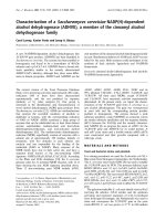
Ribbon representation showing the MAp17 and the N-termi-nal CAp24 domain of the unprocessed Pr55GagFigure 1
Ribbon representation showing the MAp17 and the
N-terminal CAp24 domain of the unprocessed
Pr55Gag. Ribbon diagram of the MAp17 [33] and CAp24
[34] depicting the structural rearrangemts that takes place in
the N-terminal domain (NTD) of CAp24 upon proteolytic
processing at the MAp17-CAp24 junction (indicated with a
sax). The model to the right represents a processed NTD
CAp24 showing the β-hairpin formation which is stabilized by
the salt-bridge formation between the imino terminal Pro 1
and Asp 51. For clarity, Por 1 and Asp 51 are shown as filled
circles. The ribbon diagrams were generated with the
PyMOL [35] and modified with Adobe Photoshop software.
Retrovirology 2007, 4:69 />Page 3 of 12
(page number not for citation purposes)
ized amounts of culture supernatants were precipitated
with Viraffinity and detected with immunoblotting using
both monoclonal and polyclonal anti-CAp24 antibodies
(data not shown) and a pool of HIV-positive sera from
two individuals (Figure 2C). Mature CAp24 represented
the major product of the precipitated material. However,
the level of this protein in both D51N and D51Q mutants
was significantly reduced relative to the wild type and
D51E mutant, correlating with the lower intracellular
CAp24 levels. A comparable level of the viral glycoprotein
(gp120) incorporation into released virions was observed
with all mutants and the wild type virus (Figure 2C). A
similar result was also obtained with a V3 loop-specific
monoclonal anti-glycoprotein antibody (data not
shown).
The Pr55Gag expression and processing pattern was fur-
ther characterized by transfecting HeLa-tat III, 293T and
COS7 cells with the wild type and mutant pNL4-3 expres-
sion plasmids and detected with immunoblotting using a
pool of HIV-positive human sera from two individuals
(Figure 3). With HeLa-tat III cells (Figure 3), the levels of
CAp24 detected with the D51N and D51Q were largely
identical with those in HeLa-tat cells detected with a rab-
bit anti-CAp24 antibody (Figure 2B). Additionally, fully
processed Pr55Gag proteins, as well as, the surface glyco-
proteins could be detected with all mutants when using a
pool of HIV-positive human sera. Further reduction or
absence of cell-associated CAp24 of the D51N and D51Q
mutants was observed in both 293T and COS7 cells.
Whereas no CAp24 was detected with the D51N mutant,
significantly reduced level of this protein was observed
with the D51Q mutant in both 293T and COS7 cells. Sim-
ilar results were also obtained when using both mono-
clonal and polyclonal antibodies directed against CAp24
or the surface glycoprotein gp120/gp160, respectively
(data not shown). With the wild type control, fully proc-
essed HIV-1 Gag proteins were detected in all three trans-
fected cell lines. As an internal control, the level of cell
associated cyclophilin A and calnexin were probed with
polyclonal antibodies directed against these two proteins
(Figure 3, lower panels).
In vitro CAp24 assembly
Turbidity assay is a valuable technique used to study a
salt-induced self-assembly process of CAp24 by monitor-
ing polymerization of CAp24 spectrophotometrically, as
the rate of CAp24 tube formation can be seen as an
increase in sample turbidity over time. One-hundred μM
of each CAp24 was mixed with NaH
2
PO
4
(pH 8.0) buffer
and polymerization was induced by addition of concen-
trated NaCl solution. The rate of CAp24 tube formation
was then measured spectrophotometrically (at 350 nm)
over time. As shown in Figure 4, an increase in sample tur-
bidity was observed for both D51N and D51E mutant
CAp24 proteins. However, as expected, the kinetics of
CAp24 assembly was lower than that of the wild type con-
trol. In marked contrast, the rate of sample turbidity
increase for the D51Q mutant CAp24 was higher than for
the wild type control. This was quite surprising to us, as
the increase in OD should be proportional to the total
Western blot analysis of transfected HeLa-tat cell and precip-itated virusesFigure 2
Western blot analysis of transfected HeLa-tat cell
and precipitated viruses. HeLa-tat cells were transfected
with the plasmids indicated using the non-liposomal transfec-
tion reagent. Forty-eight hrs post-transfection, cells were
washed and harvested in 1× RIPA buffer. Particles released
into the culture supernatant were also clarified and filtered of
cell debris and precipitated with Viraffinity (CPG) as recom-
mended by the manufacturer. Denatured cell (A and B) and
viral lysates (C) were then separated by SDS-PAGE, trans-
ferred onto a nitrocellulose membrane and detected with a
rabbit anti-HIV glycoprotein (A), a pool of anti-CAp24 and
anti-calnexin (B), and anti-CAp24 (C) antibodies. The posi-
tions of specific viral proteins are indicated to the left and the
numbers to the right depict positions of molecular mass
markers (in kDa). NT, a mock control; WT, wild type; and
D51N, D51E, and D51Q are the three CAp24 mutants.
Retrovirology 2007, 4:69 />Page 4 of 12
(page number not for citation purposes)
number of CAp24 proteins assembled into tubular struc-
tures [9].
Morphological analysis of structures formed by
recombinant HIV CAp24 in vitro
To determine the effects of CAp24 mutations on in vitro
capsid assembly, thin-sections of the polymerized mate-
rial used in turbidity assay was prepared and analyzed by
transmission electron microscopy. As shown in Figure 5,
long tubular structures were observed in both D51N and
D51E mutant CAp24 proteins induced by addition of 2.0
M NaCl solution. Additionally, the morphology of the
tubes formed by these two was comparable to the struc-
tures formed by wild type CAp24, both in terms of exter-
nal diameter and length of the tubes. In contrast, no
structure that resembled CAp24 tubular formation was
observed with the D51Q mutant CAp24 protein under the
same conditions.
Analysis of virus release and infectivity
The effects of CAp24 mutations on Pr55Gag assembly and
virus particle release was also analyzed by measuring the
CAp24 antigen contents released into the culture medium
of transfected HeLa-tat III, 293T and COS7 cells. As shown
in Figure 6A, the CAp24 antigen levels in the culture
supernatant of D51N and D51Q transfected cells were
negligible in all three cell lines, whereas the virus produc-
Turbidity assay showing the effects of CAp24 mutations on in vitro CA assemblyFigure 4
Turbidity assay showing the effects of CAp24 muta-
tions on in vitro CA assembly. Turbidity assay showing
the increase in light absorbance after addition of 2.0 M NaCl
to recombinantly produced mutant and wild type CAp24
protein (100 μM) reflecting the assembly of the CAp24 pro-
tein into tubular structures. Green, D51E; red, D51Q; blue,
wild type; pink, D51N. The structures of polymerized CAp24
structures were also analyzed by transmission electron
microscopy (Figure 5).
Western blot analysis of cell-type dependent expression of HIV-1 proteinsFigure 3
Western blot analysis of cell-type dependent expression of HIV-1 proteins. HeLa-tat III, 293T and COS7 cells were
transfected as described above with mutant and wild type proviral DNA constructs. Forty-eight hrs post-transfection, cells
were washed and harvested in 1× RIPA buffer. Denatured cell lysates were then resolved by SDS-PAGE, transferred to a nitro-
cellulose membrane and immunoblotted with a pool of two HIV-1 positive sera (A), rabbit anti-cyclophilin A (B), and anti-cal-
nexin (C) antibodies. Positions of specific viral and cellular proteins are indicated on the right.
Retrovirology 2007, 4:69 />Page 5 of 12
(page number not for citation purposes)
tion of the D51E mutant was reduced by 2- to 6-fold as
compared to the wild type.
The effect of the three CAp24 mutations on virus infectiv-
ity was then assessed with culture supernatants from
transfected HeLa-tat III, 293T and COS7 cells. MT4 cells
were infected with equal amount of cleared and filtered
culture supernatants (normalized for CAp24 antigen) and
assayed for CAp24 antigen contents with a CAp24-ELISA
three days post-infection (Figure 6B). While none of the
three mutant viruses were able to replicate, as expected,
the wild type virus replicated in this cell line. Similar
results were also seen when the infectivity of mutant
viruses was tested in H9 cells (data not shown). We kept
the infected H9 cell cultures for more than 25 days with-
out detecting virus replication with the mutants. No rever-
tants to wild type virus were observed.
Single cell cycle infectivity of HIV-1 CAp24 mutant virions
Since the infectivity of all three CAp24 mutants were
reduced or completely absent when assayed in MT4 cells,
we analyzed the infectivity of these viruses produced from
three different cell lines in a single cell cycle infectivity
assay using the TZM-bl reporter cell line [10]. In this assay,
expression of the reporter luciferase gene is under the con-
Virus release from transfected cells and their infectivityFigure 6
Virus release from transfected cells and their infec-
tivity. HeLa-tat III, 293T, and COS7 cells were transfected
with mutant and wild type proviral DNAs as indicated. (A)
Three days post-transfection, culture supernatants were col-
lected and analyzed by CAp24-ELISA. (B) Normalized
amounts of cleared and filtered culture supernatants from
the above transfected cells were then used to infect MT4
cells (1 × 10
5
cells per well in 48-well plate) using 100 ng of
CAp24 antigen. The bars indicate infectivity of the virus par-
ticles produced from the three different cell lines monitored
by CAp24-ELISA.
Morphological analysis of in vitro assembled mutant CAp24 proteinsFigure 5
Morphological analysis of in vitro assembled mutant
CAp24 proteins. Mutant and wild type CAp24 proteins
were induced for in vitro CAp24 tubular formation (see Fig.
3). At the end of the experiment, the proteins were fixed in
freshly prepared 2.5% glutaraldehyde. The electron micro-
graphs show negatively stained thin-sections of the in vitro
assembled CAp24 tubular structures used in turbidity assay.
Micrographs of the CAp24 mutant D51N (A), D51E (B),
D51Q (C) and the wild type CAp24 (D). Bars indicate 100
nm.
Retrovirology 2007, 4:69 />Page 6 of 12
(page number not for citation purposes)
trol of Tat protein that is activated by Tat protein synthe-
sized from the infecting virus. While the Tat-induced
luciferase activity could not be detected in cells infected
with mutant D51N and D51Q virions, only a subtle
amount of luciferase activity was observed repeatedly in
cells infected with the D51E virions (Figure 7). On the
other hand, the level of Tat-induced luciferase activity was
significantly higher in cells infected with the wild type
virus.
Immunofluorescence analysis of viral protein expression in
transfected cells
The viral protein expression profiles were further investi-
gated by their staining patterns using monoclonal anti-
body directed against CAp24. All mutants displayed
strong specific signals (indicated with arrows in Figure 8)
concentrated near or at the plasma membrane. This fea-
ture was most pronounced in cells transfected with the
three capsid mutants and not with the wild type pNL4-3
transfected cells. The staining pattern seen with the wild
type control was mostly throughout the whole cytoplasm
and the plasma membrane (Figure 8, panel WT). A repre-
sentative staining pattern of each mutant and the wild
type control is shown.
Effect of HIV-1 CAp24 mutations on virion morphology
Morphogenesis of all mutant viruses and the wild type
control were analyzed by transmission electron micros-
copy. The D51N and D51Q mutant virions showed
mostly particles devoid of the typical HIV-1 core structure
(Figure 9, panel D51N and D51Q). Instead, heterogene-
ous virus populations with aberrant core structures were
observed. Additionally, the D51N virions showed a large
pool of intra-vesicular viruses that were deficient of the
electron dense core structure. Most strikingly, no mature
virus particles with conical core structures were detected
with these two mutants. A limited number of immature-
like viruses and occasionally mature-like viruses but with
aberrant cores were observed with the D51E mutant. Only
the wild type control produced viruses with typical imma-
ture- and mature-like HIV-1 virions (Figure 9, panel WT).
Similar results were also observed when virus infected Jur-
kat-tat cells were analyzed (data not shown).
Immunofluorescence analysis of transfected HeLa-tat III cellsFigure 8
Immunofluorescence analysis of transfected HeLa-
tat III cells. HeLa-tat III cells were transfected with mutant
and wild type proviral DNA constructs. Forty-eight hrs post-
transfection, cells were fixed and stained with a mouse anti-
CAp24 monoclonal antibody. As a secondary antibody, FITC-
conjugated (green) rabbit anti-mouse IgG was used. DAPI
(4',6-diamidino-2-phenylindole dihydrochloride) was used to
stain cell nuclei. The images in the right column represent an
overlay of anti-CAp24 and DAPI stained images.
Single cell cycle infectivity of mutant and wild type virus parti-cles on TZM-bl reporter cell linesFigure 7
Single cell cycle infectivity of mutant and wild type
virus particles on TZM-bl reporter cell lines. For rela-
tive viral infectivity assay, TZM-bl reporter cell lines were
seeded one day before infection. Following day, medium was
removed and target cells were inoculated by adding equal
amounts of mutant and wild type NL4-3 virus produced from
transfected HeLa-tat III, 293T, and COS7 cells. In this assay,
expression of the reporter luciferase gene is under the con-
trol of Tat protein that is activated by Tat protein synthe-
sized from the infecting virus. Infected cells were then
analyzed 24 hrs post-infection with the luciferase assay kit
obtained from Promega and as recommended by the manu-
facturer. RLU, relative light unit.
Retrovirology 2007, 4:69 />Page 7 of 12
(page number not for citation purposes)
Discussion
Proper structural rearrangement of capsid (CAp24) after
Pr55Gag cleavage is a highly conserved feature in most ret-
roviruses [11]. As a result of this process, a β-hairpin struc-
ture formed by a salt-bridge between Pro1 and Asp51
(D51) of HIV-1 is important for conformational stability
of the N-terminal CAp24 structure [6]. Thus, mutations of
D51 in HIV-1 CAp24, and likewise Asp54 in murine
leukemia virus (MLV) or human T-cell leukemia virus-1
(HTLV-1), has been shown to disrupt formation of this β-
hairpin structure [6,8,12].
Structural and mutagenesis studies of D51A mutation in
HIV-1 CAp24 has previously shown this invariable resi-
due to be essential for tube formation in vitro, and for the
replication and capsid formation in cultured virus [6]. We
here demonstrated that substitution of D51 with gluta-
mate (D51E), asparagine (D51N), but not glutamine
(D51Q) (three amino acids which in proteins have simi-
lar properties as aspartate; Glu > Asn > Gln) could partly
restore in vitro CAp24 assembly but not the infectivity of
the virus particles.
Whereas generally the total protein contents produced by
transfected 293T and COS7 cells were reduced as com-
pared to HeLa-tat or HeLa-tat III cells, similar Pr55Gag-
processing patterns was repeatedly observed in all mutant
and wild type proviral DNA transfected cells. However,
intracellular concentrations of CAp24 protein in any of
the cells transfected with D51N and D51Q were generally
reduced. This could not be explained by the lack of recog-
nition by the antibody used for immunoblotting, since
detection with mouse anti-CAp24, rabbit anti-CAp24 or a
pool of sera from HIV-infected patients gave similar
results. Additionally, analysis with CAp24-ELISA using a
different rabbit anti-CAp24-specific antibody also gave
similar results. TEM analysis revealed that all mutants
were assembly competent but produced virus particles
with aberrant core morphology. The virus particles were
also able to incorporate HIV-1 glycoprotein but the infec-
tivity of the virus particles was severely reduced or absent
suggesting that there was no defect at binding or internal-
ization of these mutants although this was not specifically
tested for. Whereas no infectivity was observed with the
D51N and D51Q virions, a subtle amount was seen with
the D51E viruses in a single cell cycle infectivity assay.
Further analysis of cytoplasmic versus cell membrane
CAp24 distribution was also performed with indirect
immunofluorescence staining using mouse anti-CAp24
antibody. This analysis revealed a strong staining pattern
near or at the plasma membrane (PM) of cells transfected
with the three mutants, indicating that there was no defect
in intracellular transport of the Pr55Gag precursor to its
steady-state destination [13] where activation of the viral
protease takes place [14,15]. However, all mutants dis-
played a decreased cytoplasmic staining as compared to
the wild type CAp24 control, which showed a diffuse
cytoplasmic staining of non-membrane bound Pr55Gag/
CAp24. Perhaps mutated Pr55Gag trafficking and/or
assembly is slowed down, or even blocked close to or at
the PM in agreement with low levels of mutant particles
released. It is also possible that the virus release may have
been blocked as a result of inability to form the stabilizing
β-hairpin structure in the N-terminal domain of CAp24
upon proteolytic maturation which is necessary for assem-
bly and release of virions [6].
Self-associative properties of many viral CAp24 proteins
have been previously reported [16-19]. However, depend-
ing on the protein concentration, salt, and the buffering
pH [9,20,21], the morphology of the assembled structures
or the rate of assembly may be variable. The effects of D51
mutations on in vitro CAp24 assembly was monitored
spectrophotometrically, and as expected, the assembly
rate of both D51N and D51E mutants were substantially
reduced relative to the wild type protein, although the
ability of these mutants to form tubular structures was
shown by thin-section transmission electron microscopy
(TEM). Thus, it seems likely that the D51N and D51E
mutations impose less structural changes than the D51A
TEM analysis of mutant virus particlesFigure 9
TEM analysis of mutant virus particles. Electron micro-
graphs of mutant and wild type virus particles. Mutants D51N
and D51Q showed mostly heterogenous populations of par-
ticles with varying size and morphology (panels D51Q and
inset in panel D51N). No virus particles with conical core
structures were observed with these two mutants. Addition-
ally, a large pool of virus-like structures inside vesicles
released from transfected cells were observed in D51N
mutant. With the wild type and D51E virions particles repre-
senting immature-like viruses are shown (panels D51E and
WT). Mature viruses with conical structures were seen only
in the wild type control virus. Occasionally, D51E virions
resembling the mature wild type morphology but with aber-
rant core structure was also observed. Bars indicate 100 nm.
Retrovirology 2007, 4:69 />Page 8 of 12
(page number not for citation purposes)
mutation described earlier [6]. Remarkably, although no
tubular structure was observed with the D51Q mutant by
TEM analysis, an increased optical density measurement
that reflects the assembly kinetics was repeatedly
observed. We cannot explain this, but, it is possible that
the increased OD may result as a consequence of amor-
phous aggregates that are resistant for stable higher-order
CAp24 tube formation.
In a recent study that was published after the present work
was performed, Leschonsky et al [22] described the two
single amino acid substitution mutations, a D183E and
D183N, in an infectious provirus clone HX10. In contrast
to our results, they found no effect on extracellular level of
the CAp24 protein produced from H1299 cells transfected
with the D183E mutant. Additionally, they found no
effect on the intracellular level of the CAp24 protein in
H1299 cells transfected with the D51N mutant. This may
have been owing to the different cell type used. However,
we analyzed the viral protein expression profiles in four
different cell lines and found similar results.
Lastly, in order to correlate the lack of infectivity with
morphological appearances of the viruses, electron micro-
scopy analysis was performed. Only the D51E mutant par-
ticles were partially able to form immature- and mature-
like viruses that resembled the wild type morphology.
Importantly, despite the ability to form wild type-like
viruses, the infectivity of D51E virions was significantly
reduced, indicating the importance of optimal HIV-1 core
stability [23]. With the two other non-infectious mutants,
particles with aberrant core structures, either hollow-
shaped spherical structures in endosomal vesicles (D51N)
or particles with distorted core morphology (D51Q) were
seen.
Taken together, our data and the other previously pub-
lished observations [6,22,24] suggest that the invariable
D51 residue of HIV is crucial for formation of the β-hair-
pin structure in matured CAp24 protein. Additionally,
even substitution of D51 with such a similar residue as
with glutamate could not restore the integrity of this struc-
ture. Furthermore, although our results demonstrated that
the D51N and D51E substitutions could restore the in
vitro tubular formation, the infectivity of all D51 muta-
tion were rendered non-infectious indicating that this res-
idue is indispensable.
Methods
Cells and reagents
HeLa-tat, 293T, COS7, and TZM-bl cell lines were main-
tained in Dulbecco modified Eagle medium (DMEM)
supplemented with 10% fetal bovine serum (FBS), peni-
cillin and streptomycin sulphate (Sigma, St Louis MO).
H9, Jurkat-tat and MT4 cells were maintained in RPMI
1640 medium (Gibco, Grand Island, NY) supplemented
with 10% fetal bovine serum (FBS; GIBCO), penicillin
(100 U/ml), and streptomycin (100 μg/ml). DEAE-dex-
tran was purchased from Sigma, rabbit polyclonal anti-
bodies against calnexin from Santa Cruz Biotechnology
(catalogue no. sc-11397). The following reagents were
obtained through the AIDS Research and Reference Rea-
gent Program, Division of AIDS, NIAID, NIH: All adher-
ent cell lines, the protease inhibitor indinavir sulphate
(catalogue no. 8145) and TZM-bl cells (catalogue no.
8129) contributed by Dr. John C Kappes [10].
Plasmid DNA construction
The polymerase chain reaction (PCR) was utilized to
develop all plasmids in the study and all constructs were
derivatives of the HIV-1 molecular clone pNL4-3 [25]. The
HIV-1 CA coding sequence was amplified using PCR and
cloned into the prokaryotic expression vector pET11a
(Novagen Inc.) essentially as described elsewhere [21,26].
Briefly, the primer pair 5'-ATG GAT CCA TAT GCC TAT
AGT GCA GAA CCT CC-3' and 5'-ATG GAT CCT ATC ACA
AAA CTC TTG CTT TAT GGC C-3' containing the BamHI/
NdeI and BamHI, respectively, were used for amplification
of the CA sequence (BamHI/NdeI and BamHI sites are
shown in bold). In addition, a translational start codon at
the 5' end (ATG) and two stop codons (TGA/TAG) at the
3' end of the sequence were added. The PCR product was
subcloned into the TA cloning vector (Invitrogen), trans-
formed in DH5α E. coli (Escherichia coli), purified and
confirmed by sequencing (Cybergene, Sweden). The vec-
tor was then digested with NdeI and BamHI and the DNA
fragment encoding CA gene was isolated, purified and
cloned directionally into the pET11a vector, digested with
the same restriction enzymes. Standard procedures were
used for restriction digestion. The resulting plasmid was
designated pET11a-CA and verified by sequencing.
The three HIV-1 CAp24 mutants, D51N, D51E, and
D51Q, in the pET11a-CA vector were then engineered by
site-directed mutagenesis using the Stratagene's Quick-
Change™ Site Directed Mutagenesis Kit (Stratagene) as
recommended by the manufacturer. The primer pair used
for creating the mutations is listed in Table 1.
The same mutations were also introduced into the HIV-1
molecular clone pNL4-3Δenv using the same mutagenic
primers described above. QuickChange II XL site-directed
mutagenesis kit (Stratagene) was used to create the point
mutations in the CA sequence. All plasmid DNAs were
then propagated in E. coli XL10-Gold and purified by Max-
iprep Purification kit (Qiagen). The identity of each muta-
tion was confirmed by sequencing and the resulting
plasmids were digested with BssHII and ApaI. The 1295 bp
BssHII/ApaI DNA fragments of the mutated CA sequences
were then isolated, purified and cloned directionally into
Retrovirology 2007, 4:69 />Page 9 of 12
(page number not for citation purposes)
the pNL4-3 vector, digested with the same restriction
enzymes. The resulting plasmids were propagated in
DH5α competent E. coli, purified using Maxiprep purifica-
tion kit and verified by sequencing.
Capsid protein expression and purification
Competent E. coli Origami (DE3) cells were transformed
with the three mutants or the wild-type pET11a-CA
expression plasmid, expressed and purified essentially as
described elsewhere [26]. Briefly, a single colony from a
freshly streaked plate was initially grown in 50 ml LB-
medium containing 100 μl Ampicillin (stock 100 mg/ml)
and cultured at 37°C shaken at 220 r.p.m. Upon reaching
optical cell densities at 600 nm (OD
600
) ~0.6–1.0, the
cells culture was saved at 4°C overnight. The following
day, 10 ml of culture was added to 1 litre of LB-medium
containing ampicillin and incubated with shaking at
37°C until the OD
600
was ~0.7–1.0. Protein expression
was then induced by addition of isopropylthio-β-D-galac-
toside (IPTG) to a final concentration of 1 mM. After a 4
hrs incubation period at 37°C, the cells were harvested by
centrifugation at 4000 r.p.m. for 10 min (Megafuge 2.0 R,
rotor #8155, Kendro). The cell pellet was resuspended in
6 M Guanidine-HCl (pH 6.5) and stirred for 3 hrs at room
temperature before being centrifuged at 10000 r.p.m. for
10 min at 4°C (Beckman Avanti J30-I, rotor 25.50, Beck-
man Coulter). Fifty ml of nuclease-free water was slowly
added to the supernatant giving a final concentration of 1
M Guanidine-HCl to the protein solution. The protein
solutions were put in four 15 cm long dialysis tubings
(Spectrpor, MWCO 6–8000, 1.7 ml/cm) and dialyzed
against 50 mM Tris pH 8.0 overnight at room tempera-
ture. Next, the contents of the dialysis tubings were
pooled and centrifuged at 10000 r.p.m. for 10 min at 4°C
(Beckman Avanti J30-I, rotor 25.50, Beckman Coulter) to
remove precipitated proteins. The CAp24 proteins were
then precipitated by addition of saturated (NH
4
)
2
SO
4
to a
final concentration of 30% and incubated on ice for 1 h.
The CAp24 proteins were then collected by centrifugation
at 20000 r.p.m. for 20 min at 4°C (Beckman Avanti J30-I,
rotor 25.50, Beckman Coulter). Finally, the protein pre-
cipitate was dissolved in a buffer containing 50 mM Tris-
HCl pH 8, 30 mM NaCl and 1 mM EDTA, and purified on
ÄKTA FPLC chromatography system (Amersham Biose-
cience). The protein samples were initially loaded onto an
anion-exchange column, HiTrap DEAE 1 ml FF, with a
mobile phase of 50 mM Tris pH8.0, 30 mM NaCl, and 1
mM EDTA and flow rate of 1 ml/min. The absorbance was
measured at 280 nm. The peak fractions containing the
CAp24 proteins were pooled and precipitated with 50%
saturated (NH
4
)
2
SO
4
on ice for 1 h. The solution was then
centrifuged at 20000 r.p.m. for 20 min at 4°C (Beckman
Avanti J30-I, rotor 25.50, Beckman Coulter) and the pre-
cipitate was resupsended in 50 mM Tris pH8.0, 30 mM
NaCl, and 1 mM EDTA. The purity and integrity of each
CAp24 protein was finally analyzed by SDS-PAGE. In
order to increase the purity of the CAp24 protein, the sam-
ples were loaded onto a gel filtration column, HiLoad 16/
60 Superdex 75 prep grade, and run with the same mobile
phase and as above but with a flow rate of 1.5 ml/min.
The peak fractions containing the CAp24 proteins were
pooled and concentrated by Amicon Ultra Centrifugal fil-
ters (Millipore; MWCO 5 k) and saved in aliquots at -
80°C. A small aliquot (10 μl) was run on SDS-PAGE gel
and the protein concentration was determined with a Bio-
Rad DC Protein Assay Kit.
Transfection procedure
Transfection was performed in a 6-well culture plate using
the non-liposomal FuGENE 6 transfection reagent
(Roche). Approximately 1 × 10
5
adherent cells (HeLa-tat,
293T, and COS7) were seeded one day before and trans-
fected with 2 μg of each plasmid DNA mixed with 6 μl
FuGENE 6 transfection reagent. Forty-eight to seventy-two
hrs post-transfection, cells were washed in cold PBS and
harvested in 1× RIPA buffer [50 mM Tris (pH 7.4), 150
mM NaCl, 1% Triton X-100, 1% Na-deoxycholate, and
0.1% SDS] supplemented with a complete protease inhib-
itor cocktail obtained from Roche.
Virus stock preparation
Wild type and mutant virus stocks were prepared essen-
tially as described before [27]. Briefly, HeLa-tat, COS7,
and 293T cells were transfected as described above and
three days post-transfection, culture supernatants were
clarified from cell debris by centrifugation at 1200 r.p.m.
for 7 min, and filtered through 0.45 μm filters. Cleared
culture supernatants were then treated or not with DNase
I (Roche Applied Science) at 20 μg/ml final concentra-
tions at 37°C for 1 h and saved at -80°C until needed. The
CAp24 antigen contents of each culture supernatant was
determined by an in-house HIV-1 CAp24 antigen ELISA as
previously described [27,28].
Virus precipitation
HeLa-tat, COS7, and 293T cells were transfected with the
wild type and mutant proviral DNAs as described above.
Approximately seventy-two hrs post-transfection, virion-
associated viral proteins were prepared from cell culture
Table 1: Primers used to create the D51N, D51E and D51Q
CAp24 mutants
5' primer 3' primer
D51E GCCACCCCACAAGAGT
TAAATACCATG
CATGGTATTTAACTCTT
GTGGGGTGGC
D51Q GCCACCCCACAACAAT
TAAATACCATG
CATGGTATTTAATTGTT
GTGGGGTGGC
D51N GCCACCCCACAAAATT
TAAATACCATG
CATGGTATTTAAATTTT
GTGGGGTGGC
Retrovirology 2007, 4:69 />Page 10 of 12
(page number not for citation purposes)
supernatants by removal of cellular debris by centrifuga-
tion at 1 200 r.p.m. for 7 min and filtering through a 0.45-
μm-pore-size membrane. The virus particles in the culture
supernatants were then concentrated by centrifugation
using Viraffinity (CPC Inc.) essentially as described before
[29]. Briefly, clarified culture supernatants were mixed
with Viraffinity (3:1) and the mixture was incubated at
room temperature for 5 min. They were then centrifuged
at 1 000 × g for 10 min and viral pellets washed twice in a
buffer containing 60 mM HEPES, 150 mM NaCl, pH 6.5.
Finally, the viral pellets were dissolved in 1× RIPA buffer,
mixed with 2× SDS sample buffer and boiled for 5 min
before being subjected to sodium dodecyl sulphate-poly-
acrylamide gel electrophoresis (SDS-PAGE).
Western blot
Denatured whole cell extracts or viral lysates were sepa-
rated on 10–20% SDS-PAGE gels (Invitrogen), transferred
onto a nitrocellulose membrane (Amersham Bioscience)
overnight at 4°C and detected either with monoclonal
anti-CAp24 antibody (kindly provided by Dr Hinkula J),
polyclonal anti-CAp24, anti-cyclophilin A, anti-calnexin
(Santa Cruz) antibodies or a cocktail of three different
HIV-1 positive human sera. As a secondary antibody,
appropriate horseradish peroxidase-conjugated anti-
mouse (DAKO; 1:4000), anti-rabbit (Sigma; 1:40000), or
anti-human (Pierce; 1:20 000) IgG antibody was used.
Viral infectivity assay
The mutant and wild type HIV-1 virus stocks were pre-
pared as described above and 100 ng CAp24 antigen
equivalents were used to infect MT4 cells. Briefly, 1 × 10
5
cells were infected with normalized amounts of virus for
3 hrs at 37°C. The cells were then pelleted, residual virus
was removed, and the cell cultures were incubated in fresh
complete medium supplemented with FBS and antibiot-
ics at 37°C in 5% CO
2
. Three days post-infection, the
CAp24 antigen contents in the culture supernatants were
then processed for CAp24-ELISA.
Single cell cycle infectivity assay
TZM-bl cells (6 × 10
4
cells per 12-well plate) [10] were
seeded one day before infection. Following day, medium
was removed and cells were infected with mutant and
wild type NL4-3 virus. The cells were infected with a virus
stock corresponding to 50 ng CAp24 antigen per well with
20 μg/ml DEAE-dextran in a total volume of 300 μl. After
adsorption period of 3 hrs, input virus was removed and
cells were fed with a complete DMEM containing 10 μM
indinavir and cultured for 24 hrs. Finally, culture superna-
tants were removed and cells were lysed with 200 μl Glo
lysis buffer (Promega). One-hundred μl of the cell lysates
were then assayed for luciferase activity using the luci-
ferase assay kit obtained from Promega as recommended
by the manufacturer. Measurement of the luminescence
was done using the Luminoskan Ascent luminometer
(ThermoLabsystem).
In vitro HIV-1 CA assembly (Turbidity assay)
Turbidity assay is a valuable technique used to study a
salt-induced self-assembly process of CAp24 by monitor-
ing polymerization of CAp24 spectrophotometrically, as
the rate of CAp24 tube formation increases sample turbid-
ity [9,30,31]. The assay was performed at room tempera-
ture using a BioSpec-1601E spectrometer (Shimadzu) and
the absorbance was set to 350 nm wavelength. One-hun-
dred μM of highly purified HIV-1 CAp24 protein of each
mutant and the wild type control was mixed with 50 mM
NaH
2
PO
4
(pH 8.0). Tubular CAp24 assembly was then
induced by addition of 2.0 M NaCl solution, and the
assembly rates was monitored by a spectrophotometer as
the rate of tube formation increases the sample turbidity.
Absorbance measurements were made every 10 s for up to
60 min. The assembly rate was then set by plotting the
absorbance versus time.
For TEM analysis, 100 μM of each mutant and the wild
type CAp24 protein was mixed with 50 mM NaH
2
PO
4
(pH 8.0) and 1.0 M NaCl solution. The mixture was then
immediately transferred to a 37°C and incubated for 1 h.
Finally, the samples were fixed with freshly made 2.5%
formaldehyde and processed for TEM analysis.
Immunofluorescence assay
HeLa-tat III cells (1.5 × 10
3
cells per well in 4-well cham-
bered slides from Nunc) were cultured one day before and
transfected with 2 μg of mutant and wild type proviral
DNA constructs. Forty-eight hrs post-transfection, cells
were fixed in aceton/methanol (1:1) for 5 min and
washed with PBS. Slides were then incubated with pri-
mary anti-CAp24 monoclonal antibody and 4',6-diamid-
ino-2-phenylindole dihydrochloride (DAPI) at 37°C for 1
h. DAPI was used to labell the cellular DNAs. Cells were
washed three times in PBS and further incubated with sec-
ondary antibody for 1 h. FITC-conjugated rabbit anti-
mouse IgG antibody (DAKO) was used as secondary anti-
body. After the final wash, slides were mounted and
flourescent images were aquired by using a Nikon Eclipse
E600 phase-contrast fluorescent microsope.
Transmission electron microscopy analysis
Cells were prepared for electron microscopy essentially as
described elsewhere [32]. Briefly, transfected HeLa-tat
cells and virus infected Jurkat-tat cells (data not shown)
were fixed by freshly made 2.5% glutaraldehyde in phos-
phate buffer and post-fixed in 1% OsO
4
. The cells were
embedded in epon and post-stained with 1% uranyl ace-
tate. Epon sections were cut at approximately 60 nm thick
to accommodate the volume of the core structure parallel
Retrovirology 2007, 4:69 />Page 11 of 12
(page number not for citation purposes)
to the section plane. Duplicate sample preparations were
done, which were then analyzed by electron microscope.
Additionally, in vitro assembled CAp24 proteins were
negatively stained with 2% ammonium molybdate at pH
8.0 to study the CAp24 tubular formation.
Competing interests
The author(s) declare that they have no competing inter-
ests.
Authors' contributions
SA and MY contributed equally to the experimental work.
SA wrote the manuscript with AV. AV is the principal
investigator. SH performed all electron microscopy analy-
sis. All authors read and approved the manuscript.
Acknowledgements
We thank Alireza Padjand for help with the cloning and initial characteriza-
tion of the prokaryotic expression plasmids. This work was supported by
grants from the Swedish Medical Research Council (grant no. K2000-06X-
09501-10B), Swedish International development Cooperation Agency,
SIDA (grant no. 2006-0011786) and Tripep AB.
References
1. Wiegers K, Rutter G, Kottler H, Tessmer U, Hohenberg H, Krauss-
lich HG: Sequential steps in human immunodeficiency virus
particle maturation revealed by alterations of individual Gag
polyprotein cleavage sites. J Virol 1998, 72:2846-2854.
2. Morikawa Y, Shibuya M, Goto T, Sano K: In vitro processing of
human immunodeficiency virus type 1 Gag virus-like parti-
cles. Virology 2000, 272:366-374.
3. Briggs JA, Simon MN, Gross I, Krausslich HG, Fuller SD, Vogt VM,
Johnson MC: The stoichiometry of Gag protein in HIV-1. Nat
Struct Mol Biol 2004, 11(7):672-675.
4. Monaco-Malbet S, Berthet-Colominas C, Novelli A, Battai N, Piga N,
Cheynet V, Mallet F, Cusack S: Mutual conformational adapta-
tions in antigen and antibody upon complex formation
between an Fab and HIV-1 capsid protein p24. Structure 2000,
8(10):1069-1077.
5. Li S, Hill CP, Sundquist WI, Finch JT: Image reconstructions of
helical assemblies of the HIV-1 CA protein. Nature 2000,
407(6802):409-413.
6. von Schwedler UK, Stemmler TL, Klishko VY, Li S, Albertine KH,
Davis DR, Sundquist WI: Proteolytic refolding of the HIV-1 cap-
sid protein amino-terminus facilitates viral core assembly.
Embo J 1998, 17(6):1555-1568.
7. Tang C, Ndassa Y, Summers MF: Structure of the N-terminal
283-residue fragment of the immature HIV-1 Gag polypro-
tein. Nat Struct Biol 2002, 9(7):537-543.
8. Mortuza GB, Haire LF, Stevens A, Smerdon SJ, Stoye JP, Taylor IA:
High-resolution structure of a retroviral capsid hexameric
amino-terminal domain. Nature 2004, 431(7007):481-485.
9. Lanman J, Sexton J, Sakalian M, Prevelige PE Jr.: Kinetic analysis of
the role of intersubunit interactions in human immunodefi-
ciency virus type 1 capsid protein assembly in vitro. J Virol
2002, 76(14):6900-6908.
10. Wei X, Decker JM, Liu H, Zhang Z, Arani RB, Kilby JM, Saag MS, Wu
X, Shaw GM, Kappes JC: Emergence of resistant human immu-
nodeficiency virus type 1 in patients receiving fusion inhibi-
tor (T-20) monotherapy. Antimicrob Agents Chemother 2002,
46(6):1896-1905.
11. Coffin JM: Genetic diversity and evolution of retroviruses.
Curr
Top Microbiol Immunol 1992, 176:143-164.
12. Bouamr F, Cornilescu CC, Goff SP, Tjandra N, Carter CA: Struc-
tural and dynamics studies of the D54A mutant of human T
cell leukemia virus-1 capsid protein. J Biol Chem 2005,
280(8):6792-6801.
13. Finzi A, Orthwein A, Mercier J, Cohen EA: Productive Human
Immunodeficiency Virus Type 1 Assembly Takes Place at
the Plasma Membrane. 2007, 81(14):7476-7490.
14. Kaplan AH, Manchester M, Swanstrom R: The activity of the pro-
tease of human immunodeficiency virus type 1 is initiated at
the membrane of infected cells before the release of viral
proteins and is required for release to occur with maximum
efficiency. J Virol 1994, 68(10):6782-6786.
15. Vogt VM: Proteolytic processing and particle maturation. Curr
Top Microbiol Immunol 1996, 214:95-131.
16. Burnette WN, Holladay LA, Mitchell WM: Physical and chemical
properties of Moloney murine leukemia virus p30 protein: a
major core structural component exhibiting high helicity
and self-association. J Mol Biol 1976, 107(2):131-143.
17. Ehrlich LS, Krausslich HG, Wimmer E, Carter CA: Expression in
Escherichia coli and purification of human immunodefi-
ciency virus type 1 capsid protein (p24). AIDS Res Hum Retrovi-
ruses 1990, 6(10):1169-1175.
18. Pinter A, Fleissner E: Structural studies of retroviruses: charac-
terization of oligomeric complexes of murine and feline
leukemia virus envelope and core components formed upon
cross-linking. J Virol 1979, 30(1):157-165.
19. Pepinsky RB, Cappiello D, Wilkowski C, Vogt VM: Chemical
crosslinking of proteins in avian sarcoma and leukemia
viruses. Virology 1980, 102(1):205-210.
20. Ehrlich LS, Liu T, Scarlata S, Chu B, Carter CA: HIV-1 capsid pro-
tein forms spherical (immature-like) and tubular (mature-
like) particles in vitro: structure switching by pH-induced
conformational changes. Biophys J 2001, 81(1):586-594.
21. Gross I, Hohenberg H, Krausslich HG: In vitro assembly proper-
ties of purified bacterially expressed capsid proteins of
human immunodeficiency virus. Eur J Biochem 1997,
249(2):592-600.
22. Leschonsky B, Ludwig C, Bieler K, Wagner R: Capsid stability and
replication of human immunodeficiency virus type 1 are
influenced critically by charge and size of Gag residue 183.
2007, 88(1):207-216.
23. Forshey BM, von Schwedler U, Sundquist WI, Aiken C: Formation
of a human immunodeficiency virus type 1 core of optimal
stability is crucial for viral replication. J Virol 2002,
76(11):5667-5677.
24. Tang S, Murakami T, Agresta BE, Campbell S, Freed EO, Levin JG:
Human immunodeficiency virus type 1 N-terminal capsid
mutants that exhibit aberrant core morphology and are
blocked in initiation of reverse transcription in infected cells.
J Virol 2001, 75(19):9357-9366.
25. Adachi A, Gendelman HE, Koenig S, Folks T, Willey R, Rabson A, Mar-
tin MA: Production of acquired immunodeficiency syndrome-
associated retrovirus in human and nonhuman cells trans-
fected with an infectious molecular clone. J Virol 1986,
59(2):284-291.
26. Ehrlich LS, Agresta BE, Carter CA: Assembly of recombinant
human immunodeficiency virus type 1 capsid protein in
vitro. J Virol 1992, 66(8):4874-4883.
27. Abdurahman S, Hoglund S, Hoglund A, Vahlne A: Mutation in the
loop C-terminal to the cyclophilin A binding site of HIV-1
capsid protein disrupts proper virus assembly and infectivity.
Retrovirology 2007, 4(1):19.
28. Horal P, Svennerholm B, Jeansson S, Rymo L, Hall WW, Vahlne A:
Continuous epitopes of the human immunodeficiency virus
type 1 (HIV-1) transmembrane glycoprotein and reactivity
of human sera to synthetic peptides representing various
HIV-1 isolates. J Virol 1991, 65(5):2718-2723.
29. Abdurahman S, Hoglund S, Goobar-Larsson L, Vahlne A: Selected
amino acid substitutions in the C-terminal region of human
immunodeficiency virus type 1 capsid protein affect virus
assembly and release. J Gen Virol 2004, 85(Pt 10):2903-2913.
30. Douglas CC, Thomas D, Lanman J, Prevelige PE Jr.: Investigation of
N-terminal domain charged residues on the assembly and
stability of HIV-1 CA. Biochemistry 2004, 43(32):10435-10441.
31. Tang C, Loeliger E, Kinde I, Kyere S, Mayo K, Barklis E, Sun Y, Huang
M, Summers MF: Antiviral inhibition of the HIV-1 capsid pro-
tein.
J Mol Biol 2003, 327(5):1013-1020.
32. Hoglund S, Su J, Reneby SS, Vegvari A, Hjerten S, Sintorn IM, Foster
H, Wu YP, Nystrom I, Vahlne A: Tripeptide interference with
Publish with BioMed Central and every
scientist can read your work free of charge
"BioMed Central will be the most significant development for
disseminating the results of biomedical research in our lifetime."
Sir Paul Nurse, Cancer Research UK
Your research papers will be:
available free of charge to the entire biomedical community
peer reviewed and published immediately upon acceptance
cited in PubMed and archived on PubMed Central
yours — you keep the copyright
Submit your manuscript here:
/>BioMedcentral
Retrovirology 2007, 4:69 />Page 12 of 12
(page number not for citation purposes)
human immunodeficiency virus type 1 morphogenesis. Anti-
microb Agents Chemother 2002, 46(11):3597-3605.
33. Massiah MA, Starich MR, Paschall C, Summers MF, Christensen AM,
Sundquist WI: Three-dimensional Structure of the Human
Immunodeficiency Virus Type 1 Matrix Protein. Journal of
Molecular Biology 1994, 244(2):198-223.
34. Gamble TR, Vajdos FF, Yoo S, Worthylake DK, Houseweart M, Sun-
dquist WI, Hill CP: Crystal structure of human cyclophilin A
bound to the amino-terminal domain of HIV-1 capsid. Cell
1996, 87(7):1285-1294.
35. DeLano WL: The PyMOL Molecular Graphics System. 2002.