Báo cáo y học: "Requirements for the selective degradation of CD4 receptor molecules by the human immunodeficiency virus type 1 Vpu protein in the endoplasmic reticulum" doc
Bạn đang xem bản rút gọn của tài liệu. Xem và tải ngay bản đầy đủ của tài liệu tại đây (1.11 MB, 15 trang )
Retrovirology
BioMed Central
Open Access
Research
Requirements for the selective degradation of CD4 receptor
molecules by the human immunodeficiency virus type 1 Vpu protein
in the endoplasmic reticulum
Julie Binette1,2, Mathieu Dubé1,2, Johanne Mercier1, Dalia Halawani3, Martin
Latterich4 and Éric A Cohen*1,2
Address: 1Laboratory of Human Retrovirology, Institut de Recherches Cliniques de Montréal, 110 Avenue des Pins Ouest, Montreal, Quebec H2W
1R7, Canada, 2Department of Microbiology and Immunology, Université de Montréal, 2900, Édouard-Montpetit, Montreal, Quebec H3T 1J4,
Canada, 3Department of Anatomy and Cell Biology, McGill University, 3640 University Street Montreal, Quebec H3A 2B2, Canada and 4Faculty
of Pharmacy, Université de Montréal, 2900, Édouard-Montpetit, Montreal, Quebec H3T 1J4, Canada
Email: Julie Binette - ; Mathieu Dubé - ; Johanne Mercier - ; Dalia
Halawani - ; Martin Latterich - ; Éric A Cohen* -
* Corresponding author
Published: 15 October 2007
Retrovirology 2007, 4:75
doi:10.1186/1742-4690-4-75
Received: 23 July 2007
Accepted: 15 October 2007
This article is available from: />© 2007 Binette et al; licensee BioMed Central Ltd.
This is an Open Access article distributed under the terms of the Creative Commons Attribution License ( />which permits unrestricted use, distribution, and reproduction in any medium, provided the original work is properly cited.
Abstract
Background: HIV-1 Vpu targets newly synthesized CD4 receptor for rapid degradation by a
process reminiscent of endoplasmic reticulum (ER)-associated protein degradation (ERAD). Vpu is
thought to act as an adaptor protein, connecting CD4 to the ubiquitin (Ub)-proteasome
degradative system through an interaction with β-TrCP, a component of the SCFβ-TrCP E3 Ub ligase
complex.
Results: Here, we provide direct evidence indicating that Vpu promotes trans-ubiquitination of
CD4 through recruitment of SCFβ-TrCP in human cells. To examine whether Ub conjugation occurs
on the cytosolic tail of CD4, we substituted all four Ub acceptor lysine residues for arginines.
Replacement of cytosolic lysine residues reduced but did not prevent Vpu-mediated CD4
degradation and ubiquitination, suggesting that Vpu-mediated CD4 degradation is not entirely
dependent on the ubiquitination of cytosolic lysines and as such might also involve ubiquitination
of other sites. Cell fractionation studies revealed that Vpu enhanced the levels of ubiquitinated
forms of CD4 detected in association with not only the ER membrane but also the cytosol.
Interestingly, significant amounts of membrane-associated ubiquitinated CD4 appeared to be fully
dislocated since they could be recovered following sodium carbonate salt treatment. Finally,
expression of a transdominant negative mutant of the AAA ATPase Cdc48/p97 involved in the
extraction of ERAD substrates from the ER membrane inhibited Vpu-mediated CD4 degradation.
Conclusion: Taken together, these results are consistent with a model whereby HIV-1 Vpu
targets CD4 for degradation by an ERAD-like process involving most likely poly-ubiquitination of
the CD4 cytosolic tail by SCFβ-TrCP prior to dislocation of receptor molecules across the ER
membrane by a process that depends on the AAA ATPase Cdc48/p97.
Page 1 of 15
(page number not for citation purposes)
Retrovirology 2007, 4:75
Background
CD4 is a 55-kDa class I integral membrane glycoprotein
that serves as the primary co-receptor for human immunodeficiency virus type 1 (HIV-1) entry into cells [1]. CD4
consists of a large lumenal domain, a transmembrane
portion, and a 38-residues cytoplasmic tail. It is expressed
primarily on the surface of a subset of T lymphocytes that
recognizes major histocompatibility complex (MHC)
class II-associated peptides and plays a major role in the
development and maintenance of the immune system.
Despite the critical role played by CD4 during HIV-1
entry, it is well established that HIV-1 down-regulates cell
surface expression of its cognate receptor (reviewed in reference [2]). It is believed that this process prevents superinfection and promotes production of fully infectious
virions [3,4]. Down-regulation of CD4 in HIV-1-infected
cells is mediated through different independent mechanisms involving the activity of three viral proteins: Nef,
Env and Vpu. Early in infection, Nef removes CD4 molecules that are already present at the cell surface by enhancing their endocytosis and subsequent degradation in
lysosomes [5]. At later stages of the infection, the envelope
precursor gp160, through its high receptor binding affinity and inefficient vesicular transport [6], sequesters newly
synthesized CD4 in the endoplasmic reticulum (ER) in
the form of Env-CD4 complexes and prevents its transport
and maturation to the cell surface [7]. The accessory protein Vpu induces a rapid degradation of newly synthesized
CD4 molecules bound to gp160 in the ER [8].
Vpu is an 81-amino acids class I integral membrane protein of 16 kDa that is unique to HIV-1 and simian immunodeficiency virus isolated from chimpanzee (SIVcpz)
and a few other monkey species ([9-11] and reviewed in
reference [12]). The protein consists of an N-terminal
hydrophobic membrane anchor domain of 27 amino
acids and a charged C-terminal hydrophilic domain of 54
residues that extends into the cytoplasm [13]. This
cytosolic domain contains a highly conserved dodecapeptide sequence encompassing residues 47–58 which comprises a pair of serine residues (S52 and S56) that are
phosphorylated by casein kinase II [14,15]. Besides its
ability to mediate the rapid degradation of CD4 molecules complexed with Env gp160 in the ER, Vpu was also
found to promote efficient release of progeny HIV-1
viruses in different human cell types, including T cells and
macrophages, by a mechanism that appears to involve the
inactivation of a putative host cell factor that restricts viral
particle release in a cell-type dependent manner [10,1619].
From a mechanistic point of view, HIV-1 Env is not absolutely required for Vpu-mediated CD4 degradation. The
role of Env appears to be limited to its ability to retain
/>
CD4 in the ER, given that efficient CD4 degradation can
be observed in the absence of Env as long as CD4 is
retained in the ER through the presence of an ER retention
sequence or treatment of cells with Brefeldin A (BFA), a
fungal metabolite known to block protein sorting from
the ER to the Golgi apparatus [20]. The degradation of
CD4 mediated by Vpu involves multiple steps that are initiated by the direct physical binding of Vpu to the cytoplasmic tail of CD4 in the ER [21]. Although the binding
of Vpu to CD4 is necessary to induce CD4 degradation, it
is not sufficient. Indeed, studies aimed at identifying Vpu
partners by two-hybrid screens led to the identification of
a host cellular co-factor, β-TrCP, which plays a critical role
in Vpu-mediated CD4 degradation by interacting with
Vpu in a phosphorylation-dependent manner [22]. The
human F-box protein β-TrCP functions as a substrate recognition receptor for the multi-subunit ubiquitin ligase
(E3) SCFβ-TrCP involved in the ubiquitin (Ub) conjugating
pathway (reviewed in reference [23]). The interaction
between Vpu and β-TrCP is essential for Vpu-mediated
CD4 degradation since substitution mutations of Vpu
phospho-acceptor sites, S52 and S56, prevent association
with β-TrCP and abolish the effect of Vpu on CD4 turnover [22]. These findings have established a link between
the machinery responsible for the ubiquitination of proteins destined for degradation by the proteasome and the
enhanced CD4 turnover in presence of Vpu. Indeed, further lines of evidence for an involvement of the Ub-proteasome system in Vpu-mediated CD4 degradation were
also reported: 1) Vpu-mediated CD4 degradation is not
observed in a mammalian cell line expressing a temperature-sensitive Ub activating enzyme (E1), a key component of the machinery involved in the covalent
attachment of Ub to target proteins [24]; 2) over-expression of a mutant Ub (Ub K48/R), which prevents the formation of poly-Ub chains, impairs Vpu-mediated CD4
degradation [24]; 3) Vpu-mediated CD4 degradation is
inhibited by specific proteasome inhibitors [24].
Vpu-induced CD4 degradation is reminiscent of ER-associated protein degradation (ERAD), a quality control
process in the ER that ensures that only proteins with a
native folded conformation leave the organelle for other
destinations across the secretory pathway [25]. Misfolded
proteins that cannot reach their native state are transferred
from the ER to the cytosol by a multi-step process called
retro-translocation or dislocation which is thought to
involve pore complexes formed by proteins such as Derlin-1 [26,27] or by multi-spanning transmembrane E3
ligases such as Hrd1 [28]. ERAD substrates exposed to the
cytosol are acted upon by ER-associated components of
the Ub conjugation machinery, extracted from the ER
membrane by the AAA ATPase Cdc48/p97 and its associated cofactors Ufd1p and Np14p and degraded by the 26
S proteasome (reviewed in reference [25]). This cellular
Page 2 of 15
(page number not for citation purposes)
Retrovirology 2007, 4:75
pathway has been co-opted by some viruses to selectively
destroy cellular proteins required for immune defense of
the host. For example, two human cytomegalovirus
(HCMV) proteins, US2 and US11, are able to target newly
synthesized class I MHC (MHC-I) heavy chains (HC) for
dislocation from the ER, leading to complete extraction of
MHC-I HC from the ER membrane into the cytosol followed by proteasomal destruction [29,30]. Most ERAD
substrates are poly-ubiquitinated while undergoing dislocation although the details of recognition, timing and
post-translational modification of dislocation substrates
can vary depending on the substrates [31-33].
/>
antibodies was performed to ensure that CD4 was specifically degraded by Vpu in this system. Fig. 1 reveals that
CD4 turnover was significantly accelerated in presence of
Although part of the molecular machinery that is recruited
by Vpu to target CD4 for degradation is reasonably well
defined, several aspects of Vpu-mediated CD4 degradation still remain unclear. In particular, direct evidence of
CD4 ubiquitination in presence of Vpu in human cells has
not been demonstrated. Furthermore, it is unclear
whether ER-associated CD4 encounters the cytoplasmic
proteasome by a process involving dislocation of CD4
molecules across the ER membrane as described for ERAD
substrates. Finally, the role of CD4 ubiquitination in processes underlying Vpu-mediated CD4 degradation remains
to be specified. Meusser and Sommer have reconstituted
the process of Vpu-mediated CD4 degradation in Saccharomyces cerevisiae by expressing human CD4 together with
Vpu and human β-TrCP and have provided evidence suggesting that Vpu-mediated proteolysis strictly relies on
ubiquitination of CD4 at cytosolic lysine residues prior to
export of receptor molecules from the ER membrane [34].
In this study, we have analyzed the process of Vpu-mediated CD4 degradation in human cells. The data presented
here provide evidence suggesting that Vpu promotes ubiquitination of CD4 cytosolic tail by SCFβ-TrCP and mediates
dislocation of the viral receptor across the ER membrane
in human cells by a process that might depend on the AAA
ATPase Cdc48/p97. Interestingly, in contrast to previous
results, Vpu-mediated CD4 degradation and ubiquitination were not found to be entirely dependent on cytosolic
lysine residues, raising the possibility that ubiquitination
at sites other than lysines might also be involved.
Results
Poly-ubiquitination of CD4 is required for Vpu-mediated
CD4 degradation
In order to study processes involved in Vpu-mediated
CD4 degradation, we established a transient expression
system whereby CD4 and Vpu are expressed in trans in
SV40-transformed human embryonic kidney fibroblasts
(HEK 293T) cells. CD4- and Vpu-expressing cells were
treated with BFA in order to retain CD4 in the ER before
and during metabolic labeling. Pulse-chase radio-labeling
analysis followed by immunoprecipitation with anti-CD4
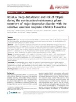
Poly-ubiquitination of CD4 is required for Vpu-mediated
Figure 1
CD4 degradation
Poly-ubiquitination of CD4 is required for Vpu-mediated CD4 degradation. A. HEK 293T cells were mocktransfected or co-transfected with 1.5 µg of SVCMV CD4 wt
and 8 µg of SVCMV Vpu+ (Vpu+) or the phosphorylationdefective Vpu mutant SVCMV Vpu S52,56/N (Vpu S52,56/N).
In parallel, CD4/Vpu transfectants were co-transfected with
8 µg of plasmids encoding his(6)/c-myc-Ub wt (myc-Ub wt)
or the TDN mutant of ubiquitin his(6)/c-myc-Ub K48/R
(myc-Ub K48/R). Transfected cells were treated with BFA,
pulse-labeled with [35S]methionine and [35S]cysteine and
chased with complete media for the indicated time intervals.
Cells were then lysed and immunoprecipitated sequentially
with anti-CD4 monoclonal and polyclonal antibodies first and
then with anti-Vpu and anti-myc antibodies. B. Using quantitative scanning of CD4 bands from three independent experiments, the percentage of CD4 remaining over time as
compared to time 0 is plotted for each transfection. C. HEK
293T cells were mock-transfected or co-transfected as
described in A. Cell transfectants were treated for two hours
with BFA prior to lysis. Steady state levels of CD4, actin and
tagged ubiquitin were analysed by western-blot. D. Quantitative analysis from three independent experiments showing
the level of CD4 relative to CD4 expressed with Vpu S52,56/
N (arbitrarily set at 100%) for each transfectant.
Page 3 of 15
(page number not for citation purposes)
Retrovirology 2007, 4:75
/>
Vpu. Furthermore, the effect of Vpu on CD4 was specific
since expression of a phospho-acceptor sites mutant, Vpu
S52,56/N, which is unable to interact with the E3 Ub
ligase complex SCFβ-TrCP [22] did not mediate CD4 degradation (Fig. 1A, compare lanes 5–8 with lanes 9–12 and
Fig. 1B). Moreover, as previously reported [24,35], addition of specific proteasome inhibitor, such as MG-132, to
HEK 293T cells expressing CD4 and Vpu inhibited Vpumediated CD4 degradation (data not shown).
We also tested whether poly-ubiquitination of CD4 was
required for Vpu-mediated CD4 degradation in HEK 293T
cells. For this purpose, we co-expressed CD4 and Vpu with
a N-terminal his(6)/c-myc tagged form of wild-type (wt)
Ub or a N-terminal his(6)/c-myc tagged form of a
transdominant negative (TDN) mutant of Ub, Ub K48/R,
that is unable to form poly-Ub chains required for proteasomal degradation [36]. This Ub mutant acts as a chain
terminator in the process of poly-ubiquitination since the
Ub-acceptor lysine residue at position 48 is mutated for
an arginine. In agreement with previous reported data
[24], results of Fig. 1A (compare lanes 9–12 with lanes
17–20) and B reveal that expression of tagged-Ub K48/R
markedly reduced the rate of Vpu-mediated CD4 degradation, thus suggesting that poly-ubiquitination of CD4 via
K48 linkage of Ub moieties was required for Vpu-mediated CD4 degradation. Although expression of wt taggedUb had some attenuating effect on Vpu-mediated CD4
degradation (compare lanes 13–16 with lanes 9–12 and
Fig. 1B), it was clearly less pronounced than with the TDN
tagged-Ub K48/R mutant. In that regard, wt tagged Ub has
been previously reported to decrease the rate of degradation of some substrate by the Ub-proteasome system
given that fusion of the his-myc tag at the N-terminal of
Ub renders poly-Ub-protein conjugates less recognizable
by the proteasome [37]. All of these results were also confirmed by analyzing steady-state levels of CD4 by westernblot in Vpu-expressing HEK 293T cells (Fig. 1C and 1D).
Overall, these results provide evidence that this expression
system in HEK 293T cells supports an efficient degradation of CD4 that is Vpu-specific, depends on the recruitment of β-TrCP, necessitates an active proteasome and
requires poly-ubiquitination of CD4.
Vpu induces ubiquitination of CD4 molecules
Having established that over-expression of Ub K48/R
inhibited Vpu-mediated CD4 degradation in HEK 293T
cells, we investigated whether we could isolate and
directly detect ubiquitinated forms of CD4 that are
expected to accumulate under these conditions. Towards
this goal, we first analyzed CD4 expression at steady state
in Vpu/CD4 HEK 293T transfectants in presence or
absence of tagged-Ub K48/R (Fig. 2A). In these experiments, transfected cells were treated with BFA during 2 h
prior to lysis to retain newly synthesized CD4 in the ER.
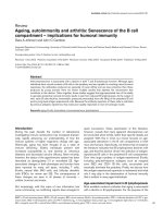
Figure Vpu on CD4 ubiquitination
Effect of2
Effect of Vpu on CD4 ubiquitination. A. Vpu-mediated
ubiquitination of CD4 wt when CD4 is retained in the ER
through treatment with BFA. HEK 293T cells were mocktransfected or co-transfected with 1 µg of SVCMV CD4 wt,
8 µg of SVCMV Vpu+ or the phosphorylation-defective Vpu
mutant SVCMV Vpu S52,56/N and 8 µg of the TDN mutant
his(6)/c-myc-Ub K48/R. Samples were then treated as
described in the materials and methods section. CD4 molecules were immunoprecipitated with anti-CD4 polyclonal
antibodies prior to western-blot analysis with anti-myc monoclonal antibodies. (triangle) indicates the position of the
heavy chains of anti-CD4 antibodies. B. Quantitative analysis
of ubiquitinated CD4 conjugates. (asterisk) represents the
area of the autoradiogram that was used for quantitation of
CD4-Ub conjugates. The histogram shows the relative levels
of ubiquitinated CD4 conjugates in presence or absence of a
functional Vpu. Relative CD4-Ub conjugate levels were evaluated by quantitation of the signal detected in the area delineated on the autoradiogram relative to total CD4 as
determined by quantitation of the band detected with the
anti-CD4 antibodies on whole cell lysate. The relative level of
ubiquitinated CD4 detected in absence of Vpu was arbitrarily
set at 1. The data represent results from seven experiments.
C. Vpu-mediated ubiquitination of CD4 wt in condition
where CD4 is retained in the ER through binding with HIV-1
Env. HEK 293T cells were mock-transfected or co-transfected with 1 µg of pHIV CD4 wt, 10 µg of provirus encoding
Vpu- (HxBH10-vpu-) or Vpu+ (HxBH10-vpu+) and 20 µg of
his(6)/c-myc-Ub K48/R. Samples were then treated as in A
but in absence of BFA. D. Quantitative analysis showing the
relative levels of ubiquitinated CD4 detected in two independent experiments. Relative levels of ubiquitinated CD4
conjugates were determined as described in B.
Page 4 of 15
(page number not for citation purposes)
Retrovirology 2007, 4:75
Fig. 2A reveals that CD4 levels at steady-state were significantly reduced in presence of Vpu (compare lanes 2 and
4). As expected, expression of tagged-Ub K48/R suppressed the effect of Vpu on CD4 and re-established the
amounts of CD4 to levels comparable to those detected in
absence of Vpu (compare lanes 5 and 2). To detect CD4Ub conjugates, cell lysates were first immunoprecipitated
with anti-CD4 polyclonal antibodies and the resulting
CD4-containing immunocomplexes were subsequently
analyzed for the presence of CD4-Ub conjugates by western-blot using anti-myc antibodies. Ubiquitinated forms
of CD4 were detected as a typical smear in presence of Vpu
(lane 5). Although background high molecular weight
ubiquitinated forms of CD4 could still be detected in
absence of Vpu (lane 3) or in presence of the non-functional Vpu S52,56/N mutant (lane 7), their levels were not
as elevated as in presence of wt Vpu (lane 5). Indeed,
quantitative analysis revealed that levels of CD4-Ub conjugates were approximately 6-fold higher in presence than
in absence of a functional Vpu (Fig. 2B). The detection of
a smear of high molecular weight proteins in presence of
Vpu is suggestive of poly-ubiquitination of CD4. Polyubiquitination is still possible even if Ub K48/R is overexpressed because cells are expressing endogenous wt Ub
that can initiate poly-Ub chains before a molecule of Ub
K48/R can prematurely terminate the chain.
Finally, we examined whether we could extend this
enhancing effect of Vpu on CD4 ubiquitination to a more
physiological system where CD4 is retained in the ER
through the formation of complexes with Env glycoproteins instead of BFA treatment. In this system, initially
described by Willey and co-workers [20], Vpu and Env
glycoproteins are co-expressed from a proviral construct
while CD4, that is under HIV-1 long terminal repeat control (pHIV CD4) [24], is expressed only in cells expressing
Vpu and Env. Results of Fig. 2C and 2D show that even in
a system where CD4 is naturally retained in the ER
through binding to HIV-1 Env, Vpu expression increases
substantially (approximately 8-fold) the level of CD4
molecules undergoing ubiquitination (compare the levels
of CD4-Ub conjugates in lanes 4 and 2 (upper panel) relative to their respective CD4 steady state levels (lower
panel) and Fig. 2D).
Overall these results indicate that Vpu promotes polyubiquitination of CD4 molecules that are targeted for degradation by the proteasome through the recruitment of
the SCFβ-TrCP E3 Ub ligase.
Vpu-mediated CD4 degradation and ubiquitination are
not strictly dependent on CD4 cytosolic lysines
CD4 contains four potential Ub acceptor lysine residues
in its cytoplasmic domain. To determine whether ubiquitination of the cytosolic tail was required for Vpu-medi-
/>
ated CD4 degradation, we analyzed a CD4 mutant, CD4
KRcyto, in which all four cytoplasmic lysine residues were
replaced by arginines. The stability of CD4 wt and CD4
KRcyto was first assessed in cells expressing a provirus
encoding either wt Vpu (HxBH10-vpu+) or Vpu S52,56/D
(HxBH10-vpu S52,56/D) as described above in Fig. 2C.
Results of Fig. 3A clearly show that both CD4 wt and CD4
KRcyto were unstable in Vpu expressing cells as observed
by the decreased recovery of CD4 molecules over the
chase period (lanes 9–12 and lanes 13–16). Quantification of CD4 turnover over several experiments indicated
an attenuation of the degradation kinetic of CD4 KRcyto
as compared to CD4 wt but the protein was clearly susceptible to Vpu-induced degradation (Fig. 3B). In contrast,
both CD4 wt and CD4 KRcyto remained stable over the
entire 7 h chase period in cells expressing the phosphorylation mutant Vpu S52,56/D (Fig. 3A, lanes 1–4 and
lanes 5–8 and Fig. 3B).
Given that previous studies had shown that Vpu-mediated
CD4 degradation strictly relied on cytosolic lysine residues in mammalian cells and yeast [24,34], we analyzed
the steady-state levels of CD4 wt or CD4 KRcyto in HEK
293T expressing Vpu+ or Vpu- provirus by western-blot.
Similar to what we found in pulse-chase experiments, we
repeatedly observed a difference in sensitivity to Vpumediated degradation between CD4 wt and CD4 KRcyto
(Fig. 3C, compare lanes 14 and 16 with lanes 10 and 12
and Fig. 3D, right panel) but clearly, the absence of
cytosolic Ub acceptor lysine residues was not entirely preventing the effect of Vpu on CD4 degradation. Similar
results were also obtained when steady-state levels of CD4
wt and CD4 KRcyto were analyzed in BFA-treated HEK
293T cells expressing Vpu+ or Vpu- provirus lacking Env
(Fig. 3C, compare lanes 2 and 4 with lanes 6 and 8, and
Fig. 3D, left panel).
Given that Vpu was reported to interact with the cytoplasmic tail of CD4 in a region (EKKT, residues 416–419 of
CD4) that encompasses some of the lysine residues
mutated in the CD4 KRcyto mutant (K417, K418), we further tested whether this difference in susceptibility to Vpumediated CD4 degradation could be explained by a
diminished ability of CD4 KRcyto to associate with Vpu.
Binding experiments were performed as described in
materials and methods using the Vpu S52,56/N mutant,
which binds CD4 as efficiently as Vpu wt but is unable to
mediate CD4 degradation [21]. Results from these experiments reveal that CD4 KRcyto associates with Vpu at least
as efficiently as CD4 wt, thus ruling-out that the decreased
sensitivity of CD4 KRcyto to Vpu-mediated degradation
results from reduced Vpu binding efficiency [Additional
file 1]. These results were also confirmed by immunoprecipitation of CD4 followed by western-blot using antiVpu antibodies (data not shown).
Page 5 of 15
(page number not for citation purposes)
Retrovirology 2007, 4:75
/>
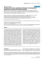
Figure Vpu on CD4 molecules lacking lysine residues in the cytoplasmic tail
Effect of3
Effect of Vpu on CD4 molecules lacking lysine residues in the cytoplasmic tail. A. Analysis of CD4 wt and CD4
KRcyto turnover in presence or absence of functional Vpu by pulse-chase labeling and immunoprecipitation. HEK 293T cells
were mock-transfected or co-transfected with 2 µg of pHIV CD4 wt or pHIV CD4 KRcyto and 20 µg of provirus encoding
Vpu+ (HxBH10-vpu+) or phosphorylation-defective Vpu mutant (HxBH10-vpu S52,56/D). Cells were pulse-labeled with
[35S]methionine and [35S]cysteine and chased in complete medium for the indicated time intervals. Cells were then lysed and
immunoprecipitated sequentially with anti-CD4 antibodies first (polyclonal and monoclonal) and then with anti-Vpu antibodies.
B. Using quantitative scanning of CD4 bands from two independent experiments, the percentage of CD4 remaining over time
as compared to time 0 is plotted for each transfection. C. Effect of Vpu on steady-state CD4 wt and CD4 KRcyto levels. HEK
293T cells were mock-transfected or co-transfected with 1 µg of pHIV CD4 wt or pHIV CD4 KRcyto and 10 µg of proviruses
encoding Vpu- or Vpu+ in addition to 25 µg of the his(6)/c-myc-Ub K48/R expressor. In the left panel (Env-), a similar experiment was performed except that HEK 293T cells were co-transfected with 10 µg of envelope-defective provirus (HxBc2-pr-,
vpu-, env- or HxBH10-pr-, vpu+, env-) and treated with BFA for 2 h prior to lysis. Cell lysates were then treated as described in
the materials and methods section. D. Quantitative analysis of steady-state CD4 levels. CD4 levels in presence of absence of
his(6)/c-myc-Ub K48/R were arbitrarily set at 100%. The levels of CD4 in presence of Vpu are shown relative to the corresponding controls. These results are representative of the data obtained in three independent experiments for Env- and five
independent experiments for Env+.
Page 6 of 15
(page number not for citation purposes)
Retrovirology 2007, 4:75
Given that CD4 KRcyto was still susceptible to Vpu-mediated degradation, we next evaluated whether CD4 KRcyto
could undergo ubiquitination in presence of Vpu. To optimize the recovery of CD4-Ub conjugates, Vpu/CD4 or
Vpu/CD4 KRcyto HEK 293T transfectants were made to
co-express the TDN Ub K48/R mutant. Analysis of CD4Ub and CD4 KRcyto-Ub conjugates levels in presence or
absence of Vpu was performed as described above for Fig.
2B. Fig. 4A reveals that even though CD4 KRcyto is less
susceptible to Vpu-mediated degradation as compared to
CD4 wt (compare lanes 1 and 3 with lanes 5 and 7, middle panel), it still undergoes enhanced ubiquitination in
presence of Vpu (compare lane 6 and lane 8). However, it
is important to note that the relative level of recovered
CD4 KRcyto-Ub conjugates was decreased as compared to
CD4-Ub conjugates. In fact, quantitative analysis of ubiquitinated CD4 conjugate levels reveals that Vpu enhanced
ubiquitination of CD4 KR by approximately 3-fold while
it increased ubiquitination of wt CD4 by 8-fold. Altogether, these results suggest that lysine residues in the
cytosolic domain of CD4 are not absolutely essential for
ubiquitination and degradation of the viral receptor in
presence of Vpu. Even-though optimal Vpu-mediated
CD4 ubiquitination most probably involves cytosolic
lysine residues there must be other sites that are also targeted during Vpu-induced ubiquitination.
Figure Vpu on CD4 KRcyto poly-ubiquitination
Effect of4
Effect of Vpu on CD4 KRcyto poly-ubiquitination. A.
HEK 293T cells were mock-transfected or co-transfected
with 1 µg of pHIV CD4 wt or pHIV CD4 KRcyto, 10 µg of
provirus encoding Vpu- (HxBH10-vpu-) or Vpu+ (HxBH10vpu+) and 25 µg of the TDN mutant of Ub his(6)/c-myc-Ub
K48/R. Transfected cells were not treated with BFA prior to
lysis. Samples were then treated as described in the materials
and methods. B. Quantitative analysis of the relative levels of
ubiquitinated CD4 conjugates for CD4 wt and CD4 KRcyto
in two independent experiments. (asterisk) represents the
area of the autoradiogram that was used for the quantitation
of CD4-Ub conjugates. Relative levels of ubiquitinated CD4
conjugates were determined as described in Fig. 2B.
/>
Vpu-mediated CD4 degradation involves the dislocation of
ubiquitinated CD4 conjugates across the ER membrane
To examine whether CD4 undergoes a process of dislocation across the ER membrane during Vpu-mediated degradation, we conducted subcellular fractionation studies. To
optimize recovery and detection of dislocated forms of
CD4 targeted for degradation by the cytosolic proteasome, we performed these cell fractionation experiments
in conditions where CD4 degradation was inhibited by
over-expression of the TDN Ub K48/R mutant. BFAtreated HEK 293T cells expressing CD4/Ub K48/R and
Vpu or CD4/Ub K48/R alone were fractionated by
mechanical lysis into membrane and cytosolic fractions
and each resulting fraction was directly analyzed for the
presence of CD4, Vpu and membrane or cytosolic markers, such as calnexin and actin respectively, by westernblot as described in materials and methods. Furthermore,
the presence of poly-ubiquitinated forms of CD4 in membrane or cytosolic fractions was determined by immunoprecipitation/western-blot analysis. In contrast to Fig. 2
and 4 and because of technical reasons, ubiquitinated
CD4 molecules were detected in these experiments by performing immunoprecipitation using anti-Myc antibodies
followed by western-blot using anti-CD4 antibodies. As
expected, Vpu and calnexin were detected exclusively in
association with membrane fractions (Fig. 5A, lane 5 for
Vpu and lanes 1, 3, 5 and 7 for calnexin) whereas actin
(lanes 2, 4, 6, 8) or Ub (lanes 4, 6, 8) were recovered in a
very large proportion in the cytosolic fractions, thus demonstrating that the fractionation procedure was almost
free of membrane or cytosolic contaminations. CD4 molecules were found in the membrane fraction in presence
or absence of Vpu (lanes 3 and 5). We could repeatedly
recover and detect CD4-Ub conjugates, represented as a
smear signal, predominantly in the membrane fraction
but also in the cytosolic fraction in absence and in presence of Vpu (Fig. 5A); in some instances, depending on
the experiments, we also detected discrete high molecular
bands in addition to the smear signal [lane 5 of Additional file 2A and lane 6 of Additional file 2B]. Interestingly, the absolute signal associated with membrane and
cytosolic fractions was always more intense in presence
than in absence of Vpu (Fig. 5A, compare lanes 3, 5 and 7
as well as lanes 4, 6 and 8, upper panel). The specific levels
of CD4-Ub conjugates associated with membrane and
cytosolic fractions in absence and in presence of Vpu were
calculated relative to the amount of CD4 detected directly
by western-blot. As shown in Fig. 5B, quantitative analysis
revealed that in presence of Vpu there was approximately
a six-fold increase in membrane-associated CD4-Ub conjugate levels relative to the negative control without Vpu
(Vpu-); in the cytosolic fractions, the levels of CD4-Ub
conjugates detected in presence of Vpu were approximately two-fold higher relative to the Vpu- control (Fig.
5B).
Page 7 of 15
(page number not for citation purposes)
Retrovirology 2007, 4:75
/>
Figure 5
Vpu-mediated CD4 degradation involves dislocation of ubiquitinated CD4 conjugates from the ER membrane to the cytosol
Vpu-mediated CD4 degradation involves dislocation of ubiquitinated CD4 conjugates from the ER membrane
to the cytosol. HEK 293T cells were mock-transfected or co-transfected with 1 µg of pHIV CD4 wt, 10µg of envelope-defective provirus (HxBc2-pr-, vpu-, env- or HxBH10-pr-, vpu+, env-) and 15 µg of his(6)/c-myc-Ub K48/R expression plasmid where
indicated. Cells were treated with BFA for 2 h before mechanical lysis. CD4-Ub conjugates were immunoprecipitated with
anti-myc monoclonal antibodies prior to western-blot analysis with anti-CD4 polyclonal antibodies while control proteins in
each fraction were revealed by western-blot. Actin and calnexin were used as cytosolic and membrane controls, respectively.
A. Membrane (M) and cytosolic (C) fractions were separated and treated as described in the materials and methods section. B.
Quantitative analysis of the relative amounts of ubiquitinated CD4 molecules present in each fraction relative to the amounts
measured in absence of Vpu (arbitrarily set at 1). (asterisk) represents the area of the autoradiogram that was used for the
quantitation of CD4-Ub conjugates. Non-specific background signal detected in lanes 7 and 8 was subtracted. Relative levels of
ubiquitinated CD4 conjugates were determined as described in the legend of Fig. 2B. Error bars reflect standard deviations
from duplicate independent experiments. C. Membrane (M) fractions were treated with Na2CO3 (pH 11) as described in materials and methods. Treated membrane and supernatant (S) were subsequently recovered by centrifugation. Fractions were analyzed as described above in A. D. Quantitative analysis of the relative amounts of ubiquitinated CD4 molecules (as described in
the legend of Fig. 2B) present in each fraction relative to the amounts measured in absence of Vpu (arbitrarily set at 1). (asterisk) represents the area that was used for the quantitation of CD4-Ub conjugates. Non-specific background signal detected in
lanes 7 and 8 was subtracted. Error bars reflect standard deviations from duplicate independent experiments.
Page 8 of 15
(page number not for citation purposes)
Retrovirology 2007, 4:75
To determine whether membrane-associated CD4-Ub
conjugates represent CD4 molecules that are still embedded in the membrane while undergoing dislocation or if
some of these conjugates are fully dislocated but stay tethered to the cytosolic face of the membrane, we treated
membrane fractions with 100 mM sodium carbonate at
basic pH (pH 11) (Fig. 5C) and analyzed the treated membrane and resulting supernatant for the presence of CD4Ub conjugates as described in Fig. 5A. Salt-wash at basic
pH (Na2CO3) but not at neutral pH (NaCl) was previously shown to remove peripheral proteins that are associated with membranes [38]. In this experiment, Vpu (Fig.
5C, lane 5) and calnexin (lanes 1, 3, 5 and 7) were exclusively recovered in the membrane fractions after Na2CO3
treatment, thus confirming that the integrity of microsomes was maintained during the procedure. Surprisingly, we repeatedly detected small amounts of CD4 in the
salt-wash supernatant (lanes 4 and 6, WB anti-CD4 panel)
that perhaps represent population of CD4 molecules that
are dislocated prior to ubiquitination. Quantitative analysis of the relative CD4-Ub conjugates signal associated
with membrane and supernatant fractions revealed that
approximately 50% of the membrane-associated signal
could be salt washed at basic pH (Fig. 5C, compare lane 3
to lane 4 and lane 5 to lane 6), thus indicating that part of
the membrane-associated CD4-Ub signal represents dislocated ubiquitinated forms of CD4 that are associated with
the cytosolic face of the membrane. Importantly, in presence of Vpu we detected a 2-3-fold increase in the relative
levels of CD4-Ub conjugates associated with the treated
membrane fraction and salt-washed supernatant (Fig.
5D). As expected, control experiments where membranes
were washed with sodium chloride at neutral pH (pH 7)
did not lead to any recovery of CD4-Ub in the supernatant
[Additional file 2A]. Conversely, treatment of membranes
with RIPA-DOC lysis buffer solubilized CD4-Ub conjugates, which were detected almost completely in the
supernatant [Additional file 2B]. As expected, in both conditions the absolute levels of detected CD4-Ub conjugates
was more elevated in presence than in absence of Vpu.
Overall, these results suggest that Vpu targets CD4 for
cytosolic proteasomal degradation by enhancing dislocation of receptor molecules across the ER membrane.
Expression of a transdominant negative mutant of p97
inhibits Vpu-mediated CD4 degradation
To further confirm that Vpu-mediated CD4 degradation
involves a dislocation step, we examined the implication
of the Cdc48/p97 ATPase in this process. Mammalian p97
plays an important role in dislocation of ERAD substrates
presumably by binding poly-ubiquitinated substrates in
conjunction with its cofactors, including Ufd1 and Npl4
[39], and mediating a process of extraction that is energydependent [40]. The p97 protein has two ATPase domains
and mutants affected in their ability to bind or hydrolyze
/>
ATP are no longer able to perform their function in retrotranslocation [41]. We took advantage of a well-described
p97 TDN ATP binding mutant (p97 AA) [41] and tested
its effect on Vpu-mediated CD4 degradation. HEK 293T
cells were co-transfected with expression plasmids encoding CD4, Vpu and FLAG-tagged p97 wt or FLAG-tagged
p97 TDN mutant and the levels of CD4 were analyzed at
steady-state by western-blot. As shown in Fig. 6, expression of the p97 TDN mutant strongly inhibited Vpu-mediated CD4 degradation while wt p97 had no significant
inhibitory effect on Vpu ability to degrade CD4 (compare
lanes 3 and 5 with lanes 2 and 4). These results were also
confirmed by pulse-chase labeling experiments where
CD4 turnover was evaluated in presence of Vpu and the
p97 TDN mutant or wt p97 (data not shown). Since p97
is directly involved in the dislocation of several ERAD substrates, these results provide additional evidence suggesting that Vpu targets the CD4 receptor for cytosolic
proteasomal degradation by a process that involves a dislocation step across the ER membrane.
Figure
dation a
Effect of6 TDN mutant of p97 on Vpu-mediated CD4 degraEffect of a TDN mutant of p97 on Vpu-mediated
CD4degradation. HEK 293T cells were mock-transfected
or co-transfected with 1.5 µg of SVCMV CD4 wt, 12 µg of
SVCMV Vpu- or Vpu+ and 1 µg of an expression plasmid
encoding a FLAG-tagged version of p97 wt or the TDN
mutant p97 AA. Cells were treated with BFA for 2 h prior to
lysis. Cell lysates were then analyzed by western-blot as
described in materials and methods. These results are representative of the data obtained in two independent experiments.
Page 9 of 15
(page number not for citation purposes)
Retrovirology 2007, 4:75
Discussion
In the present study, we have conducted a detailed analysis of processes involved in the ER-associated degradation
of CD4 receptor molecules induced by the HIV-1 Vpu
accessory protein in human cells. Using a TDN mutant of
Ub, Ub K48/R, which acts as a poly-Ub chain terminator,
we have confirmed previous findings [24] suggesting that
poly-ubiquitination of CD4 is required for Vpu-mediated
CD4 degradation (Fig. 1). Based on these observations,
we attempted to directly detect ubiquitinated forms of
CD4, which are expected to accumulate under conditions
where Ub K48/R is over-expressed. A similar approach
was successfully used to facilitate the isolation and detection of substrates of the Ub pathway such as APOBEC3G
in presence of HIV-1 Vif [42]. Under these conditions, we
could demonstrate an increased accumulation of high
molecular weight CD4-Ub conjugates, typical of polyubiquitinated protein targets, in presence of Vpu (Fig. 2).
Direct detection of ubiquitinated forms of CD4 in presence of Vpu was achieved both in conditions where CD4
retention in the ER was produced through short treatment
of cells with BFA or through formation of Env/CD4 complexes, thus demonstrating that both systems could be
used to analyze Vpu-mediated CD4 degradation. Some
high molecular weight ubiquitinated CD4 conjugates
could be detected in absence or presence of a non functional Vpu mutant unable to recruit the SCFβ-TrCP E3 ligase
complex, except that their levels were significantly lower
than those found in presence of Vpu. It is likely that ubiquitinated CD4 conjugates detected at steady-state in
absence of Vpu or in presence of inactive Vpu represent
intermediates resulting from the relatively low but normal
degradation of misfolded CD4 molecules that occurs
through the ERAD pathway in condition of transient
ectopic over-expression. Together, these findings provide
direct evidence that Vpu promotes trans-ubiquitination of
CD4 through recruitment of the SCFβ-TrCP complex in
human cells.
CD4, as a type 1 integral membrane protein, consists of a
38-amino acid cytosolic domain that contains four lysine
residues (amino acid positions: K411, KK417-418, and
K428) that could serve as acceptor sites for ubiquitination.
Ubiquitin conjugation of lysine residues accessible from
the cytosol through recruitment of the specific SCFβ-TrCP
E3 ligase complex by Vpu may represent a very early step
in the process of CD4 degradation and precede the transport of the viral receptor through the ER membrane for
proteolytic degradation by the cytosolic proteasome. To
investigate the role of cytosolic lysine residues in Vpumediated CD4 degradation, we used a CD4 mutant in
which all four lysines were replaced by arginine residues.
In contrast to earlier observations made in HeLa cells [24],
replacement of lysine residues in the CD4 cytoplasmic tail
did not strictly prevent CD4 degradation by Vpu in HEK
/>
293T cells. In our conditions, even though we detected a
significant difference in the protein turnover (Fig. 3A–B)
as well as in the steady-state levels (Fig. 3C–D) of CD4
KRcyto and CD4 wt in presence of Vpu, our data also
revealed that CD4 KRcyto was still susceptible to Vpumediated CD4 degradation. These results suggest that
ubiquitination of the cytosolic tail at lysine acceptor sites
by the SCFβ-TrCP E3 ligase is not strictly required for Vpumediated CD4 degradation and, therefore, does not
appear to constitute an essential early signal that triggers
CD4 targeting to the cytosolic proteasome. Given that
poly-ubiquitination of CD4 appears to be required for
Vpu-mediated CD4 degradation (Fig. 1), our findings
raise the possibility that ubiquitination may occur at sites
other than cytosolic lysines. Consistent with this possibility, CD4 molecules lacking cytosolic lysine Ub acceptor
sites (CD4 KRcyto) are still capable of undergoing ubiquitination in presence of Vpu, albeit to levels that are lower
than wt CD4 (Fig. 4). One possible explanation for Vpumediated ubiquitination of cytosolic lysine-less CD4 is
that a partial dislocation of the receptor N-termini to the
cytosolic side may be required, so that lysine residues in
the lumenal domain of CD4 may be accessible for ubiquitination by the cytosolic ubiquitination machinery
recruited by Vpu. In that regard, in the specific case of
HCMV US2-induced ERAD of MHC-I HC, the replacement of cytosolic tail lysine residues did not affect MHCI HC dislocation and degradation while internal lysine
residues were found to be required for these processes.
These results have raised the possibility that US2 could
induce a partial dislocation of part of the heavy chain into
the cytosol, resulting in cytosolic deposition of lumenal
lysine residues [43]. Although, this possibility cannot be
completely excluded at this point for Vpu-mediated CD4
degradation, we believe that this scenario is unlikely since
replacement of cytosolic lysine residues led to an attenuation of CD4 degradation and to a substantial decrease of
CD4 ubiquitination by the SCFβ-TrCP E3 ligase (Fig. 4);
these observations suggests that Vpu-mediated CD4 degradation involves most probably ubiquitination of the
receptor cytosolic tail.
An alternative explanation for the ubiquitination and degradation of cytosolic tail lysine-less CD4 molecules by
Vpu is that ubiquitination may occur via non-lysine residues. Interestingly, recent evidence indicate that the
mouse gamma herpesvirus (γ-HSV) mK3 E3 Ub ligase,
which targets nascent MHC-I HC for degradation by
ERAD, mediates ubiquitination via serine, threonine or
lysine on the HC tail, each of which was found to be sufficient to induce rapid degradation of HC [44]. The γ-HSV
mK3 E3 Ub ligase was found to have the ability to mediate
the formation of ester bonds that covalently linked Ub to
serine or threonine in the tail of the HC substrate. Unlike
MIR 1 (also called kK3), an E3 ligase of Kaposi's sarcoma-
Page 10 of 15
(page number not for citation purposes)
Retrovirology 2007, 4:75
associated herpesvirus that requires cysteine residues to
ubiquitinate MHC-I [45], a cysteine residue in the HC tail
was not required for HC to be a substrate for mK3induced ubiquitination and degradation. Interestingly,
the CD4 cytoplasmic tail contains three serine, three threonine and four cysteine residues in addition to four
lysines that could potentially serve as ubiquitin acceptor
sites. Vpu-mediated ubiquitination of CD4 cytosolic tail
at serine, threonine, cysteine or lysine ubiquitin acceptor
sites would obviate the need for a partial dislocation of
CD4 before ubiquitination and would suggest a vectorial
exit of CD4 from the ER in presence of Vpu. On the basis
of these novel findings, a systematic analysis of the role of
cytosolic serine, threonine, cysteine and lysine residues in
Vpu-mediated CD4 ubiquitination and degradation is
warranted.
Our cell fractionation studies reveal that Vpu promoted
dislocation of CD4 across the ER membrane since levels
of poly-ubiquitinated CD4 molecules found associated
with membrane and cytosolic fractions were found to be
significantly increased in presence of Vpu (Fig. 5A–B).
Furthermore, larger amounts of membrane-associated
CD4-Ub conjugates, which likely represent exported ubiquitinated CD4 intermediates still attached to the cytosolic
surface of the membrane, were recovered following salt
wash treatment in presence of Vpu (Fig. 5D). These
cytosolic- and membrane-associated poly-ubiquitinated
CD4 molecules represent very likely substrates for the
cytosolic 26 S proteasome. Importantly, the process
underlying Vpu-mediated CD4 degradation appears to
depend on the AAA ATPase Cdc48/p97 since over-expression of a TDN mutant of p97 inhibits efficiently the degradation of the receptor (Fig. 6). Although we cannot ruleout that the effect of the p97 TDN mutant might be indirect, we believe that this result combined with the subcellular fractionation studies provide evidence consistent
with a model whereby Vpu targets CD4 for cytosolic proteasomal degradation by a process involving dislocation
of the receptor across the ER membrane.
Our findings contrast in part with observations made by
Meusser and Sommer in S. cerevisiae where they have
reconstituted the process of Vpu-mediated CD4 degradation by expressing human CD4 together with Vpu and
human β-TrCP [34]. They found that Vpu-mediated proteolysis of CD4 involved dislocation of ubiquitinated
intermediates in the cytosol as we found in the present
study. However, in their reconstituted biological system
this process relied strictly on prior ubiquitination of CD4
at cytosolic lysine residues. Their findings based on data
obtained in yeast display one basic difference compared
to our results, which indeed suggest that the process of
CD4 degradation mediated by Vpu involves a dislocation
of CD4 across the ER membrane that is not entirely
/>
dependent on prior ubiquitination of CD4 at cytosolic
lysine Ub acceptor sites. Even though cytosolic lysine-less
CD4 molecules displayed a substantial reduction of Vpumediated ubiquitination, they were still substrate susceptible to Vpu-mediated degradation. This apparent discrepancy may reflect differences between human cells and
yeast where indeed the CD4 receptor is not normally
expressed. Indeed, human CD4, which is a stable protein
when expressed in mammalian cells, was found to be rapidly degraded in yeast in the absence of Vpu. On the other
hand, we cannot rule-out that Vpu-mediated degradation
and ubiquitination of cytosolic lysine-less CD4 may
indeed represent a forced pathway used by substrate having no available lysine residues in the cytoplasmic tail.
Nevertheless, both studies provide evidence suggesting
that Vpu-mediated ubiquitination of the CD4 cytosolic
tail represents an early signal triggering dislocation of
receptor molecules across the ER membrane for proteolysis by the cytosolic proteasome.
The recruitment of an E3 ubiquitin ligase complex by Vpu
that is distinct from those used in classical ERAD raises the
possibility that Vpu might target CD4 to a distinct ERAD
pathway. Interestingly, three major pathways of ERAD are
now emerging, including ERAD-C, ERAD-L and more
recently ERAD-M [46-48]. These pathways, which were
mostly characterized in S. cerevisiae, are involved in the
degradation of substrates that display misfolded cytosolic,
lumenal or transmembrane domains, respectively. Even
though these pathways have been found to involve ERassociated dislocation and ubiquitination machineries of
different protein composition, they all appear to rely on
the presence of the Cdc48/p97 ATPase complex [49]. It is
believed that the ERAD-M and ERAD-C pathways involve
dislocation of substrate membrane-anchored portion to
the cytosol before the lumenal domain whereas in the
ERAD-L pathway the lumenal domain of the misfolded
substrate is dislocated first through a channel before the
membrane-anchored portion is released in the cytosol.
The situation is thought to be similar in mammalian cells
but less is known about the different protein complexes
involved in the different ERAD pathways. Interestingly,
the well-characterized degradation of MHC-I HC by the
HCMV proteins US11 and US2 involve different protein
complexes and distinct requirements for cytoplasmic
lysine residues for dislocation, ubiquitination and degradation. Indeed, MHC-I HC degradation by HCMV US11
involves the recruitment of Derlin-1 [26,27] whereas US2
does not need this interaction to mediate degradation of
MHC-I HC [26]. The specific ERAD pathway recruited by
Vpu to target the CD4 receptor for degradation by the proteasome remains to be identified. More studies in this area
will not only shed light on the molecular mechanism
underlying Vpu-mediated CD4 degradation but will also
Page 11 of 15
(page number not for citation purposes)
Retrovirology 2007, 4:75
enhance our understanding of ER-associated protein quality control pathway in mammalian cells.
Conclusion
Our data provide evidence supporting a model whereby
HIV-1 Vpu targets CD4 to the ubiquitin-proteasome degradative machinery by a process involving most likely
poly-ubiquitination of the CD4 cytosolic tail by the SCFβTrCP E3 ligase prior to dislocation of CD4 through the ER
membrane. Given that lysine residues in the cytosolic
domain of CD4 are not absolutely essential for ubiquitination and degradation of the viral receptor in presence of
Vpu, there might be sites, other than lysines, that are also
targeted during Vpu-induced CD4 ubiquitination.
Methods
DNA constructions
SVCMV CD4 was constructed by inserting a XbaI-XbaI
cDNA fragment encoding CD4 into the corresponding
sites of the expression vector SVCMV expa as described
previously [50]. Plasmid pHIV CD4 KRcyto has already
been described [24] and is a kind gift from Dr. Klaus
Strebel (NIAID, NIH, Bethesda). The four cytoplasmic
lysine residues of CD4 were replaced by arginines in pHIV
CD4 KRcyto. pHIV CD4 was constructed by inserting a
NheI-BamHI fragment from the CD4 cDNA derived from
the pT4B expression plasmid [51] into the corresponding
sites of pHIV CD4 KRcyto plasmid, thus creating the wildtype (wt) counterpart of pHIV CD4 KRcyto. The plasmid
SVCMV CD4 KRcyto was constructed by subcloning a
PCR-generated fragment from pHIV CD4 KRcyto into
SVCMV CD4.
The Vpu expression plasmid, SVCMV Vpu has been
described previously [52]. SVCMV Vpu S52,56/N expressing the corresponding Vpu substitution mutant was generated by PCR-based site-directed mutagenesis as
described previously [53].
HxBH10-vpu+ (LTR-gag+, pol+, vif+, vpr-, tat+, rev+, vpu+,
env+, nef--LTR) and HxBH10-vpu- (LTR-gag+, pol+, vif+,
vpr-, tat+, rev+, vpu-, env+, nef--LTR) are two isogenic infectious molecular clones of HIV-1 that differ only in their
ability to express Vpu [16]. The phosphorylation mutant
of Vpu, HxBH10-vpu S52,56/D, was created from
HxBH10-vpu+ using PCR-based mutagenesis. HxBc2-pr-,
vpu-, env- was obtained by replacing the SalI-BamHI fragment of HxBc2-pr-, vpu- [54] with the corresponding fragment from HxBc2-vpu-, env-fs [55]. HxBH10-pr-, vpu+,
env- was obtained by replacing the SalI-BamHI fragment
of HxBH10-pr-, vpu+ with the corresponding fragment
from HxBH10-vpu+, env-fs [55].
The expression plasmids pCW7 and pCW8 encoding wt
Ub and the Ub K48/R transdominant negative (TDN)
/>
mutant respectively, have been described previously [56]
and were kindly provided by Dr. Ron Kopito (Department
of Biological Sciences, Stanford University, Stanford).
They both encode N-terminal his(6)/c-myc tagged forms
of yeast Ub, which is almost identical to the mammalian
counterpart. The plasmids encoding the FLAG-tagged versions of p97 wt and the TDN mutant p97AA were kindly
provided by Dr. Martin Latterich (Faculty of Pharmacy,
Université de Montréal, Montreal). The TDN p97AA
mutant was described previously [40]. The nucleotide
sequence of all plasmids was confirmed by automatic
DNA sequencing.
Cell lines and transfections
SV40-transformed human embryonic kidney fibroblasts
(HEK 293T) were obtained from the American Type Culture Collection (ATCC, Rockville, MD) and cultured in
Dulbecco's modified Eagle medium (Wisent Inc., SaintBruno, QC) supplemented with 5% of fetal bovine serum
(FBS) (Wisent Inc.) (DMEM+5%). For transfections, 100mm petri dishes were seeded with 1 or 2 million cells and
cultured overnight in DMEM+5%. Cells were then cotransfected with a mixture of the indicated DNA plasmids
by the calcium-phosphate method.
Antibodies and chemical compounds
The anti-CD4 (OKT4) and anti-myc (9E10) monoclonal
antibodies were derived from ascitic fluids of Balb/c mice
that were injected with the OKT4 or 9E10 hybridoma
respectively. The OKT4 and 9E10 hybridomas were
obtained from the ATCC. Rabbit polyclonal anti-CD4
(CD4 H-370) antibodies were purchased from Santa Cruz
Biotechnology Inc. (Santa Cruz, CA). Rabbit anti-Vpu
serum was raised by immunization of rabbits with a synthetic peptide corresponding to amino acids 73–81 of the
HIV-1 BH10 Vpu protein [9]. Rabbit polyclonal anti-actin,
anti-calnexin and anti-FLAG (M2) antibodies as well as
BFA were obtained from Sigma Chemical Co (Saint-Louis,
MO). BFA was stored as a stock solution of 10 mM in ethanol at -20°C. MG-132 was purchased from Peptide International (Louisville, KY) and was stored as a 10 mM stock
solution in DMSO at -20°C.
Metabolic labeling and radio-immunoprecipitation
Pulse-chase analysis of CD4 degradation experiments
were all performed 48 hours post-transfection. Transfected cells were starved in methionine-free DMEM+5% in
the presence of 10 µM BFA for 30 min before labeling.
Cells were then pulse-labeled for 30 min with 800 µCi/ml
of [35S]methionine and [35S]cysteine ([35S] Protein Labeling mix, Perkin Elmer, Waltham, MA) and chased in
complete DMEM+5% supplemented with 10 µM BFA. At
the indicated time periods, radio-labeled cells were lysed
in radio-immunoprecipitation assay (RIPA-DOC) buffer
(140 mM NaCl, 8 mM Na2HPO4, 2 mM NaH2PO4, 1%
Page 12 of 15
(page number not for citation purposes)
Retrovirology 2007, 4:75
Nonidet-P40, 0.5% sodium dodecyl sulfate, 1.2 mM
deoxycholate (DOC), pH 7.2) supplemented with a cocktail of protease inhibitors (Complete, Roche Diagnostics,
Laval, QC). For CD4 degradation experiments where Vpu
was expressed from HxBH10 provirus, no BFA was added
since CD4 ER retention was achieved through HIV-1 Env
glycoproteins.
CD4-Vpu binding experiments were performed 48 hours
post-transfection. Transfected cells expressing CD4 and a
phosphorylation mutant of Vpu (Vpu S52,56/N) were
first starved in methionine-free DMEM+5% for 30 min.
Cells were then labeled for 2.5 h with 400 µCi/ml of
[35S]methionine and [35S]cysteine and lysed in CHAPS
buffer (50 mM Tris, 5 mM EDTA, 100 mM NaCl, 0.5%
CHAPS, pH 7.2) supplemented with a cocktail of protease
inhibitors.
Following lysis, labeled protein lysates were sequentially
immunoprecipitated with anti-CD4 OKT4 monoclonal
antibodies only (for binding experiments) or a mixture of
OKT4 and CD4 H-370 antibodies (for CD4 degradation
experiments) and subsequently with a rabbit anti-Vpu
serum as described previously [57]. When indicated, antimyc antibodies (9E10) were mixed with anti-Vpu antibodies in order to detect his(6)/c-myc Ub fusion proteins.
Immunoprecipitates were resolved on a 12.5% SDS-polyacrylamide tricine gel and analyzed by autoradiography.
Scanning of the autoradiograms was performed on an
AGFA Duoscan T1200 scanner. Densitometric analysis of
autoradiograms was performed with Image Quant 5.0
from Molecular Dynamics (Sunnyvale, CA).
Protein analysis by immunoprecipitation and westernblots
All experiments were performed 48 h post-transfections.
For experiments using SVCMV expressor plasmids to
express CD4 and Vpu proteins or experiments using protease and envelope deficient proviruses (HxBH10 pr-, env), cells were pre-treated with 10 µM BFA for 2 h when indicated prior to lysis with 0.5%-1% Nonidet-P40 (10 mM
Tris, 250 mM glucose, 1 mM EDTA, 0.5–1% Nonidet-P40,
pH 7.6) for 30 min on ice. When HxBH10 was used to
express Vpu, no BFA was added to the cells. Cell lysates
were obtained after centrifugation at 10,000 g in a microcentrifuge for 30 min at 4°C. A sample of each lysate was
run directly on 12.5% SDS-poly-acrylamide tricine gel.
For detection of ubiquitinated CD4 conjugates, the
remaining portion was immunoprecipitated with antiCD4 polyclonal antibodies (CD4 H-370) and analyzed
on an 8% SDS-poly-acrylamide tricine gel. Proteins were
then electro-blotted over-night in a Bio-Rad Trans Blot
Cell on a 0.45 µm pore size nitrocellulose membrane
(Bio-Rad Laboratories, Mississauga, ON) and specific pro-
/>
teins were revealed by western-blotting using anti-CD4
polyclonal antibodies (1:1,000 dilution), anti-Vpu polyclonal antibodies (1:1,000 dilution), anti-actin polyclonal antibodies (1:1,200 dilution), anti-myc monoclonal
antibodies (1:1,500 dilution) or anti-FLAG antibodies
(1:2,000 dilution) diluted in PBS containing 0.02%
sodium azide (NaN3). Ubiquitinated forms of CD4 were
revealed by western-blotting the membrane containing
the anti-CD4 immunoprecipitates with anti-myc monoclonal antibodies. Bound antibodies were then probed
with horse-radish peroxidase-linked anti-rabbit (1:7,000
dilution) or anti-mouse (1:6,000 dilution) antibodies,
washed extensively and revealed using a standard
enhanced chemiluminescence (ECL) detection system.
Cell fractionation and salt wash experiment
Forty-eight hours post-transfection, HEK 293T cells were
treated with 10 µM BFA for 2 h, washed in cold PBS, resuspended in 500 µl of hypotonic buffer (10 mM Tris, 250
mM glucose, 1 mM EDTA) and incubated on ice for 30
min. Cells were then lysed mechanically with a type B
Dounce homogeneizer on ice (70 strokes). Cell lysates
were centrifuged twice at 250 g in a microcentrifuge at
4°C for 15 min to eliminate unlysed cells and then centrifuged at 10,000 g for 30 min at 4°C to isolate the membrane fraction. The supernatant (cytosolic fraction) was
ultra-centrifuged at 100,000 g at 4°C for 1.5 h to eliminate remaining membrane contaminants and then
adjusted with lysis buffer (10 mM Tris, 250 mM glucose,
1 mM EDTA, 4% Nonidet-P40, pH 7.6) to a final concentration of 1% Nonidet-P40. The pellet (membrane fraction) of the 10,000 g centrifugation was washed with the
hypotonic buffer 4 times prior to lysis in 1% Nonidet-P40
lysis buffer. After lysis, a sample of each fraction was run
directly on 12.5% SDS-poly-acrylamide tricine gel while
the remaining portion was immunoprecipitated first with
anti-myc 9E10 monoclonal antibodies before analysis on
an 8% SDS-poly-acrylamide tricine gel. Analysis of proteins in lysates was performed as described above while
detection of ubiquitinated CD4 conjugates was performed by western-blotting using anti-CD4 polyclonal
antibodies. Polyclonal anti-calnexin antibodies were
diluted 1:7,000.
For the salt wash experiment, the cytosolic fraction was
discarded and the membrane fraction was either treated
with 100 µl of Na2CO3 (100 mM, pH 11), NaCl (100
mM) or with RIPA-DOC lysis buffer during 10 min on ice.
After treatment, samples were centrifuged at 10,000 g for
30 min at 4°C to isolate the membrane fraction (M) and
the supernatant (S) was recovered and adjusted to a final
volume of 1 ml with 1% Nonidet-P40 lysis buffer. The
remaining membrane fraction was lysed with 1 ml of 1%
Nonidet-P40 lysis buffer. Each fraction was then analyzed
as described above.
Page 13 of 15
(page number not for citation purposes)
Retrovirology 2007, 4:75
Competing interests
The author(s) declare that they have no competing interests.
Authors' contributions
JB designed and performed all the experiments and contributed to the writing of the manuscript. MD provided
reagents, participated in the design of some experiments
and in the revision of the manuscript. JM participated in
the execution of several experiments. DH and ML provided original reagents. EAC conceived the study, participated to data analysis and contributed to the writing of
the manuscript. All authors read and approved the final
manuscript.
/>
This work was performed by JB in partial fulfillment of her doctoral thesis
and was supported by grants from the Canadian Institutes of Health
Research (CIHR) (MOP-14228) and from the Fonds de la Recherche en
Santé du Québec (FRSQ) to EAC. JB and MR are the recipients of studentships from the FRSQ and the CIHR strategic training program in cancer
research, respectively. EAC holds the Canada Research Chair in Human
Retrovirology.
References
1.
2.
3.
Additional material
4.
Additional file 1
Analysis of Vpu binding to CD4 wt or CD4 KRcyto. HEK 293T cells were
mock-transfected, co-transfected with 1.5 µg of SVCMV CD4 wt or
SVCMV CD4 KRcyto and 12 µg of a plasmid encoding a phosphorylationdefective Vpu mutant (SVCMV Vpu S52,56/N) or with 12 µg of SVCMV
Vpu S52.56/N alone. Cells were labeled with [35S]methionine and
[35S]cysteine, lysed and sequentially immunoprecipitated with anti-CD4
OKT4 monoclonal antibodies first to observe bound Vpu and then with
anti-Vpu antibodies to recover the unbound Vpu proteins. B. Quantitative
analysis of the bands in A showing the percentage of binding of CD4
KRcyto to Vpu S52,56/N relative to CD4 wt (arbitrary set at 100%).
Error bars reflect standard deviations from duplicate independent experiments.
Click here for file
[ />
5.
6.
7.
8.
9.
10.
Additional file 2
Salt-wash experiment controls. HEK 293T cells were mock-transfected or
co-transfected with 1 µg of pHIV CD4 wt, 10 µg of envelope-defective
provirus (HxBc2-pr-, vpu-, env- or HxBH10-pr-, vpu+, env-) and 15 µg of
his(6)/c-myc-Ub K48/R expression plasmid where indicated. Cells were
treated with BFA for 2 h prior to mechanical lysis. Membrane (M) were
treated with either NaCl (pH 7.0) (panel A.) or RIPA-DOC (panel B.)
as described in materials and methods. The treated membranes (M) and
supernatants (S) were subsequently isolated by centrifugation. CD4-Ub
conjugates were immunoprecipitated with anti-myc monoclonal antibodies prior to western-blot using anti-CD4 polyclonal antibodies whereas
control proteins in each fraction were directly revealed by western-blot.
Calnexin was used as a membrane-associated protein control. (asterisk)
represents the area of the autoradiogram that we considered as poly-ubiquitinated CD4 molecules.
Click here for file
[ />
11.
12.
13.
14.
15.
16.
17.
18.
Acknowledgements
We want to thank Klaus Strebel for kindly providing the pHIV CD4 KRcyto
plasmid, and Ron Kopito for the kind gift of pCW7 and pCW8 ubiquitin
expressors.
19.
Klatzmann D, Champagne E, Chamaret S, Gruest J, Guetard D, Hercend T, Gluckman JC, Montagnier L: T-lymphocyte T4 molecule
behaves as the receptor for human retrovirus LAV. Nature
1984, 312:767-768.
Levesque K, Finzi A, Binette J, Cohen EA: Role of CD4 receptor
down-regulation during HIV-1 infection. Curr HIV Res 2004,
2:51-59.
Levesque K, Zhao YS, Cohen EA: Vpu exerts a positive effect on
HIV-1 infectivity by down-modulating CD4 receptor molecules at the surface of HIV-1-producing cells. J Biol Chem 2003,
278:28346-28353.
Wildum S, Schindler M, Munch J, Kirchhoff F: Contribution of Vpu,
Env, and Nef to CD4 down-modulation and resistance of
human immunodeficiency virus type 1-infected T cells to
superinfection. J Virol 2006, 80:8047-8059.
Schwartz O, Dautry-Varsat A, Goud B, Marechal V, Subtil A, Heard
JM, Danos O: Human immunodeficiency virus type 1 Nef
induces accumulation of CD4 in early endosomes. J Virol 1995,
69:528-533.
Willey RL, Bonifacino JS, Potts BJ, Martin MA, Klausner RD: Biosynthesis, cleavage, and degradation of the human immunodeficiency virus 1 envelope glycoprotein gp160. Proc Natl Acad Sci
USA 1988, 85:9580-9584.
Crise B, Buonocore L, Rose JK: CD4 is retained in the endoplasmic reticulum by the human immunodeficiency virus type 1
glycoprotein precursor. J Virol 1990, 64:5585-5593.
Willey RL, Maldarelli F, Martin MA, Strebel K: Human immunodeficiency virus type 1 Vpu protein regulates the formation of
intracellular gp160-CD4 complexes. J Virol 1992, 66:226-234.
Cohen EA, Terwilliger EF, Sodroski JG, Haseltine WA: Identification of a protein encoded by the vpu gene of HIV-1. Nature
1988, 334:532-534.
Strebel K, Klimkait T, Martin MA: A novel gene of HIV-1, vpu, and
its 16-kilodalton product. Science 1988, 241:1221-1223.
Huet T, Cheynier R, Meyerhans A, Roelants G, Wain-Hobson S:
Genetic organization of a chimpanzee lentivirus related to
HIV-1. Nature 1990, 345:356-359.
Binette J, Cohen EA: Recent advances in the understanding of
HIV-1 Vpu accessory protein functions. Curr Drug Targets
Immune Endocr Metabol Disord 2004, 4:297-307.
Maldarelli F, Chen MY, Willey RL, Strebel K: Human immunodeficiency virus type 1 Vpu protein is an oligomeric type I integral membrane protein. J Virol 1993, 67:5056-5061.
Schubert U, Schneider T, Henklein P, Hoffmann K, Berthold E, Hauser
H, Pauli G, Porstmann T: Human-immunodeficiency-virus-type1-encoded Vpu protein is phosphorylated by casein kinase II.
Eur J Biochem 1992, 204:875-883.
Friborg J, Ladha A, Gottlinger H, Haseltine WA, Cohen EA: Functional analysis of the phosphorylation sites on the human
immunodeficiency virus type 1 Vpu protein. J Acquir Immune
Defic Syndr Hum Retrovirol 1995, 8:10-22.
Terwilliger EF, Cohen EA, Lu YC, Sodroski JG, Haseltine WA: Functional role of human immunodeficiency virus type 1 vpu. Proc
Natl Acad Sci USA 1989, 86:5163-5167.
Klimkait T, Strebel K, Hoggan MD, Martin MA, Orenstein JM: The
human immunodeficiency virus type 1-specific protein vpu is
required for efficient virus maturation and release. J Virol
1990, 64:621-629.
Varthakavi V, Smith RM, Bour SP, Strebel K, Spearman P: Viral protein U counteracts a human host cell restriction that inhibits
HIV-1 particle production. Proc Natl Acad Sci USA 2003,
100:15154-15159.
Schubert U, Clouse KA, Strebel K: Augmentation of virus secretion by the human immunodeficiency virus type 1 Vpu protein is cell type independent and occurs in cultured human
Page 14 of 15
(page number not for citation purposes)
Retrovirology 2007, 4:75
20.
21.
22.
23.
24.
25.
26.
27.
28.
29.
30.
31.
32.
33.
34.
35.
36.
37.
38.
39.
primary macrophages and lymphocytes.
J Virol 1995,
69:7699-7711.
Willey RL, Maldarelli F, Martin MA, Strebel K: Human immunodeficiency virus type 1 Vpu protein induces rapid degradation
of CD4. J Virol 1992, 66:7193-7200.
Bour S, Schubert U, Strebel K: The human immunodeficiency
virus type 1 Vpu protein specifically binds to the cytoplasmic
domain of CD4: implications for the mechanism of degradation. J Virol 1995, 69:1510-1520.
Margottin F, Bour SP, Durand H, Selig L, Benichou S, Richard V, Thomas D, Strebel K, Benarous R: A novel human WD protein, hbeta TrCp, that interacts with HIV-1 Vpu connects CD4 to
the ER degradation pathway through an F-box motif. Mol Cell
1998, 1:565-574.
Fuchs SY, Spiegelman VS, Kumar KG: The many faces of betaTrCP E3 ubiquitin ligases: reflections in the magic mirror of
cancer. Oncogene 2004, 23:2028-2036.
Schubert U, Anton LC, Bacik I, Cox JH, Bour S, Bennink JR, Orlowski
M, Strebel K, Yewdell JW: CD4 glycoprotein degradation
induced by human immunodeficiency virus type 1 Vpu protein requires the function of proteasomes and the ubiquitinconjugating pathway. J Virol 1998, 72:2280-2288.
Meusser B, Hirsch C, Jarosch E, Sommer T: ERAD: the long road
to destruction. Nat Cell Biol 2005, 7:766-772.
Lilley BN, Ploegh HL: A membrane protein required for dislocation of misfolded proteins from the ER. Nature 2004,
429:834-840.
Ye Y, Shibata Y, Yun C, Ron D, Rapoport TA: A membrane protein complex mediates retro-translocation from the ER
lumen into the cytosol. Nature 2004, 429:841-847.
Gauss R, Sommer T, Jarosch E: The Hrd1p ligase complex forms
a linchpin between ER-lumenal substrate selection and
Cdc48p recruitment. EMBO J 2006, 25:1827-1835.
Wiertz EJHJ, Tortorella D, Bogyo M, Yu J, Mothes W, Jones TR, Rapoport TA, Ploegh HL: Sec61-mediated transfer of membrane
protein from the endoplasmic reticulum to the proteasome
for destruction. Nature 1996, 384:432-438.
Wiertz EJHJ, Jones TR, Sun L, Bogyo M, Geuze HJ, Ploegh HL: The
Human Cytomegalovirus US11 Gene Product Dislocates
MHC Class I Heavy Chains from the Endoplasmic Reticulum
to the Cytosol. Cell 1996, 84:769-779.
Yang M, Omura S, Bonifacino JS, Weissman AM: Novel aspects of
degradation of T cell receptor subunits from the endoplasmic reticulum (ER) in T cells: importance of oligosaccharide
processing, ubiquitination, and proteasome-dependent
removal from ER membranes. J Exp Med 1998, 187:835-846.
Furman MH, Loureiro J, Ploegh HL, Tortorella D: Ubiquitinylation
of the cytosolic domain of a type I membrane protein is not
required to initiate its dislocation from the endoplasmic
reticulum. J Biol Chem 2003, 278:34804-34811.
Shamu CE, Flierman D, Ploegh HL, Rapoport TA, Chau V: Polyubiquitination is required for US11-dependent movement of
MHC class I heavy chain from endoplasmic reticulum into
cytosol. Mol Biol Cell 2001, 12:2546-2555.
Meusser B, Sommer T: Vpu-mediated degradation of CD4
reconstituted in yeast reveals mechanistic differences to cellular ER-associated protein degradation. Mol Cell 2004,
14:247-258.
Fujita K, Omura S, Silver J: Rapid degradation of CD4 in cells
expressing human immunodeficiency virus type 1 Env and
Vpu is blocked by proteasome inhibitors. J Gen Virol 1997,
78(Pt 3):619-625.
Chau V, Tobias JW, Bachmair A, Marriott D, Ecker DJ, Gonda DK,
Varshavsky A: A multiubiquitin chain is confined to specific
lysine in a targeted short-lived protein.
Science 1989,
243:1576-1583.
Ellison MJ, Hochstrasser M: Epitope-tagged ubiquitin. A new
probe for analyzing ubiquitin function. J Biol Chem 1991,
266:21150-21157.
Doolman R, Leichner GS, Avner R, Roitelman J: Ubiquitin is conjugated by membrane ubiquitin ligase to three sites, including
the N terminus, in transmembrane region of mammalian 3hydroxy-3-methylglutaryl coenzyme A reductase. J Biol Chem
2004, 279:38184-38193.
Meyer HH, Shorter JG, Seemann J, Pappin D, Warren G: A complex
of mammalian ufd1 and npl4 links the AAA-ATPase, p97, to
/>
40.
41.
42.
43.
44.
45.
46.
47.
48.
49.
50.
51.
52.
53.
54.
55.
56.
57.
ubiquitin and nuclear transport pathways. EMBO J 2000,
19:2181-2192.
Ye Y, Meyer HH, Rapoport TA: The AAA ATPase Cdc48/p97
and its partners transport proteins from the ER into the
cytosol. Nature 2001, 414:652-656.
Ye Y, Meyer HH, Rapoport TA: Function of the p97-Ufd1-Npl4
complex in retrotranslocation from the ER to the cytosol:
dual recognition of nonubiquitinated polypeptide segments
and polyubiquitin chains. J Cell Biol 2003, 162:71-84.
Mehle A, Strack B, Ancuta P, Zhang C, McPike M, Gabuzda D: Vif
overcomes the innate antiviral activity of APOBEC3G by
promoting its degradation in the ubiquitin-proteasome
pathway. J Biol Chem 2004, 279:7792-7798.
Hassink GC, Barel MT, Van Voorden SB, Kikkert M, Wiertz EJ: Ubiquitination of MHC class I heavy chains is essential for dislocation by human cytomegalovirus-encoded US2 but not US11.
J Biol Chem 2006, 281:30063-30071.
Wang X, Herr RA, Chua WJ, Lybarger L, Wiertz EJ, Hansen TH:
Ubiquitination of serine, threonine, or lysine residues on the
cytoplasmic tail can induce ERAD of MHC-I by viral E3 ligase
mK3. J Cell Biol 2007, 177:613-624.
Cadwell K, Coscoy L: Ubiquitination on nonlysine residues by a
viral E3 ubiquitin ligase. Science 2005, 309:127-130.
Huyer G, Piluek WF, Fansler Z, Kreft SG, Hochstrasser M, Brodsky
JL, Michaelis S: Distinct machinery is required in Saccharomyces cerevisiae for the endoplasmic reticulum-associated degradation of a multispanning membrane protein and a soluble
luminal protein. J Biol Chem 2004, 279:38369-38378.
Vashist S, Ng DT: Misfolded proteins are sorted by a sequential
checkpoint mechanism of ER quality control. J Cell Biol 2004,
165:41-52.
Carvalho P, Goder V, Rapoport TA: Distinct ubiquitin-ligase
complexes define convergent pathways for the degradation
of ER proteins. Cell 2006, 126:361-373.
Ismail N, Ng DT: Have you HRD? Understanding ERAD is
DOAble! Cell 2006, 126:237-239.
Yao XJ, Friborg J, Checroune F, Gratton S, Boisvert F, Sekaly RP,
Cohen EA: Degradation of CD4 induced by human immunodeficiency virus type 1 Vpu protein: a predicted alpha-helix
structure in the proximal cytoplasmic region of CD4 contributes to Vpu sensitivity. Virology 1995, 209:615-623.
Maddon PJ, Littman DR, Godfrey M, Maddon DE, Chess L, Axel R:
The isolation and nucleotide sequence of a cDNA encoding
the T cell surface protein T4: a new member of the immunoglobulin gene family. Cell 1985, 42:93-104.
Yao XJ, Gottlinger H, Haseltine WA, Cohen EA: Envelope glycoprotein and CD4 independence of vpu-facilitated human
immunodeficiency virus type 1 capsid export. J Virol 1992,
66:5119-5126.
Tiganos E, Friborg J, Allain B, Daniel NG, Yao XJ, Cohen EA: Structural and functional analysis of the membrane-spanning
domain of the human immunodeficiency virus type 1 Vpu
protein. Virology 1998, 251:96-107.
Gottlinger HG, Dorfman T, Cohen EA, Haseltine WA: Vpu protein
of human immunodeficiency virus type 1 enhances the
release of capsids produced by gag gene constructs of widely
divergent retroviruses.
Proc Natl Acad Sci USA 1993,
90:7381-7385.
Lavallee C, Yao XJ, Ladha A, Gottlinger H, Haseltine WA, Cohen EA:
Requirement of the Pr55gag precursor for incorporation of
the Vpr product into human immunodeficiency virus type 1
viral particles. J Virol 1994, 68:1926-1934.
Ward CL, Omura S, Kopito RR: Degradation of CFTR by the
ubiquitin-proteasome pathway. Cell 1995, 83:121-127.
Tiganos E, Yao XJ, Friborg J, Daniel N, Cohen EA: Putative alphahelical structures in the human immunodeficiency virus type
1 Vpu protein and CD4 are involved in binding and degradation of the CD4 molecule. J Virol 1997, 71:4452-4460.
Page 15 of 15
(page number not for citation purposes)